Back to Journals » Pediatric Health, Medicine and Therapeutics » Volume 10
Triple A syndrome (Allgrove syndrome): improving outcomes with a multidisciplinary approach
Authors Flokas ME , Tomani M, Agdere L, Brown B
Received 14 February 2019
Accepted for publication 11 July 2019
Published 29 August 2019 Volume 2019:10 Pages 99—106
DOI https://doi.org/10.2147/PHMT.S173081
Checked for plagiarism Yes
Review by Single anonymous peer review
Peer reviewer comments 2
Editor who approved publication: Professor Roosy Aulakh
Myrto Eleni Flokas, Michael Tomani, Levon Agdere, Brande Brown
Department of Pediatrics, NewYork-Presbyterian Brooklyn Methodist Hospital, Brooklyn, NY, USA
Correspondence: Brande Brown
Department of Pediatrics, NewYork-Presbyterian Brooklyn Methodist Hospital, 506, 6th Street, Brooklyn, NY 11215, USA
Tel +1 718 780 5260
Fax +1 718 780 3266
Email [email protected]
Abstract: Allgrove syndrome or triple A (3A) syndrome is a multisystem disorder which classically involves the triad of esophageal achalasia, alacrima, and adrenal insufficiency due to adrenocorticotropin hormone insensitivity. It follows an autosomal recessive pattern of inheritance and is associated with mutations in the AAAS (achalasia–addisonianism–alacrima syndrome) gene. Since its first description in 1978, the knowledge on clinical and genetic characteristics has been expanding; however, the current literature is limited to case reports and case reviews. Early recognition of the syndrome is challenging, given the rarity of the condition and high phenotypic heterogeneity even among members of kin. The coordination of care for these patients requires a multidisciplinary team of specialists, including endocrinologists, neurologists, gastroenterologists, ophthalmologists, developmental specialists, dentists, geneticists, and surgeons. In this review, we aim to summarize the current recommendations for the diagnosis, management, and follow-up of patients with 3A syndrome.
Keywords: AAA, guidelines, alacrima, achalasia, adrenal failure
Introduction
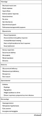
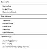
Triple A (3A) syndrome or Allgrove syndrome is a multisystem disorder first described in 1978, which classically involves the triad of esophageal achalasia, alacrima, and adrenocorticotropin hormone (ACTH)-resistant adrenal insufficiency.1 While the complete triad is present in approximately 70% of patients,2 dysfunction of the autonomic nervous system is also seen in about one-third of the patients,3 leading some authors to use the term 4A syndrome (achalasia, alacrima, adrenal insufficiency, and autonomic abnormalities).4 Additional symptoms reported in the literature include other neurological and dermatological manifestations,5 short stature, microcephaly, osteoporosis, and dysmorphic features.6 Given this wide array of phenotypes (Table 1), a genotype–phenotype correlation consequently is yet to be established.6 The exact burden of this disease is unknown. 3A syndrome has an estimated prevalence of 1 in 1 million, though it has been suggested to be very underreported due to missed diagnosis.7 This rare autosomal recessive disorder has been linked with mutations in the AAAS (achalasia–addisonianism–alacrima syndrome) gene. The AAAS gene product is a 546-amino acid protein called alacrima–achalasia–adrenal insufficiency neurologic disorder (ALADIN), which belongs to the WD-repeat family of proteins and exhibits wide functional diversity.8 Allgrove syndrome features great variability in presentation with discordance between phenotypes and genotypes even among members of the same family.9 Although genetic testing is essential in revealing the final diagnosis, DNA studies are not useful in the prediction of phenotype or prognosis of this disorder.10
 |  |  |
Table 1 Manifestations of triple A syndrome* |
Given that the presence of 2 of the 3 cardinal clinical entities strongly suggests the diagnosis of 3A, the recognition of the clinical syndrome is a challenge at the onset of the disease, when only one presenting symptom is typically observed. Differential diagnosis includes other causes of adrenal insufficiency including familial glucocorticoid deficiency. In adrenoleukodystrophy, similar to 3A, patients may present with impaired glucocorticoid function with minimal or no dysfunction in mineralocorticoid production. Elevated levels of very-long-chain fatty acids in the plasma are pathognomonic. There is a broad differential for neurological symptoms (see the section “Neurology”). Finally, 3A syndrome is sometimes confused with Sjogren symptom when the presenting symptoms are alacrima and xerostomia (see the section “Oral Health”).
Long-term follow-up recommendations for patients with 3A are limited, as current literature consists mainly of case reports and case series. With no definitive treatment for the condition, management focuses on individual presenting signs and symptoms. The prognosis of 3A syndrome is highly dependent on early diagnosis in order to prevent life-threatening adrenal crises—a challenging feat given the rarity of the condition and its high phenotypic heterogeneity. Early diagnosis may also prevent unnecessary investigations and inappropriate treatments.11 In this review, we present an analytical, multidisciplinary approach for the diagnosis, management, and follow-up of patients with 3A syndrome.
Genetics
In 3A syndrome, approximately 90% of the mutations involve altered genetic modification, leading to a repeat of the AAAS gene on chromosome 12q13.12 The AAAS gene encodes for a 546-amino acid polypeptide, known as ALADIN, which is involved with signal transduction, RNA processing, and transcription, as well as nuclear pore complex targeting.8,9 In addition, the ALADIN gene has been reported to play a role in redox homeostasis in human adrenal cells and to inhibit steroidogenesis.13 Krumbholz et al have shown that most mutations cause mislocalization of the mutant ALADIN proteins in the cytoplasm, as a result of inhibition of the correct targeting of ALADIN to nuclear pore complexes.14 High expression of this protein is seen in the adrenal gland, brain, and gastrointestinal tract, the organs in which the main pathologic manifestations of disease occur.3,8,15–18
Patients with 3A display a variety of homozygous or heterozygous compound mutations, often resulting in a shortened overall protein.19 In one study spanning 30 years, 47 different mutations were described including 20 splice site or frameshift mutations (43%), 16 nonsense mutations (34%), 10 missense mutations (21%), and one Alu-mediated intragenic 3,2 kb deletion (2%).11 It is also important to note that while the diagnosis of 3A syndrome could be made on the basis of the molecular genetic analysis of the AAAS gene, some individuals do not present with an AAAS gene mutation, suggesting additional genetic mechanisms involved.5 Recently two new genes, GMPPA and TRAPPC11, have been associated with “triple-A-like” syndrome phenotypes.20,21
Identification of the mutation(s) is important, as there is a 25% recurrence risk in future pregnancies in line with an autosomal recessive mode of inheritance. DNA confirmation in the proband also allows for early identification of currently asymptomatic siblings at risk in order to provide proper monitoring and treatment.7 Prenatal diagnosis by chorionic villus sampling or amniocentesis, as well as preimplantation diagnosis, is also possible.7
Achalasia
Achalasia is an esophageal motility disorder characterized by the failure of the lower esophageal sphincter to relax.22 It is an uncommon disorder with a prevalence of 10 cases per 100,000 individuals.23 Esophageal achalasia in childhood is rare, with less than 5% of patients presenting under the age of 15 years.24 Symptoms of achalasia most often include regurgitation, dysphagia, weight loss, and/or failure to thrive.25 Patients with achalasia may additionally present with pulmonary symptoms, including cough, aspiration, hoarseness, dyspnea, wheezing, or sore throat in up to 40% of the cases.26 In some cases, rare or recurrent pneumonias have alerted the medical team, triggering further investigations and subsequent diagnosis of 3A syndrome.27–29
The exact mechanism of achalasia is not fully understood. Achalasia present in 3A is caused by absent lower esophageal sphincter relaxation and impaired esophageal motility, compared to also rare cricopharyngeal achalasia, which is caused by isolated upper esophageal sphincter dysfunction. The gold standard of diagnosing achalasia is manometry25 and is established by the presence of aperistalsis in the distal two-thirds of the esophagus and incomplete lower esophageal sphincter relaxation. Signs of dysphagia and regurgitation may be present for years until the diagnosis of achalasia is established,6 with patients often being misdiagnosed and treated for gastroesophageal reflux.24,30 In a retrospective study that analyzed clinical data from children with idiopathic achalasia and patients with 3A syndrome, the presentation of the symptoms was similar among patients.30 Manometry, however, showed high lower esophageal sphincter pressures more frequently among 3A patients, which was associated with treatment failure.30 In a retrospective analysis of children with achalasia from various etiologies, children with 3A developed symptoms at a younger age compared to patients that did not have a genetic syndrome or a recognized chromosomal abnormality.31 However, the time interval from the development of symptoms to diagnosis was significantly longer for 3A patients in comparison and 3A patients were significantly more underweight at the time of the diagnosis.31 Furthermore, although the treatment outcomes were comparable, children with 3A had significantly less weight gain postintervention, which may indicate that they may represent a subgroup in need of closer follow-up.31
For those with a presumptive or established diagnosis of 3A, we recommend routine inquiry of symptoms that may be related to achalasia and prompt referral to gastroenterology and surgical specialties when indicated. Heller’s cardiomyotomy, medical treatment (calcium channel blockers, botulinum toxin, or nitrates) as well as pneumatic esophageal dilatation have been reported as successful interventions for achalasia.30–32 There is no consensus regarding the first line of treatment for achalasia and specific management and follow-up protocols, especially given the rarity of achalasia among the pediatric population. However, there is a tendency for preference of surgical treatment when radical treatment is desired.32,33 To reduce the risk of esophageal reflux post Heller’s cardiomyotomy, it is suggested to perform a fundopliclation.34 There is no current literature that suggests deviating from standard practices for achalasia with regard to the management of 3A patients.31
Alacrima
Alacrima is consistently reported to be the most common early presenting sign of 3A, although its significance is usually not recognized and medical assistance is not sought until other symptoms develop.5 It is the most consistent finding, with prevalence reaching >90% of the patients affected.6,35,36 Some case reports have shown that decreased tear production was either present from birth or was noted within the first year of life.5,9 In addition, various cases and reports have studied the prevalence of alacrima with relation to achalasia, as achalasia is an extremely rare finding in infancy.37 It was found that alacrima can be as high as 42% in patients with early-onset achalasia.37
As lacrimation is under parasympathetic control, it has been suggested that alacrima may be considered part of autonomic dysfunction.38,39 Orbital CT scans reveal the absence or decrease of lacrimal glands and a depletion of secretory granules in the acinar cell.19 The presence of optic nerve atrophy, expressed as optic disc pallor with either delayed timing or reduced amplitude on visual evoked potential, may represent another sign of nerve degeneration.40 As part of the initial investigation, Schirmer’s test confirms the presence of reduced or absent tears. Administration of artificial tears and lubricants help relief the sensation of dryness. If left untreated, alacrima may lead to keratopathy and corneal ulceration.19 As such, monitoring by an ophthalmologist with visual acuity assessment, ocular surface study, tonometry, and fundus examination is recommended at least yearly.39 While 3A syndrome is a rare condition, it should be taken into consideration in every child with alacrima. In addition, we would like to stress the importance of routinely inquiring about tear production in order to diagnose and properly manage these complex patients.
Endocrine
Adrenal insufficiency due to deficient glucocorticoid secretion affects up to 85% of patients with 3A and manifests during the first or more rarely second decade of life,11 but does not appear to be congenital.6 It is the main cause of mortality among patients with 3A, mostly due to severe hypoglycemia.41 In most cases, adrenocortical insufficiency manifests acutely as a crisis with associated hypoglycemia and/or hypotension.19 Alternatively, investigation may commence for recurrent vomiting, hyperpigmentation of the skin and mucous membranes, or developmental delays.42 A cortisol level at 8 am along with concomitant ACTH should be measured. The expected findings in ACTH sensitivity are low cortisol combined with markedly high ACTH.43 Confirmation should be established using IV or IM corticotropin stimulation test.44 When the stimulation test results are conflicting, insulin-induced hypoglycemia may confirm the diagnosis; however, it is not recommended as a first-line test as it is laborious and contraindicated in patients with a history of seizures or cardiovascular disease.45 The inadequate response of cortisol to administration of glucose in a hypoglycemic state confirms the diagnosis. Though mineralocorticoid deficiency is reported in only a minority of patients,2,46 concurrent aldosterone and renin levels should be drawn to assess deficiency.
Declining or borderline adrenal function has been detected in patients at the time of diagnosis of 3A syndrome, with normal cortisol levels but elevated ACTH and appropriate cortisol increase after ACTH challenge.2,47 In long-term follow-up, however, only some patients eventually develop adrenal insufficiency.2 Although there are no specific guidelines for the surveillance of patients diagnosed with 3A syndrome, we recommend lifelong follow-up of patients with initial normal or borderline function by an endocrinologist. In patients with adrenal insufficiency, we recommend initial visits every 3 months with hormone and serum chemistry monitoring. For patients that are clinically stable, based on clinical symptoms and laboratory values, we recommend follow-up every 6 months and educating patients and families about symptoms concerning for adrenal crisis. The short-acting glucocorticoid hydrocortisone is the treatment of choice in patients with adrenal insufficiency; however, in some cases, fludrocortisone may also be required. 44 In addition, serum chemistry should be monitored during episodes of acute illness in anticipation of adrenal crisis.
Osteoporosis, confirmed by measurement of low bone density, and presenting as early as childhood, has been reported in sporadic cases.48,49 It has been suggested that steroid supplementation alone could not explain the finding. The cause may be multifactorial, given decreased or lack of physical activity and sun exposure due to immobilization in progressive neurological disease, malnutrition secondary to achalasia, and low levels of androgens.48 As such, patients and families should be counseled on preventive measures, screening, and nutritional supplementation.
Neurology
There is a great variety of neurologic symptoms associated with 3A syndrome that involve the central, the peripheral, as well as the autonomic nervous system. A variety of neurological signs and symptoms have been described (see Table 1).3,50,51 In about one-third of patients, autonomic dysfunction symptoms include postural hypotension, abnormal cardiovascular responses and arrhythmias, anisocoria, abnormal pupillary reflexes, impaired or increased sweating, and impotence.6 While seizures have been described, those are hypoglycemic in origin.52 In a single case report, a patient with 3A syndrome was found to have Arnold Chiari malformation and syringomyelia, and Bizzari et al suggest screening diagnosed patients with magnetic resonance imaging of brain and spine, even without the presence of neurologic symptoms.52
The pathophysiology of the neurologic symptoms is not known. When performed, nerve conduction velocity tests typically exhibit axonal motor neuropathy, with selective involvement of the ulnar nerve being characteristic.53 Few published results on nerve biopsy studies show normal or nonspecific findings.17,54 Muscle biopsy results have shown evidence of neurogenic degeneration,54 nonspecific myopathy process,55 or mixed pathology.56
Neurological symptoms typically manifest in the second decade of life and adulthood; however, in some reports, it may be among the presenting symptoms in childhood. 3A syndrome should be part of the differentials in cases of early neurological dysfunction and developmental delay and assessment of adrenal function should be performed.51,57 Patients with an incomplete triad and early neurologic manifestations often undergo an extensive workup and are misdiagnosed as juvenile amyotrophic lateral sclerosis, adrenoleukodystrophy, Charcot–Marie–Tooth, spinocerebellar ataxia, spinal muscular amyotrophy, mitochondriopathy, and multiple sclerosis among others.51,53
Some evidence has shown that neurological symptoms in 3A syndrome may be slowly progressing, reaching a steady phase in adulthood.42 Potential symptoms may be debilitating or life-threatening.12,54 Glucocorticoid supplementation does not seem to affect the development and course of neurologic symptoms,43 however, tailored physical therapy sessions may help improve endurance and balance in patients who experience neuromuscular symptoms.58
Neuropsychology
The most frequent cognitive deficit in 3A consists of mild intellectual disability.13 Cognitive problems may be secondary to recurrent hypoglycemia in patients, and subsequent neuron damage, in patients with adrenal insufficiency.19 However, reports show that the cognitive function may be affected even in patients with normal adrenal function.19,47
Cognitive deficiencies have been reported in many pediatric patients with 3A in literature,19,42,59 with some showing IQ to gradually decrease over time.17 These findings are usually noticed clinically and then confirmed by neuropsychometry.59 Such data further support the notion to consider impaired intelligence as one of the most consistent clinical signs of 3A syndrome.59 The extent of deficits ranges from learning disabilities and poor school performance to slight and moderate intellectual disability.42,53 Mazzone et al conducted an extensive neuropsychological assessment of a 4-year-old male patient diagnosed with 3A and found the subject to be deficient in a variety of areas with an adaptive level lower than chronological age.60 Significant deficits in planning competence and attention abilities have also been associated with 3A syndrome.60
A variety of the disabling physiological and neurologic manifestations often lead to neuropsychological impairments. Additional literature states that patients affected by adrenal insufficiency (Addison’s disease) have increased the levels of anxiety and a higher risk for affective and mood disorder.43,61,62 Such extensive psychological investigations have not been performed on 3A patients and should be the focus of future studies.
As intellectual disability and other multisystem neurological symptoms can arise over time,63 assessment of neuropsychological and psychopathological features should be performed in patients with this disease highlighting the need for a multidisciplinary approach and tailored neuropsychological therapies.
Oral health
Xerostomia has been described in some case reports as a complaint among patients with 3A syndrome, and confirmatory sialometry has showed decreased salivary secretion.12,64–67 Hyposalivation has also been identified as a cause of dysphagia after correction of achalasia.12 A small, spastic, fissured tongue is characteristic of the syndrome.68 Premature tooth loss has been described. In some cases this was attributed to decreased saliva production and subsequent tooth decay;64,65,69 however, there have been reports describing permanent tooth loss without an underlying gum, salivary, bone, or tooth pathology.59 Furthermore, gastroesophageal reflux, which is prevalent in patients with 3A, has been shown to contribute to tooth erosion.70
Moreover, it has been suggested that there is significant overlap in the symptomatology of patients with 3A and Sjogren syndromes, more specifically in patients that may have been diagnosed with “achalasia sicca”.64,66 The presence of anti-Ro and anti-La antibodies may make the differentiation between the conditions difficult, and in those cases, salivary gland biopsy may prove useful.66 We recommend the periodical assessment of adrenal function for patients with unclear diagnosis.
The investigation of salivary function is recommended in patients with 3A syndrome, in order to alleviate symptoms, decrease oral infections, and prevent tooth decay. For patients with decreased salivary function, artificial saliva has been proven effective, and in cases of oral candidiasis, treatment with antimycotics should be intiated.64,65 For those with normal salivary function, we recommend strict adherence to dental visits at least every 6 months, as recommended for the general population,71 as well as routine preventive measures such as meticulous oral hygiene, fluoride-containing toothpaste, and low-sugar diet. Signs and symptoms of decreased salivary flow and tooth erosion should be evaluated at every visit and should prompt appropriate investigation.
Perioperative management
Preoperative evaluation for patients with known or suspected 3A syndrome should include serum electrolytes, 9 am serum cortisol and ACTH levels, ACTH stimulation test, Schirmer test, as well as investigation for autonomic neuropathy.72 Adrenal insufficiency should be recognized and adequately treated with a stress dose of glucocorticoids the morning of the procedure, to prevent perioperative crisis,72–75 while long procedures may require continuous glucocorticoid infusion in the operating room.72 During the procedure, blood glucose should be monitored, as steroid administration could lead to significant hyperglycemia warranting the administration of insulin.72–74 In patients with impairment of autonomic responses, hemodynamic stability may be fragile and requires close cardiopulmonary monitoring for hypotension and arrhythmias, practicing caution during induction and position changes.73–75 Protection for the eyes in the form of lubrication should be provided, in order to avoid keratopathy and corneal ulcer, in the setting of alacrima and absent protective corneal responses.72,73 For those who have undergone myotomy for the correction of achalasia, there is increased risk for gastroparesis, and aspiration related to incisions on the lower esophageal sphincter.72–74 Expert recommendations include the administration of antiemetics, antacids, and head elevation for these patients.72
Conclusion
Given the rarity of the condition and the potentially detrimental adrenal insufficiency that is associated with 3A syndrome, we suggest investigation of children presenting with alacrima, achalasia, or other cardinal symptoms of the syndrome. Increased awareness of the disease, followed by vigilant surveillance for the development of adrenal insufficiency, is warranted to improve survival rates. The general practitioner should be able to recognize the diagnosis early, refer patients to the appropriate specialists, coordinate the care of these complex patients, as well as screen for new signs and symptoms at each encounter.
Acknowledgment
The authors would like to thank Kavitha Vemuri for her assistance in literature search.
Disclosure
The authors report no conflicts of interest in this work.
References
1. Allgrove J, Clayden GS, Grant DB, Macaulay JC. Familial glucocorticoid deficiency with achalasia of the cardia and deficient tear production. Lancet. 1978;1(8077):1284–1286. doi:10.1016/s0140-6736(78)91268-0
2. Roucher-Boulez F, Brac de la Perriere A, Jacquez A, et al. Triple-A syndrome: a wide spectrum of adrenal dysfunction. Eur J Endocrinol. 2018;178(3):199–207. doi:10.1530/EJE-17-0642
3. Huebner A, Yoon SJ, Ozkinay F, et al. Triple A syndrome–clinical aspects and molecular genetics. Endocr Res. 2000;26(4):751–759.
4. Gazarian M, Cowell CT, Bonney M, Grigor WG. The “4A” syndrome: adrenocortical insufficiency associated with achalasia, alacrima, autonomic and other neurological abnormalities. Eur J Pediatr. 1995;154(1):18–23.
5. Brooks BP, Kleta R, Stuart C, et al. Genotypic heterogeneity and clinical phenotype in triple A syndrome: a review of the NIH experience 2000–2005. Clin Genet. 2005;68(3):215–221. doi:10.1111/j.1399-0004.2005.00482.x
6. Huebner A, Elias LL, Clark AJ. ACTH resistance syndromes. J Pediatr Endocrinol Metab. 1999;12(Suppl 1):277–293.
7. Brown B, Agdere L, Muntean C, David K. Alacrima as a harbinger of adrenal insufficiency in a child with Allgrove (AAA) syndrome. Am J Case Rep. 2016;17:703–706. doi:10.12659/AJCR.899546
8. Handschug K, Sperling S, Yoon SJ, Hennig S, Clark AJ, Huebner A. Triple A syndrome is caused by mutations in AAAS, a new WD-repeat protein gene. Hum Mol Genet. 2001;10(3):283–290. doi:10.1093/hmg/10.3.283
9. Prpic I, Huebner A, Persic M, Handschug K, Pavletic M. Triple A syndrome: genotype-phenotype assessment. Clin Genet. 2003;63(5):415–417.
10. Milenkovic T, Koehler K, Krumbholz M, Zivanovic S, Zdravkovic D, Huebner A. Three siblings with triple A syndrome with a novel frameshift mutation in the AAAS gene and a review of 17 independent patients with the frequent p.Ser263Pro mutation. Eur J Pediatr. 2008;167(9):1049–1055. doi:10.1007/s00431-007-0640-7
11. Milenkovic T, Zdravkovic D, Savic N, et al. Triple A syndrome: 32 years experience of a single centre (1977–2008). Eur J Pediatr. 2010;169(11):1323–1328. doi:10.1007/s00431-010-1222-7
12. Dumic M, Barisic N, Kusec V, et al. Long-term clinical follow-up and molecular genetic findings in eight patients with triple A syndrome. Eur J Pediatr. 2012;171(10):1453–1459. doi:10.1007/s00431-012-1745-1
13. Prasad R, Metherell LA, Clark AJ, Storr HL. Deficiency of ALADIN impairs redox homeostasis in human adrenal cells and inhibits steroidogenesis. Endocrinology. 2013;154(9):3209–3218. doi:10.1210/en.2013-1241
14. Krumbholz M, Koehler K, Huebner A. Cellular localization of 17 natural mutant variants of ALADIN protein in triple A syndrome - shedding light on an unexpected splice mutation. Biochem Cell Biol. 2006;84(2):243–249. doi:10.1139/o05-198
15. Gilio F, Di Rezze S, Conte A, et al. Case report of adult-onset Allgrove syndrome. Neurol Sci. 2007;28(6):331–335. doi:10.1007/s10072-007-0848-3
16. Stratakis CA, Lin JP, Pras E, Rennert OM, Bourdony CJ, Chan WY. Segregation of Allgrove (triple-A) syndrome in Puerto Rican kindreds with chromosome 12 (12q13) polymorphic markers. Proc Assoc Am Physicians. 1997;109(5):478–482.
17. Houlden H, Smith S, De Carvalho M, et al. Clinical and genetic characterization of families with triple A (Allgrove) syndrome. Brain. 2002;125(Pt 12):2681–2690. doi:10.1093/brain/awf270
18. Hadj-Rabia S, Salomon R, Pelet A, et al. Linkage disequilibrium in inbred North African families allows fine genetic and physical mapping of triple A syndrome. Eur J Hum Genet. 2000;8(8):613–620. doi:10.1038/sj.ejhg.5200508
19. Moore PS, Couch RM, Perry YS, Shuckett EP, Winter JS. Allgrove syndrome: an autosomal recessive syndrome of ACTH insensitivity, achalasia and alacrima. Clin Endocrinol (Oxf). 1991;34(2):107–114.
20. Gold WA, Sobreira N, Wiame E, et al. A novel mutation in GMPPA in siblings with apparent intellectual disability, epilepsy, dysmorphism, and autonomic dysfunction. Am J Med Genet A. 2017;173(8):2246–2250. doi:10.1002/ajmg.a.38292
21. Koehler K, Milev MP, Prematilake K, et al. A novel TRAPPC11 mutation in two Turkish families associated with cerebral atrophy, global retardation, scoliosis, achalasia and alacrima. J Med Genet. 2017;54(3):176–185. doi:10.1136/jmedgenet-2016-104108
22. Furuzawa-Carballeda J, Torres-Landa S, Valdovinos MA, Coss-Adame E, et al. New insights into the pathophysiology of achalasia and implications for future treatment. World J Gastroenterol. 2016;22(35):7892–7907. doi:10.3748/wjg.v22.i35.7892
23. Sadowski DC, Ackah F, Jiang B, Svenson LW. Achalasia: incidence, prevalence and survival. A population-based study. Neurogastroenterol Motil. 2010;22(9):e256–e261. doi:10.1111/j.1365-2982.2010.01511.x
24. Hallal C, Kieling CO, Nunes DL, et al. Diagnosis, misdiagnosis, and associated diseases of achalasia in children and adolescents: a twelve-year single center experience. Pediatr Surg Int. 2012;28(12):1211–1217. doi:10.1007/s00383-012-3214-3
25. Sarathi V, Shah NS. Triple-A syndrome. Adv Exp Med Biol. 2010;685:1–8. doi:10.1007/978-1-4419-6448-9_1
26. Sinan H, Tatum RP, Soares RV, Martin AV, Pellegrini CA, Oelschlager BK. Prevalence of respiratory symptoms in patients with achalasia. Dis Esophagus. 2011;24(4):224–228. doi:10.1111/j.1442-2050.2010.01126.x
27. Parhizkar B, Maghsoodi N, Forootan M, Entezari AH. A 12 year old boy with recurrent episodes of pneumonia: triple A syndrome. Gastroenterol Hepatol Bed Bench. Spring 2012;5(2):112–115.
28. Ledoyen A, Bresson V, Deneux I, et al. [Bronchiectasis revealing triple A syndrome]. Arch Pediatr. 2015;22(7):746–749. doi:10.1016/j.arcped.2015.03.023
29. Emiralioglu N, Ersoz DD, Oguz B, et al. Pulmonary mycobacterium abscessus infection in a patient with triple A syndrome. J Trop Pediatr. 2016;62(4):324–327. doi:10.1093/tropej/fmv104
30. Alhussaini B, Gottrand F, Goutet JM, et al. Clinical and manometric characteristics of Allgrove syndrome. J Pediatr Gastroenterol Nutr. 2011;53(3):271–274. doi:10.1097/MPG.0b013e31821456ba
31. Meyer A, Catto-Smith A, Crameri J, et al. Achalasia: outcome in children. J Gastroenterol Hepatol. 2017;32(2):395–400. doi:10.1111/jgh.13484
32. Torab FC, Hamchou M, Ionescu G, Al-Salem AH. Familial achalasia in children. Pediatr Surg Int. 2012;28(12):1229–1233. doi:10.1007/s00383-012-3186-3
33. Nakamura J, Hikichi T, Inoue H, et al. Per-oral endoscopic myotomy for esophageal achalasia in a case of Allgrove syndrome. Clin J Gastroenterol. 2018;11(4):273–277. doi:10.1007/s12328-018-0819-7
34. Patti MG, Albanese CT, Holcomb GW
35. Patt H, Koehler K, Lodha S, et al. Phenotype-genotype spectrum of AAA syndrome from Western India and systematic review of literature. Endocr Connect. 2017;6(8):901–913. doi:10.1530/EC-17-0255
36. Kallabi F, Belghuith N, Aloulou H, et al. Clinical and genetic characterization of 26 Tunisian patients with Allgrove syndrome. Arch Med Res. 2016;47(2):105–110. doi:10.1016/j.arcmed.2016.04.004
37. Kim HM. Is alacrima so prevalent in patients with early-onset achalasia? J Neurogastroenterol Motil. 2011;17(3):330.
38. Babu K, Murthy KR, Babu N, Ramesh S. Triple A syndrome with ophthalmic manifestations in two siblings. Indian J Ophthalmol. 2007;55(4):304–306. doi:10.4103/0301-4738.33048
39. Aragona P, Rania L, Roszkowska AM, et al. 4A syndrome: ocular surface investigation in an Italian young patient. BMC Ophthalmol. 2014;14:155. doi:10.1186/1471-2415-14-155
40. Villanueva-Mendoza C, artinez-Guzman O, Rivera-Parra D, Zenteno JC. Triple A or Allgrove syndrome. A case report with ophthalmic abnormalities and a novel mutation in the AAAS gene. Ophthalmic Genet. 2009;30(1):45–49. doi:10.1080/13816810802502962
41. Thomas J, Subramanyam S, Vijayaraghavan S, Bhaskar E. Late onset adrenal insufficiency and achalasia in Allgrove syndrome. BMJ Case Rep. 2015;2015. doi:10.1136/bcr-2014-208900
42. Grant DB, Barnes ND, Dumic M, et al. Neurological and adrenal dysfunction in the adrenal insufficiency/alacrima/achalasia (3A) syndrome. Arch Dis Child. 1993;68(6):779–782. doi:10.1136/adc.68.6.779
43. Clark AJL, Weber A. Adrenocorticotropin insensitivity syndromes. Endocr Rev. 1998;19(6):828–843. doi:10.1210/edrv.19.6.0351
44. Bornstein SR, Allolio B, Arlt W, et al. Diagnosis and treatment of primary adrenal insufficiency: an endocrine society clinical practice guideline. J Clin Endocrinol Metab. 2016;101(2):364–389. doi:10.1210/jc.2015-1710
45. Salehi M, Houlden H, Sheikh A, Poretsky L. The diagnosis of adrenal insufficiency in a patient with Allgrove syndrome and a novel mutation in the ALADIN gene. Metabolism. 2005;54(2):200–205. doi:10.1016/j.metabol.2004.08.013
46. Collares CV, Antunes-Rodrigues J, Moreira AC, et al. Heterogeneity in the molecular basis of ACTH resistance syndrome. Eur J Endocrinol. 2008;159(1):61–68. doi:10.1530/EJE-08-0079
47. Ismail EA, Tulliot-Pelet A, Mohsen AM, Al-Saleh Q. Allgrove syndrome with features of familial dysautonomia: a novel mutation in the AAAS gene. Acta Paediatr. 2006;95(9):1140–1143. doi:10.1080/08035250500538999
48. Dumic M, Putarek NR, Kusec V, Barisic N, Koehler K, Huebner A. Low bone mineral density for age/osteoporosis in triple A syndrome-an overlooked symptom of unexplained etiology. Osteoporos Int. 2016;27(2):521–526. doi:10.1007/s00198-015-3265-0
49. Ozgen AG, Ercan E, Ozutemiz O, Hamulu F, Bayraktar F, Yilmaz C. The 4A syndrome association with osteoporosis. Endocr J. 1999;46(1):227–230.
50. Dixit A, Chow G, Sarkar A. Neurologic presentation of triple A syndrome. Pediatr Neurol. 2011;45(5):347–349. doi:10.1016/j.pediatrneurol.2011.07.003
51. Dumic M, Barisic N, Rojnic-Putarek N, et al. Two siblings with triple A syndrome and novel mutation presenting as hereditary polyneuropathy. Eur J Pediatr. 2011;170(3):393–396. doi:10.1007/s00431-010-1314-4
52. Bizzarri C, Benevento D, Terzi C, Huebner A, Cappa M. Triple A (Allgrove) syndrome: an unusual association with syringomyelia. Ital J Pediatr. 2013;39:39. doi:10.1186/1824-7288-39-39
53. Vallet AE, Verschueren A, Petiot P, et al. Neurological features in adult Triple-A (Allgrove) syndrome. J Neurol. 2012;259(1):39–46. doi:10.1007/s00415-011-6115-9
54. Kimber J, McLean BN, Prevett M, Hammans SR. Allgrove or 4 “A” syndrome: an autosomal recessive syndrome causing multisystem neurological disease. J Neurol Neurosurg Psychiatry. 2003;74(5):654–657. doi:10.1136/jnnp.74.5.654
55. Koehler K, Brockmann K, Krumbholz M, et al. Axonal neuropathy with unusual pattern of amyotrophy and alacrima associated with a novel AAAS mutation p.Leu430Phe. Eur J Hum Genet. 2008;16(12):1499–1506. doi:10.1038/ejhg.2008.132
56. Reimann J, Kohlschmidt N, Tolksdorf K, Weis J, Kuchelmeister K, Roos A. Muscle pathology as a diagnostic clue to Allgrove Syndrome. J Neuropathol Exp Neurol. 2017;76(5):337–341. doi:10.1093/jnen/nlx016
57. Jerie M, Vojtech Z, Malikova H, Prochazkova S, Vackova Z, Rolfs A. Allgrove syndrome with prominent neurological symptoms. Case Report. Neuro Endocrinol Lett. 2016;37(3):184–188.
58. Adams JPDN. Clinical decision making and application of an active rehabilitation program for a person with the neuromuscular symptoms of Allgrove syndrome: a case report. Physiother Theory Pract. 2018;1–8. doi:10.1080/09593985.2018.1548049
59. Razavi Z, Taghdiri MM, Eghbalian F, Bazzazi N. Premature loss of permanent teeth in Allgrove (4A) syndrome in two related families. Iran J Pediatr. 2010;20(1):101–106.
60. Mazzone L, Postorino V, De Peppo L, et al. Longitudinal neuropsychological profile in a patient with triple a syndrome. Case Rep Pediatr. 2013;2013:604921.
61. Thomsen AF, Kvist TK, Andersen PK, Kessing LV. The risk of affective disorders in patients with adrenocortical insufficiency. Psychoneuroendocrinology. 2006;31(5):614–622. doi:10.1016/j.psyneuen.2006.01.003
62. Printha K, Hulathduwa SR, Samarasinghe K, Suh YH, De Silva KR. Apoptosis in subicular neurons: a comparison between suicide and Addison’s disease. Indian J Psychiatry. 2009;51(4):276–279. doi:10.4103/0019-5545.58293
63. Yassaee VR, Soltani Z, Ardakani BM. Mutation spectra of the AAAS gene in Iranian families with Allgrove Syndrome. Arch Med Res. 2011;42(2):163–168. doi:10.1016/j.arcmed.2011.02.006
64. Dumic M, Mravak-Stipetic M, Kaic Z, et al. Xerostomia in patients with triple A syndrome–a newly recognised finding. Eur J Pediatr. 2000;159(12):885–888.
65. Vucicevic-Boras V, Juras D, Gruden-Pokupec JS, Vidovic A. Oral manifestations of triple A syndrome. Eur J Med Res. 2003;8(7):318–320.
66. Onat AM, Pehlivan Y, Buyukhatipoglu H, et al. Unusual presentation of triple A syndrome mimicking Sjogren’s syndrome. Clin Rheumatol. 2007;26(10):1749–1751. doi:10.1007/s10067-006-0498-5
67. Chu ML, Berlin D, Axelrod FB. Allgrove syndrome: documenting cholinergic dysfunction by autonomic tests. J Pediatr. 1996;129(1):156–159. doi:10.1016/s0022-3476(96)70205-6
68. Houlden H. The small, spastic, and furrowed tongue of Allgrove syndrome. Neurology. 2009;72(15):1366. doi:10.1212/WNL.0b013e3181a9fad1
69. Vahedi M, Fathi S, Allahbakhshi H. Edentulous child with Allgrove syndrome: a rare case report. Korean J Pediatr. 2016;59(11):456–459. doi:10.3345/kjp.2016.59.11.456
70. Sarath Kumar KS, Mungara J, Venumbaka NR, Vijayakumar P, Karunakaran D. Oral manifestations of gastroesophageal reflux disease in children: a preliminary observational study. J Indian Soc Pedod Prev Dent. 2018;36(2):125–129. doi:10.4103/JISPPD.JISPPD_1182_17
71. Riley P, Worthington HV, Clarkson JE, Beirne PV. Recall intervals for oral health in primary care patients. Cochrane Database Syst Rev. 2013;12:Cd004346.
72. Arun B, Deepak B, Chakravarthy MR. Anaesthetic management of a patient with Allgrove syndrome. Indian J Anaesth. 2014;58(6):736–738. doi:10.4103/0019-5049.147168
73. Dhar M, Verma N, Singh RB, Pai VK. Triple A to triple S: from diagnosis, to anesthetic management of Allgrove syndrome. J Clin Anesth. 2016;33:141–143. doi:10.1016/j.jclinane.2016.02.035
74. Ozer AB, Erhan OL, Sumer C, Yildizhan O. Administration of anesthesia in a patient with allgrove syndrome. Case Rep Anesthesiol. 2012;2012:109346.
75. Pino Gomez S, Parrado Figueroa A, Blanco del Val B, Nuno Gil J. [Anaesthetic management of a patient with Allgrove’s syndrome]. Rev Esp Anestesiol Reanim. 2013;60(1):55–57. doi:10.1016/j.redar.2012.05.021
 © 2019 The Author(s). This work is published and licensed by Dove Medical Press Limited. The full terms of this license are available at https://www.dovepress.com/terms.php and incorporate the Creative Commons Attribution - Non Commercial (unported, v3.0) License.
By accessing the work you hereby accept the Terms. Non-commercial uses of the work are permitted without any further permission from Dove Medical Press Limited, provided the work is properly attributed. For permission for commercial use of this work, please see paragraphs 4.2 and 5 of our Terms.
