Back to Journals » Clinical Ophthalmology » Volume 17
Treatment of Non-Infectious Posterior Uveitis with Dexamethasone Intravitreal Implants in a Real-World Setting
Authors Butt F, Devonport H
Received 29 October 2022
Accepted for publication 13 January 2023
Published 16 February 2023 Volume 2023:17 Pages 601—611
DOI https://doi.org/10.2147/OPTH.S393662
Checked for plagiarism Yes
Review by Single anonymous peer review
Peer reviewer comments 3
Editor who approved publication: Dr Scott Fraser
Farhat Butt, Helen Devonport
Opthalmology Department, Bradford Royal Infirmary, Duckworth Lane, BD9 6RJ, UK
Correspondence: Farhat Butt, Bradford Royal Infirmary, Duckworth Lane, BD9 6RJ, UK, Tel +44 7834 922022, Fax +44 1274 364786, Email [email protected]
Purpose: The present study aimed to assess the efficacy and safety associated to the treatment of patients with non-infectious posterior uveitis with intravitreal dexamethasone (DEX) implants in a real-world clinical setting.
Patients and Methods: This is a retrospective, single center analysis of the data from 29 patients with non-infectious posterior uveitis in whom 38 eyes were treated with dexamethasone intravitreal implants in routine clinical practice between January 2012 and October 2017. The parameters of visual acuity (VA), intraocular pressure (IOP) and central retinal thickness (CRT) were recorded 6 weeks after the first implant was administered, in accordance with the clinical guidelines for the use of these implants, and after a 6-month follow-up period. In addition, the formation of cataracts was evaluated at 12 months.
Results: Treatment with the DEX implant caused a significant improvement in the VA from baseline at 6 weeks in eyes treated with 2– 6 implants and for eyes without cataracts. A significant decrease in CRT was observed relative to the baseline at 6 weeks for eyes treated with 1 and 2– 6 implants, which was maintained at 6 months for those eyes treated with 2– 6 implants. This significant improvement in CRT at 6 weeks and 6 months was evident in eyes with and without cataracts. During the study period, the IOP was found to increase significantly from baseline at 6 weeks in some eyes but this was managed topically, and no surgical intervention was necessary.
Conclusion: Intravitreal DEX implants represent an effective and safe therapy for the treatment of non-infectious uveitis in routine clinical practice, producing favorable visual and anatomical outcomes after the administration of just 2– 6 DEX implants.
Keywords: visual acuity, intraocular pressure, central retinal thickness, cataracts, retrospective analysis
Plain Language Summary
Patients with non-infectious posterior uveitis may benefit from treatment with intravitreal dexamethasone (DEX) implants, although their efficacy and safety must first be demonstrated in a real-world clinical setting. As such, we assessed the efficacy of this therapy in a 6-month period after administering the first implant to 38 eyes of 29 patients with non-infectious posterior uveitis treated at our hospital between January 2012 and October 2017. We assessed efficacy through the improvement in visual acuity (VA) and central retinal thickness (CRT), as well as recording any common adverse events associated with the treatment, such as increased intraocular pressure (IOP) or cataract development in the 12 months after the first treatment. In eyes that received 2–6 DEX implants, VA improved significantly after 6 weeks and the CRT decreased (reflecting less inflammation of the retina), effects that persisted for 6 months in these eyes. Only a mild increase in IOP could be detected, which did not require surgery to be adequately managed, and cataract induction was not exacerbated by the DEX implants. Thus, intravitreal DEX implants are an effective and safe treatment for non-infectious posterior uveitis in routine clinical practice, producing favorable visual and anatomical outcomes after administering just 1–6 DEX implants.
Introduction
Uveitis refers to a group of infectious and non-infectious intraocular inflammatory diseases that lead to irreversible blindness in approximately 35% of patients, and that account for up to 15% of blindness in the developed world.1,2 Unlike other ocular diseases, uveitis may occur at any age, although it most commonly affects adults of working-age,1,3 with the ensuing social and economic impact.4–6 The estimated average annual incidence of uveitis is 50.45 per 100,000,1,7,8 with a geographic variation in prevalence from 9 to 730 cases per 100,000 individuals.9 The main causes of the visual loss associated with uveitis are cystoid macular edema (CME),3,4,10 which can provoke structural changes in the retina and cataracts, and complications associated with increased intraocular pressure (IOP).
The majority of uveitis cases (67–90%) are non-infectious,7,11–13 making this the third-leading cause of preventable blindness worldwide.14 Uveitis is classified into anterior, intermediate and posterior according to the part of the uvea that is affected.1 Posterior uveitis affects the choroid at the back of the eye, although it can also affect the retina and optic nerve, accounting for 15–22% of all uveitis cases15 and representing the type of uveitis most strongly associated with a loss of vision.16,17 The treatments available for non-inflammatory posterior uveitis are either topical or systemic corticosteroid or immunosuppressor administration to control ocular inflammation.18 However, the systemic and ocular side-effects associated with prolonged administration limit their use. Topical corticosteroids are better tolerated than systemic corticosteroids but compliance is an issue, and therapeutic concentrations are unlikely to reach the posterior portions of the eye.15,19 Accordingly, sustained-release corticosteroid implants have been developed to maintain steroid release to the posterior eye segment over prolonged periods of time.20,21
The Dexamethasone intravitreal implant (0.7 mg, DEX implant, Ozurdex®: Allergan, Inc., CA, USA) is a biodegradable, sustained-release drug delivery system that secretes low doses of dexamethasone into the vitreous humor for up to 6 months,22 and it can be easily administered in an outpatient setting. DEX implants were first approved by the FDA and EMA (US Food and Drug Administration and the European Medicines Agency) for the treatment of macular edema (ME) secondary to Retinal Vein Occlusion (RVO) based on the conclusions of a Phase III, randomized, controlled clinical trial: the GENEVA study.23,24 Subsequently, it was approved as a safe and effective treatment for non-infectious intermediate and posterior uveitis based on the results of the HURON study25 and more recently, for the treatment of diabetic macular edema (DME) based on the results of the MEAD study.26 In the UK, Ozurdex® was approved in 2011 for the treatment of adult patients with ME following either Branch or Central RVO (BRVO or CRVO),27 which was extended to the treatment of posterior eye segment inflammation presenting as non-infectious uveitis in 2017.27
Treatment with intravitreal DEX implants reduces sight threatening intraocular inflammation with a low risk of IOP spike requiring surgical intervention.25 However, there is limited data available regarding the effects of DEX implants in patients with non-infectious posterior uveitis in real-world settings, with most studies involving relatively small cohorts of patients with different types of uveitis.28–30 A retrospective study on the long-term outcomes of eyes with non-infectious intermediate and posterior uveitis after repeated treatment with DEX implants31 reported visual and anatomical improvements (resolved ocular inflammation and central retinal thickness – CRT) within 1–2 months of each injection, which were sustained for about 6 months, and no eyes required surgery for increased IOP or cataract progression. Favorable visual outcomes were also reported in the largest study to date on the effect of intravitreal DEX implants in non-infectious uveitis in routine clinical practice, supporting their use as either single therapy or as a co-adjuvant to systemic treatment, but requiring repeated injections to achieve a sustained improvement in visual acuity (VA) at 12 months.32
Here, we present data from a cohort of 29 patients with non-infectious posterior uveitis, in which 38 eyes were treated with DEX implants in routine clinical practice and in accordance with the UK’s NICE (National Institute for Health and Care Excellence) guidance (https://www.nice.org.uk/guidance/ta460). The results were consistent with those reported previously on patients with non-infectious uveitis in real-world settings, emphasizing the benefits and safety of this therapy to treat non-infectious posterior uveitis in routine clinical practice.
Materials and Methods
Study Design
A retrospective, single center audit study was carried out on patients with non-infectious posterior uveitis attended at the Bradford Hospital (Bradford Teaching Hospitals National Health Service Foundation Trust, Bradford, UK). Based on their clinical records, 31 patients were identified that had been treated with intravitreal DEX implants between January 2012 and October 2017, and followed for at least 6 months after receiving the first implant. Two of these patients were excluded from the study, one due to the lack of information available after administering the implant and the other due to complicated cataract surgery undertaken at a different clinical center. Informed consent was obtained from all patients included in the study in accordance with the Helsinki Declaration for medical research involving human subjects.
Clinical Data Collection and Outcome Measurements
Data were retrieved by retrospective review of non-infectious posterior uveitis patients’ electronic medical records (Medisoft). A total of 38 eyes (from 29 patients) that received at least one intravitreal DEX implant were evaluated in this study. In addition to the patient’s demographic data, the clinical variables assessed included VA, IOP, CRT, lens status and the presence of a cataract, the number of DEX implant injections, and whether the patient was on immunosuppressor therapy or medication to lower their IOP. The VA, IOP and CRT at baseline were recorded, as well as 6 weeks and 6 months after the first implant was administered. Lens status was measured at baseline and at 12 months after the injection of the DEX implants, with eyes with a pseudophakic lens at 12 months but that were phakic at baseline flagged as having developed a cataract.
All the data were gathered in a routine clinical setting, with IOP measured in mmHg by Goldmann tonometry. VA scores were recorded on the Snellen scale and since all the line steps were not equal in size, the measurements were converted to the logMAR scale. The analysis of eyes with a low VA was undertaken by substituting counting fingers (CF) and hand movement (HM) with 1.9 and 3.0 logMAR, respectively.33 The CRT was measured by optical coherence tomography (OCT). No missing value substitutions were performed in patients where data were not available for a particular visit or that were lost during the follow-up.
Statistical Analysis
Standard summary statistics were used for the baseline characteristics of the patients, and for the VA, IOP and CRT outcomes at the follow-up time points, stratified according to the number of DEX implants and the presence or absence of a cataract. A Wilcoxon signed-rank test was used to compare the pre- and post-implant values for VA, IOP and CRT. The p-values were not adjusted for multiple testing and a p-value <0.05 was considered significant in these exploratory analyses. Patients with missing data at any of the follow-up points were not included in the Wilcoxon signed-rank tests. The statistical package R (R Core Team (2019), URL https://www.R-project.org/) was used for all statistical analyses.
Results
Baseline Patient Demographic and Study Eye Characteristics
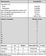
A total of 38 eyes from 29 patients were included in this study and the characteristics of the affected eyes were recorded (Table 1). While all the patients were diagnosed with non-infectious posterior uveitis, the underlying causes in the audit were: Idiopathic (63%); Sarcoidosis (8%); Retinitis Pigmentosa (8%); MS/Demyelination (5%); Birdshot chorioretinopathy (5%); Eales disease (3%); Tubulointerstitial nephritis and uveitis syndrome (TINU, 3%); Punctate inner chorioretinopathy (3%); Vogt-Koyanagi-Harada (VKH) disease (2%). At the time of the first implant (baseline), 10 eyes were being treated with immunosuppressor therapy (26.3%) and 8 were receiving IOP lowering medication, and some patients may also have been receiving a low dose systemic steroid. None of these additional treatments were stopped during the period of the study. At baseline, the mean VA was 0.7 (±0.5, logMAR) and the mean CRT was 439.5 (±117.8 μm).
 |
Table 1 The Characteristics of the Patients and the Affected Eyes at Baseline, the Number of DEX Implants, Unstratified and Stratified According to the Presence or Absence of Cataract |
Treatment
The patients received between 1 and 26 DEX implants over the 5-years of the study and of the 38 eyes in total, 13 (34.2%) received a single injection, 9 (23.7%) required 2 injections, while 16 (42.1%) required between 3 and 26 injections (Table 2). Since in some cases the number of eyes receiving ≥3 DEX implants was relatively low, three groups of eyes were established for the analyses according to the number of implants received: 1 (n = 13), 2–6 (n = 18), or 9–26 (n = 7). These groups were established not only to facilitate the capture and analysis of the data but also, with a view to gaining potentially relevant information regarding cataract development. No significant differences were found in the number of DEX implants received between eyes with cataract and without cataract at 12 months (Table 3).
 |
Table 3 CRT (µm) Outcomes After DEX Implants at the Follow-Up Time Points, Stratified According to the Number of DEX Implants Received and According to the Presence or Absence of a Cataract |
Efficacy Analysis
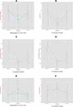
In general terms, in the three groups of eyes analyzed administering DEX implants improved the VA from baseline at 6 weeks, which reverted a little at 6 months (Figure 1 and Table 2). However, this improvement was only significant at 6 weeks for the group of eyes treated with 2–6 implants (p = 0.009: see Figure 1 and Table 2). When considering the presence or absence of a cataract at baseline, a similar pattern of change in VA was observed over time for the two groups of eyes, with and without cataracts. As such, VA improved at 6 weeks and by 6 months it had deteriorated, even to the median baseline value in the case of the group of eyes with cataracts. The improvement in VA observed was only significant at 6 weeks for the group of eyes without cataracts (p = 0.009: see Figure 1 and Table 2).
The effect of DEX implants on CRT was also studied over the follow-up period (Figure 1 and Table 3), producing a significant decrease in CRT relative to the baseline at 6 weeks for the groups of eyes treated with 1 (p = 0.003) and 2–6 implants (p < 0.001). This decrease from baseline was maintained at 6 months for the group of eyes treated with 2–6 implants (p = 0.001) and although not significantly, also for the group of eyes that received only 1 implant (p = 0.054). When considering the presence or absence of cataract at baseline, a similar change in CRT over time was observed for the two groups of eyes. Indeed, the CRT decreased significantly at 6 weeks (p < 0.001 for the eyes with no cataract, p = 0.004 for the eyes with cataracts: see Figure 1 and Table 3), later stabilizing at the 6-month follow-up point for eyes with or without a cataract (p = 0.006 for the eyes with no cataract, p = 0.004 for the eyes with a cataract: Figure 1 and Table 3).
Safety Analysis
At baseline, 8 eyes (21.1%) were receiving IOP lowering medication (Table 1). During the study period, IOP generally increased from baseline to 6 weeks and then decreased again at 6 months in the three groups of eyes analyzed (Figure 1 and Table 4). However, the increase at 6 weeks was only significant for the group of eyes treated with 1 implant (p = 0.024: see Table 4). When stratified according to the presence or absence of a cataract at baseline, a similar pattern in the IOP over time was found for the group of eyes without cataracts, with a significant increase in IOP at 6 weeks (p = 0.006: see Figure 1 and Table 4), decreasing at 6 months. Nevertheless, in all patients the increase in IOP was managed topically with no long-term sequelae or the need for surgical intervention.
 |
Table 4 IOP (mmHg) After DEX Implants at the Follow-Up Time Points, Stratified According to the Number of DEX Implants Received and According to the Presence or Absence of a Cataract |
In terms of other adverse events (AEs) associated with the use of DEX implants, cataracts developed in 9 of the 24 phakic eyes at baseline (37.5%), a figure that was in line with the cataract development previously associated with DEX implant use.34–36 Apart from this, 2 patients required anti-VEGF therapy within 12 months due to choroidal neovascularization and one patient developed seclusio pupillae requiring a Peripheral Iridotomy (PI), both of which are generally considered to be associated with the progression of uveitis and unlikely to have been provoked by DEX implant administration. No other AEs of note were reported.
Discussion
This retrospective study focused on the response of patients with non-infectious posterior uveitis to treatment with DEX implants in a clinical setting from a single center in the United Kingdom. The majority of these patients required less than four injections during the six-month study, with 2–6 DEX implants leading to significant VA improvement from 6 weeks after the first injection, particularly in eyes that did not develop a cataract. Treatment with 1–6 DEX implants improved CRT 6 weeks after the first injection and this was maintained at 6 months, particularly in those eyes treated with 2–6 implants. This improved CRT was not influenced by whether or not a cataract developed in the eye studied. Thus, DEX implants seem to represent an effective and safe treatment for non-infectious posterior uveitis in real-world clinical practice, the administration of just 2–6 DEX implants producing favorable visual and anatomical outcomes over 6 months.
The DEX implant was well tolerated in these patients, with IOP only increasing significantly from baseline at 6 weeks for eyes treated with 1 implant alone, and with no patients developing rapid onset cataract. The increased IOP observed was controlled with topical medication in all cases. Among the phakic eyes that received implants, the incidence of cataracts was in line with that observed elsewhere, given that the risk of cataract with a single DEX implant is 10% and uveitis alone is a significant cause of cataracts.1,3,11,37 Hence, DEX implant administration does not appear to induce this AE beyond the levels expected for intravitreal corticosteroid treatment and to a lesser extent than other corticosteroids used to treat this or other conditions.15,19,38 All the AEs reported were predictable and manageable, consistent with the safety profile established for intravitreal DEX implant injection.39
The real-world data presented here are consistent with that obtained from clinical trials on intravitreal DEX use to treat uveitis and from reports on smaller patient cohorts.25,28,29,32 The benefits of using the DEX implants in non-infectious intermediate and posterior uveitis were demonstrated over a decade ago in the largest clinical trial to date (Identifier NCT00333814), improving vision and reducing inflammation with a favorable safety profile.25 These features were largely replicated in our real-world study exclusively on non-infectious posterior uveitis patients. In neither case did the implants produce important AEs, with similar effects on the proportion and severity of increased IOP in both studies, not requiring surgery and controlled adequately with medication when necessary. The incidence of cataracts was slightly higher here than in the Huron trial,25 although the longer period over which this was evaluated in our study (12 months as opposed to 26 weeks) may in part explain this increase in the absence of other clear explanations (eg, age bias, environmental factors, other known risk factors, etc.). Indeed, a similar number of cataracts developed here to that seen in non-infectious intermediate uveitis patients35 and fewer than when non-infectious uveitis in general was studied.36 In this latter study, the follow-up period varied from 12 to 64 months, further suggesting that the longer the follow-up the higher the prevalence of cataracts.
The DEX implants used here are biodegradable and produce delayed drug release. This offers benefits over systemic or topical treatments, not least due to the poor penetration of the latter that is particularly relevant in posterior non-infectious uveitis, while also avoiding the more general side-effects and complications of the long-term use of the former. The DEX implants also ensure that steroid concentrations do not drop quite as rapidly between administrations, a decrease that could provoke a recurrence of inflammation as the concentration falls and hence, an increased risk of visual loss. These implants also reduce other risks associated with repeated intravitreal injection and those associated with the need to remove non-biodegradable implants, including hemorrhage, retinal detachment or endophthalmitis, none of which were observed here.
Local administration of other corticosteroids to treat non-infectious uveitis has been assessed, particularly triamcinolone acetonide (TA) and fluocinolone acetonide (FA).15 Intravitreal TA administration produces similar short-term effects to DEX administration but sustained over a shorter period with the formulations used, limiting its clinical use due to the need for more frequent injections.15 The FA implants currently available are only indicated for quiescent eyes and they are non-biodegradable, requiring surgery to be removed when they need to be replaced with the risks associated with such interventions.27 In addition to the patient discomfort caused, a slightly higher risk of AEs is associated with the use of these implants, such as surgery for increased IOP or cataract formation.21,29,38,40 Hence, DEX biodegradable implants currently seem to offer the best benefits and safety to treat active, non-infectious posterior uveitis with local corticosteroids.
Little information is available regarding the use of other intravitreal treatments used to manage ophthalmic inflammation in the treatment of non-infectious posterior uveitis. While anti-VEGF agents studied are safe and appear to offer benefits for this condition, their anti-inflammatory effects are limited and their short half-life requires frequent repeat injections.15,40,41 Similarly, the short half-life of sirolimus limits its therapeutic use despite its good safety profile, and it is not clear if it produces real improvements in VA or CMT. It remains to be seen if the biodegradable intravitreal implants under development to administer these agents offer improvements.15,40,41 There is also evidence that intravitreal methotrexate may be a good treatment for non-infectious posterior uveitis although this requires a longer lag period to produce a response, and it may be associated with other contraindications and safety issues.15,40,41 There are little data regarding the intravitreal use of non-steroidal anti-inflammatory drugs (NSAIDs) although it may be an interesting future prospect.42,43 Advances in immunotherapy involving the use of different antibodies and adenoviral delivery systems may also improve the treatment of non-infectious posterior uveitis in the forthcoming years and as such, these should be borne in mind.44,45
Here, we did not assess whether the effectiveness of DEX implants may be improved when combined with any other systemic immunomodulatory or other treatment, an issue that could be evaluated in the future. Moreover, we found no evidence suggesting the use of DEX intravitreal implants might be incompatible with other therapies that limit its use. Indeed, the importance of studying the effects of such therapies in real-world settings is that the patients studied may be using multiple and diverse medications for other conditions, despite being a cohort restricted to one particular condition (ie, posterior non-infectious uveitis). This provides more realistic information on the benefits to be gained with a specific therapy despite the use of common concomitant medications.
The main weakness of the present analysis is the retrospective, observational nature of the study, meaning that many items are not defined and some data was lacking for a few patients. However, as the data is obtained from a cohort of patients outside of a clinical trial, it better reflects the patients seen in daily clinical practice. A detailed comparison of the data obtained in the present study with that from the HURON trial and other retrospective studies is complicated by the fact that they involve different patient groups and eyes with different baseline characteristics, particularly in terms of the type of non-infectious uveitis. Indeed, in many previous studies the cohorts are a mix of non-infectious uveitis patients, whereas here only patients with non-infectious posterior uveitis were included. There are also differences in terms of prior treatments, the number of repeated DEX implant injections and the intervals between them.
The results of the present study indicate that DEX implant therapy is a safe and effective therapy for non-infectious posterior uveitis in routine clinical practice. It will be of interest to follow the future developments regarding the development of novel therapeutic agents and new delivery systems, especially if these can reduce the incidence of AEs, although it will be some time until such developments will be available for use in clinical practice. As such, this study supports the continued and more extended use of intravitreal DEX implants to treat non-infectious posterior uveitis.
Conclusion
Intravitreal DEX implants represent an effective and safe therapy for the treatment of non-infectious uveitis in routine clinical practice, producing favorable visual and anatomical outcomes after the administration of just 2–6 DEX implants.
Abbreviations
AE, adverse event; BRVO, Branch Retinal Vein Occlusion; CF, counting fingers; CME, cystoid macular edema; CRT, central retinal thickness; CRVO, Central Retinal Vein Occlusion; DEX, Dexamethasone; DME, diabetic macular edema; EMA, European Medicines Agency; FA, fluocinolone acetonide; FDA, US Food and Drug Administration; HM, hand movement; IOP, intraocular pressure; ME, macular edema; NSAIDs, non-steroidal anti-inflammatory drugs; OCT, optical coherence tomography; PI, peripheral iridotomy; RVO, Retinal Vein Occlusion; TA, triamcinolone acetonide; VA, visual acuity.
Data Sharing Statement
All the data included in this work will be made available upon reasonable request to the authors.
Ethics Approval and Informed Consent
This was a retrospective study carried out on data obtained from patients who had previously provided their informed consent for their data to be used in accordance with the Helsinki Declaration for medical research involving human subjects. This non-interventional audit study used data from the standard UK NHS system to manage electronic medical records (Medisoft), such that according to national law ethical approval is not required.
Acknowledgments
The authors would like to acknowledge the assistance of the staff at the Ophthalmology Department of the Bradford Infirmary in carrying out this work, and the patient’s that willingly participated in the study.
Funding
The authors received an unrestricted grant from Abbvie to assist them with the preparation of the manuscript (medical writing) and to cover the costs of publication. In no way has this party influenced the data collection or the interpretation of the data, and they have not influenced the decision to publish the manuscript or its preparation.
Disclosure
Miss Helen Devonport reports having received personal fees from Abbvie outside the submitted work. The author reports no other conflicts of interest in this work.
References
1. Durrani OM. Degree, duration, and causes of visual loss in uveitis. Br J Ophthalmol. 2004;88(9):1159–1162. doi:10.1136/bjo.2003.037226
2. Rosenbaum JT, Bodaghi B, Couto C, et al. New observations and emerging ideas in diagnosis and management of non-infectious uveitis: a review. Semin Arthritis Rheum. 2019;49(3):438–445. doi:10.1016/j.semarthrit.2019.06.004
3. Rothova A, Suttorp-van Schulten MS, Frits Treffers W, Kijlstra A. Causes and frequency of blindness in patients with intraocular inflammatory disease. Br J Ophthalmol. 1996;80(4):332–336. doi:10.1136/bjo.80.4.332
4. Durrani OM, Meads CA, Murray PI. Uveitis: a potentially blinding disease. Ophthalmologica. 2004;218(4):223–236. doi:10.1159/000078612
5. Tallouzi MO, Moore DJ, Bucknall N, et al. Outcomes important to patients with non-infectious posterior segment-involving uveitis: a qualitative study. BMJ Open Ophthalmol. 2020;5(1):e000481. doi:10.1136/bmjophth-2020-000481
6. Naik RK, Rentz AM, Foster CS, et al. Normative comparison of patient-reported outcomes in patients with noninfectious uveitis. JAMA Ophthalmol. 2013;131(2):219. doi:10.1001/2013.jamaophthalmol.102
7. Joltikov KA, Lobo-Chan AM. Epidemiology and risk factors in non-infectious uveitis: a systematic review. Front Med. 2021;8:695904. doi:10.3389/fmed.2021.695904
8. Tsirouki T, Dastiridou A, Symeonidis C, et al. A focus on the epidemiology of uveitis. Ocul Immunol Inflamm. 2018;26(1):2–16. doi:10.1080/09273948.2016.1196713
9. García-Aparicio Á, García de Yébenes MJ, Otón T, Muñoz-Fernández S. Prevalence and incidence of uveitis: a systematic review and meta-analysis. Ophthalmic Epidemiol. 2021;28(6):461–468. doi:10.1080/09286586.2021.1882506
10. Lardenoye CWTA, van Kooij B, Rothova A. Impact of macular edema on visual acuity in uveitis. Ophthalmology. 2006;113(8):1446–1449. doi:10.1016/j.ophtha.2006.03.027
11. Bodaghi B, Cassoux N, Wechsler B, et al. Chronic severe uveitis: etiology and visual outcome in 927 patients from a single center. Medicine. 2001;80(4):263–270. doi:10.1097/00005792-200107000-00005
12. Gritz D. Incidence and prevalence of uveitis in Northern California The Northern California Epidemiology of Uveitis Study. Ophthalmology. 2004;111(3):491–500. doi:10.1016/j.ophtha.2003.06.014
13. Thorne JE, Suhler E, Skup M, et al. Prevalence of non-infectious uveitis in the United States: a claims-based analysis. JAMA Ophthalmol. 2016;134(11):1237–1245. doi:10.1001/jamaophthalmol.2016.3229
14. Siddique SS, Suelves AM, Baheti U, Foster CS. Glaucoma and uveitis. Surv Ophthalmol. 2013;58(1):1–10. doi:10.1016/j.survophthal.2012.04.006
15. Tan HY, Agarwal A, Lee CS, et al. Management of noninfectious posterior uveitis with intravitreal drug therapy. Clin Ophthalmol. 2016;10:1983–2020. doi:10.2147/OPTH.S89341
16. de Smet MD, Taylor SRJ, Bodaghi B, et al. Understanding uveitis: the impact of research on visual outcomes. Prog Retin Eye Res. 2011;30(6):452–470. doi:10.1016/j.preteyeres.2011.06.005
17. Suttorp-Schulten MS, Rothova A. The possible impact of uveitis in blindness: a literature survey. Br J Ophthalmol. 1996;80(9):844–848. doi:10.1136/bjo.80.9.844
18. Rosenbaum J, Pasadhika S. Update on the use of systemic biologic agents in the treatment of noninfectious uveitis. Biol Targets Ther. 2014;67. doi:10.2147/BTT.S41477
19. Hunter RS, Lobo AM. Dexamethasone intravitreal implant for the treatment of noninfectious uveitis. Clin Ophthalmol. 2011;1613. doi:10.2147/OPTH.S17419
20. Abdulla D, Ali Y, Menezo V, Taylor SRJ. The use of sustained release intravitreal steroid implants in non-infectious uveitis affecting the posterior segment of the eye. Ophthalmol Ther. 2022;11(2):479–487. doi:10.1007/s40123-022-00456-4
21. Cabrera M, Yeh S, Albini TA. Sustained-release corticosteroid options. J Ophthalmol. 2014;2014:1–5. doi:10.1155/2014/164692
22. Chang-Lin JE, Attar M, Acheampong AA, et al. Pharmacokinetics and pharmacodynamics of a sustained-release dexamethasone intravitreal implant. Invest Ophthalmol Vis Sci. 2011;52(1):80–86. doi:10.1167/iovs.10-5285
23. Haller JA, Bandello F, Belfort R, et al. Randomized, sham-controlled trial of dexamethasone intravitreal implant in patients with macular edema due to retinal vein occlusion. Ophthalmology. 2010;117(6):1134–1146.e3. doi:10.1016/j.ophtha.2010.03.032
24. Haller JA, Bandello F, Belfort R, et al. Dexamethasone intravitreal implant in patients with macular edema related to branch or central retinal vein occlusion. Ophthalmology. 2011;118(12):2453–2460. doi:10.1016/j.ophtha.2011.05.014
25. Lowder C. Dexamethasone intravitreal implant for non-infectious intermediate or posterior uveitis. Arch Ophthalmol. 2011;129(5):545. doi:10.1001/archophthalmol.2010.339
26. Boyer DS, Yoon YH, Belfort R, et al. Three-year, randomized, sham-controlled trial of dexamethasone intravitreal implant in patients with diabetic macular edema. Ophthalmology. 2014;121(10):1904–1914. doi:10.1016/j.ophtha.2014.04.024
27. The National Institute for Health and Care Excellence (NICE). Adalimumab and dexamethasone for treating non-infectious uveitis. Available from: https://www.nice.org.uk/guidance/ta460/chapter/1-Recommendations.
28. Adán A, Pelegrín L, Rey A, et al. Dexamethasone intravitreal implant for treatment of uveitic persistent cystoid macular edema in vitrectomized patients. Retina. 2013;33(7):1435–1440. doi:10.1097/IAE.0b013e31827e247b
29. Arcinue CA, Cerón OM, Foster CS. A comparison between the fluocinolone acetonide (Retisert) and dexamethasone (Ozurdex) intravitreal implants in uveitis. J Ocul Pharmacol Ther. 2013;29(5):501–507. doi:10.1089/jop.2012.0180
30. Saincher SS, Gottlieb C. Ozurdex (dexamethasone intravitreal implant) for the treatment of intermediate, posterior, and panuveitis: a systematic review of the current evidence. J Ophthalmic Inflamm Infect. 2020;10(1):1. doi:10.1186/s12348-019-0189-4
31. Tomkins-Netzer O, Taylor SRJ, Bar A, et al. Treatment with repeat dexamethasone implants results in long-term disease control in eyes with noninfectious uveitis. Ophthalmology. 2014;121(8):1649–1654. doi:10.1016/j.ophtha.2014.02.003
32. Zarranz-Ventura J, Carreño E, Johnston RL, et al. Multicenter study of intravitreal dexamethasone implant in noninfectious uveitis: indications, outcomes, and reinjection frequency. Am J Ophthalmol. 2014;158(6):1136–1145.e5. doi:10.1016/j.ajo.2014.09.003
33. Lange C, Feltgen N, Junker B, Schulze-Bonsel K, Bach M. Resolving the clinical acuity categories “hand motion” and “counting fingers” using the Freiburg Visual Acuity Test (FrACT). Graefes Arch Clin Exp Ophthalmol. 2009;247(1):137–142. doi:10.1007/s00417-008-0926-0
34. Tufail A, Lightman S, Kamal A, et al. Post-marketing surveillance study of the safety of dexamethasone intravitreal implant in patients with retinal vein occlusion or noninfectious posterior segment uveitis. Clin Ophthalmol. 2018;12:2519–2534. doi:10.2147/OPTH.S181256
35. Palla S, Biswas J, Nagesha C. Efficacy of Ozurdex implant in treatment of noninfectious intermediate uveitis. Indian J Ophthalmol. 2015;63(10):767. doi:10.4103/0301-4738.171505
36. Hasanreisoğlu M, Özdemir HB, Özkan K, et al. Intravitreal Dexamethasone Implant in the Treatment of Non-infectious Uveitis. Turk J Ophthalmol. 2019;49(5):250–257. doi:10.4274/tjo.galenos.2019.81594
37. European Medicines Agency. Ozurdex: EPAR - product information. European Medicines Agency; 2022. Available from: https://www.ema.europa.eu/documents/product-information/ozurdex-epar-product-information_en.pdf.
38. Brady CJ, Villanti AC, Law HA, et al. Corticosteroid implants for chronic non-infectious uveitis. Cochrane Database Syst Rev. 2016;2:CD010469. doi:10.1002/14651858.CD010469.pub2
39. Fassbender Adeniran JM, Jusufbegovic D, Schaal S. Common and rare ocular side-effects of the dexamethasone implant. Ocul Immunol Inflamm. 2016;1–7. doi:10.1080/09273948.2016.1184284
40. Modugno RL, Testi I, Pavesio C. Intraocular therapy in noninfectious uveitis. J Ophthalmic Inflamm Infect. 2021;11(1):37. doi:10.1186/s12348-021-00267-x
41. Mikhail M, Sallam A. Novel intraocular therapy in non-infectious uveitis of the posterior segment of the eye. Med Hypothesis Discov Innov Ophthalmol J. 2013;2(4):113–120.
42. Soheilian M, Eskandari A, Ramezani A, Rabbanikhah Z, Soheilian R. A pilot study of intravitreal diclofenac versus intravitreal triamcinolone for uveitic cystoid macular edema. Ocul Immunol Inflamm. 2013;21(2):124–129. doi:10.3109/09273948.2012.745883
43. Kianersi F, Rezaeian-Ramsheh A, Pourazizi M, Kianersi H. Intravitreal diclofenac for treatment of refractory uveitis-associated cystoid macular oedema: a before and after clinical study. Acta Ophthalmol. 2018;96(3):e355–e360. doi:10.1111/aos.13604
44. Zaki AM, Suhler EB. Biologics in non-infectious uveitis past, present and future. Ann Eye Sci. 2021;6:20. doi:10.21037/aes-20-80
45. He Y, Jia SB, Zhang W, Shi JM. New options for uveitis treatment. Int J Ophthalmol. 2013;6(5):702–707. doi:10.3980/j.issn.2222-3959.2013.05.29
 © 2023 The Author(s). This work is published and licensed by Dove Medical Press Limited. The full terms of this license are available at https://www.dovepress.com/terms.php and incorporate the Creative Commons Attribution - Non Commercial (unported, v3.0) License.
By accessing the work you hereby accept the Terms. Non-commercial uses of the work are permitted without any further permission from Dove Medical Press Limited, provided the work is properly attributed. For permission for commercial use of this work, please see paragraphs 4.2 and 5 of our Terms.


