
Back to Journals » Clinical, Cosmetic and Investigational Dentistry » Volume 16
Treatment Alternative of Molar Incisor Hypomineralisation for Young Permanent Teeth: A Scoping Review
Authors Jayanti CNR  , Riyanti E
Received 28 June 2024
Accepted for publication 16 August 2024
Published 14 September 2024 Volume 2024:16 Pages 337—348
DOI https://doi.org/10.2147/CCIDE.S479103
Checked for plagiarism Yes
Review by Single anonymous peer review
Peer reviewer comments 4
Editor who approved publication: Professor Christopher E. Okunseri
Claudia Nur Rizky Jayanti, Eriska Riyanti
Department of Paediatric Dentistry, Universitas Padjadjaran, Bandung, West Java, Indonesia
Correspondence: Eriska Riyanti, Department of Paediatric Dentistry, Universitas Padjadjaran, Bandung, West Java, Indonesia, Email [email protected]
Background: Treatment of Molar-incisor hypomineralisation (MIH) poses significant challenges for pediatric dentists due to its varied clinical manifestations and treatment needs. Understanding and evaluating different treatment options can improve patient outcomes. This study aimed to analyze available evidence on treatment options for restoring MIH-affected young permanent teeth.
Methods: This scoping review followed the Preferred Reporting Items for Systematic Reviews and Meta-analyses extension for Scoping Reviews (PRISMA-ScR) guidelines. A systematic literature search was conducted using Scopus, PubMed, and Science Direct databases, covering publications from 2014 to 2024. The search focused on therapies for young permanent teeth with MIH in children, employing predefined keywords and the Population, Concept, and Context framework.
Results: A total of 20 studies were included from Turkiye, Brazil, Syria, Germany, Egypt, and India. Thirteen articles examined first molars, five focused on incisors, and two covered both. The population studied ranged from 6 to 18 years old, involving up to 281 children and between 30 to 326 teeth. The study provides insights into various management and treatment approaches for MIH-affected teeth, along with the effectiveness and long-term stability of different methods and materials.
Conclusion: Materials such as resin infiltration, SDF, HVGI, full metal crowns, SSC, lithium disilicate, zirconia crowns, and CAD/CAM ceramic restorations offer greater longevity and require less retreatment in managing MIH-affected teeth.
Keywords: treatment alternatives, hypomineralization, incisor molars, young permanent teeth
Introduction
Many children experience challenges with emerging first permanent molars (FPM) due to the pain caused by the reduced mineralization of the enamel, often resulting in decay after the molars emerge.1 In 2001, Karin Weerheijm coined the term Molar-Incisor Hypomineralization (MIH) in a workshop at the European Academy of Paediatric Dentistry (EAPD). This condition can affect the four first permanent molars (FPMs) with different levels of severity. In some cases, this condition can also affect the incisors.1–3 MIH is characterized by a developmental problem in which the enamel lacks proper mineralization and inorganic components, resulting in discoloration and potential cracking of the affected tooth.4
The causes of MIH are complex and influenced by a number of genetic, epigenetic and systemic factors. These factors play an important role in the stages of enamel development, especially during pregnancy and the first three years of life, resulting in enamel that is less mineralized and less mature but of normal thickness.5
Factors that occur before and after birth such as hypoxia, delivery by caesarean section, and babies born prematurely, as well as diseases after birth such as measles, urinary tract infections, bronchitis, otitis media, stomach disorders, kidney disease, pneumonia, and Asthma is believed to have a more potential role in the development of MIH compared to factors that occur during pregnancy.2,4 Globally, the average incidence rate ranges from around 13.1–14.2%.6
The diagnostic criteria established by the EAPD have been largely unchanged since 2003. Since 2010, these criteria involve identifying at least one affected first permanent molar (with or without affected incisors), observing demarcated opacities in the enamel, noting potential post-eruption cracks in the affected enamel, and recognize the increased clinical sensitivity of the affected tooth. These criteria contribute to the complexity of treatment, as the EAPD also mentions the use of unconventional restorations in teeth affected by MIH and sometimes premature removal of these teeth as part of the diagnostic process.2
MIH is also identified clinically by the presence of marked changes in tooth enamel translucency, with opacity greater than 1 mm. The color of these opacities can vary from creamy white to yellow to brown, and are usually found on the smooth buccal or lingual surfaces of the first permanent molars or incisors. These changes provide unique visual cues for MIH.7
The Würzburg concept was developed in 2016 for German-speaking countries. This concept includes a classification index (MIH Treatment Need Index - MIH-TNI) and a treatment plan based on this index. The MIH-TNI categorizes MIH into four categories based on the presence and extent of damage and hypersensitivity. As knowledge about MIH has increased, this concept was updated to version 2.0 to include non-invasive strategies and temporary therapy options, as well as treatment approaches for incisors.
The concept consists of six therapies. Therapy A involves prevention and regeneration, including the use of fluoride-containing toothpaste and fluoride varnish in the clinic. Therapy B encompasses non-invasive therapies such as sealants for molars and various whitening techniques and microabrasion for incisors. Therapy C consists of short-term temporary therapy using glass ionomer cement (GIC) with or without Silver Diamine Fluoride (SDF). Therapy D involves long-term temporary therapy, such as stainless-steel crowns or zirconia crowns. Therapy E is permanent therapy with direct (composite) or indirect restorations. Therapy F is extraction as a last resort if the damage is extremely severe. Although the Würzburg concept provides an easy-to-use clinical index and a comprehensive treatment plan for MIH, further clinical evidence is still needed to validate the effectiveness of the various recommended treatment approaches.6
Treatment of MIH is a major challenge for paediatric dentists due to the varied clinical manifestations and different treatment needs. Understanding the various treatment options and evaluating their effectiveness can improve clinical outcomes for these patients.8
This article aims to collect and analyse the existing evidence regarding various treatment options for restoring teeth affected by MIH in young permanent dentition.
Method
The type of research carried out was scoping review research using the PRISMA-ScR (Preferred Reporting Items for Systematic Reviews and Meta-analyses extension for Scoping Reviews) instrument. The search and analysis of articles was carried out from February 2024 to May 2024. The first step taken was to develop a Population-Concept-Context (PCC) framework to assist in searching for research materials. This PCC framework will be reflected in the inclusion criteria. A search using all identified keywords (Table 1) and index terms will then be conducted across all included databases. These databases will include Scopus, PubMed, and Science Direct. The types of articles that are the basic material for research are articles with the inclusion and exclusion criteria (Table 2).
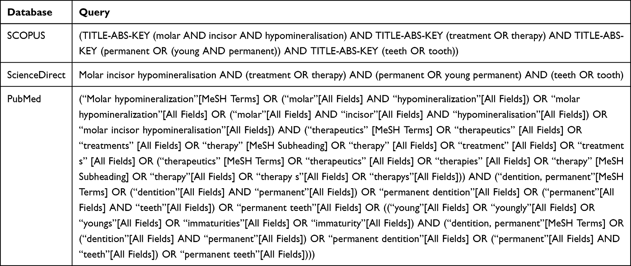 |
Table 1 Keywords |
 |
Table 2 Inclusion Criteria and Exclusion |
This scoping review aims to find out what therapy is carried out on young permanent teeth with MIH in children. The initial stage of the study involved searching for articles using three search engines with predefined keywords. A total of 150 articles were collected in the initial stage, 113 articles on Scopus, 12 articles on PubMed, and 25 articles on Science Direct. The next step was to remove duplicate articles which resulted in 139 articles remaining after 11 articles were eliminated. Then, further filtering was carried out based on the title and abstract, leaving 35 articles and eliminating 104 articles. The final process was filtering articles based on full-text, resulting in 20 articles that met the inclusion criteria and 15 articles that did not. The selection and screening process of these articles is illustrated in the PRISMA-ScR diagram (Figure 1).
 |
Figure 1 PRISMA-ScR diagram. |
Result
This scoping review limits the articles included within the last 10 years, from 2014 to 2024. The articles studied have various types of research, 3 articles are cohort studies, 2 articles are non-randomized experimental studies, 1 article is a longitudinal study and 14 articles were randomized control trials.
The majority of the research covered in the article was conducted in Brazil, with a total of 10 studies, while the rest were conducted in other countries. There were 6 studies in Turkiye, 1 in India, 1 in Germany, 1 in Egypt, and 1 study in Syria. There are 13 articles examining first molars, 5 articles examining incisor teeth, and 2 articles examining both.
Discussion
Based on the studies contained in the literature Table 3, the use of resin infiltration is highly effective for aesthetic restoration and concealment of enamel lesions for both hypomineralized and demineralized enamel.9 Aesthetic treatment of hypomineralized opacities in front teeth with resin infiltration has a positive impact on parents and children. Resin infiltration significantly reduces the color difference between opacity and healthy enamel after a minimum application time of fifteen minutes.10 Repeated application of the Icon-Etch cycle on white or cream opacities showed the best results overall, whereas repeated application of the Icon-Etch cycle on yellow/brown opacities only improved color matching with the surrounding healthy tooth enamel.11 Resin infiltration shows stable results after three to six months.11,12
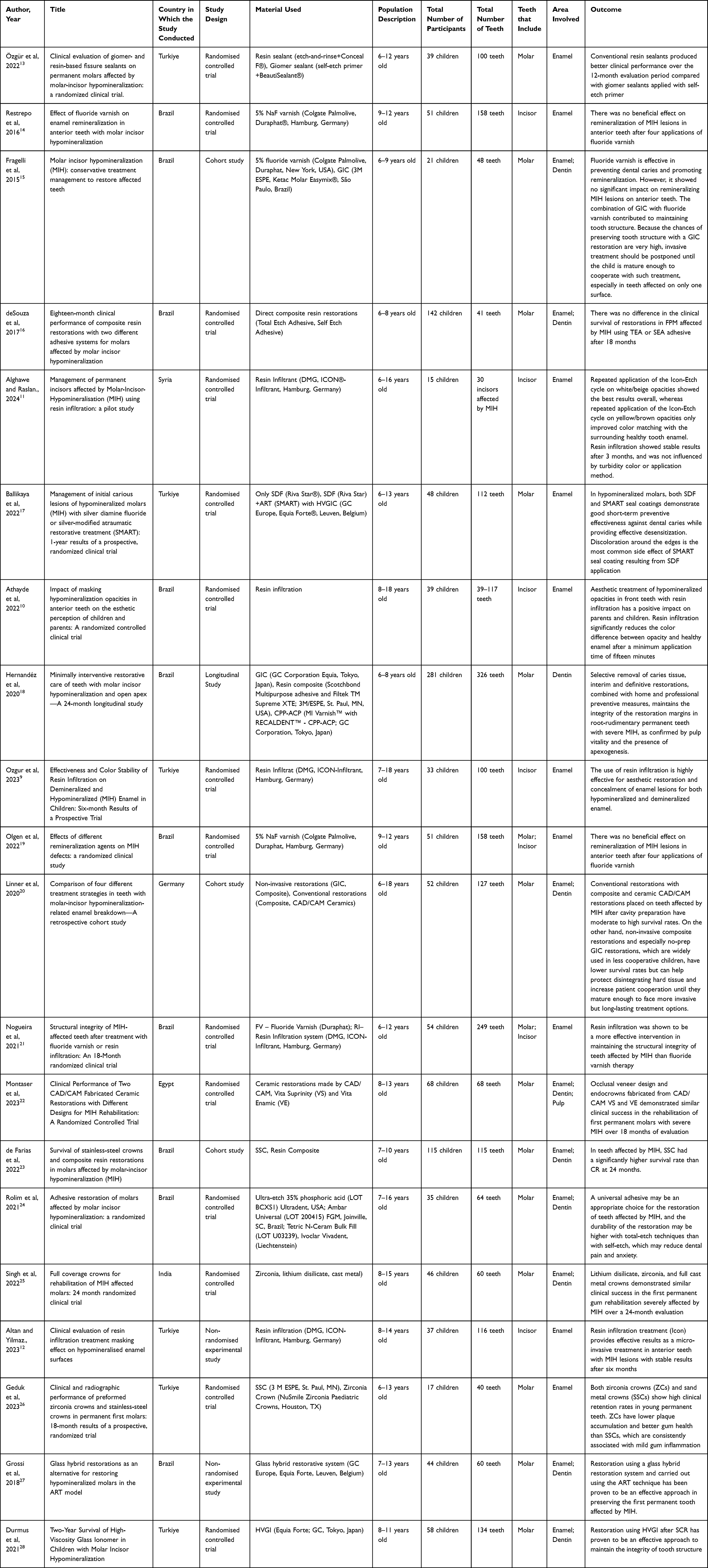 |
Table 3 Literature Presentation Data |
Individuals suffering from MIH have a higher incidence of caries and require more frequent and intensive dental care. Because of the high caries risk associated with MIH, it is important to plan a more intensive preventive approach as soon as lesions are detected in the first permanent teeth that are emerging. Fissure sealants have been suggested for mild cases of MIH in which the FPM does not show PEB (Post-Eruptive Breakdown) but experiences increased sensitivity to external stimuli such as air or water.13
In the pursuit of better glass-ionomer-like restoratives, manufacturers have developed “Giomer”, a new class of restorative materials. Giomers combine the properties of glass-ionomers (fluoride release and recharge) with those of resin composites (excellent aesthetics, easy polishability, and biocompatibility). Giomer is a tooth-colored restorative material that uses a resin base and a new class of fluoride-releasing resin materials with “Pre Reacted Glass” (PRG), offering improved color matching, reduced micro-leakage, and enhanced fluoride release compared to other resin materials. The PRG filler is created by reacting fluoride-containing acid-reactive glass with polyalkenoic acid in water before incorporating it into the resin.29
Conventional resin sealant produced better clinical performance over a twelve-month evaluation period compared with giomer sealant applied with self-etch primer. Recent clinical trial results also reported poor retention rates of giomer sealant in healthy molars after 18 months.13
Several non-invasive and minimally invasive procedures have been proposed to strengthen and protect tooth structure in hypomineralized molars, serving as preventive measures against caries.14,17 Fluoride is the most effective remineralizing agent, as it promotes the deposition of minerals similar to calcium fluoride and fluorapatite, which prevent demineralization and enhance remineralization.14
However, four applications of fluoride varnish showed no beneficial effect on the remineralization of MIH lesions on anterior teeth.14,19 Resin infiltration has proven to be more effective in maintaining the structural integrity of MIH-affected teeth than fluoride therapy.21
Silver diamine fluoride (SDF) effectively stabilizes active carious lesions due to its fluoride remineralization and silver’s antibacterial properties. SDF is non-invasive and maintains tooth structure both alone and combined with glass ionomer cement for caries treatment.17
SMART (silver-modified atraumatic restorative treatment) is a method where caries lesions are initiated using SDF and then closed/restored using standard glass ionomer cement or high viscosity glass ionomer cement (HVGIC). HVGIC adheres to tooth hard tissue through chemical and micromechanical adhesion and releases fluoride which helps reduce biofilm formation and recurrent caries. SDF and SMART seal coatings show good effectiveness in preventing caries and provide effective desensitization of molars by hypomineralization. Discoloration on the edges of the teeth is a side effect that often occurs due to the application of SDF to SMART sealants.17
Recent studies have evaluated the success of restorations in young permanent teeth using minimally invasive techniques like Selective Caries Tissue Removal (SCR) and Atraumatic Restorative Treatment (ART). Introduced in 2007, high viscosity glass ionomer (HVGI) cement serves as an alternative to composite resins for class I and II restorations in back teeth. Initially a temporary restorative material for caries-affected dentin, HVGI mineralizes over time, becoming harder and suitable for permanent restoration with minimal tissue reduction. This technique is particularly effective for managing difficult patient behavior. Using HVGI for ART over twelve months or SCR for six to twenty-four months has proven effective in maintaining the structural integrity of teeth with MIH.27,28
Non-invasive restorative procedures are essential to meet patient preferences and conceal defects in sensitive hard tissue, aiming to reduce sensitivity, protect hypomineralized tissue, ensure children’s cooperation, and enhance oral health quality of life. To achieve this, direct restorative materials like glass ionomer cement (GIC) or adhesive-applied composites are used clinically to protect and repair hypomineralized enamel.18,20
In 2015, Fragelli et al conducted a cohort study evaluating the performance of ionomer cement restorations on teeth with MIH over 12 months. The study found that molars affected by MIH and restored with GIC maintained high structural integrity, particularly in single-surface restorations. Based on these findings, along with the age and developmental stage of the affected tooth, it is recommended to postpone invasive procedures, such as complete removal of the affected area, until the child is mature enough to understand and cooperate with more complex treatments.15,18
Glass ionomer cement plays a crucial role in treating caries, serving as a fluoride reservoir and mechanical barrier against bacteria. Its key property is providing a durable seal even in challenging clinical situations. It is often used temporarily to protect dentin before a definitive restoration, such as composite installation, or as a final restorative material. Although not recommended for high-stress areas like the occlusal surfaces of hypomineralized molars, it remains effective until a definitive restoration is applied. In composite restorations, several adhesives, including the three-step total-etch adhesive and self-etch primer, have consistently shown successful enamel bonding results after eighteen months.16 Research by Roslim et al obtained results that were no different from the total ingredients -etch, self-etch, and composite resin.24
Composite resins have been frequently evaluated in clinical studies and have survival rates varying from 59% to 96%. As a direct restorative material, composite resin is recommended for best long-term performance in fully erupted molars experiencing MIH.23
MIH is a complex condition affecting one or more ridges of a tooth, sometimes involving the pulp, with limited treatment options. Severely Affected Hypomineralized Teeth (SAHT) are prone to wear and tear at the cavosurface margin, necessitating frequent replacements. Leaving seemingly healthy opaque enamel during cavity preparation can hinder proper bonding of restorative materials to hypomineralized enamel, leading to restoration failure. SAHT often requires full coverage crowns, which have a higher clinical success rate than multi-surface restorations. Stainless steel crowns are commonly used for MIH-affected teeth in children due to their quick application, strength, low cost, and immediate relief of tooth sensitivity. However, these crowns are temporary, and newer materials are needed for better treatment of severely hypomineralized teeth.25
De Farias et al conducted a study to evaluate the 24-month survival rate of restorations for the first permanent tooth affected by MIH, comparing stainless steel crowns (SSC) and composite resin (CR) crowns. Both restorative procedures used a minimally invasive approach. For SSC, no tooth preparation was needed, only unsupported structures were removed if necessary, and the crowns were installed. For CR, selective removal of hypomineralized structures was performed. There is ongoing debate in the literature about the necessity of completely removing hypomineralized structures to achieve better adhesion to healthy enamel, considering the ultrastructural changes in MIH-affected teeth. The study found that SSC had a significantly higher survival rate than CR after 24 months. SSC placement without tooth preparation is a feasible and promising temporary option for severe MIH cases using a minimally invasive approach, but strong scientific evidence is needed to evaluate its therapeutic impact.23,26
Singh et al conducted a study to evaluate and compare the clinical performance of zirconia, lithium disilicate and cast metal crowns as full coverage restorations in first permanent teeth affected by MIH (hypomineralization of the first molars). The results of the study were that lithium disilicate, zirconia, and full cast metal crowns demonstrated comparable clinical results in restoring severely affected first permanent teeth with MIH over a twenty-four-month evaluation period. Clinical success does not depend on the type of restorative material. Based on favorable clinical and radiographic performance, these crowns can also be used to restore hypomineralized teeth seen in amelogenesis imperfecta, enamel hypoplasia, and severe dental fluorosis. Full metal crowns (FMCs) provide an anatomical representation of the missing portion of the tooth, and are generally considered the gold standard in the field of dental restoration due to their superior strength, retention, durability and wear resistance.25
Montaser et al conducted a randomized clinical trial to evaluate the clinical performance of two CAD/CAM ceramic restorations, namely occlusal and endocrown veneers, for rehabilitating the first permanent tooth affected by MIH. The study found that both lithium silicate-based CAD/CAM restorations reinforced with zirconia and hybrid ceramic restorations are reliable materials for repairing severely affected first permanent teeth, with consistent performance over 18 months to three years.20,22 Both occlusal veneer and endocrown restorations have been clinically accepted for treating MIH-affected first permanent teeth. CAD/CAM technology has been increasingly used to manufacture dental prostheses, and industrially manufactured CAD/CAM ceramic blocks have been developed to enhance the mechanical properties of restorations.22
When treating hypomineralized enamel, it’s crucial to note that the defect is localized and distinct from healthy enamel, making extensive preparation unnecessary. For young permanent teeth affected by MIH, occlusal veneers are suitable within the concept of minimal intervention. Newly erupted hypomineralized teeth needing endodontic treatment face challenges such as minimal interocclusal space and inadequate coronal structure, which reduce the effectiveness of full coverage restorations. In such cases, endocrowns are appropriate as they provide retention through the pulp chamber. Preserving tooth structure is essential for maintaining biological, mechanical, and aesthetic balance, leading to changes in restoration placement protocols. New cuspal coverage restorations, such as occlusal veneers and endocrowns, can be applied without extensively reducing the axial surface or subgingival margin for severe MIH defects. Occlusal veneers offer a conservative alternative for correcting defects in the occlusal portion with a thin overlay that requires minimal retention. Endocrown restorations, or adhesive endodontic restorations, are recommended for teeth with significant coronal structural damage and pulp involvement, as they adhere to the internal part of the pulp chamber and cavity edge.22
The advantage of this scoping review is that the search is carried out on a trusted database. This review provides more articles with randomized control trials on different treatment options and the use of different materials for the therapy of teeth affected by MIH. We provide a comprehensive overview of studies on this subject that have developed over the past decade, including diverse young populations from different countries and age ranges. Most of the included studies had long follow-up periods and large samples.
A limitation of this study is that we only included studies published in English which may lead to reporting bias as other studies on this subject may have been published in other languages that were not included. The concentration of studies in Brazil (10 out of 20) may introduce a regional bias, potentially limiting the generalizability of the findings.
For the future, further research involving larger clinical trials with more rigorous methodologies is necessary to strengthen the existing evidence. Future reviews should include a balanced mix of study designs to capture a broader range of insights. Incorporating a meta-analysis could offer more quantitative insights into the effectiveness of various treatments. The development and refinement of materials and treatment techniques are also crucial to ensure the long-term effectiveness of various MIH treatment alternatives.
Conclusion
The findings of this review highlight the importance of early diagnosis and preventive strategies in managing MIH, as well as the need for personalized treatment approaches based on the severity and individual condition of the patients. Additionally, the integration of non-invasive techniques and interim therapies in treatment protocols shows potential for improving clinical outcomes. We advocate against the use of all available treatment tools to manage dental MIH, taking into account the need for a painless and effective treatment plan, as well as the dental, oral, medical and social well-being of paediatric patients.
Every clinician wishes to preserve the first permanent tooth affected by MIH because of its important role in the oral cavity and the impact if the tooth is lost. Therefore, materials that offer greater longevity and require less retreatment are highly desirable. Materials such as resin infiltration, SDF, HVGI, full metal crowns, SSC, lithium disilicate and zirconia crowns, as well as CAD/CAM ceramic restorations offer greater longevity and require less retreatment in the management of teeth affected by MIH.
Data Sharing Statement
Data sharing is not applicable to this article as no new data were created or analysed in this study.
Acknowledgments
The authors have no funding to report.
Author Contributions
All authors made a significant contribution to the work reported, whether that is in the conception, study design, execution, acquisition of data, analysis and interpretation, or in all these areas; took part in drafting, revising or critically reviewing the article; gave final approval of the version to be published; have agreed on the journal to which the article has been submitted; and agree to be accountable for all aspects of the work.
Disclosure
The authors declare no conflicts of interest in this work.
References
1. Hajdarević A, Čirgić E, Robertson A, Sabel N, Jälevik B. Treatment choice for first permanent molars affected with molar-incisor hypomineralization, in patients 7–8 years of age: a questionnaire study among Swedish general dentists, orthodontists, and pediatric dentists. Eur Arch Paediatr Dent. 2024;25(1):93–103. doi:10.1007/s40368-023-00860-9
2. Georgina-Pérez L, Ribas-Pérez D, Dehesa-Santos A, Mendoza-Mendoza A. Relationship between the TGFBR1 Gene and Molar Incisor Hypomineralization. J Pers Med. 2023;13(5):777. doi:10.3390/jpm13050777
3. Weerheijm KL, Jälevik B, Alaluusua S, Molar-Incisor Hypomineralisation. Caries Res. 2001;35(5):390–391. doi:10.1159/000047479
4. Alzahrani AY, Alamoudi NMH, Meligy OAES E. Contemporary Understanding of the Etiology and Management of Molar Incisor Hypomineralization: a Literature Review. Dent J. 2023;11(7):157. doi:10.3390/dj11070157
5. Berenstein Ajzman G, Dagon N, Iraqi R, Blumer S, Fadela S. The Prevalence of Developmental Enamel Defects in Israeli Children and Its Association with Perinatal Conditions: a Cross-Sectional Study. Children. 2023;10(5):903. doi:10.3390/children10050903
6. Bekes K, Steffen R, Krämer N. Update of the molar incisor hypomineralization: würzburg concept. Eur Arch Paediatr Dent. 2023;24(6):807–813. doi:10.1007/s40368-023-00848-5
7. Enax J, Amaechi BT, Farah R, Liu JA, Schulze Zur Wiesche E, Meyer F. Remineralization Strategies for Teeth with Molar Incisor Hypomineralization (MIH): a Literature Review. Dent J. 2023;11(3):80. doi:10.3390/dj11030080
8. Tituana-Yupangui DM, Cuaspud OJ, Montesinos-Guevara C. Rehabilitation Treatment in Pediatric Patients with Molar Incisor Hypomineralization: a Scoping Review. Pesqui Bras Odontopediatria Clin Integr. 2023;23:e220112. doi:10.1590/pboci.2023.038
9. Ozgur B, Unverdi GE, Ertan AA, Cehreli ZC. Effectiveness and Color Stability of Resin Infiltration on Demineralized and Hypomineralized (MIH) Enamel in Children: six-month Results of a Prospective Trial. Oper Dent. 2023;48(3):258–267. doi:10.2341/22-041-C
10. Athayde GDS, Reis PPGD, Jorge RC, Americano GCA, Fidalgo TKDS, Soviero VM. Impact of masking hypomineralization opacities in anterior teeth on the esthetic perception of children and parents: a randomized controlled clinical trial. J Dent. 2022;123:104168. doi:10.1016/j.jdent.2022.104168
11. Alghawe S, Raslan N. Management of permanent incisors affected by Molar-Incisor-Hypomineralisation (MIH) using resin infiltration: a pilot study. Eur Arch Paediatr Dent. 2024;25(1):105–116. doi:10.1007/s40368-024-00861-2
12. Altan H, Yilmaz RE. Clinical evaluation of resin infiltration treatment masking effect on hypomineralised enamel surfaces. BMC Oral Health. 2023;23(1). doi:10.1186/s12903-023-03140-6
13. Özgür B, Kargın ST, Ölmez MS. Clinical evaluation of giomer- and resin-based fissure sealants on permanent molars affected by molar-incisor hypomineralization: a randomized clinical trial. BMC Oral Health. 2022;22(1):275. doi:10.1186/s12903-022-02298-9
14. Restrepo M, Jeremias F, Santos-Pinto L, Cordeiro RCL, Zuanon ACC. Effect of fluoride varnish on enamel remineralization in anterior teeth with molar incisor hypomineralization. J Clin Pediatr Dent. 2016;40(3):207–210. doi:10.17796/1053-4628-40.3.207
15. Fragelli CMB, de Souza JF, Jeremias F, de Cássia Loiola Cordeiro R, Santos-Pinto L. Molar incisor hypomineralization (MIH): conservative treatment management to restore affected teeth. Braz Oral Res. 2015;29(1):1–7. doi:10.1590/1807-3107BOR-2015.vol29.0076
16. de Souza JF, Fragelli CB, Jeremias F, Paschoal MAB, Santos-Pinto L, de Cássia Loiola Cordeiro R. Eighteen-month clinical performance of composite resin restorations with two different adhesive systems for molars affected by molar incisor hypomineralization. Clin Oral Investig. 2017;21(5):1725–1733. doi:10.1007/s00784-016-1968-z
17. Ballikaya E, Ünverdi GE, Cehreli ZC. Management of initial carious lesions of hypomineralized molars (MIH) with silver diamine fluoride or silver-modified atraumatic restorative treatment (SMART): 1-year results of a prospective, randomized clinical trial. Clin Oral Investig. 2022;26(2):2197–2205. doi:10.1007/s00784-021-04236-5
18. Hernandéz P, Serrano CR, da Silva LAB, et al. Minimally interventive restorative care of teeth with molar incisor hypomineralization and open apex—A 24-month longitudinal study. Int J Paediatr Dent. 2020;30(1):4–10. doi:10.1111/ipd.12581
19. Olgen IC, Sonmez H, Bezgin T. Effects of different remineralization agents on MIH defects: a randomized clinical study. Clin Oral Investig. 2022;26(3):3227–3238. doi:10.1007/s00784-021-04305-9
20. Linner T, Khazaei Y, Bücher K, Pfisterer J, Hickel R, Kühnisch J. Comparison of four different treatment strategies in teeth with molar-incisor hypomineralization-related enamel breakdown—A retrospective cohort study. Int J Paediatr Dent. 2020;30(5):597–606. doi:10.1111/ipd.12636
21. Nogueira VKC, Mendes Soares IP, Fragelli CMB, et al. Structural integrity of MIH-affected teeth after treatment with fluoride varnish or resin infiltration: an 18-Month randomized clinical trial. J Dent. 2021:105:103570. doi:10.1016/j.jdent.2020.103570.
22. Montaser AG, Hashem SN, Ali MAS, Fathy NA, Safwat HA, Eldehna AM. Clinical Performance of Two CAD/CAM Fabricated Ceramic Restorations with Different Designs for MIH Rehabilitation: a Randomized Controlled Trial. Open Dent J. 2023;17:1 doi:10.2174/0118742106268968231101065907
23. de Farias AL, Rojas-Gualdrón DF, Mejía JD, Bussaneli DG, Santos-Pinto L, Restrepo M. Survival of stainless-steel crowns and composite resin restorations in molars affected by molar-incisor hypomineralization (MIH). Int J Paediatr Dent. 2022;32(2):240–250. doi:10.1111/ipd.12849
24. Rolim TZC, da Costa TRF, Wambier LM, et al. Adhesive restoration of molars affected by molar incisor hypomineralization: a randomized clinical trial. Clin Oral Investig. 2021;25(3):1513–1524. doi:10.1007/s00784-020-03459-2
25. Singh SK, Goyal A, Gauba K, Bhandari S, Kaur S. Full coverage crowns for rehabilitation of MIH affected molars: 24 month randomized clinical trial. Eur Arch Paediatr Dent. 2022;23(1):147–158. doi:10.1007/s40368-021-00657-8
26. Geduk N, Ozdemir M, Erbas Unverdi G, Ballikaya E, Cehreli ZC. Clinical and radiographic performance of preformed zirconia crowns and stainless-steel crowns in permanent first molars: 18-month results of a prospective, randomized trial. BMC Oral Health. 2023;23(1). doi:10.1186/s12903-023-03501-1
27. Grossi JA, Cabral RN, Ribeiro APD, Leal SC. Glass hybrid restorations as an alternative for restoring hypomineralized molars in the ART model. BMC Oral Health. 2018;18(1). doi:10.1186/s12903-018-0528-0
28. Durmus B, Sezer B, Tugcu N, Caliskan C, Bekiroglu N, Kargul B. Two-Year Survival of High-Viscosity Glass Ionomer in Children with Molar Incisor Hypomineralization. Med Princ Pract. 2021;30(1):73–79. doi:10.1159/000508676
29. Quader SMA, Alam MS, Bashar AKM, Gafur A, Al-Mansur MA. Compressive Strength, Fluoride Release and Recharge of Giomer. Updat Dent Coll J. 2012;2(2):28–37. doi:10.3329/updcj.v2i2.15533
 © 2024 The Author(s). This work is published and licensed by Dove Medical Press Limited. The
full terms of this license are available at https://www.dovepress.com/terms
and incorporate the Creative Commons Attribution
- Non Commercial (unported, 3.0) License.
By accessing the work you hereby accept the Terms. Non-commercial uses of the work are permitted
without any further permission from Dove Medical Press Limited, provided the work is properly
attributed. For permission for commercial use of this work, please see paragraphs 4.2 and 5 of our Terms.
© 2024 The Author(s). This work is published and licensed by Dove Medical Press Limited. The
full terms of this license are available at https://www.dovepress.com/terms
and incorporate the Creative Commons Attribution
- Non Commercial (unported, 3.0) License.
By accessing the work you hereby accept the Terms. Non-commercial uses of the work are permitted
without any further permission from Dove Medical Press Limited, provided the work is properly
attributed. For permission for commercial use of this work, please see paragraphs 4.2 and 5 of our Terms.