Back to Journals » Clinical, Cosmetic and Investigational Dermatology » Volume 12
Topical effects of SCA® (Cryptomphalus aspersa secretion) associated with regenerative and antioxidant ingredients on aged skin: evaluation by confocal and clinical microscopy
Authors Addor FA ![]()
Received 17 October 2018
Accepted for publication 19 December 2018
Published 14 February 2019 Volume 2019:12 Pages 133—140
DOI https://doi.org/10.2147/CCID.S191153
Checked for plagiarism Yes
Review by Single anonymous peer review
Peer reviewer comments 6
Editor who approved publication: Dr Jeffrey Weinberg
Flavia Alvim Sant’Anna Addor
Clinical Research Department, Medcin Pesquisa, Osasco, Sao Paulo, Brazil
Purpose: This was an open-label, single-center clinical study to evaluate a topical association of SCA® (Cryptomphalus aspersa secretion) with regenerative and antioxidant ingredients, according to the type and area of the face, on the improvement of signs of skin aging.
Patients and methods: One hundred and twenty female participants aged between 40 and 65 years, with facial aging complaints (presence of static, dynamics wrinkles, loss of elasticity, and skin firmness) were randomized into two groups according to the type of skin: normal–oily and normal–dry, and 40 participants were randomized for evaluation of the periocular area (with the presence of wrinkles, expression lines, and dark circles) with both types of skin. The groups received serum for normal–oily skin, a cream for normal–dry skin, and a cream for periocular and eyelid skin (eye area). All the participants were evaluated by a dermatologist and submitted to hydration evaluation by corneometry, elasticity and firmness measures complementarily, and images were collected in confocal reflectance microscopy.
Results: Topical skin treatment with the association of SCA with antioxidant ingredients (green coffee oil, olive oil, ectoine, hyaluronic acid, and peptides) was able to promote significant clinical and subjective improvement of all signs of skin aging. This improvement was presented at the epidermal level, with improved hydration levels measured by corneometry and epidermal thickness, and at the dermal level, with improvement of the firmness and elasticity parameters, measured by cutometry, from 45 days of use.
Conclusion: All the evaluated topical formulations seemed to be an effective alternative for the progressive treatment of signs of skin aging, since they demonstrate a real improvement of dermal–epidermal structure and function with high safety margin for long-term use.
Keywords: skin aging, confocal microscopy, photoaging, collagen
Introduction
The structural and functional decline of skin, characteristic of the chronological aging process, is represented by clinical signs, such as sagging and thinning of skin, where dermal loss is the main phenomenon. The extrinsic factors associated with skin aging, associated to the environment and lifestyle, aggravate and accelerate the clinical findings of intrinsic aging; in addition to the widely documented solar radiation, other factors, such as ultraviolet radiation, air pollution, injuries, smoking habits, and metabolites of ingested or inhaled substances, cause molecular changes and induce changes in the morphological and biophysical properties of skin, such as dermis’ elastosis and atrophy with reduction of collagen, leading to greater skin fragility.1
The oxidative phenomenon is a common denominator in this extrinsic and intrinsic processes, playing a crucial role in the process of skin aging; along with the physiological decline of endogenous antioxidant systems, there is an endogenous increase in oxidative stress and reduction of protective and neutralizing barriers of exogenous injuries, such as solar radiation and pollution.2
Therefore, the intrinsic and extrinsic phenomena lead to greater oxidative damage, with degradation of the dermal and epidermal components. It should be considered within the most modern conception of extrinsic aging, not only solar radiation, but also all types of skin exposures, which involve all harmful cumulative interactions to the health of the skin throughout the life.3
The clinical manifestations corresponding to these damages are hyperpigmentation, wrinkles, change in skin texture, and formation of comedones. Extrinsic aging is an intensifier of chronological aging, which is the reason for the exposed and unexposed areas to ultraviolet radiation presenting very different aspects.4
Topical formulations containing combinations of protective, antioxidant, and cellular modulating ingredients can aid in the recovery of aged skin, recovering its physiological functions, favoring its defenses, and preventing tissue damage.
SCA® (Cryptomphalus aspersa secretion) is a purified glycoprotein compound with a widely proven regenerative action since the 1970s, being successfully used in patients with radiodermatitis and chronic wounds. Later, in vitro studies demonstrated an antioxidant activity, such as scavenger of oxygen-free species, regulator of loricrin and desmoglein 1, inhibitor of plasmin, in addition to inducing the proliferation and metabolic stimulation to the fibroblasts, which also inspired its use in the approach of skin aging.5
The aim of this study was to investigate the efficacy of a formulation associating SCA topically with regenerative (hyaluronic acid, peptides) and antioxidant ingredients (ectoine, coffeeberry oil, and olive oil), acting synergistically on the pathophysiology of aging on two phenomena – intrinsic and extrinsic mechanisms – according to the type and area of the face, and may interfere positively in the improvement of its signs and symptoms.
Patients and methods
This was an open-label, single-center clinical study with noninvasive clinical and instrumental evaluations, lasting 12 weeks.
Ethical aspects
The study was conducted in accordance with the Good Clinical Practice guidelines, after approval issued by the Research Ethics Committee of the University of San Francisco on November 10, 2017, CAAEs: 79418517.9.0000.5514; 79424717.4.0000.5514; 79423017.0.0000.5514.
Population
A total of 120 female participants aged between 40 and 65 years old, with facial aging complaints (presence of static, dynamics wrinkles, loss of elasticity, and skin firmness), without facial skin disorders, were invited and subsequently selected. The following exclusion criteria were considered: gestation/lactation; use of immunosuppressant drugs, antihistamines, nonhormonal anti-inflammatories, and corticosteroids up to 30 days prior to screening; atopic or allergic history to cosmetic products; use of cosmetics, food supplements, drugs, or cosmetic procedures until 4 weeks prior to enrolment; drugs or active systemic diseases that may interfere with the study, such as endocrinopathies; and intentional sun exposure up to 2 weeks prior to study.
Methodology
This was a single-center, blind, and noncomparative clinical study to evaluate the efficacy of products intended to improve signs of facial aging, lasting 12 weeks (90±3 days), with an average temperature of 26°C and an air relative humidity of 65%.
The study protocol was initiated on December 12, 2017, and ended on March 20, 2018, at a Private Clinical Research Laboratory (Medcin Pesquisa, São Paulo, Brazil), and supported by Farmoquimica S.A. (Rio de Janeiro, Brazil).
After signing the informed consent form, all participants were evaluated by a dermatologist, being randomized into two groups, according to the clinical skin features: normal–oily and normal–dry; then 40 participants were randomized for evaluation of the periocular area (with the presence of wrinkles, expression lines, and dark circles) with both types of skin. Thus, three groups have been established, according to the vehicle and area to be treated:
- For use in monotherapy of a lipid-free serum (containing SCA, low molecular weight hyaluronic acid, epidermal peptides, coffee oil, and olive oil) for normal–oily skin.
- For use in monotherapy of an oil in water cream (containing SCA, ectoine, low molecular weight hyaluronic acid, epidermal peptides, coffee oil, and olive oil) for normal–dry skin.
- For use in monotherapy of an oil in water cream (containing SCA, ectoine, low molecular weight hyaluronic acid, epidermal peptides, coffee oil, and olive oil) for periocular and eyelid skin (eye area).
In the initial clinical evaluation, signs of hydration, softness, firmness, elasticity, lining, fine lines (static), nasolabial grooves, and expression lines (dynamics) were verified by a 5-point categorical scale, and, for group 3, periorbital hyperchromia (dark circles) was also evaluated; an investigation on the history of systemic diseases (cardiovascular, respiratory, etc.) and habits (alcoholism, smoking) was also carried out, and the body mass index was evaluated.
They were then acclimatized in a room with controlled temperature and humidity (T=20°C ±2°C and RH =50%±5%). During the conditioning period, the participants answered a questionnaire about their perception of the same signs of aging clinically evaluated, as mentioned above.
After the conditioning period, hydration measurements were performed by corneometry (Corneometer® MPA 580, Courage and Khazaka, Köln, Germany) and elasticity and firmness measures by cutometry (Cutometer® MPA 580, Courage and Khazaka) in the right temporal region; the product referring to each group was then applied at the site and a new measure was collected in the left contralateral hemi-face, which, without the application of any product, was used as control.
Images were collected with confocal reflectance microscopy (Vivascope 1500-Caliber®, Rochester, NY, USA). This equipment can be used to analyze epidermis and dermis elements.6
The group that used the product for the periorbital region alternately performed the colorimetry measurements to evaluate the coloration of the lower periorbital area of the “dark circles” (Chroma meter, Minolta)
All participants then received the product corresponding to their group, along with the method of use, which was two times daily (12/12 hours), and were instructed to visit the Center within 45 and 90 days, where new clinical and subjective (5-point categorical scale, where 1 was a significant worsening and 5 was a significant improvement) and instrumental evaluations were performed. They were instructed not to use any other product than the evaluation samples.
Statistical evaluation
For the variables analyzed in the clinical and subjective evaluation, the statistical improvement after 45 and 90 days was verified in relation to the baseline (D0) through the Wilcoxon test for paired data.
For the instrumental evaluations, the Anderson–Darling normality test was applied to identify whether the data followed a normal distribution or not. According to the normal distribution of the samples, the differences between the experimental times (D45 and D90) and the baseline (D0) were verified through the following tests:
- •t-student for paired data (parametric test more suitable for comparing two groups from dependent samples) if the normality hypothesis is not rejected
- •Wilcoxon for paired data (nonparametric test more suitable for comparing two groups from dependent samples) if normality is rejected.
The hypothesis tests used in the study have a significance level of 5% and a statistical power of 80%.
Results
Of the 120 participants who participated in the study, 108 completed 45 study days and 107 completed the study.
The average age of the sample was 51.6 years. Among the habits, 9.1% were smokers; 23.3% consume alcohol at least monthly; 8% had any cardiovascular background (high blood pressure, arrhythmia); 36.6% were overweight, and 32.5% were obese.
Safety
The only adverse reaction occurred was of mild intensity and did not require study interruption, being unrelated to the treatment used. One participant in group 2 experienced an episode of nausea and vomiting that evolved with total regression, being characterized as a food intoxication; throughout the study, no cutaneous reactions were noted clinically or reported to have occurred.
Comedogenicity and acnegenicity were evaluated, with count of comedones and possible inflammatory lesions. There was no significant increase in both parameters in any of the evaluated groups, demonstrating that none of the three evaluated products showed a comedogenic or acnegenic potential.
Efficacy
Group 1 (normal–oily skin)
Demographic data
A total of 40 participants aged between 40 and 65 years, with an average age of 52 years, were recruited. Of these, 2 participants gave up for reasons unrelated to the study and 4 had their data excluded due to low compliance/loss to follow-up to the protocol, which ended with 34 participants with valid data.
Subjective evaluation
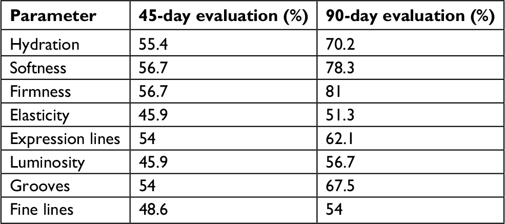
After 45 and 90 days of use, a statistically significant improvement (P<0.05) was observed in all parameters evaluated: hydration, softness, firmness, elasticity, lining, expression lines, nasolabial grooves, and fine lines (Table 1).
 | Table 1 Percentage of participants with perception of significant improvement (P<0.05; n=34) |
Clinical evaluation
There was a statistically significant improvement (P<0.05) in all clinical parameters evaluated: hydration (70.6%), softness (73.5%), firmness (26.4%), wrinkles (17.6%), fine lines (29.4%), nasolabial groove (17.6%), expression lines (20.5%), lining (55.8%), and elasticity (23.5%), after 45 days (D45) with progressive improvement at 90 days (D90) of use, where all parameters showed a statistically significant improvement, suggesting progressive effect: hydration (91.2%), softness (85.3%), firmness (41.2%), wrinkles (26.4%), fine lines (44.1%), nasolabial groove (17.6%), expression lines (26.4%), lining (73.5%), and elasticity (44.1%).
Evaluation of hydration by Corneometer
The improvement presented in a single application, after 2 hours, was 11.2%, being statistically significant. After 45 and 90 days of continuous use, improvement was 12.9% at 45 days and reached 25% after 90 days of use, both statistically significant (P<0.05).
Evaluation of elasticity and firmness by Cutometer
There was a statistically significant increase (P=0.001) in firmness measured at 45 days, of 39%, which was maintained throughout time at 90 days (27%); the elasticity improved 44%, a statistically significant result (P=0.001) at 45 days and remained throughout time, with an average of 42%, when measured at 90 days.
Confocal microscopy
The evaluated elements demonstrated progressive improvement throughout time, being statistically significant (P=0.004) at 90 days for the epidermal thickness (10.6%); organization of dermal collagen identified as an organization of the reticular collagen fibers and parallelism pattern showed an increase of 16.6% and 66.14%, respectively, at 90 days. Collagen production was also evaluated through brightness (mild/moderate/intense), showing a collagen increase of moderate to intense brightness 2.3 times after 90 days, being statistically significant (P=0.008; Figure 1).
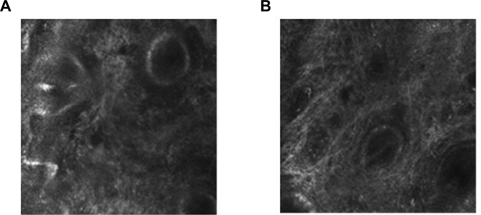 | Figure 1 Photographs of dermis with confocal reflectance microscopy. Notes: (A) Initial. (B) After 90 days of use. Observe greater parallelism of more evident collagen fibers in B. |
Group 2 (normal–dry skin)
Demographic data
A total of 40 participants aged between 42 and 65 years, with an average age of 53 years, were recruited. Of these, 2 participants gave up for reasons unrelated to the study and 1 had their data excluded due to low compliance to the protocol, which ended with 37 participants with valid data.
Subjective evaluation
After 45 and 90 days of use, there was a statistically significant improvement (P<0.05) of all evaluated parameters: hydration, softness, firmness, elasticity, lining, expression lines, grooves, and fine lines (Table 2).
 | Table 2 Percentage of participants with perception of significant improvement (P<0.05; n=37) |
Clinical evaluation
There was a statistically significant improvement (P<0.05) in all clinical parameters evaluated: hydration (72.9%), softness (54%), firmness (21.6%), wrinkles (35.1%), fine lines (43.2%), nasolabial groove (24.3%), expression lines (21.6%), and lining (54%), except for elasticity, after 45 (D45) with progressive improvement at 90 days (D90) of use, where all parameters showed a statistically significant improvement: hydration (86.4%), softness (78.3%), firmness (35.1%), wrinkles (45.9%), fine lines (54.5%), nasolabial groove (43.2%), expression lines (45.9%), lining (72.9%), and elasticity (32.4%).
Evaluation of hydration by Corneometer
The improvement presented in a single application, after 2 hours, was of the order of 8%, being statistically significant. After 45 and 90 days of continuous use, improvement was 15.7% at 45 days and reached 33.1% after 90 days of use, both of which were statistically significant (P<0.05).
Evaluation of elasticity and firmness by Cutometer
There was a statistically significant increase (P=0.001) in firmness measured at 45 days, of 30.9%, which was maintained throughout time in 90 days (28.8%); the elasticity improved 66.6%, a statistically significant result (P=0.001) at 45 days and progressive throughout time, of 76.8%, when measured at 90 days.
Confocal microscopy
The evaluated elements demonstrated progressive improvement throughout time, being statistically significant (P=0.004) at 90 days for the epidermal thickness (12.7%); the organization of dermal collagen identified as an organization of the reticular collagen fibers and parallelism pattern showed an increase of 21.4% and 7.14%, respectively, at 90 days. Collagen production was also evaluated through brightness (mild/moderate/intense), showing an increase of collagen from moderate to intense brightness three times after 90 days, being statistically significant (P=0.001; Figure 2).
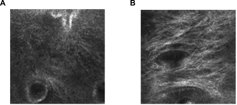 | Figure 2 Photographs of dermis with confocal reflectance microscopy. Notes: (A) Initial. (B) After 90 days of use. Observe higher brightness suggesting the presence of more collagen fibers. |
Group 3 (eye area)
Demographic data
A total of 40 participants aged between 40 and 65 years, with an average age of 50 years, were recruited. Of these, 3 participants gave up for reasons unrelated to the study and 1 had their data excluded due to low compliance to the protocol, which ended with 36 participants with valid data.
Efficacy
Subjective evaluation
After 45 and 90 days of use, a statistically significant improvement (P<0.05) was observed in all evaluated parameters: hydration, softness, firmness, elasticity, lining, expression lines, and dark circles (Table 3).
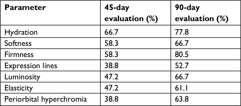 | Table 3 Percentage of participants with a perception of significant improvement (P<0.05; n=36) |
Clinical evaluation
There was a statistically significant improvement (P<0.05) in the clinical parameters – hydration (91.7%), softness (91.7%), and lining (38.8%) – after 45 days of use (D45). After 90 days (D90), there was a statistically significant improvement (P<0.05) in the parameters of hydration (94.4%), softness, (94.45%) firmness (55.5%), expression lines (52.7%), lining (69.4%), elasticity (52.7%), and periorbital hyperchromia (44.4%).
Evaluation of hydration
There was a significant increase of 22% (P<0.05) in skin hydration after 2 hours of single application. After 45 and 90 days of continuous use, improvement was 32% at 45 days and reached 45% after 90 days of use, both statistically significant (P<0.05).
Colorimetric evaluation: infraorbital area
There was a progressive increase in the parameter of skin luminosity, reaching statistical significance (P<0.05) in 90 days of use, with a 17.9% improvement in infraorbital hyperchromia.
Discussion
The results demonstrate that the topical skin treatment with the association of SCA with antioxidant ingredients (green coffee oil, olive oil, ectoine, hyaluronic acid, and peptides), peptides, squalane, and hyaluronic acid was able to promote significant clinical and subjective improvement of all signs of skin aging. This improvement was presented at the epidermal level, with improved hydration levels measured by corneometry and epidermal thickness, demonstrated in both versions of the facial formulations, and at the dermal level, with improvement of the firmness and elasticity parameters, measured by cutometry, from 45 days of use. This clinical improvement seems to be associated with better organization of collagen fibers observed in confocal microscopy, and correlated with the cutometer measurement, also demonstrated for both groups of facial products.
The product used for the eye area, which contains the mentioned association, has also been shown to promote an improvement in periorbital hyperchromia, both clinically and colorimetrically, with improved skin surface over time.
SCA is an ingredient that has already been used in formulations for aged skin, since its regenerative capacity has already been documented from studies on its positive effects on skin with radiodermatitis.5,7
In addition to demonstrating activity on the proliferation and differentiation of keratinocytes, thus improving the barrier function,8 its major action on the mechanisms of skin aging is on the fibroblasts: its direct action on the functionality of primary and senescent dermal fibroblasts is dose- and time-dependent, promoting the production of fibroblastic extracellular components, such as collagen and vimentin. Its use has also been shown to reduce the expression of aging-related markers, such as p53 and beta galactosidase, increasing cell survival after UVB radiation.9
Another study revealed other aspects of the SCA’s mechanism of action, demonstrating its ability to induce differentiation of mesenchymal cells, promote the migration of dermal fibroblasts and keratinocytes, and regulate metalloproteinases.7
SCA also exhibits cytoprotective activity against UVA and antioxidant radiation, neutralizing UV-dependent damages and acting in conjunction with endogenous systems.10
All these findings demonstrate a broad action on all findings related to skin aging, both in its intrinsic and extrinsic components.
A clinical study with a formulation containing C. aspersa and associations, conducted in 40 participants of both genders, all of them with signs of aging, for 12 weeks showed a progressive improvement of elasticity at 8 weeks (8%) and 12 weeks (39%), in addition to the improvement of luminosity in 26% and lightening in 12%), results similar to those obtained in this study.11
Improvement of periocular wrinkles showed significant improvement in another clinical study after 12 weeks of use, and the skin texture remained improved even after 2 weeks of withdrawal.10
Ectoine, also present in the formulations studied, is a molecule naturally synthesized by microorganisms under extreme environmental conditions (temperature and aridity), having the role of maintaining cellular homeostasis through the preservation of intercellular water, due to physical-chemical qualities, such as high hygroscopy and osmolarity.12
Topically applied to the skin, ectoine not only exerts a skin protective barrier function by enhancing levels of hydration, but is also able to mitigate DNA damage by ultraviolet, infrared, and visible radiations, the effect of which is well documented, but still not fully understood.13,14
By these properties, ectoine is considered an environmental “shield”, also reducing the damage caused by oxidative and inflammatory agents. Combinations of ectoine and hydroxyectoin have been studied in vitro demonstrating a protective ability against pollutant particles, stabilizing the cell membrane, and significantly reducing inflammation. Its protective activity against the damage from solar radiation associated with pollution was evidenced by the preservation of Langerhans cells and reduction of inflammatory infiltrate induced by photochemical reactions with pollutants.15,16
Low molecular weight hyaluronic acid has been used in topical formulations for its multiple mechanisms of action: through the hydration provided, acting as a hygroscopic agent, it is also able to penetrate deeper layers of epidermis and up to dermis, modulating inflammation, repairing processes, and17,18 sebogenesis regulation.19
Green coffee oil is known to be a potent antioxidant because of its composition, especially on lipid peroxidation.20
More recent studies demonstrate a stimulatory action of fibroblasts, increasing collagenase, synthesis of elastin, and glycosaminoglycans.21
In addition to increasing the expression of growth factors and aquaporin 3, thus exerting a protective and restorative barrier action,22 the formulation also contains a predominantly epidermal dipeptide (syn up®) capable of regulating epidermal proinflammatory enzymes and cytokines, reducing plasmin and proteases levels, maintaining the skin barrier and reducing signs, such as dryness and hyperreactivity.23
Finally, addition of olive oil associated to olivates provides antioxidant action against lipid peroxidation, in addition to helping to stabilize the formulation as a whole, promoting a biomimetic film-forming effect, known as “second skin”.24 This effect provides high hydration and inhibition of interleukin IL-1 alpha.25,26
The antioxidant and repairing effects of ingredients in combination can protect and even reduce the impact of environmental damage, but also of the oxidative effects caused by habit-related factors; the studied sample shows that almost 70% of the women are overweight or obese, data that coincide with the incidence found in the literature,27,28 an oxidative factor that is also capable of promoting changes related to skin aging.29–31
The study had the following limitations: low number of patients and lack of placebo control.
Conclusion
Association of ingredients with synergistic effects in topical formulations seems to be an effective alternative for the progressive treatment of signs of skin aging, since they demonstrate a real improvement of dermal–epidermal structure and function with high safety margin for long-term use.
Ingredients from formulations studied have been shown to reduce the signs of skin aging by the multiple extrinsic factors known today as ultraviolet, visible, and infrared solar radiation; pollutants; aridity conditions; or even endogenous factors, such as dietary factors. Furthermore, it is able to stimulate physiological protecting and repairing mechanisms, both epidermal and dermal. SCA combined with repairing and antioxidant ingredients has allowed for the extensive protection and restructuring of aged skin.
Acknowledgments
I acknowledge Gisele Gargantini Rezze, MD PhD, who collected and analyzed all the confocal images for this investigation. The study was conducted in MEDCIN Clinical Trials Institute and was sponsored by Farmoquímica S.A.
Disclosure
The author reports no conflicts of interest in this work. .
References
Krutmann J, Bouloc A, Sore G, Bernard BA, Passeron T. The skin aging exposome. J Dermatol Sci. 2017;85(3):152–161. | ||
Kammeyer A, Luiten RM. Oxidation events and skin aging. Ageing Res Rev. 2015;21:16–29. | ||
Addor FAS. Beyond photoaging: additional factors involved in the process of skin aging. Clin Cosmet Investig Dermatol. 2018;11:437–443. | ||
Stoebner PE, Meunier L. Photoaging of face. Ann Dermatol Venereol. 2008;1351S(1 Pt2):21–26. | ||
Brieva A, Philips N, Tejedor R, et al. Molecular basis for the regenerative properties of a secretion of the mollusk Cryptomphalus aspersa. Skin Pharmacol Physiol. 2008;21(1):15–22. | ||
Bernstein EF, Chen YQ, Kopp JB, et al. Long-term sun exposure alters the collagen of the papillary dermis. Comparison of sun-protected and photoaged skin by northern analysis, immunohistochemical staining, and confocal laser scanning microscopy. J Am Acad Dermatol. 1996;34(2 Pt1):209–218. | ||
Brieva A, Guerrero A, Pivel JP. Mecanismos bioquímicos y farmacológicos relacionados con la actividad de la secreción de Cryptomphalus aspersa (SCA) (Endocare®) en radiodermitis [Biochemical and pharmacological mechanisms related to the activity of Cryptomphalus aspersa (SCA) (Endocare®) in radiodermitis]. Dermatol Cosmet. 2001;11(2):71–75. Spanish. | ||
Cruz MC, Sanz-Rodríguez F, Zamarrón A, et al. A secretion of the mollusc Cryptomphalus aspersa promotes proliferation, migration and survival of keratinocytes and dermal fibroblasts in vitro. Int J Cosmet Sci. 2012;34(2):183–189. | ||
Espada J, Matabuena M, Salazar N, et al. Cryptomphalus aspersa mollusc eggs extract promotes migration and prevents cutaneous ageing in keratinocytes and dermal fibroblasts in vitro. Int J Cosmet Sci. 2015;37(1):41–55. | ||
Fabi SG, Cohen JL, Peterson JD, Kiripolsky MG, Goldman MP. The effects of filtrate of the secretion of the Cryptomphalus aspersa on photoaged skin. J Drugs Dermatol. 2013;12(4):453–457. | ||
Draelos ZD. The role of a natural mollusk egg-derived ingredient in facial appearance. J Drugs Dermatol. 2017;16(7):678–681. | ||
Graf R, Anzali S, Buenger J, Pfluecker F, Driller H. The multifunctional role of ectoine as a natural cell protectant. Clin Dermatol. 2008;26(4):326–333. | ||
Schröter MA, Meyer S, Hahn MB, Solomun T, Sturm H, Kunte HJ. Ectoine protects DNA from damage by ionizing radiation. Sci Rep. 2017;7(1):15272. | ||
Buenger J, Driller H. Ectoin: an effective natural substance to prevent UVA-induced premature photoaging. Skin Pharmacol Physiol. 2004;17(5):232–237. | ||
Botta C, di Giorgio C, Sabatier AS, de Méo M. Genotoxicity of visible light (400-800 nm) and photoprotection assessment of ectoin, L-ergothioneine and mannitol and four sunscreens. J Photochem Photobiol B. 2008;91(1):24–34. | ||
Schagen S, Overhagenn S, Grether-Beck S, Krutmannn J, Bilstein A. 28Extremoin®: randomized study data confirm antipollution activity. Euro Cosm. 2016;7/8:22–25. | ||
Kavasi RM, Berdiaki A, Spyridaki I, et al. HA metabolism in skin homeostasis and inflammatory disease. Food Chem Toxicol. 2017;101:128–138. | ||
Price RD, Myers S, Leigh IM, Navsaria HA. The role of hyaluronic acid in wound healing: assessment of clinical evidence. Am J Clin Dermatol. 2005;6(6):393–402. | ||
Schlesinger T, Rowland Powell C. Efficacy and safety of a low-molecular weight hyaluronic acid topical gel in the treatment of facial seborrheic dermatitis. J Clin Aesthet Dermatol. 2012;5(10):20–23. | ||
Kroyer GT, Kretschmer L, Washiittl J. Antioxidant properties of tea and coffee extracts. In: Agricultural Food Chemistry and the Consumer, Proceedings of the 5th European Conference on Food Chemistry. 1989;2:433–437. | ||
Nosari ABFL, Lima JF, Serra OA, Freitas LAP. Improved green coffee oil antioxidant activity for cosmetical purpose by spray drying microencapsulation. Revista Brasileira de Farmacognosia. 2015;25(3):307–311. | ||
Velazquez Pereda MC, Dieamant GC, Eberlin S, et al. Effect of green Coffea arabica L. seed oil on extracellular matrix components and water-channel expression in in vitro and ex vivo human skin models. J Cosmet Dermatol. 2009;8(1):56–62. | ||
Voegeli R, Rawlings AV, Doppler S, Heiland J, Schreier T. Profiling of serine protease activities in human stratum corneum and detection of a stratum corneum tryptase-like enzyme. Int J Cosmet Sci. 2007;29(3):191–200. | ||
Addy J, Oliphant T, Harper R. A botanically derived skin surface lipid mimetic based on the composition of healthy 22-year-old females. J Cosmet Sci. 2017;68(1):59–67. | ||
Lin T-K, Zhong L, Santiago J. Anti-inflammatory and skin barrier repair effects of topical application of some plant oils. Int J Mol Sci. 2017; 19(1):70. | ||
Ah M, Silva CO, Nicolai M, et al. Design and evaluation of novel topical formulation with olive oil as natural functional active. Pharm Dev Technol. 2017;3:1–12. | ||
Verkouter I, Noordam R, de Roos A, et al. Adult weight change in relation to visceral fat and liver fat at middle age: the Netherlands epidemiology of obesity study. Int J Obes. 2018;364. | ||
Figueiredo AC, Ferreira RN, Duarte MA, Coelho AF, Cabral KMAPrevalência da obesidade em mulheres tratadas de câncer de mama numa UNACOM em Juiz de Fora [Prevalence of obesity in women treated for breast cancer at UNACOM in Juiz de Fora]. Rev Bras Mastol. 2016;26(4):169–174. Spanish. | ||
Pearce LR, Atanassova N, Banton MC, et al. KSR2 mutations are associated with obesity, insulin resistance, and impaired cellular fuel oxidation. Cell. 2013;155(4):765–777. | ||
Dorjgochoo T, Gao YT, Chow WH, et al. Obesity, age, and oxidative stress in middle-aged and older women. Antioxid Redox Signal. 2011;14(12):2453–2460. | ||
Ibuki A, Kuriyama S, Toyosaki Y, et al. Aging-like physiological changes in the skin of Japanese obese diabetic patients. SAGE Open Med. 2018;6:2050312118756662. |
 © 2019 The Author(s). This work is published and licensed by Dove Medical Press Limited. The
full terms of this license are available at https://www.dovepress.com/terms
and incorporate the Creative Commons Attribution
- Non Commercial (unported, 3.0) License.
By accessing the work you hereby accept the Terms. Non-commercial uses of the work are permitted
without any further permission from Dove Medical Press Limited, provided the work is properly
attributed. For permission for commercial use of this work, please see paragraphs 4.2 and 5 of our Terms.
© 2019 The Author(s). This work is published and licensed by Dove Medical Press Limited. The
full terms of this license are available at https://www.dovepress.com/terms
and incorporate the Creative Commons Attribution
- Non Commercial (unported, 3.0) License.
By accessing the work you hereby accept the Terms. Non-commercial uses of the work are permitted
without any further permission from Dove Medical Press Limited, provided the work is properly
attributed. For permission for commercial use of this work, please see paragraphs 4.2 and 5 of our Terms.