
Back to Journals » International Journal of Nanomedicine » Volume 21
Therapeutic Potential of Silver Nanoparticles in Hepatocellular Carcinoma: From Pathogenesis to Clinical Perspectives
Authors Alawi MM  , Raahim M, Kumar M, Chin KL , Aramwit P, Mahmood S
Received 29 December 2025
Accepted for publication 27 February 2026
Published 7 March 2026 Volume 2026:21 573137
DOI https://doi.org/10.2147/IJN.S573137
Checked for plagiarism Yes
Review by Single anonymous peer review
Peer reviewer comments 3
Editor who approved publication: Prof. Dr. Anderson Oliveira Lobo
Marwa MK Alawi,1 Mohammad Raahim,2 Mohit Kumar,3 Kim Ling Chin,4 Pornanong Aramwit,5 Syed Mahmood1,5
1Department of Pharmaceutical Chemistry, Faculty of Pharmacy, Universiti Malaya, Kuala Lumpur, 50603, Malaysia; 2Department of Pharmaceutical Technology, Faculty of Pharmacy, Universiti Malaya, Kuala Lumpur, 50603, Malaysia; 3Chitkara College of Pharmacy, Chitkara University, Rajpura, Punjab, 140401, India; 4Department of Medical Microbiology, Faculty of Medicine, Universiti Malaya, Kuala Lumpur, 50603, Malaysia; 5Faculty of Pharmaceutical Sciences, Chulalongkorn University, Pathum Wan, Bangkok, 10330, Thailand
Correspondence: Syed Mahmood, Department of Pharmaceutical Technology, Faculty of Pharmacy, Universiti Malaya, Kuala Lumpur, 50603, Malaysia, Email [email protected]
Abstract: Hepatocellular carcinoma (HCC) is one of the deadliest malignancies worldwide, characterised by late-stage diagnosis, high recurrence rates, and limited responsiveness to conventional therapeutic strategies. Despite advancements in surgical interventions, locoregional therapies, and targeted drugs, survival outcomes remain unsatisfactory due to systemic toxicity, drug resistance, and tumour heterogeneity. In this context, nanotechnology-based therapeutic approaches have attracted considerable interest, particularly silver nanoparticles (AgNPs), owing to their unique physicochemical properties and multifaceted biological activity. AgNPs demonstrate distinct anticancer effects in hepatic cancer models through mechanisms involving reactive oxygen species generation, mitochondrial dysfunction, DNA damage, cell cycle arrest, and activation of apoptosis-related signalling pathways. Additionally, advances in green and biogenic synthesis methods have improved the biocompatibility and safety profile of AgNPs, enhancing their suitability for biomedical applications. Tumour-targeting strategies, including passive accumulation via the enhanced permeability and retention effect and active ligand-mediated targeting, further improve therapeutic selectivity in HCC. The emerging evidence also highlights the potential of AgNP-based systems in combination therapies and stimuli-responsive platforms to overcome therapeutic resistance. However, despite their promising anticancer activity, AgNPs exhibit dose-dependent toxicity profiles characterised by hepatic accumulation, oxidative stress induction, mitochondrial dysfunction, inflammatory cytokine release, and potential off-target organ deposition, particularly in the liver, spleen, and kidneys. Silver ion (Ag⁺) release kinetics, particle size, surface chemistry, and repeated exposure significantly influence systemic toxicity. While short-term studies often report tolerable safety margins at therapeutic concentrations, concerns regarding chronic accumulation, redox imbalance, immunotoxicity, and long-term hepatic injury remain incompletely resolved. Therefore, comprehensive pharmacokinetic evaluation and standardised toxicological profiling are essential for safe clinical translation. In this review, silver nanoparticles represent a promising yet safety-dependent nanoplatform for hepatic cancer therapy, warranting further investigation to facilitate their integration into future HCC treatment paradigms.
Keywords: hepatocellular carcinoma, silver nanoparticles, oxidative stress, apoptosis, targeted drug delivery
Introduction
Hepatocellular carcinoma (HCC) is a highly prevalent and deadly form of primary liver cancer. As such, it is one of the top health challenges worldwide due to its rising incidence and poor treatment outcomes.1–3 The majority of patients with HCC developed their disease as a result of chronic liver injury from hepatitis B and hepatitis C viruses, alcohol-associated liver injury, and metabolic syndromes such as non-alcoholic steatohepatitis, which can lead to progressive inflammation, fibrosis, and cirrhosis (Figure 1).4–6 In recent years, surgical resection, liver transplantation, locoregional treatments, and molecularly targeted therapy developments have resulted in improved treatment options for HCC patients; however, the survival of HCC patients is dismal. Several factors, including delay in diagnosis, tumour heterogeneity, and frequent recurrence, as well as systemic toxicity and development of drug resistance, significantly limit the effectiveness of conventional treatment modalities.7–9 Therefore, new therapeutic modalities must be developed to provide innovative treatment options for HCC.
 |
Figure 1 Schematic representation of multi-stage HCC development from common etiological factors. |
Recent advancements in cancer treatment have led to the development of new methods for delivering drugs into tumours, providing better access for cancer cells to absorb the drug, and allowing for targeted delivery of the drug to the tumour.10–12 Silver nanoparticles (AgNPs) are extensively studied for their anticancer activity and due to their nanosized properties and ability to target cancer selectively. AgNPs have many unique properties that make them target selective: they are nanoscale (1 nanometer = 1 billionth of a meter); they have a very large surface area; they are highly reactive; and they can be easily modified13,14 Although silver has a long history of being used as an antimicrobial agent, AgNPs are also recognised for their ability to kill a variety of cancer cells, including HCC.15 Research has shown that AgNPs can kill HCC cells through several mechanisms. Examples of those methods include the generation of free radicals that cause oxidative stress, mitochondrial disruption, enzyme inhibition, DNA damage, and activation of the intrinsic apoptosis pathway.16,17 The ability of AgNPs to have multiple mechanisms of action gives AgNPs the potential to be effective against many of the significant characteristics of cancer cells: uncontrolled growth, the ability to defeat apoptosis, and the ability to spread.15,18 These characteristics make AgNPs a potentially highly effective treatment for patients with HCC.
The use of biogenic and green synthesis methods, including natural reducing agents and plant extracts, enhances the biocompatibility and safety of AgNPs, making them much better suited for biomedical use. The structure of the liver’s unique vascular system and the enhanced permeability and retention (EPR) effect result in enhanced accumulation of AgNPs within the tumour tissue.19,20 Thus, AgNP therapy has a distinct advantage over many types of cancer therapies for HCC. The ability to functionalize AgNP with phytochemicals, targeting ligands, or chemotherapeutic drugs opens many new avenues for the development of synergistic/targeting anticancer strategies.21 This review will provide a detailed summary of the pathophysiology of hepatocellular carcinoma, an analytical critique of AgNP anticancer mechanisms, as well as discuss the latest advances from a preclinical perspective, discuss the major strategies to target HCC with AgNP-based products, and provide insights into future clinical use of these products for the treatment of HCC.
This review aims to provide a critical and integrative analysis of the therapeutic potential of silver nanoparticles in hepatocellular carcinoma. Specifically, we evaluate mechanistic evidence underlying AgNP-induced cytotoxicity, compare tumour-targeting strategies, examine biodistribution and hepatic microenvironmental interactions, and discuss translational, regulatory, and long-term safety considerations. Particular emphasis is placed on distinguishing experimental promise from clinically actionable evidence to define realistic pathways toward future application.
Epidemiology of HCC
HCC is a significant public health concern due to its increasing incidence and fatality rates globally. In 2022, HCC ranked as the third leading cause of cancer-related mortality and the sixth most common cancer, with 865,269 new liver cancer diagnoses (predominantly HCC) and 757,948 deaths, as illustrated in Figure 2A.22 Projections further indicate that, if current prevention and management efforts remain unchanged, annual incidence is expected to rise substantially approaching 1.52 million cases by 2050 (Figure 2B) with over 1 million additional cases anticipated by 2025.23 Geographical disparities are pronounced: because endemic hepatitis B virus (HBV) infection contributes to more than 50% of cases in East Asia and sub-Saharan Africa, these regions continue to show the highest incidence rates.23 In contrast, in Western countries such as the United States, HCC prevalence has tripled since the 1980s, driven largely by alcohol-related liver disease (ALD), non-alcoholic steatohepatitis (NASH), and metabolic dysfunction-associated steatotic liver disease (MASLD).24 Asia accounts for over half of global HCC cases and deaths, underscoring substantial regional concentration, which is represented in Figure 2C using the minimum split implied by the text.25 Moreover, Sex-based differences are also evident, with men affected two to four times more frequently than women, consistent with the male: female ratio scenarios depicted in Figure 2D. Late-stage diagnosis and limited access to curative treatment remain key determinants of the poor 5-year survival rate (15–20%), particularly in low- and middle-income countries with inadequate screening and treatment infrastructure. Addressing this expanding burden requires comprehensive prevention strategies, including antiviral therapy, HBV immunisation, and interventions targeting alcohol use and obesity.26
 |
Figure 2 Based summary of key HCC burden and distribution indicators. (A) Global HCC burden in 2022, new cases vs deaths. (B) Projected increase in annual HCC incidence. (C) Regional concentration of HCC cases. (D) sex distribution implied by male: female incidence ratios. |
Mechanistic Approach to Anti-Cancer Activity of Silver Nano-Biomaterials Toward Hepatic Cancer Cells
The precise molecular pathways underlying the anticancer effects of biosynthesized silver nanoparticles (AgNPs) against hepatic cancer cells remain incompletely understood. A significant cause of AgNP cytotoxicity is thought to be oxidative stress (OS), or the increase in the body’s cellular levels of reactive oxygen species (ROS) due to exposure to AgNPs and the reduction in adenosine triphosphate (ATP) production caused by AgNPs. The presence of both ROS and decreased ATP levels will impair mitochondrial function and trigger the intrinsic apoptotic pathway by disrupting the mitochondrial respiratory chain. In addition to mitochondrial dysfunction, AgNP exposure has also been shown to induce the secretion of pro-inflammatory cytokines such as IL-1, IL-6 and TNF-α.27,28 Elevated levels of these cytokines are associated with increased ROS, which can damage cellular DNA through oxidative stress and promote unstable genomes.29 The effects of AgNPs on biological systems depend on their physicochemical properties, such as size, charge, and shape. These physical characteristics will determine how AgNPs can bind to biomolecules within cells, ultimately creating a variety of consequences within cells, such as enzyme inhibition, ionic imbalance, triggering cell death signalling pathways, as well as being potentially mutagenic. There is evidence suggesting that AgNPs may affect cell membranes, allowing excess calcium to enter the cell and thereby increasing the generation of reactive oxygen species, ultimately leading to apoptotic cell death. This effect on membrane structure is thought to be due to AgNP’s ability to strongly interact with different types of thiol-containing groups (cysteines) that are found within membrane phospholipids and proteins and therefore create interactions between AgNPs and the cell membrane, ultimately causing structural damage to both.30,31 Figure 3 represents anticancer mechanisms of biogenic AgNPs. In a research study, Yassin et al demonstrated a significant increase in intracellular reactive oxygen species (ROS) levels in HepG2 cells following exposure to biogenically synthesized silver nanoparticles. This elevation in ROS was identified as a key event contributing to AgNP-induced cytotoxicity, suggesting that oxidative stress plays a central role in mediating the anticancer effects of biogenic AgNPs in liver cancer cells.32
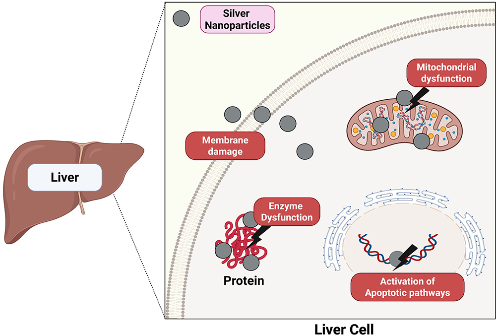 |
Figure 3 Schematic anticancer mechanisms of biogenic AgNPs. |
Mechanistic Variability Across Studies
Silver nanoparticle (AgNP)–induced cytotoxicity in hepatocellular carcinoma (HCC) models is most frequently attributed to oxidative stress, mitochondrial dysfunction, DNA damage, and activation of apoptotic signalling cascades.33–35 However, a critical evaluation of the literature reveals substantial variability in the magnitude and dominant pathways of these effects across studies. This heterogeneity appears to arise from differences in nanoparticle physicochemical properties, experimental design, and cellular context.36,37
One major determinant is particle size. Several investigations report enhanced intracellular uptake and greater reactive oxygen species (ROS) production with smaller AgNPs (<20 nm), likely due to increased surface area-to-volume ratios and higher rates of silver ion (Ag⁺) dissolution.35,38,39 In contrast, larger particles may demonstrate reduced cellular penetration but prolonged extracellular interaction, potentially shifting toxicity mechanisms toward membrane-associated damage rather than intracellular mitochondrial disruption 81,106. These size-dependent differences are not consistently controlled or standardised across studies, complicating direct mechanistic comparisons.37
Surface chemistry and coating composition further modulate biological responses. Stabilising agents such as polyvinylpyrrolidone (PVP), citrate, polyethylene glycol (PEG), or plant-derived biomolecules influence protein corona formation, cellular internalisation pathways, and immune recognition.40–42 Biogenic AgNPs are often reported to exhibit enhanced biocompatibility;43,44 however, comparative mechanistic studies directly contrasting chemically synthesised and green-synthesised nanoparticles under identical conditions remain limited.38,45 Consequently, whether observed differences in cytotoxicity reflect intrinsic nanoparticle properties or residual phytochemical contributions is not always clearly delineated.
Silver ion release kinetics represent another critical variable. While many studies attribute cytotoxicity primarily to ROS-mediated apoptosis,33,35,38 the relative contribution of particulate versus ionic silver remains debated.46,47 Increased Ag⁺ dissolution in acidic intracellular compartments (eg, lysosomes) may amplify oxidative stress and mitochondrial membrane depolarisation.33,46 Yet, few studies quantitatively measure ion release alongside mechanistic endpoints, limiting definitive conclusions regarding causality.37
Cellular model differences also contribute to inconsistent findings. Reported IC50 values for AgNPs in HCC cell lines such as HepG2 and Huh7 vary substantially, even within similar size ranges.45,48,49 These discrepancies may reflect differences in metabolic activity, p53 status, antioxidant capacity, and basal redox regulation between cell lines.50
Moreover, many investigations do not include parallel assessment in normal hepatocytes, making it difficult to distinguish cancer-selective cytotoxicity from non-specific oxidative injury.38,39
Importantly, while apoptosis is frequently reported as the predominant mode of cell death,34,35,39 some studies demonstrate concurrent necrosis, autophagy modulation, or cell-cycle arrest.38,51 The relative dominance of intrinsic (mitochondrial) versus extrinsic (death receptor–mediated) apoptotic pathways is rarely compared systematically.33,48 In addition, few studies perform time-course analyses to determine whether oxidative stress precedes mitochondrial collapse or represents a secondary consequence of cellular damage.37
Collectively, these observations indicate that AgNP-induced anticancer activity in HCC is highly context-dependent and influenced by nanoparticle design, exposure conditions, and cellular phenotype. The lack of standardised experimental frameworks and cross-study comparability currently limits definitive mechanistic generalisation.36,37 Future investigations would benefit from harmonised reporting of nanoparticle characterisation (size distribution, zeta potential, ion release kinetics), inclusion of appropriate normal-cell controls, and integration of quantitative pathway analyses to enable more robust comparative evaluation.40,47
While AgNP-induced ROS generation and apoptosis are widely reported in HCC models,33–35 it is critical to distinguish tumour-selective cytotoxicity from non-specific oxidative injury. Malignant hepatocytes often exhibit elevated basal ROS levels and dysregulated redox homeostasis, which may increase their susceptibility to further oxidative stress.16,17 However, several studies demonstrate ROS induction and cytotoxicity in non-malignant cells at higher concentrations, indicating that dose-dependent non-specific toxicity remains a concern.39,46 Importantly, only a subset of investigations includes parallel assessment in normal liver cell lines or report selectivity indices.45,52 This limitation complicates definitive claims regarding cancer-specific targeting and underscores the necessity for systematic comparative toxicity profiling using matched malignant and non-malignant hepatocyte models.
Hepatocellular Carcinoma Pathogenesis and Molecular Mechanisms
Hepatocellular carcinoma (HCC) develops through a multistep process driven by chronic liver injury, sustained inflammation, fibrogenesis, and progressive genomic instability.4–6,53 Repeated hepatocyte damage induced by viral hepatitis (HBV/HCV), alcohol-associated liver disease, and metabolic dysfunction-associated steatotic liver disease promotes compensatory regeneration within a pro-inflammatory and pro-oxidative microenvironment.4–6,53 Over time, persistent oxidative stress, telomere dysfunction, and impaired DNA repair mechanisms facilitate clonal expansion of genetically altered hepatocytes, ultimately resulting in malignant transformation.16,17,54
This chronic inflammatory–oxidative axis is central to HCC biology and has important therapeutic implications, as redox imbalance and mitochondrial vulnerability are also key mechanisms underlying silver nanoparticle (AgNP)–mediated cytotoxicity.16,17,33,37
Genetic and Epigenetic Alterations
HCC is characterised by recurrent driver mutations and epigenetic dysregulation that collectively promote proliferation, survival, and immune evasion.54 Telomerase reverse transcriptase (TERT) promoter mutations represent one of the earliest and most frequent genetic events, enabling replicative immortality.25,55 Tumour suppressor inactivation—most prominently TP53 mutation—impairs DNA damage response and apoptotic regulation.25 Activating mutations in CTNNB1 stabilise β-catenin and drive Wnt-dependent transcription of proliferative genes, including CCND1 and MYC.25 Additional alterations affecting chromatin remodelling (ARID1A/ARID2), DNA damage signalling (ATM/ATR), and Wnt regulation (AXIN1) further contribute to tumour heterogeneity.25
Beyond sequence mutations, epigenetic reprogramming plays a critical role in hepatocarcinogenesis.54 Promoter hypermethylation of tumour suppressor genes (eg, CDKN2A, RASSF1A, APC), global DNA hypomethylation, and dysregulated histone modifications reshape transcriptional landscapes.54 Non-coding RNAs also exert strong regulatory influence: miR-122 is frequently downregulated, whereas oncogenic miRNAs such as miR-21 and miR-221 are upregulated, promoting proliferation and resistance to apoptosis.56 Long non-coding RNAs, including HULC, further enhance oncogenic signalling through post-transcriptional mechanisms.56
Dysregulated Signalling Pathways
HCC progression is sustained by aberrant activation of interconnected signalling pathways.53 The Wnt/β-catenin pathway promotes transcription of genes governing cell cycle progression and stemness.25 The PI3K/AKT/mTOR axis enhances survival, angiogenesis, and metabolic adaptation, frequently activated via PTEN loss or growth factor receptor stimulation.53,57 Parallel activation of the RAS/RAF/MAPK cascade further drives mitogenic signalling and tumour growth.58
Inflammation-associated pathways also play a central role. IL-6–mediated JAK/STAT activation supports tumour proliferation and immune modulation.1,53 Developmental signalling pathways such as Hedgehog have also been implicated in tumour progression and stem-like phenotypes.53 In HBV-related HCC, viral integration events and HBx-mediated transactivation of oncogenic pathways, including NF-κB, amplify tumour-promoting transcriptional programs.2 HCV non-structural proteins promote oxidative stress, endoplasmic reticulum stress, and epigenetic alterations, thereby accelerating genomic instability.38,56,59 In metabolic liver disease–associated HCC, lipotoxicity and insulin resistance activate NF-κB and JNK signalling, further increasing reactive oxygen species (ROS) production and DNA damage.53,60
Collectively, these signalling perturbations reinforce sustained proliferative signalling, apoptotic resistance, metabolic reprogramming, and chronic oxidative stress.16,17,53
Tumour Microenvironment Role
HCC arises within a complex and chronically inflamed hepatic microenvironment characterised by immune dysregulation and stromal remodelling.53 Chronic inflammation remodels hepatic architecture and establishes a tumour microenvironment enriched with tumour-associated macrophages (TAMs), cancer-associated fibroblasts (CAFs), hepatic stellate cells, and myeloid-derived suppressor cells.61 These stromal populations promote angiogenesis, extracellular matrix remodelling, epithelial–mesenchymal transition (EMT), and immune suppression.61
Hypoxia within tumour nodules activates hypoxia-responsive pathways and upregulates pro-angiogenic mediators such as vascular endothelial growth factor (VEGF), thereby sustaining neovascularisation.53 Concurrently, immunosuppressive cytokines and checkpoint ligand expression attenuate cytotoxic T-cell activity, enabling immune evasion and tumour persistence.62
Importantly, the oxidative and inflammatory milieu of the HCC microenvironment may enhance susceptibility of malignant hepatocytes to additional redox stress. However, neighbouring non-malignant hepatocytes exposed to similar conditions may also exhibit heightened sensitivity to oxidative injury, underscoring the importance of therapeutic selectivity.16,33,46
Angiogenesis, Metastasis, and Immune Evasion
Advanced HCC is characterised by amplified angiogenesis, driven predominantly by VEGF-mediated signalling.53,63 Invasive behaviour is facilitated through EMT-associated transcriptional reprogramming and increased matrix metalloproteinase activity, enabling extracellular matrix degradation and metastatic dissemination.61
Immune escape mechanisms further sustain tumour progression. Upregulation of immune checkpoint pathways, including PD-L1 expression, recruitment of regulatory T cells, and persistent inflammatory signalling contribute to suppression of antitumour immunity.62,63 These mechanisms complicate therapeutic responses and contribute to resistance against systemic therapies.1,62
Toxicity and Safety Considerations in Normal Hepatic Cells
While AgNPs demonstrate cytotoxic efficacy in HCC models primarily through ROS amplification and mitochondrial disruption, these mechanisms are not intrinsically tumour-selective.33,37 Non-malignant hepatocytes may also undergo oxidative stress, glutathione depletion, mitochondrial membrane depolarisation, and inflammatory activation following AgNP exposure, particularly at higher concentrations or prolonged exposure durations.46,47
Experimental models report dose-dependent elevations in hepatic enzymes (ALT, AST), histopathological alterations, and redox imbalance following systemic silver nanoparticle administration.64,65 Particle size and surface chemistry critically influence hepatotoxic potential; smaller nanoparticles with increased Ag⁺ dissolution rates exhibit enhanced cellular uptake but may also increase oxidative injury risk.35,37
Although malignant hepatocytes often display elevated basal ROS levels and impaired redox buffering capacity, potentially increasing their susceptibility to additional oxidative stress,16,17 the therapeutic window remains concentration-dependent and requires careful optimisation.37,46 Chronic hepatic silver accumulation within the reticuloendothelial system further underscores the necessity for long-term safety evaluation.47,64
Therefore, precise control of nanoparticle physicochemical properties, dosing regimens, and biodistribution profiling is essential to maximise tumour-selective cytotoxicity while minimising collateral hepatocellular injury.
Tumour Targeting Strategies of Silver Nanoparticles
Passive Targeting
Passive targeting describes the tendency of nanosystems to accumulate in tumours without requiring a specific ligand–receptor interaction. It is commonly explained by the enhanced permeability and retention (EPR) effect, where newly formed tumour vessels are structurally abnormal (more permeable) and lymphatic drainage is inefficient (greater retention). Together, these features can favour the extravasation and prolonged residence of nanoparticles within tumour tissue compared with healthy organs.66
In hepatocellular carcinoma (HCC), passive targeting is influenced by the liver’s unique biology. Although HCC lesions may present leaky and heterogeneous vasculature that can support EPR-mediated entry, the liver is also a major site of nanoparticle sequestration. Circulating nanoparticles are readily opsonised and taken up by the mononuclear phagocyte system, particularly Kupffer cells, which can reduce the amount of material that remains available to reach tumour nodules. For this reason, “hepatic accumulation” should not be automatically interpreted as “tumour accumulation”, and the passive delivery efficiency can vary substantially between models and disease states.67
Particle size is a central determinant of passive targeting. In general, nanoscale materials are often designed within a size window where they can remain in circulation long enough to encounter tumour vasculature while still being small enough to extravasate through vascular defects. Very small particles (hydrodynamic diameters around a few nanometres) may be eliminated rapidly via renal filtration, limiting exposure time. Conversely, larger particles can be cleared more aggressively by liver and spleen macrophages, especially when surface properties promote protein adsorption. These competing processes create a practical design trade-off: sizes in the tens of nanometres are frequently used to balance circulation stability with the ability to permeate tumour tissue after extravasation.
For silver nanoparticles (AgNPs), size-dependent biodistribution is particularly important because silver can localise strongly in clearance organs, and its biological effects depend on particle dimensions, coating, and the extent of silver ion release. From a passive targeting standpoint, the objective is typically to maintain colloidal stability and adequate systemic residence time while enabling tumour entry and intratumoural spread. Accordingly, the selection of an AgNP size range (for example, tens of nanometres) should be justified in relation to the intended administration route, the expected HCC microenvironment, and the broader formulation strategy.36
Active Targeting
Active targeting is achieved when silver nanoparticles (AgNPs) are functionalised with ligands that recognise receptors or antigens enriched on hepatocellular carcinoma (HCC) cells (and, in some cases, tumour-associated endothelium). In practice, the ligand first enables selective binding at the tumour cell surface and then promotes receptor-mediated internalisation (commonly via clathrin- or caveolae-associated pathways), thereby increasing intracellular delivery of silver (or silver-linked payloads) compared with non-targeted AgNPs.68 Because uncoated AgNPs are highly reactive in biological fluids, active targeting is typically implemented on top of a stabilising surface “shell” (eg, citrate, PVP, PEG, protein, silica, or polymer coatings) that maintains colloidal stability and provides functional groups for ligand attachment. Common coupling strategies include Ag–S anchoring of thiolated ligands (eg, HS–PEG–ligand) and amide-bond formation via EDC/NHS chemistry on carboxylated coatings. Importantly, ligand density and spacer length are critical formulation parameters, as they can strongly influence serum protein adsorption, cellular uptake, and cytotoxicity in AgNP systems.69
Several ligand classes have been used for HCC-oriented targeting. ASGPR-directed targeting uses terminal galactose or GalNAc motifs to bind the asialoglycoprotein receptor (ASGPR) and promote hepatocyte/HCC uptake through receptor-mediated endocytosis; performance can vary with tumour differentiation and receptor expression.
Folate receptor targeting employs folic acid to enhance uptake in folate receptor-positive tumour cells; it is widely used due to the ligand’s small size and straightforward conjugation chemistry, although receptor expression remains tumour-dependent. Transferrin receptor targeting leverages overexpression of TfR/CD71 in rapidly proliferating cells to drive transferrin–TfR binding and endocytosis, thereby increasing nanoparticle internalisation.
Glycyrrhizin/glycyrrhetinic-acid family targeting uses licorice-derived ligands (commonly glycyrrhetinic acid) to bias uptake toward hepatoma cells via receptor-associated pathways described in liver-directed nanoplatforms.68 Finally, aptamer targeting applies nucleic-acid ligands that bind HCC-associated surface markers (including AFP/HepG2-related targets), enabling constructs that support therapeutic delivery and, where appropriate, diagnostic imaging (theranostics).70
Combination and Stimuli-Responsive Approaches
Recent AgNP platforms increasingly employ multifunctional designs to enhance tumour selectivity and therapeutic efficacy in HCC by combining pharmacological synergy with triggered activation in the tumour microenvironment. In combination therapy, AgNPs may be co-delivered or chemically conjugated with chemotherapeutics or biomolecules to produce synergistic cytotoxicity and increase intracellular drug exposure; for instance, AgNP–doxorubicin systems have been reported to improve intracellular accumulation and help counter multidrug resistance (MDR) mechanisms in HCC-relevant models.71
Although combination approaches involving AgNPs and chemotherapeutic agents demonstrate enhanced cytotoxicity in HCC models,71–73 most studies report percentage reductions in cell viability without formal quantitative assessment of drug–nanoparticle interaction. True mechanistic synergy requires mathematical evaluation using established pharmacodynamic models such as the Chou–Talalay combination index or Bliss independence analysis. In the absence of such modelling, observed improvements in cytotoxicity may represent additive effects rather than genuine synergistic interaction.37 Furthermore, few investigations differentiate between enhanced intracellular drug delivery, ROS amplification, and independent parallel cytotoxic mechanisms when interpreting combination outcomes. Future studies should incorporate rigorous combination modelling frameworks to substantiate claims of synergy and clarify the mechanistic basis of combined AgNP-based therapies.
To minimise off-target effects, AgNP formulations can also be engineered as internal stimuli-responsive systems that preferentially activate under tumour conditions; because tumours and intracellular vesicles (endosomes/lysosomes) are relatively acidic, pH-responsive AgNPs may accelerate Ag⁺ release and/or payload liberation in these environments, thereby improving treatment specificity compared with physiological pH.74 In addition, AgNPs can be incorporated into external stimuli-responsive systems by pairing them with photoactive agents for photothermal therapy (PTT) and/or photodynamic therapy (PDT). Upon light exposure, these platforms generate localised heat (PTT) and/or reactive oxygen species (ROS) (PDT), enabling spatially controlled tumour damage and, in some cases, image-guided and minimally invasive intervention.37
The principal active and passive targeting strategies explored for AgNP delivery in hepatocellular carcinoma are summarised in Table 1.
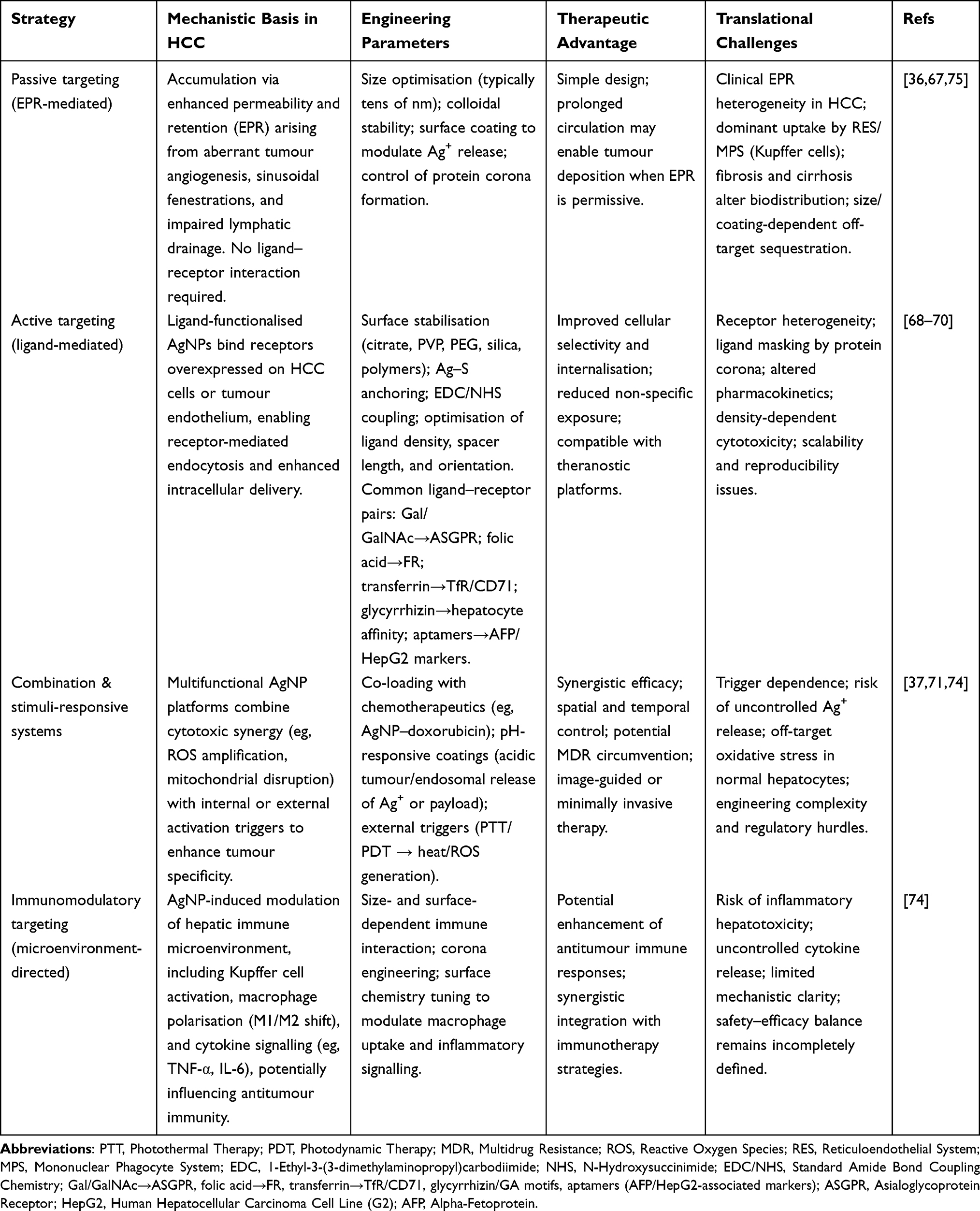 |
Table 1 Targeting Strategies for Silver Nanoparticles in Hepatocellular Carcinoma: Design Principles, Mechanisms, Advantages, and Translational Limitations |
While both passive and active targeting strategies are proposed to enhance AgNP accumulation in HCC, their relative contributions remain difficult to isolate.68 The enhanced permeability and retention (EPR) effect may facilitate tumour deposition in murine models;66 however, EPR variability in human tumours is significantly higher and often less pronounced than in preclinical systems.66 Moreover, hepatic sequestration by the reticuloendothelial system (RES), particularly Kupffer cells, can dominate nanoparticle uptake regardless of targeting ligand presence.37,64,65 Active targeting strategies—such as ligand-mediated ASGPR or transferrin receptor engagement—may enhance cellular internalisation following tumour exposure68,70 50–52, yet they do not fully overcome systemic clearance mechanisms or macrophage-mediated sequestration.37,47 Therefore, the translational impact of ligand-mediated targeting in human HCC requires cautious interpretation and further validation in clinically relevant model.
Blood-Brain and Liver Microenvironmental Interactions
The distinct liver tumour microenvironment in the case of HCC has been identified to have a pivotal role in the biodistribution and therapeutic efficacy of silver nanoparticles (AgNPs). The HCC liver microenvironment is distinguished by the presence of chronic inflammation, hypoxia, highly fibrotic tissue, and the immunosuppressive tumour microenvironment.
Liver Microenvironment and AgNP Dynamics
HCC TME is characterized and enriched with TAMs, CAFs, HSCs, and MDSCs, and all these cells support immunosuppression, angiogenesis, and ECM remodelling.33 The liver offered a natural reticuloendothelial system (RES) seclusion to a major fraction of extravasated nanoparticles, leading to natural hepatic tropism of AgNPs. It was supplemented with a prominent change associated with a permeable vascularity found within HCC, because of which there was a prominent accumulation of NPs at HCC nodules.76
AgNPs can modulate the tumour microenvironment (TME) through multiple complementary mechanisms, including reprogramming tumour-associated macrophages (TAMs) from an immunosuppressive M2 phenotype toward a pro-inflammatory M1 phenotype, which is associated with reduced vascular endothelial growth factor (VEGF) signalling and inhibition of tumour neoangiogenesis.64 They may also inhibit hepatic stellate cells (HSCs) and cancer-associated fibroblasts (CAFs), downregulate matrix metalloproteinases (MMP-2 and MMP-9), and thereby reduce extracellular matrix (ECM) stiffness, epithelial–mesenchymal transition (EMT), and metastatic potential.61,76
In addition, AgNPs can promote selective oxidative stress within hypoxic tumour regions, leveraging the heightened susceptibility of cancer cells to reactive oxygen species (ROS). Collectively, these immunoregulatory and stromal-modulating effects support the potential use of AgNPs as an adjunct strategy to promote immunogenic conversion of otherwise immunosuppressive HCC.64
Cross-Talk Between Liver and Systemic Barriers
Despite their substantial hepatocellular accumulation, nanoparticles smaller than 20 nm may still cross the blood–brain barrier (BBB), potentially via receptor-mediated transcytosis or transient disruption of tight junctions; this penetration can trigger neuroinflammation through elevated pro-inflammatory cytokines/mediators and may induce oxidative stress in neural tissues.40 In the context of targeted HCC therapy, such BBB permeability is a safety concern—particularly with repeated or frequent administrations—because it may increase the risk of neurotoxicity. To mitigate these risks, liver-specific targeting ligands (eg, glycyrrhizin or asialoglycoprotein receptor ligands) and related guidance strategies are employed to preferentially confine AgNP circulation and uptake to the hepatoportal system.26
Immunological Responses
The interactions between AgNP and immune cells present in the liver are dose-dependent:
Additionally, the activation of Kupffer cells has been shown to improve the phagocytosis of tumour debris and antigen presentation, hence stimulating anti-tumour immunity. Overstimulation can lead to overproduction of pro-inflammatory cytokines (TNF-α Inhibition of hepatic stellate cells reduces fibrogenesis and increases nanoparticle access to the fibrotic areas.25,77
Immunological and Oxidative Responses
The toxicity of AgNPs is closely related to the disruption of the redox balance through the Nrf2/Keap1 pathway. Keap1 binds Nrf2 in the cytoplasm under physiological conditions. The generated ROS from AgNPs triggers the release and translocation of Nrf2 to the nucleus to initiate the transcription of antioxidant proteins (HO1 and NQO1). This mechanism is overpowered in HCC cells at therapeutic levels of AgNPs and results in apoptosis through the mitochondrial route; however, normal cells are less affected and have the capability to balance redox. The complex interplay between AgNPs and the liver microenvironment in HCC reveals their potential in acting as both cytotoxic agents and modulators of the TME. Surface engineering and combination therapy are critical in exploiting their hepatic specificity while countering systemic challenges associated with immunotoxicity.33,46
Beyond direct cytotoxicity, AgNP exposure influences immunological dynamics within the liver microenvironment.37,64 Activation of Kupffer cells and modulation of macrophage polarisation (M1 vs M2 phenotypes) have been reported following nanoparticle uptake, where M1 macrophages are generally associated with pro-inflammatory, antitumour immune responses, while M2 phenotypes are linked to anti-inflammatory and tissue-repair functions, contributing to both antitumour immune stimulation and inflammatory injury.37,46 Cytokine release, including TNF-α and IL-6, may further amplify oxidative stress signalling and tumour–stroma interactions.46 In addition, nanoparticle–protein corona formation influences complement activation, immune recognition, and macrophage-mediated clearance.41 Recent immunological analyses highlight the dualistic nature of nanoparticle–immune interactions in hepatic disease, emphasising the need to balance immune activation with potential immunotoxicity.78 The complex interplay between AgNP-mediated redox stress and immune modulation in HCC remains incompletely characterised and warrants systematic investigation in clinically relevant models.
Pharmacokinetics and Biodistribution of Silver Nanoparticles
Therapeutics based on nanotechnology, especially silver nanoparticles (AgNPs), have become viable options for treating HCC. AgNPs have special physicochemical and biological characteristics that allow for targeted administration and increased anticancer action, such as strong surface reactivity, regulated release, and preferential accumulation in tumour tissues.36 Determining the pharmacokinetics, tumour targeting strategies, and interactions in the hepatic milieu is crucial for creating AgNP-based treatments for HCC. The ensuing segments emphasise these facets, connecting basic biopharmaceutical principles with prospective applications in liver cancer.
Absorption, Distribution, Metabolism and Excretion (ADME)
The absorption, distribution, metabolism/biotransformation, and excretion (ADME) fate of AgNPs in HCC therapy is summarised schematically in Figure 4.
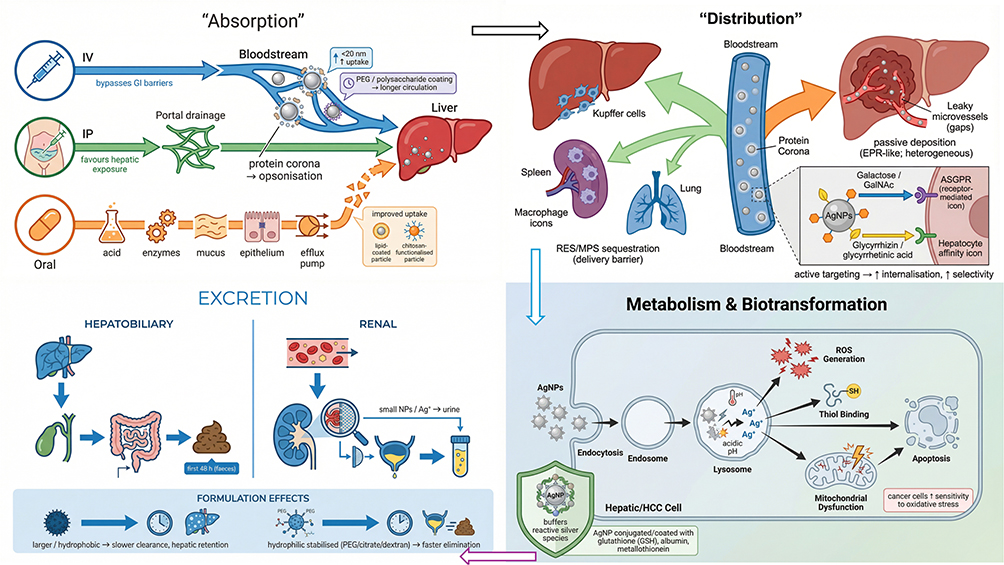 |
Figure 4 Schematic overview of the absorption, distribution, metabolism/biotransformation, and excretion (ADME) of silver nanoparticles (AgNPs) in hepatocellular carcinoma (HCC). |
Absorption
AgNP bioavailability and hepatic deposition in HCC therapy are strongly dependent on the administration route. Parenteral delivery (particularly intravenous and intraperitoneal administration) generally provides the most reliable systemic exposure by bypassing gastrointestinal degradation and first-pass absorption barriers. Following injection, nanoparticles can reach the liver through hepatic blood supply; intraperitoneal dosing may additionally favour hepatic exposure via peritoneal absorption pathways that drain into portal circulation, whereas intravenous dosing distributes through the systemic circulation with subsequent hepatic uptake.79 In contrast, oral administration typically yields low AgNP absorption because of gastric/intestinal instability, mucus barriers, limited epithelial transport, and efflux mechanisms. Nevertheless, appropriately engineered nanoformulations—such as lipid-coated systems or chitosan-functionalized AgNPs—may enhance gastrointestinal stability and mucosal interaction, thereby improving uptake relative to unmodified AgNPs.41 Once in the bloodstream, AgNPs rapidly adsorb plasma proteins to form a protein corona, which effectively defines their “biological identity” and plays a major role in opsonization, macrophage recognition, and downstream hepatic sequestration. Size is also a critical determinant of cellular uptake: smaller AgNPs (eg, <20 nm) often display higher apparent internalization in hepatocytes and HCC cells, consistent with more favorable endocytic processing and intracellular trafficking compared with larger particles.65 Finally, surface engineering—such as PEGylation or coating with natural polysaccharides—is commonly used to improve colloidal stability, prolong circulation, and reduce premature clearance by the mononuclear phagocyte system, thereby increasing the probability of tumour exposure.41
Distribution
In HCC, the reticuloendothelial/mononuclear phagocyte system (RES/MPS)—particularly Kupffer cells—is a dominant determinant of AgNP biodistribution after systemic dosing. Once in blood, AgNPs are rapidly opsonised and cleared by macrophage-rich organs, which explains why in vivo studies repeatedly report major deposition in liver and spleen, with measurable uptake also observed in the lungs depending on particle size, coating, and agglomeration state. This RES-driven sequestration is a key “delivery barrier” because it can limit the fraction of the injected dose that remains available to reach tumour nodules.37 At the same time, tumour-bearing livers can display vascular abnormalities that support passive tumour deposition. Analogous to the enhanced permeability and retention (EPR) effect, HCC-associated angiogenesis and altered sinusoidal/tumour microvasculature can increase endothelial permeability and reduce effective clearance from tumour interstitium, thereby promoting nanoparticle accumulation within tumour regions (noting that this effect is heterogeneous across models and tumour differentiation).75 To shift uptake from non-specific RES capture toward cell-selective internalisation, AgNPs can be surface-engineered with hepatocyte/HCC-targeting ligands. Examples include galactose (or GalNAc-like motifs) that bind the asialoglycoprotein receptor (ASGPR) and promote receptor-mediated endocytosis, as well as glycyrrhizin/glycyrrhetinic-acid–related ligands reported to increase affinity toward hepatocyte/hepatoma-associated recognition pathways. By leveraging receptor overexpression (or receptor accessibility) on malignant hepatocytes, these approaches can enhance intracellular delivery at the tumour site and, in principle, reduce systemic toxicity by improving therapeutic selectivity.75
Long-term accumulation of silver within hepatic tissue represents a significant safety consideration.37,64 Experimental models have demonstrated persistence of silver deposits in the liver and spleen following systemic administration, reflecting reticuloendothelial system (RES) sequestration and limited clearance.46,47 Such accumulation may promote chronic oxidative stress, mitochondrial dysfunction, and sustained inflammatory activation in hepatic tissue.33,46 Kupffer cell engagement and prolonged redox imbalance may further contribute to hepatocellular injury at higher or repeated dosing.37,64 Recent pharmacokinetic evaluations emphasise the importance of longitudinal biodistribution assessment and organ-specific toxicity profiling to clarify accumulation patterns and safety margins.64 While short-term studies often report tolerable toxicity profiles at therapeutic concentrations, comprehensive chronic exposure investigations, particularly in cirrhotic or inflamed hepatic microenvironments, remain limited.78
Metabolism and Biotransformation
Reactive oxygen species (ROS) can be generated when AgNPs undergo oxidative dissolution within hepatic cells, releasing Ag⁺ ions that bind to intracellular thiol-containing proteins, disrupt redox homeostasis, and trigger apoptotic pathways.54 This redox-mediated cytotoxicity may provide a degree of tumour selectivity because malignant cells are often more vulnerable to oxidative stress than normal hepatocytes. The lysosomal compartment is considered a primary site for AgNP biotransformation, where the acidic environment promotes Ag⁺ release and can facilitate secondary nanoparticle formation.79 To moderate toxicity while maintaining anticancer activity, AgNPs may be conjugated with biomolecules such as glutathione, metallothioneins, or albumin, which can buffer reactive silver species. Overall, leveraging these regulated intracellular processes can support safer and more selective AgNP-based approaches for HCC therapy.54
Excretion
The principal routes for AgNP elimination are hepatobiliary excretion and renal clearance. Experimental detection of silver in animal models supports that a substantial fraction of hepatic elimination occurs through biliary secretion, with silver subsequently recovered in faeces, including within the first 48 hours after dosing.56 In parallel, the kidneys can contribute to elimination, particularly for smaller nanoparticles and ionic silver species that are sufficiently small (or have been transformed into filterable forms) to pass through the glomerular filtration barrier and be excreted in urine.50 Clearance kinetics are strongly influenced by surface chemistry and colloidal properties. Coatings such as PEG, citrate, or dextran can alter protein adsorption and aggregation behaviour, thereby shifting organ sequestration and excretion. In general, larger and more hydrophobic AgNP formulations tend to show slower clearance and prolonged hepatic retention, whereas more hydrophilic, stabilised surfaces are associated with relatively improved renal handling and faster systemic elimination.47 These pharmacokinetic considerations are critical for HCC-directed applications, because they help guide formulation choices that achieve sufficient tumour silver exposure while minimising the risk of long-term liver accumulation and related safety concerns.66
Toxicity Profile of Silver Nanoparticles in HCC Context
Silver nanoparticles exhibit a complex toxicity profile that is strongly influenced by physicochemical parameters including particle size, morphology, surface charge, coating composition, and silver ion (Ag⁺) dissolution kinetics.33,37,47 In hepatic systems, AgNPs preferentially accumulate within the liver due to reticuloendothelial system (RES) sequestration, particularly by Kupffer cells.37,64 This accumulation may induce oxidative stress, mitochondrial membrane depolarisation, DNA damage, and activation of pro-inflammatory pathways, including TNF-α and IL-6 signalling.33,46
Dose-dependent hepatotoxicity has been reported in animal models, characterised by elevated liver enzymes (ALT, AST), histopathological alterations, and redox imbalance.47,65 Smaller nanoparticles (<20 nm) generally demonstrate increased cellular uptake and higher Ag⁺ release rates, which may enhance anticancer activity but also increase cytotoxic risk.35,37 Surface modifications such as PEGylation or biomolecule capping can partially mitigate rapid clearance and excessive inflammatory activation, yet long-term silver persistence in hepatic and splenic tissues remains a concern.47,64
Importantly, while malignant hepatocytes often exhibit heightened susceptibility to ROS-mediated apoptosis, non-malignant hepatocytes may also experience oxidative injury at higher exposure levels.46,52 Current evidence suggests that therapeutic windows exist; however, chronic exposure studies and cirrhotic-liver models remain limited. Therefore, careful dose optimisation, longitudinal biodistribution analysis, and standardised toxicological frameworks are essential prerequisites for clinical development of AgNP-based HCC therapeutics.
Silver Nanoparticles as a Nanotechnological Platform in HCC Therapy
Hepatocellular carcinoma (HCC) remains a leading cause of cancer-related mortality worldwide, largely due to late diagnosis, tumour heterogeneity, and limited responsiveness to systemic therapies.60,80 Conventional chemotherapeutics are frequently constrained by systemic toxicity, suboptimal tumour selectivity, and acquired resistance.7–9 These limitations have driven increasing interest in nanotechnology-based strategies capable of enhancing tumour-specific cytotoxicity while reducing off-target injury.10–12
Silver nanoparticles (AgNPs) have emerged as multifunctional nanomaterials with intrinsic anticancer properties, attributable to their high surface reactivity, tunable physicochemical characteristics, and ability to induce oxidative stress–mediated apoptosis.36,37,81,82 In HCC models, AgNPs consistently demonstrate cytotoxic effects through reactive oxygen species (ROS) generation, mitochondrial membrane depolarisation, DNA damage, cell cycle arrest, and activation of intrinsic apoptotic pathways involving Bax, caspases, and cytochrome c.33,35,48
Green and Chemically Synthesised AgNPs: Comparative Evidence
Green-synthesised AgNPs using plant or microbial extracts are frequently reported to exhibit enhanced biocompatibility due to phytochemical capping agents that improve colloidal stability and modulate protein corona formation.43,44 Reported IC50 values for plant-mediated AgNPs in HepG2 models range widely—from approximately 3–80 µg/mL depending on synthesis source, particle size, and experimental design.59,83–86 A representative example of green-synthesised AgNP characterisation and corresponding cytotoxic evaluation in HepG2 cells is illustrated in Figure 5. A comparative summary of representative AgNP-based therapeutic systems investigated in hepatocellular carcinoma models is presented in Table 2.
However, chemically synthesised AgNPs demonstrate overlapping cytotoxic ranges in comparable HCC models.35,48 Because particle size, zeta potential, and silver ion release kinetics are rarely standardised across comparative studies, it remains unclear whether observed efficacy differences are attributable to synthesis methodology or physicochemical variability.37,46 Rigorous head-to-head comparisons under controlled conditions are required to determine whether green synthesis confers measurable therapeutic advantage beyond improved formulation stability.
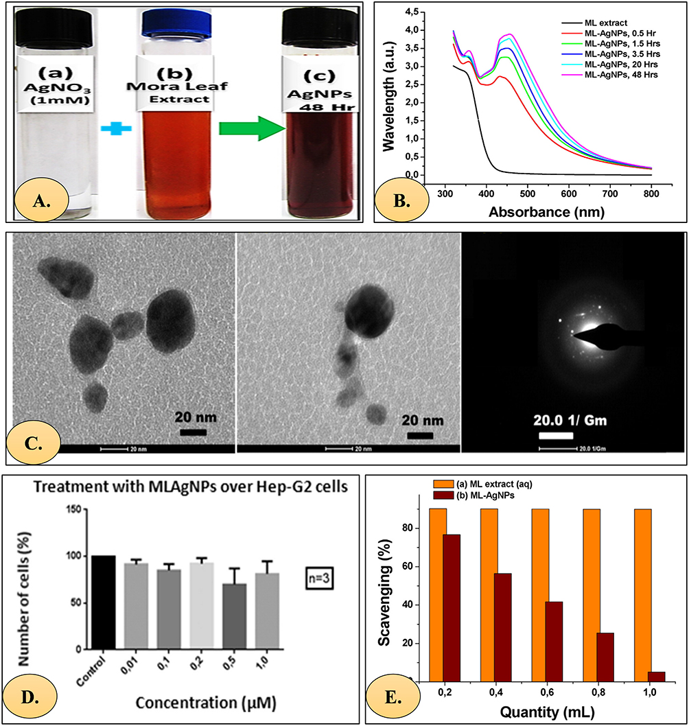 |
Figure 5 (A) 1 mM AgNO3, Andean ML extract and ML-AgNP solution after addition of extract to AgNO3 solution (B) UV–vis absorption spectra of ML-AgNPs. (C) TEM micrograph of synthesized ML-AgNPs. (D) Histogram showing cellular densities of Hep-G2 cells after exposure to different concentrations of ML-AgNPs. A control (no ML-AgNPs) is included. (E) Antioxidant activity of (a) ML extract and (b) ML-AgNPs. Reuse with permission.87 |
 |
Table 2 Therapeutic Potential of Silver Nanoparticles on the Hepatocellular Carcinoma |
Drug-Conjugated and Hybrid AgNP Systems
To enhance therapeutic specificity and potency, AgNPs have been conjugated with chemotherapeutic agents and bioactive compounds. AgNP–gemcitabine systems demonstrated enhanced apoptosis and tumour regression in chemically induced HCC rat models compared with gemcitabine alone, associated with Bax upregulation and Bcl-2 downregulation.72 The corresponding cytotoxicity profile and IC50 determination are illustrated in Figure 6. Similarly, Raptinal-loaded AgNPs showed increased caspase-3 and cytochrome c expression in murine HCC models relative to free drug administration.111
 |
Figure 6 MTT cytotoxicity assay (% of cell viability) and IC50 of different chemotherapeutic drugs on HepG2 cells (the value displayed as means ± SE (n = 3) of two independent experiments.72 |
In vitro studies further indicate that AgNP–doxorubicin constructs may improve intracellular accumulation and partially overcome multidrug resistance through modulation of ABC transporter activity.74 However, most combination studies report enhanced cytotoxicity without formal quantitative synergy modelling (eg, Chou–Talalay combination index), limiting definitive mechanistic interpretation. Distinguishing additive effects from true pharmacodynamic synergy remains an important methodological priority.
Hybrid nanoplatforms integrating AgNPs with liposomal, polymeric, or mesoporous carriers have also demonstrated improved tumour penetration and controlled silver ion release in preclinical systems.67,69 These multifunctional systems offer improved pharmacokinetic stability while preserving ROS-mediated cytotoxic mechanisms.
In vivo Evidence and Translational Constraints
Several animal studies report reduced tumour burden, improved histopathology, and modulation of inflammatory mediators following AgNP administration in chemically induced HCC models.72,112 Nevertheless, heterogeneity in dosing regimens, nanoparticle size distribution, and exposure duration complicates cross-study comparison.
Importantly, the majority of available data derive from in vitro HepG2 systems or small rodent models. Differences in tumour vasculature, immune microenvironment, and nanoparticle clearance between murine and human HCC limit direct translational extrapolation.47,66 Moreover, chronic silver accumulation within hepatic tissue and the reticuloendothelial system remains insufficiently characterised in long-term tumour-bearing models.64,65
Collectively, current evidence supports the mechanistic plausibility of AgNP-based HCC therapy but underscores the need for standardised nanoparticle characterisation, quantitative synergy assessment, longitudinal biodistribution analysis, and clinically relevant disease modelling prior to therapeutic generalisation.
Future Perspectives and Research Directions
The field of developing therapies based on silver nanoparticles (AgNPs) for hepatocellular carcinoma (HCC) is advancing rapidly; however, many obstacles remain before broad clinical use. Translation of AgNPs is hampered by low tumour selectivity, safety issues, and unpredictability in synthesis processes, despite their vigorous anticancer activity and distinctive physicochemical characteristics. Advanced targeting and intelligent carrier systems, tailored nanomedicine, integration with new biological therapies, and strong long-term clinical validation should be the main focuses of future research.113
Personalized Nanomedicine Approaches
According to the patient’s genetic, molecular, and metabolic characteristics, personalised nanomedicine seeks to customise nanoparticle-based treatment. Individualised targeting strategies are required in HCC due to molecular heterogeneity and varied receptor expression (eg, ASGPR, EGFR, or glypican-3).50 To increase selectivity and decrease off-target effects, silver nanoparticles can be engineered with ligand combinations or surface chemistries that correspond to the receptor profile of a particular tumour subtype.23 Furthermore, including biomarker-guided nanoparticle dose, such as by employing circulating AFP or microRNA signatures, can assist in tracking treatment response and making dynamic therapeutic adjustments.24 By monitoring biodistribution and treatment results in real time, emerging “theranostic” AgNP systems that combine diagnostic imaging and therapy further assist individualized management.50 The changing paradigm of personalized cancer care is in line with these precision-based approaches.
Advances in Targeted Delivery and Smart Nanocarriers
RES activity naturally causes AgNPs to accumulate in the liver; however, it remains challenging to deliver AgNPs specifically to tumours in HCC. Smart nanocarriers that respond to internal or external stimuli, such as pH, enzymes, redox gradients, or temperature, are the subject of current research.57 In acidic tumour settings, for example, pH-sensitive AgNPs preferentially release silver ions while preserving healthy hepatocytes. Drug release from enzyme-triggered coatings that use matrix metalloproteinase or hyaluronidase substrates has been developed in response to tumour-specific enzyme exposure.58 Furthermore, chemotherapeutic drugs, siRNA, or immunomodulators can be co-delivered using dual-targeting nanocarriers that combine AgNPs with polymeric or lipidic matrices. AgNP–liposome or AgNP–mesoporous silica composites are examples of hybrid nanoplatforms that have demonstrated enhanced tumour penetration, circulation time, and biostability.67 Spatiotemporal control of drug release is enabled by externally stimulus-responsive devices, such as photoactivated and magnetically guided AgNPs, which improve accuracy and reduce systemic toxicity.58,114 On-demand medication release and real-time monitoring will probably be combined in the upcoming generation of intelligent AgNP-based nanocarriers, bringing adaptive nanotherapy one step closer.
Integration with Immunotherapy and Gene Therapy
The immunosuppressive milieu in HCC limits the efficacy of genetic therapies and immune checkpoint inhibitors. When used with various techniques, silver nanoparticles may have synergistic effects. AgNPs have intrinsic immunomodulatory qualities that can boost cytotoxic T-cell activity, improve dendritic cell maturation, and improve antigen presentation.50 AgNPs and immune checkpoint inhibitors (anti-PD-1 or anti-CTLA-4) have been shown to improve tumour regression in preclinical models when administered together.57 AgNPs can also be used as gene delivery vehicles, delivering CRISPR-Cas9 components, microRNA mimics, or small interfering RNA (siRNA) straight into HCC cells.115 These platforms enhance treatment effectiveness by enabling silver-mediated apoptosis and gene silencing (eg, VEGF, Bcl-2, or MMP9) at the same time.115 AgNP integration into multimodal regimens, such as chemo-immunotherapy or immuno-gene therapy, can take advantage of complementary pathways to overcome resistance and foster long-lasting antitumour immunity.114 To prevent recurrence or progression in high-risk groups, future research should include investigating AgNP-based vaccine adjuvants that target HCC-associated antigens.53
Long-Term Safety Studies and Clinical Validation
Despite encouraging preclinical and in vitro results, AgNPs’ long-term safety and clinical validation remain incomplete. The main difficulties are chronic oxidative stress in non-tumour tissues, biodistribution heterogeneity, and possible hepatic and renal accumulation.100 To ensure reproducibility, standardization of nanoparticle manufacturing and thorough pharmacokinetic–toxicodynamic correlation studies are necessary for regulatory acceptability.64 Potential immunogenicity, chronic exposure concerns, and dose-response correlations should all be assessed in future clinical trials, including a variety of patient populations. Safer prediction of nanoparticle activity before human trials is now possible because to advance in silico toxicological models and organoid-based liver systems.40 Additionally, translation can be accelerated by synthesis that complies with Good Manufacturing Practice (GMP), biodistribution tracking using real-time imaging, and the use of safety indicators, including hepatic enzymes, oxidative stress markers, and serum Ag levels.40 Establishing standardized procedures for clinical-grade AgNP formulations will require cooperative frameworks between regulatory bodies, hepatologists, and nanotechnologists. AgNP-based treatments cannot obtain clinical integration and regulatory approval for the treatment of HCC until such thorough validation is completed.116,117
Translational Limitations of Preclinical Evidence
The majority of evidence supporting AgNP anticancer activity in HCC derives from in vitro cell culture systems or small animal models.66 However, extrapolation to human disease is constrained by interspecies differences in tumour vasculature, immune architecture, nanoparticle clearance kinetics, and hepatic microanatomy.37,64 Murine models often exhibit more pronounced and homogeneous enhanced permeability and retention (EPR) effects than human tumours, potentially leading to overestimation of nanoparticle accumulation and therapeutic efficacy.66 Furthermore, controlled laboratory dosing conditions do not fully recapitulate the complexity of chronic liver disease, viral hepatitis, and cirrhotic microenvironments that characterise clinical HCC.118 Chronic inflammation, fibrosis, and altered immune signalling may significantly influence nanoparticle biodistribution and therapeutic response.78 These translational discrepancies necessitate cautious interpretation of preclinical efficacy data and underscore the importance of clinically relevant modelling frameworks before therapeutic conclusions can be generalised to human patients.
Challenges and Innovations
Despite their potential, NP-based treatments face many obstacles. With as much as 50–70% of injected NPs accumulating in non-target liver tissue, high liver absorption by the reticuloendothelial system (RES), especially Kupffer cells, prevents tumour-specific delivery.71 Intratumoural distribution is restricted by the fibrotic HCC stroma and elevated interstitial fluid pressure, which also hinders NP penetration.119 Because of toxicity issues, including AgNP-induced hepatotoxicity through ROS production, careful dose adjustment is required.120 PEGylation, which decreases opsonisation and increases circulation half-life, and zwitterionic coatings, which reduce the development of protein corona, are examples of innovations to overcome these difficulties.42 NPs that respond to stimuli, such as redox-triggered micelles or pH-sensitive liposomes, release payloads selectively in the hypoxic or acidic (pH ~6.5) HCC milieu, increasing their effectiveness.121 In preclinical models, biomimetic coatings, such as those seen on erythrocytes or cancer cell membranes, improve tumour homing and immune evasion, improving delivery efficiency by two to three times.122 Synergistic benefits are demonstrated by nanotechnology-enabled combination therapies, such as NP co-delivery of TKIs and checkpoint inhibitors; for example, PLGA NPs co-loaded with sorafenib and anti-PD-1 antibodies decreased tumour development in HCC xenografts by 60% in comparison to monotherapies.123
Batch-to-Batch Reproducibility in Green-Synthesised AgNPs
While green and biogenic synthesis approaches are widely promoted for improved biocompatibility and reduced use of toxic reagents, reproducibility remains a significant translational challenge. Plant-mediated or microorganism-mediated reduction processes inherently depend on biological variability, including differences in phytochemical composition, seasonal variation, extraction conditions, and storage stability.43,44,82 These factors can influence nucleation kinetics, particle growth rate, final size distribution, morphology, surface chemistry, and silver ion release behaviour.13,37
Batch-to-batch heterogeneity may result in variations in zeta potential, protein corona formation, dissolution rate, and ultimately biological activity.40,47 Since AgNP cytotoxicity is highly size- and surface-dependent, even modest physicochemical deviations may significantly alter therapeutic index and toxicity profile.33,37 To improve reproducibility, several strategies have been proposed, including:
- Standardisation of extract preparation protocols and phytochemical quantification.44
- Real-time monitoring of nanoparticle nucleation using spectroscopic methods.13
- Rigorous physicochemical characterisation (eg, DLS, TEM, ICP-MS for Ag⁺ release).36,37
- And in accordance with ICH Q8–Q10 principles synthesis workflows and controlled manufacturing systems.64,66
Integration of process analytical technologies and computational optimisation approaches may further enhance batch consistency. However, until robust manufacturing standardisation is achieved, green-synthesised AgNP systems remain at an early translational stage compared with chemically controlled nanoparticle platforms.36,37
Conclusion
Hepatocellular carcinoma remains a formidable global health challenge due to its complex pathogenesis, late clinical presentation, and limited effectiveness of current therapeutic options. The growing experimental evidence discussed in this article highlights silver nanoparticles as a versatile nanotechnological platform with significant potential in hepatic cancer management. Through multiple anticancer mechanisms, while exhibiting dose-dependent redox-mediated toxicity that requires careful optimisation, silver nanoparticles effectively target key hallmarks of hepatocellular carcinoma. Advances in green and biogenic synthesis approaches have further improved their biocompatibility, reducing concerns associated with conventional chemically synthesised nanoparticles.
In addition to their intrinsic cytotoxic properties, silver nanoparticles offer substantial advantages in tumour targeting through both passive accumulation and ligand-mediated active delivery strategies. Their ability to be functionalised with chemotherapeutic agents, phytoconstituents, or stimuli-responsive coatings provides opportunities to enhance therapeutic efficacy while minimising off-target toxicity. Nevertheless, challenges related to nanoparticle stability, biodistribution, long-term safety, and reproducibility remain critical barriers to clinical translation.
Clinical translation of AgNP-based therapeutics requires rigorous compliance with Good Manufacturing Practice (GMP) standards, reproducible nanoparticle characterisation protocols, and validated long-term toxicity frameworks.37,47 Regulatory approval pathways for nanomedicine demand harmonised physicochemical characterisation, batch-to-batch consistency, and comprehensive pharmacokinetic evaluation to ensure safety and efficacy. Variability in synthesis methods—particularly between chemically reduced and green-mediated systems—currently represents a significant barrier to standardisation and regulatory approval.36,43 Emerging translational analyses emphasise the importance of integrating pharmacovigilance, immune safety assessment, and disease-specific modelling in chronic liver conditions.118 Addressing these regulatory and manufacturing challenges is essential before AgNP-based platforms can progress toward clinical implementation in HCC.
Future investigations should prioritise standardised synthesis protocols, comprehensive toxicological profiling, and well-designed in vivo and clinical studies to establish safety and therapeutic relevance. With continued interdisciplinary efforts integrating nanotechnology, molecular oncology, and translational research, silver nanoparticle–based systems may emerge as a valuable component of next-generation therapeutic strategies for hepatocellular carcinoma.
Data Sharing Statement
Data availability does not apply to this article as no new data were created or analyzed in this study.
Ethics Declarations
This article does not contain any studies with human participants performed by any of the authors.
Acknowledgments
The authors would like to express their utmost gratitude and appreciation to Universiti Malaya for funding the research project from Universiti Malaya - Grant Research Program - Research Cluster (CORG002-2025).
Disclosure
The authors declare that they have no competing financial interests, affiliations, or personal relationships that could have influenced the work reported in this manuscript.
References
1. Singal AG, Kanwal F, Llovet JM. Global trends in hepatocellular carcinoma epidemiology: implications for screening, prevention and therapy. Nat Rev Clin Oncol. 2023;20(12):864–26. doi:10.1038/S41571-023-00825-3
2. Singal AG, Llovet JM, Yarchoan M, et al. AASLD practice guidance on prevention, diagnosis, and treatment of hepatocellular carcinoma. Hepatology. 2023;78(6):1922–1965. doi:10.1097/HEP.0000000000000466
3. Chacko S, Samanta S. Hepatocellular carcinoma: a life-threatening disease. Biomed Pharmacother. 2016;84:1679–1688. doi:10.1016/j.biopha.2016.10.078
4. Suresh D, Srinivas AN, Kumar DP. Etiology of hepatocellular carcinoma: special focus on fatty liver disease. Front Oncol. 2020;10:601710. doi:10.3389/FONC.2020.601710/FULL
5. Liu SY, Tsai IT, Hsu YC. Alcohol-related liver disease: basic mechanisms and clinical perspectives. Int J Mol Sci. 2021;22(10):5170. doi:10.3390/IJMS22105170
6. Wu X, Fan X, Miyata T, et al. Recent advances in understanding of pathogenesis of alcohol-associated liver disease. Annu Rev Pathol. 2023;18(1):411–438. doi:10.1146/ANNUREV-PATHMECHDIS-031521-030435
7. Dagogo-Jack I, Shaw AT. Tumour heterogeneity and resistance to cancer therapies. Nat Rev Clin Oncol. 2018;15(2):81–94. doi:10.1038/NRCLINONC.2017.166
8. Nikolaou M, Pavlopoulou A, Georgakilas AG, Kyrodimos E. The challenge of drug resistance in cancer treatment: a current overview. Clin Exp Metastasis. 2018;35(4):309–318. doi:10.1007/S10585-018-9903-0
9. Zafar A, Khatoon S, Khan MJ, Abu J, Naeem A. Advancements and limitations in traditional anti-cancer therapies: a comprehensive review of surgery, chemotherapy, radiation therapy, and hormonal therapy. Discover Oncol. 2025;16(1). doi:10.1007/S12672-025-02198-8
10. Veselov VV, Nosyrev AE, Jicsinszky L, Alyautdin RN, Cravotto G. Targeted delivery methods for anticancer drugs. Cancers. 2022;14(3). doi:10.3390/CANCERS14030622
11. Kumari P, Ghosh B, Biswas S. Nanocarriers for cancer-targeted drug delivery. J Drug Target. 2016;24(3):179–191. doi:10.3109/1061186X.2015.1051049
12. He B, Sui X, Yu B, Wang S, Shen Y, Cong H. Recent advances in drug delivery systems for enhancing drug penetration into tumors. Drug Deliv. 2020;27(1):1474–1490. doi:10.1080/10717544.2020.1831106
13. Duman H, Eker F, Akdaşçi E, Witkowska AM, Bechelany M, Karav S. Silver nanoparticles: a comprehensive review of synthesis methods and chemical and physical properties. Nanomaterials. 2024;14(18):1527. doi:10.3390/NANO14181527
14. Beyene HD, Werkneh AA, Bezabh HK, Ambaye TG. Synthesis paradigm and applications of silver nanoparticles (AgNPs), a review. Sustainable Mater Technol. 2017;13:18. doi:10.1016/J.SUSMAT.2017.08.001
15. Takáč P, Michalková R, Čižmáriková M, Bedlovičová Z, Balážová Ľ, Takáčová G. The role of silver nanoparticles in the diagnosis and treatment of cancer: are there any perspectives for the future? Life. 2023;13(2):466. doi:10.3390/life13020466
16. Chandimali N, Bak SG, Park EH, et al. Free radicals and their impact on health and antioxidant defenses: a review. Cell Death Discovery. 2025;11(1):19. doi:10.1038/s41420-024-02278-8
17. Sadiq IZ. Free radicals and oxidative stress: signaling mechanisms, redox basis for human diseases, and cell cycle regulation. Curr Mol Med. 2023;23(1):13–35. doi:10.2174/1566524022666211222161637
18. Mikhailova EO, Mikhailova EO. Silver nanoparticles: mechanism of action and probable bio-application. J Funct Biomaterials. 2020;11(4):84. doi:10.3390/JFB11040084
19. Huang D, Sun L, Huang L, Chen Y. Nanodrug delivery systems modulate tumor vessels to increase the enhanced permeability and retention effect. J Pers Med. 2021;11(2):124. doi:10.3390/jpm11020124
20. Nel A, Ruoslahti E, Meng H. New insights into “permeability” as in the enhanced permeability and retention effect of cancer nanotherapeutics. ACS Nano. 2017;11(10):9567–9569. doi:10.1021/acsnano.7b07214
21. Chakraborty E, Sarkar D. Emerging therapies for hepatocellular carcinoma (HCC). Cancers. 2022;14(11):2798. doi:10.3390/cancers14112798
22. Chhikara BS, Parang K. Global cancer statistics 2022: the trends projection analysis. Chem Biol Lett. 2023;10(1):451.
23. Rumgay H, Arnold M, Ferlay J, et al. Global burden of primary liver cancer in 2020 and predictions to 2040. J Hepatol. 2022;77(6):1598–1606. doi:10.1016/J.JHEP.2022.08.021
24. Yang JD, Hainaut P, Gores GJ, Amadou A, Plymoth A, Roberts LR. A global view of hepatocellular carcinoma: trends, risk, prevention and management. Nat Rev Gastroenterol Hepatol. 2019;16(10):589–604. doi:10.1038/s41575-019-0186-y
25. Schulze K, Imbeaud S, Letouzé E, et al. Exome sequencing of hepatocellular carcinomas identifies new mutational signatures and potential therapeutic targets. Nat Genet. 2015;47(5):505–511. doi:10.1038/ng.3252
26. Trickler WJ, Lantz SM, Murdock RC, et al. Silver nanoparticle induced blood-brain barrier inflammation and increased permeability in primary rat brain microvessel endothelial cells. Toxicol Sci. 2010;118(1):160–170. doi:10.1093/TOXSCI/KFQ244
27. Murphy A, Casey A, Byrne G, Chambers G, Howe O. Silver nanoparticles induce pro-inflammatory gene expression and inflammasome activation in human monocytes. J Appl Toxicol. 2016;36(10):1311–1320. doi:10.1002/JAT.3315
28. Chen J, Chen X, Xuan Y, et al. Surface functionalization-dependent inflammatory potential of polystyrene nanoplastics through the activation of MAPK/NF-κB signaling pathways in macrophage Raw 264.7. Ecotoxicol Environ Saf. 2023;251:114520. doi:10.1016/J.ECOENV.2023.114520
29. Zhang XF, Shen W, Gurunathan S. Silver nanoparticle-mediated cellular responses in various cell lines: an in vitro model. Int J Mol Sci. 2016;17(10):1603. doi:10.3390/ijms17101603
30. Rodríguez-Zamora P, Cordero-Silis CA, Fabila J, et al. Interaction mechanisms and interface configuration of cysteine adsorbed on gold, silver, and copper nanoparticles. Langmuir. 2022;38(18):5418–5427. doi:10.1021/ACS.LANGMUIR.1C03298
31. Jadhav K, Deore S, Dhamecha D, et al. Phytosynthesis of silver nanoparticles: characterization, biocompatibility studies, and anticancer activity. ACS Biomater Sci Eng. 2018;4(3):892–899. doi:10.1021/ACSBIOMATERIALS.7B00707
32. Yassin AM, El-Deeb NM, Metwaly AM, El Fawal GF, Radwan MM, Hafez EE. Induction of apoptosis in human cancer cells through extrinsic and intrinsic pathways by Balanites aegyptiaca furostanol saponins and saponin-coated silvernanoparticles. Appl Biochem Biotechnol. 2017;182(4):1675–1693. doi:10.1007/s12010-017-2426-3
33. Li J, Chang X, Shang M, et al. The crosstalk between DRP1-dependent mitochondrial fission and oxidative stress triggers hepatocyte apoptosis induced by silver nanoparticles. Nanoscale. 2021;13(28):12356–12369. doi:10.1039/D1NR02153B
34. Naik J, David M. ROS mediated apoptosis and cell cycle arrest in human lung adenocarcinoma cell line by silver nanoparticles synthesized using Swietenia macrophylla seed extract. J Drug Deliv Sci Technol. 2023;80:104084. doi:10.1016/J.JDDST.2022.104084
35. Zhu B, Li Y, Lin Z, et al. Silver nanoparticles induce HePG-2 cells apoptosis through ROS-mediated signaling pathways. Nanoscale Res Lett. 2016;11(1). doi:10.1186/S11671-016-1419-4
36. Gherasim O, Puiu RA, Bîrcă AC, et al. An updated review on silver nanoparticles in biomedicine. Nanomaterials. 2020;10(11):1–44. doi:10.3390/NANO10112318
37. Kovács D, Igaz N, Gopisetty MK, et al. Cancer therapy by silver nanoparticles: fiction or reality? Int J Mol Sci. 2022;23(2):839. doi:10.3390/IJMS23020839
38. Hailan WA, Al-Anazi KM, Farah MA, et al. Reactive oxygen species-mediated cytotoxicity in liver carcinoma cells induced by silver nanoparticles biosynthesized using schinus molle extract. Nanomaterials. 2022;12(1):161. doi:10.3390/NANO12010161
39. Al-Khedhairy AA, Wahab R, Wahab R, Al-Khedhairy AA, Wahab R. Silver nanoparticles: an instantaneous solution for anticancer activity against human liver (HepG2) and breast (MCF-7) cancer cells. Metals. 2022;12(1):148. doi:10.3390/MET12010148
40. Fadeel B. Understanding the immunological interactions of engineered nanomaterials: role of the bio-Corona. Wiley Interdiscip Rev Nanomed Nanobiotechnol. 2022;14(6):e1798. doi:10.1002/WNAN.1798
41. Suk JS, Xu Q, Kim N, Hanes J, Ensign LM. PEGylation as a strategy for improving nanoparticle-based drug and gene delivery. Adv Drug Deliv Rev. 2016;99(Pt A):28–51. doi:10.1016/j.addr.2015.09.012
42. Schöttler S, Becker G, Winzen S, et al. Protein adsorption is required for stealth effect of poly(ethylene glycol)- and poly(phosphoester)-coated nanocarriers. Nat Nanotechnol. 2016;11(4):372–377. doi:10.1038/NNANO.2015.330
43. Simon S, Sibuyi NRS, Fadaka AO, et al. Biomedical applications of plant extract-synthesized silver nanoparticles. Biomedicines. 2022;10(11):2792. doi:10.3390/BIOMEDICINES10112792
44. Rónavári A, Igaz N, Adamecz DI, et al. Green silver and gold nanoparticles: biological synthesis approaches and potentials for biomedical applications. Molecules. 2021;26(4):844. doi:10.3390/MOLECULES26040844
45. Azhar NA, Abu Bakar SA, Citartan M, Ahmad NH. mRNA transcriptome profiling of human hepatocellular carcinoma cells HepG2 treated with catharanthus roseus-silver nanoparticles. World J Hepatol. 2023;15(3):393–409. doi:10.4254/wjh.v15.i3.393
46. Bobyk L, Tarantini A, Beal D, et al. Toxicity and chemical transformation of silver nanoparticles in A549 lung cells: dose-rate-dependent genotoxic impact. Environ Sci Nano. 2021;8(3):806–821. doi:10.1039/D0EN00533A
47. Lee P, Kim JK, Jo MS, et al. Biokinetics of subacutely co-inhaled same size gold and silver nanoparticles. Particle Fibre Toxicol. 2023;20(1):9. doi:10.1186/S12989-023-00515-Z
48. Ahmadian E, Dizaj SM, Rahimpour E, et al. Effect of silver nanoparticles in the induction of apoptosis on human hepatocellular carcinoma (HepG2) cell line. Mater Sci Eng C. 2018;93:465–471. doi:10.1016/j.msec.2018.08.027
49. Santana da Costa T, Rodrigues da Silva M, Jerônimo Barbosa JC, Da Silva Das Neves U, de Jesus MB, Tasic L. Biogenic silver nanoparticles’ antibacterial activity and cytotoxicity on human hepatocarcinoma cells (Huh-7). RSC Adv. 2024;14(4):2192. doi:10.1039/D3RA07733K
50. Calderaro J, Couchy G, Imbeaud S, et al. Histological subtypes of hepatocellular carcinoma are related to gene mutations and molecular tumour classification. J Hepatol. 2017;67(4):727–738. doi:10.1016/j.jhep.2017.05.014
51. Wu H, Yuan Y, Kang S, et al. Autophagy inhibitor-loaded mesoporous AgNPs@SiO2 nanoplatform for synergistically enhanced glioma radiotherapy. Sci China Mater. 2023;66(7):2902–2912. doi:10.1007/S40843-022-2395-Y
52. Alyami NM, Alyami HM, Almeer R. Using green biosynthesized kaempferol-coated sliver nanoparticles to inhibit cancer cells growth: an in vitro study using hepatocellular carcinoma (HepG2). Cancer Nanotechnol. 2022;13(1):26. doi:10.1186/S12645-022-00132-Z
53. Anstee QM, Reeves HL, Kotsiliti E, Govaere O, Heikenwalder M. From NASH to HCC: current concepts and future challenges. Nat Rev Gastroenterol Hepatol. 2019;16(7):411–428. doi:10.1038/S41575-019-0145-7
54. Liu M, Jiang L, Guan XY. The genetic and epigenetic alterations in human hepatocellular carcinoma: a recent update. Protein Cell. 2014;5(9):673–691. doi:10.1007/S13238-014-0065-9
55. Nault JC, Mallet M, Pilati C, et al. High frequency of telomerase reverse-transcriptase promoter somatic mutations in hepatocellular carcinoma and preneoplastic lesions. Nat Commun. 2013;4(1). doi:10.1038/NCOMMS3218
56. Wong CM, Tsang FH, IOL N. Non-coding RNAs in hepatocellular carcinoma: molecular functions and pathological implications. Nat Rev Gastroenterol Hepatol. 2018;15(3):137–151. doi:10.1038/NRGASTRO.2017.169
57. Bang J, Jun M, Lee S, et al. Targeting EGFR/PI3K/AKT/mTOR signaling in hepatocellular carcinoma. Pharmaceutics. 2023;15(8):2130. doi:10.3390/PHARMACEUTICS15082130
58. Delire B, Stärkel P. The Ras/MAPK pathway and hepatocarcinoma: pathogenesis and therapeutic implications. Eur J Clin Invest. 2015;45(6):609–623. doi:10.1111/ECI.12441
59. Azhar NA, Ghozali SZ, Abu Bakar SA, Lim V, Ahmad NH. Suppressing growth, migration, and invasion of human hepatocellular carcinoma HepG2 cells by Catharanthus roseus-silver nanoparticles. Toxicol In Vitro. 2020;67:104910. doi:10.1016/J.TIV.2020.104910
60. Huang DQ, Singal AG, Kono Y, Tan DJH, El-Serag HB, Loomba R. Changing global epidemiology of liver cancer from 2010 to 2019: NASH is the fastest growing cause of liver cancer. Cell Metab. 2022;34(7):969–977.e2. doi:10.1016/j.cmet.2022.05.003
61. Scheau C, Badarau IA, Costache R, et al. The role of matrix metalloproteinases in the epithelial-mesenchymal transition of hepatocellular carcinoma. Anal Cell Pathol. 2019;2019. doi:10.1155/2019/9423907
62. Hegde PS, Chen DS. Top 10 challenges in cancer immunotherapy. Immunity. 2020;52(1):17–35. doi:10.1016/J.IMMUNI.2019.12.011
63. Wang D, Ke H, Wang H, et al. Green synthesis of silver nanoparticles (CM-AgNPs) from the root of chuanminshen for improving the cytotoxicity effect in cancer cells with antibacterial and antioxidant activities. Molecules. 2024;29(23):5682. doi:10.3390/MOLECULES29235682/S1
64. Recordati C, De Maglie M, Bianchessi S, et al. Tissue distribution and acute toxicity of silver after single intravenous administration in mice: nano-specific and size-dependent effects. Particle Fibre Toxicol. 2016;13(1):12. doi:10.1186/S12989-016-0124-X
65. Salim EI, Abdel-Halim KY, El-Mahalawy ME, et al. Tissue distribution, pharmacokinetics, and effect of hematological and biochemical parameters of acute intravenous administration of silver nanoparticles in rats. Nanomaterials. 2023;14(1):29. doi:10.3390/NANO14010029
66. Sun R, Xiang J, Zhou Q, et al. The tumor EPR effect for cancer drug delivery: current status, limitations, and alternatives. Adv Drug Deliv Rev. 2022;191:114614. doi:10.1016/J.ADDR.2022.114614
67. Gomes HIO, Martins CSM, Prior JAV. Silver nanoparticles as carriers of anticancer drugs for efficient target treatment of cancer cells. Nanomaterials. 2021;11(4):964. doi:10.3390/NANO11040964
68. Zhou XQ, Li YP, Dang SS. Precision targeting in hepatocellular carcinoma: exploring ligand-receptor mediated nanotherapy. World J Hepatol. 2024;16(2):164–176. doi:10.4254/WJH.V16.I2.164
69. Montalvo-Quiros S, Aragoneses-Cazorla G, Garcia-Alcalde L, Vallet-Regí M, González B, Luque-Garcia JL. Cancer cell targeting and therapeutic delivery of silver nanoparticles by mesoporous silica nanocarriers: insights into the action mechanisms using quantitative proteomics. Nanoscale. 2019;11(10):4531–4545. doi:10.1039/C8NR07667G
70. Lai Z, Tan J, Wan R, et al. An “activatable” aptamer-based fluorescence probe for the detection of HepG2 cells. Oncol Rep. 2017;37(5):2688–2694. doi:10.3892/OR.2017.5527/ABSTRACT
71. Yang L, Gao Y, Liu J, et al. Silver-coated nanoparticles combined with doxorubicin for enhanced anticancer therapy. J Biomed Nanotechnol. 2018;14(2):312–320. doi:10.1166/JBN.2018.2481
72. Mohamed MR, Osman SA, Hassan AA, Raafat AI, Refaat MM, Fathy SA. Gemcitabine and synthesized silver nanoparticles impact on chemically induced hepatocellular carcinoma in male rats. Int J Immunopathol Pharmacol. 2024;38. doi:10.1177/03946320241263352
73. Tabbasam R, Khursid S, Ishaq Y, Farrukh SY. Synergistic cytotoxic effects of doxorubicin loaded silver, gold and zinc oxide nanoparticles in HepG2 liver cancer cells. BioNanoScience. 2024;15(1):105. doi:10.1007/S12668-024-01747-9
74. Kovács D, Szoke K, Igaz N, et al. Silver nanoparticles modulate ABC transporter activity and enhance chemotherapy in multidrug resistant cancer. Nanomedicine. 2016;12(3):601–610. doi:10.1016/J.NANO.2015.10.015
75. Fujimoto A, Furuta M, Totoki Y, et al. Whole-genome mutational landscape and characterization of noncoding and structural mutations in liver cancer. Nat Genet. 2016;48(5):500–509. doi:10.1038/ng.3547
76. Johnston HJ, Hutchison G, Christensen FM, Peters S, Hankin S, Stone V. A review of the in vivo and in vitro toxicity of silver and gold particulates: particle attributes and biological mechanisms responsible for the observed toxicity. Crit Rev Toxicol. 2010;40(4):328–346. doi:10.3109/10408440903453074
77. Grodzicki W, Dziendzikowska K, Gromadzka-Ostrowska J, et al. In vivo pro-inflammatory effects of silver nanoparticles on the colon depend on time and route of exposure. Int J Mol Sci. 2024;25(9):4879. doi:10.3390/IJMS25094879
78. Peng X, Liu Z, Luo C, et al. PSMD12 promotes hepatocellular carcinoma progression by stabilizing CDK1. Front Immunol. 2025;16:1581398. doi:10.3389/fimmu.2025.1581398
79. Morishita A, Oura K, Tadokoro T, Fujita K, Tani J, Masaki T. MicroRNAs in the pathogenesis of hepatocellular carcinoma: a review. Cancers. 2021;13(3):514. doi:10.3390/cancers13030514
80. Tan EY, Danpanichkul P, Yong JN, et al. Liver cancer in 2021: global burden of disease study. J Hepatol. 2025;82(5):851–860. doi:10.1016/J.JHEP.2024.10.031
81. Magdy G, Aboelkassim E, Abd elhaleem SM, Belal F. A comprehensive review on silver nanoparticles: synthesis approaches, characterization techniques, and recent pharmaceutical, environmental, and antimicrobial applications. Microchem J. 2024;196:109615. doi:10.1016/J.MICROC.2023.109615
82. Abbas R, Luo J, Qi X, et al. Silver nanoparticles: synthesis, structure, properties and applications. Nanomaterials. 2024;14(17). doi:10.3390/NANO14171425
83. Priya K, Vijayakumar M, Janani B. Chitosan-mediated synthesis of biogenic silver nanoparticles (AgNPs), nanoparticle characterisation and in vitro assessment of anticancer activity in human hepatocellular carcinoma HepG2 cells. Int J Biol Macromol. 2020;149:844–852. doi:10.1016/j.ijbiomac.2020.02.007
84. Vijayakumar M, Priya K, Ilavenil S, et al. Shrimp shells extracted chitin in silver nanoparticle synthesis: expanding its prophecy towards anticancer activity in human hepatocellular carcinoma HepG2 cells. Int J Biol Macromol. 2020;165(Pt A):1402–1409. doi:10.1016/j.ijbiomac.2020.10.032
85. Lu L, Zhuang Z, Fan M, et al. Green formulation of Ag nanoparticles by hibiscus rosa-sinensis: introducing a navel chemotherapeutic drug for the treatment of liver cancer. Arabian J Chem. 2022;15(2):103602. doi:10.1016/J.ARABJC.2021.103602
86. Khuda F, Gul M, Ali Khan Khalil A, et al. Biosynthesized silver nanoparticles using alnus nitida leaf extract as a potential antioxidant and anticancer agent. ACS Omega. 2023;8(33):30221–30230. doi:10.1021/ACSOMEGA.3C02928
87. Kumar B, Smita K, Seqqat R, Benalcazar K, Grijalva M, Cumbal L. In vitro evaluation of silver nanoparticles cytotoxicity on Hepatic cancer (Hep-G2) cell line and their antioxidant activity: green approach for fabrication and application. J Photochem Photobiol B. 2016;159:8–13. doi:10.1016/j.jphotobiol.2016.03.011
88. Kharwade R, Mahajan N, Ali S, Chakolkar M, Jain S, More S. Development and characterization of genistein-loaded silver nanoparticles for the induction of apoptosis on human hepatocellular carcinoma cell lines. Bionanoscience. 2025;15(2):227. doi:10.1007/s12668-025-01829-2
89. Venmani S, Kesavan MP, Ayyanaar S, Muniyappan N. Cymodocea serrulata-capped silver nanoparticles for battling human lung cancer, breast cancer, hepatic cancer: optimization by full factorial design and in vitro cytotoxicity evaluation. Heliyon. 2023;9(9):e20039. doi:10.1016/j.heliyon.2023.e20039
90. Saratale RG, Shin HS, Kumar G, Benelli G, Kim DS, Saratale GD. Exploiting antidiabetic activity of silver nanoparticles synthesized using Punica granatum leaves and anticancer potential against human liver cancer cells (HepG2). Artif Cells Nanomed Biotechnol. 2018;46(1):211–222. doi:10.1080/21691401.2017.1337031
91. Das G, Patra JK, Debnath T, Ansari A, Shin HS. Investigation of antioxidant, antibacterial, antidiabetic, and cytotoxicity potential of silver nanoparticles synthesized using the outer peel extract of ananas comosus (L.). PLoS One. 2019;14(8):e0220950. doi:10.1371/JOURNAL.PONE.0220950
92. Pandey P, Lakhanpal S, Bishoyi AK, et al. Biosynthesis of silver nanoparticles from plant extracts: a comprehensive review focused on anticancer therapy. Front Pharmacol. 2025;16:1600347. doi:10.3389/FPHAR.2025.1600347
93. Shyamalagowri S, Charles P, Manjunathan J, Kamaraj M, Anitha R, Pugazhendhi A. In vitro anticancer activity of silver nanoparticles phyto-fabricated by Hylocereus undatus peel extracts on human liver carcinoma (HepG2) cell lines. Process Biochem. 2022;116:17–25. doi:10.1016/J.PROCBIO.2022.02.022
94. Rajkumar T, Sapi A, Das G, Debnath T, Ansari A, Patra JK. Biosynthesis of silver nanoparticle using extract of Zea mays (corn flour) and investigation of its cytotoxicity effect and radical scavenging potential. J Photochem Photobiol B. 2019;193:1–7. doi:10.1016/j.jphotobiol.2019.01.008
95. Khuda F, Jamil M, Khalil AAK, et al. Assessment of antioxidant and cytotoxic potential of silver nanoparticles synthesized from root extract of Reynoutria japonica Houtt. Arabian J Chem. 2022;15(12):104327. doi:10.1016/J.ARABJC.2022.104327
96. El-Hawary AS, Ibrahim OM, Kalaba MH, El-Sehrawy MH, Ismail MKA. Limosilactobacillus fermentum-derived silver nanoparticles: biosynthesis, optimization, and biological activities. Biomass Convers Biorefin. 2025;15(7):9999–10013. doi:10.1007/S13399-024-05784-Y/FIGURES/9
97. Ahamad I, Nadeem M, Rizvi MMA, Fatma T. Bio-fabricated silver nanoparticles: therapeutic evaluation as a potential nanodrug against cervical and liver cancer cells. Discover Nano. 2025;20(1):47. doi:10.1186/s11671-025-04212-y
98. Hamouda RA, Aljohani ES, Aljohani ES, Hamouda RA, Aljohani ES. Assessment of silver nanoparticles derived from brown algae sargassum vulgare: insight into antioxidants, anticancer, antibacterial and hepatoprotective effect. Mar Drugs. 2024;22(4):154. doi:10.3390/MD22040154
99. Raj Meena P, Pratap Singh A, Kumar Tejavath K. Biosynthesis of silver nanoparticles using cucumis prophetarum aqueous leaf extract and their antibacterial and antiproliferative activity against cancer cell lines. ACS Omega. 2020;5:16. doi:10.1021/acsomega.0c00155
100. Oves M, Ahmar Rauf M, Aslam M, et al. Green synthesis of silver nanoparticles by Conocarpus Lancifolius plant extract and their antimicrobial and anticancer activities. Saudi J Biol Sci. 2022;29(1):460–471. doi:10.1016/J.SJBS.2021.09.007
101. Singh A, Dar MY, Joshi B, Sharma B, Shrivastava S, Shukla S. Phytofabrication of silver nanoparticles: novel drug to overcome hepatocellular ailments. Toxicol Rep. 2018;5:333–342. doi:10.1016/j.toxrep.2018.02.013
102. Gul H, Khan FS, Afzal U, et al. Rumex hastatus derived silver nanoparticles development and their potential applications as hepatic-protection agent along with antimicrobial activity. J King Saud Univ Sci. 2021;33(7):101587. doi:10.1016/J.JKSUS.2021.101587
103. Veeramani S, Shanmugam K. Biosynthesis and characterization of silver nanoparticles coated with metabolites extracted from clerodendron phlomoides with its biochemical studies in liver tissue. Proc Anticancer Res. 2023;7(4):1–19. doi:10.26689/par.v7i4.5049
104. Alsayed MF, Alodaini HA, Aziz IM, et al. Silver nanoparticles synthesized using aerial part of Achillea fragrantissima and evaluation of their bioactivities. Sci Rep. 2024;14(1):24703. doi:10.1038/s41598-024-75558-z
105. Javid S, Dilshad E. Artemisia carvifolia buch silver nanoparticles downregulate the Rap2A gene in liver cancer. Sci Rep. 2023;13(1):21553. doi:10.1038/s41598-023-48946-0
106. Khaled AM, Othman MS, Obeidat ST, et al. Green-synthesized silver and selenium nanoparticles using berberine: a comparative assessment of in vitro anticancer potential on human hepatocellular carcinoma cell line (HepG2). Cells. 2024;13(3):287. doi:10.3390/CELLS13030287
107. Yu R, Chen J, Yu R, et al. Arctigenin from Saussurea medusa Maxim. Targets the PI3K/AKT pathway to inhibit hepatocellular carcinoma proliferation and induces apoptosis. Nutrients. 2025;17(19):3151. doi:10.3390/NU17193151
108. Rîmbu MC, Popescu L, Mihăilă M, et al. Synergistic effects of green nanoparticles on antitumor drug efficacy in hepatocellular cancer. Biomedicines. 2025;13(3):641. doi:10.3390/BIOMEDICINES13030641/S1
109. Gupta S, Jasrotia S, Kandwal A, Rawat R, Purohit MC. Green synthesis of silver nitrate nanoparticles using L. of Pyracantha crenulata and its anticancer activity on liver cancer. J Mountain Res. 2023;18(2):273–283. doi:10.51220/jmr.v18i2.29
110. Narayanan M, Divya S, Natarajan D, et al. Green synthesis of silver nanoparticles from aqueous extract of Ctenolepis garcini L. and assess their possible biological applications. Process Biochem. 2021;107:91–99. doi:10.1016/J.PROCBIO.2021.05.008
111. Taha H, Elfar N, Haffez H, Hassan ZA. Raptinal silver nanoparticles: new therapeutic advances in hepatocellular carcinoma mouse model. Naunyn Schmiedebergs Arch Pharmacol. 2020;394(2):279–289. doi:10.1007/S00210-020-01973-4
112. Elaidy SM, Moghazy A, El-Kherbetawy MK. Evaluation of the therapeutic effects of polyvinylpyrrolidone-capped silver nanoparticles on the diethylnitrosamine/carbon tetrachloride-induced hepatocellular carcinoma in rats. Egypt J Basic Clin Pharmacol. 2017;7(2):9–24.
113. Bray F, Laversanne M, Weiderpass E, Soerjomataram I. The ever‐increasing importance of cancer as a leading cause of premature death worldwide. Cancer. 2021;127(16):3029–3030. doi:10.1002/cncr.33587
114. Vescovo T, Refolo G, Vitagliano G, Fimia GM, Piacentini M. Molecular mechanisms of hepatitis C virus–induced hepatocellular carcinoma. Clin Microbiol Infect. 2016;22(10):853–861. doi:10.1016/j.cmi.2016.07.019
115. Fan Z, Fu PP, Yu H, Ray PC. Theranostic nanomedicine for cancer detection and treatment. J Food Drug Anal. 2014;22(1):3–17. doi:10.1016/j.jfda.2014.01.001
116. Chen Y, Deng X, Li Y, et al. Comprehensive molecular classification predicted microenvironment profiles and therapy response for HCC. Hepatology. 2024;80(3):536–551. doi:10.1097/HEP.0000000000000869
117. Chan MH, Lu CN, Chung YL, et al. Magnetically guided theranostics: montmorillonite-based iron/platinum nanoparticles for enhancing in situ MRI contrast and hepatocellular carcinoma treatment. J Nanobiotechnol. 2021;19(1):308. doi:10.1186/S12951-021-01052-7
118. Ning Q, Yang T, Guo X, et al. CHB patients with rtA181T-mutated HBV infection are associated with higher risk hepatocellular carcinoma due to increases in mutation rates of tumour suppressor genes. J Viral Hepat. 2023;30(12):951–958. doi:10.1111/jvh.13886
119. Liu S, Ren Z, Yan M, Ye W, Hu Y. Strategies to enhance the penetration of nanomedicine in solid tumors. Biomaterials. 2025;321:123315. doi:10.1016/J.BIOMATERIALS.2025.123315
120. Muhamad M, Abrahim N, Wan Omar WA, Nik Mohamed Kamal NNS. Cytotoxicity and genotoxicity of biogenic silver nanoparticles in A549 and BEAS-2B cell lines. Bioinorg Chem Appl. 2022;2022(1):8546079. doi:10.1155/2022/8546079
121. Kanamala M, Wilson WR, Yang M, Palmer BD, Wu Z. Mechanisms and biomaterials in pH-responsive tumour targeted drug delivery: a review. Biomaterials. 2016;85:152–167. doi:10.1016/j.biomaterials.2016.01.061
122. Jiang Q, Xie M, Chen R, et al. Cancer cell membrane-wrapped nanoparticles for cancer immunotherapy: a review of current developments. Front Immunol. 2022;13:973601. doi:10.3389/FIMMU.2022.973601/BIBTEX
123. Kudo M, Finn RS, Qin S, et al. Lenvatinib versus sorafenib in first-line treatment of patients with unresectable hepatocellular carcinoma: a randomised Phase 3 non-inferiority trial. Lancet. 2018;391(10126):1163–1173. doi:10.1016/S0140-6736(18)30207-1
 © 2026 The Author(s). This work is published and licensed by Dove Medical Press Limited. The
full terms of this license are available at https://www.dovepress.com/terms
and incorporate the Creative Commons Attribution
- Non Commercial (unported, 4.0) License.
By accessing the work you hereby accept the Terms. Non-commercial uses of the work are permitted
without any further permission from Dove Medical Press Limited, provided the work is properly
attributed. For permission for commercial use of this work, please see paragraphs 4.2 and 5 of our Terms.
© 2026 The Author(s). This work is published and licensed by Dove Medical Press Limited. The
full terms of this license are available at https://www.dovepress.com/terms
and incorporate the Creative Commons Attribution
- Non Commercial (unported, 4.0) License.
By accessing the work you hereby accept the Terms. Non-commercial uses of the work are permitted
without any further permission from Dove Medical Press Limited, provided the work is properly
attributed. For permission for commercial use of this work, please see paragraphs 4.2 and 5 of our Terms.