Back to Journals » Journal of Inflammation Research » Volume 16
The Value of Peripheral Blood Cell Ratios in Primary Membranous Nephropathy: A Single Center Retrospective Study
Authors Zhang AH , Dai GX, Zhang QD, Huang HD, Liu WH
Received 24 January 2023
Accepted for publication 28 February 2023
Published 9 March 2023 Volume 2023:16 Pages 1017—1025
DOI https://doi.org/10.2147/JIR.S404591
Checked for plagiarism Yes
Review by Single anonymous peer review
Peer reviewer comments 2
Editor who approved publication: Professor Ning Quan
Ai-Hua Zhang,1 Guang-Xia Dai,2 Qi-Dong Zhang,1 Hong-Dong Huang,1 Wen-Hu Liu1
1Nephrology Department, Capital Medical University Affiliated Beijing Friendship Hospital, Beijing, People’s Republic of China; 2Endocrinology Department, Beijing Nanyuan Hospital, Beijing, People’s Republic of China
Correspondence: Hong-Dong Huang; Wen-Hu Liu, Nephrology Department, Capital Medical University Affiliated Beijing Friendship Hospital, No. 95 Yong An Road, Xi Cheng District, Beijing, 100050, People’s Republic of China, Tel +86-10 63138579, Fax +86-10 63139144, Email [email protected]; [email protected]
Background: Primary membranous nephropathy (PMN) is a common cause of nephrotic syndrome in adults. Forty percent of the patients continue to progress and eventually develop into chronic renal failure. Although phospholipase A2 receptor (PLA2R) is the major antigen of PMN, the clinical features do not often parallel with the antibody titers. Therefore, it is significant to find relative credible markers to predict the treatment response.
Methods: One hundred and eighteen PMN patients were recruited. The response to treatment was defined as ALB≥ 30g/L at 6 months and complete remission (CR) or not at the end of the follow-up. Renal outcome endpoint was defined as 50% or more Cr increase at the end.
Results: The patients with poor treatment effects had numerically higher platelet-lymphocytes ratio (PLR). For patients with CR or not, the difference was near to statistic significant (P=0.095). When analyzing CR or not, the fitting of the binary logistic regression model including both PLA2R Ab titer and PLR (Hosmer–Lemeshow test: χ2=8.328, P = 0.402; OR (PLA2R Ab titer) = 1.002 (95% CI 1.000– 1.004, P = 0.042); OR (PLR) = 1.006 (95% CI 0.999– 1.013, P = 0.098)) was markedly better than that with only PLA2R Ab titer (Hosmer–Lemeshow test: χ2=13.885, P = 0.016). The patients with renal function deterioration showed significantly higher monocyte-lymphocyte ratio (MLR) (0.26 (0.22– 0.31) vs 0.18 (0.13– 0.22), P = 0.012).
Conclusion: PMN patients with poor treatment response tended to have higher PLR at the time of renal biopsy, and a higher MLR was associated with poor renal outcomes. Our findings suggested that PLR and MLR might be used to predict treatment efficacy and prognosis for PMN patients, respectively.
Keywords: membranous nephropathy, peripheral blood cell ratios, platelet-lymphocytes ratio, monocyte–lymphocyte ratio, treatment response, outcome
Introduction
Membranous nephropathy (MN) is a common cause of nephrotic syndrome in adults and can be primary or secondary. The antigenic target of antibodies in 70% of primary cases is phospholipase A2 receptor (PLA2R).1 The specificity of PLA2R Ab for the lesion of MN nears 100%.2 Yet, the correlation between antibody levels and clinical features in primary MN (PMN) is not intuitive, as changes in antibody levels tend to precede and predict clinical changes but with a significant lag time.3 Early in the humoral immune response, low concentrations of autoantibody would engage podocyte antigen and rapidly be removed from the circulation, limiting detection by conventional clinical assays. The rapid elimination of low levels of PLA2R Ab from the circulation due to antigen binding has been termed the “sink hypothesis” and results from a very high affinity of PLA2R Ab for the N-terminal epitope in PLA2R.3–
9 Therefore, it is significant to find relative credible markers to predict the response to treatment in patients with PMN. Accumulating data has begun to emerge that MN is associated with high inflammatory burden.10,11 Recently, several studies have reported that some peripheral blood cell ratios, especially neutrophil–lymphocyte ratio (NLR), monocyte–lymphocyte ratio (MLR), platelet–lymphocyte ratio (PLR), and systemic immune-inflammation index (SII, calculated by platelet count × neutrophil count/lymphocyte count) serve as valuable biomarkers of inflammatory conditions and prognosis. For example, elevated NLR has been reported in inflammatory bowel disease,12 diabetes mellitus,13 gastrointestinal conditions,14 thyroiditis,15 and SARS CoV-2 infection.16 In addition, thyroid conditions,17 gastrointestinal diseases,14 cancer,18 diabetes mellitus,19 irritable bowel disease,20 and Covid-19 infection21 are characterized with high PLR levels in blood. Finally, MLR is suggested as a disease marker in malignancy,22 diabetic nephropathy,23 functional bowel conditions,20 frailty,24 gastrointestinal conditions,14 and Covid-19 infection.21 Therefore, studying these markers in membranous nephropathy makes sense due to common inflammatory pathway.
However, to our knowledge, limited studies have focused on evaluating the involvement of these markers in PMN, as well as their predictive value for treatment and progression in PMN. Therefore, the aim of this study was to evaluate whether the peripheral blood cell ratios at the renal biopsy were associated with the therapeutic effects in PMN patients.
Materials and Methods
Study Population
We retrospectively analyzed 168 patients with nephrotic syndrome (NS) and biopsy-proven PMN25 in Renal Division of Capital Medical University Affiliated Beijing Friendship Hospital from 2018 to 2019. Among them, 5 patients who received corticosteroids or other immunosuppressive therapy before the renal biopsy and 45 patients who were followed up for less than 1 year were excluded from this study. Finally, the remaining 118 patients were recruited for our following analysis (Figure 1). This study protocol was reviewed and approved by Ethics Committee of Beijing Friendship Hospital, approval number 2021-P2-269-01, and the written informed consents were not required.
 |
Figure 1 Flow diagram of the study population selection. Abbreviation: PMN, primary membranous nephropathy. |
Demographic, Clinical, and Laboratory Information
Baseline demographic and clinical information of age, sex, serum albumin and creatinine (ALB and Cr), 24-h urinary protein (UTP), and laboratory data, including white cells (WBC), neutrophils (NE), lymphocytes (LY), and platelets (PLT), were collected from the initial medical records at the renal biopsy. PLA2R Ab titer was determined a validated quantitative ELISA test kit (EUROIMMUN, Lübeck, Germany).26 NLR, MLR, PLR, and were calculated. All renal biopsy specimens were reviewed and graded by independent pathologists who were blinded to the patients’ information. The response to treatment was defined as ALB≥30g/L at 6 months and complete remission (CR) or not at the end of the follow-up visit. According to the two indices of therapeutic effects, respectively, the patients were divided into two groups (Improved ALB group/Control group, and CR group/Control group). Renal outcome endpoint was defined as 50% or more Cr increase at the end of the follow-up.
Statistical Analysis
Skewness and Kurtosis test were used to conduct normality analyses. Quantitative variables were expressed as the mean ± SD for normally distributed data or median and interquartile range (IQR) for nonnormally distributed data, and categorical data were represented as absolute frequencies or percentages. Continuous data for two groups were compared by the independent-samples t-test or the Mann–Whitney U-test when appropriate, while the chi-square test was performed for categorical data. Binary logistic regression models were used to estimate the relationship between indicators and treatment responses. Receiver operating characteristic (ROC) curves were generated to evaluate the predictive performance of indicators for renal outcomes. Statistical analysis was performed by SPSS software (version 22.0; SPSS, Chicago, IL, USA). Figures were produced by GraphPad Prism version 8.0 (GraphPad Software, San Diego, CA, USA). A two-sided P<0.05 was considered statistically significant.
Results
Characteristics of the Patients
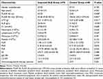
Among the 168 PMN patients, 118 patients were recruited for the following analysis. The 118 enrolled patients showed comparable demographic and clinical features with the 168 PMN patients (Table 1). Among the enrolled patients, the average age was 52.9 ± 12.1 years old at the time of renal biopsy, 70 (59.3%) were male and 48 (40.7%) were female. A total of 75 (63.6%) patients presented with positive serum PLA2R Ab, whose median titer was 99.23 (IQR 63.32–262.96) RU/mL. At the time of renal biopsy, the median 24-h proteinuria was 3.72 (IQR 2.04–5.48) g, the mean serum ALB was 24.6 ± 5.7 g/L, and the median serum Cr was 66.6 (57.0–78.0) µmol/L. Ninety-eight (83.1%) patients received corticosteroids and/or other immunosuppressive therapy. The median follow-up was 33.0 (IQR 22.5–39.0) months. At 6 months, serum ALB was elevated to more than 30g/L in 78 (66.1%) patients. At the end of the follow-up, 75 (63.6%) patients achieved CR. Seven (5.9%) patients had a 50% or more Cr increase at the end of the follow-up.
 |
Table 1 Comparison of the Included and Excluded PMN Patients |
Comparison of Improved ALB Group and Control Group
Compared to control group, PMN patients with ALB≥30g/L at 6 months had lower PLA2R Ab positive rate and titer, less UTP, higher serum ALB levels, and less C3 deposit along the glomerular basement membrane (GBM) at the time of renal biopsy (Table 2). There were no significant differences in all the peripheral blood cell ratios between the groups (Table 2). Binary logistic regression models revealed that only ALB at the renal biopsy was independently associated with ALB levels at 6 months (Hosmer–Lemeshow test: χ2=6.123, P=0.633; OR=0.867 (95% CI 0.799–0.941, P=0.001)).
 |
Table 2 Comparison of Improved ALB Group and Control Group |
Comparison of CR Group and Control Group
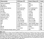
Compared to control group, PMN patients with CR at the end of the follow-up had lower PLA2R Ab positive rate and titer at the time of renal biopsy (Table 3), and they also tended to have lower PLR (P=0.095) (Table 3). However, when only PLA2R Ab titer was included into the analysis of binary logistic regression models, the fitting was not satisfying (Hosmer–Lemeshow test: χ2=13.885, P=0.016). In contrast, when both PLA2R Ab titer and PLR were included, the fitting was significantly improved (Hosmer–Lemeshow test: χ2=8.328, P=0.402). PLA2R Ab titer was identified as the independently correlate of CR (OR (PLA2R Ab titer) = 1.002 (95% CI 1.000–1.004, P=0.042)); OR (PLR) = 1.006 (95% CI 0.999–1.013, P=0.098).
 |
Table 3 Comparison of CR Group and Control Group |
Characteristics of the Patients with Cr Increase
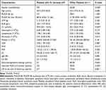
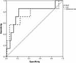
At the end of the follow-up, 7 patients had a 50% or more serum Cr increase. They were all male and had higher Cr levels and higher MLR at the renal biopsy (Table 4). Unsurprisingly, 7 patients had worse treatment response (Table 4). We additionally performed ROC curve analysis and calculated the area under the ROC curve (AUC) to evaluate the association of renal outcomes with Cr and MLR. MLR showed the larger AUC (MLR: 0.784 (95% CI 0.577–0.990, P=0.012); Cr: 0.754 (95% CI 0.520–0.988, P = 0.024)) (Figure 2).
 |
Table 4 Characteristics of Patients with a 50% or More Cr Increase |
Discussion
As well known, PMN is an autoimmune disease directed against podocytes. PLA2R is the major antigen of PMN in adults. However, the clinical remission does not often parallel with the immunological remission. The same titer of antibody may be observed in the early stages of the disease or in patients undergoing remission.5 Therefore, there is a need to find some biomarkers to evaluate patients’ inflammatory and immune status and then make treatment decision. In this study, we retrospectively compared several peripheral blood cell ratios in PMN patients with different treatment response. The results suggest that PLR might be a promising candidate to predict the effect of treatments, and that MLR may be associated with poor renal outcomes.
In-vitro and in-vivo studies have elucidated the origin of activated circulating platelets, which display numerous membrane receptors and release multiple biologically active substances from their granules that can regulate cellular interactions and contribute to immune, inflammatory, and thrombotic diseases.27–29 Circulating platelets can also interact with erythrocytes, neutrophils, and lymphocytes in the vessel lumen at sites of vascular damage.30,31 PLR has believed as an informative marker revealing shifts in platelet and lymphocyte counts due to inflammatory and prothrombotic states.32 Some studies showed that PLR could be potentially valuable in the accurate evaluation of inflammatory activity,32 and others demonstrated that high PLR was associated with venous thrombosis.33 Particularly, emerging evidence suggests that PLR has also been proposed as a marker for kidney diseases.34 In this study, we found that the patients with poor treatment effects, whose serum ALB levels were less than 30g/L at 6 months or who did not reach CR at the end of the follow-up, had numerically higher PLR. Especially for patients with CR or not, the difference was near to statistic significant. It is more noteworthy that when analyzing CR or not, the fitting of the binary logistic regression model including both PLA2R Ab titer and PLR was markedly better than that with only PLA2R Ab titer. PMN is an autoimmune disease as well as the pathology type with the highest incidence of thrombotic events in nephrotic syndrome.35 Our findings give a vital clue to determine treatment response of PMN patients at the time of renal biopsy. Besides serum ALB levels and PLA2R Ab, PLR at baseline might be a potential and good addition. This is intriguing, given that even in patients who respond to therapy, improvement of proteinuria only occurs after months, which imposes long trials of at least 2 years when testing the effectiveness of drugs.5 Kazan et al36 reviewed retrospectively 39 patients with low and moderate risk PMN and reported that the non-remission patients had significantly higher SII and pan-immune-inflammation value (calculated as (platelet count× neutrophil count× monocyte count)/lymphocyte count), which partly supported our findings. Larger studies are needed to demonstrate our hypothesis.
In the present study, only 7 (5.9%) patients had a 50% or more Cr increase at the end, which was far less than those reported by previous researches.9 The relatively short investigation period may be an important cause as 63.6% patients achieved CR during the median 33-month follow-up period, which was consistent with other studies.9 A study, whose mean duration of follow-up was 6.2 ± 3.2 years, showed that 59.6% patients reached renal endpoint (end-stage renal disease).37 Their results also revealed that both high NLR and PLR predicted poor long-term renal survivals in PMN patients, while MLR was not analyzed. In our study, the patients with Cr increase indeed tended to have higher NLR, but the difference did not reach the statistic significant (P=0.087) (Table 4). In contrast, higher MLR was significantly associated with renal function deterioration. Various metabolic stimuli lead to an increase in monocytes. These monocytes differentiate into macrophages and create an inflammatory state.34 Some kidney macrophages have been demonstrated to be able to promote kidney fibrosis.38 The prognostic value of MLR for PMN patients deserves further investigation by larger and longer-time studies.
Finally, although immunosuppressive agents did not display any beneficial effects on whether treatment response or renal outcomes in the present study, we cannot negate the value of immunosuppressive therapy in PMN patients, especially high-risk ones. We did not analyze specific therapeutic programs, and some patients were given immunosuppressive agents after several months but not at the renal biopsy. The heterogeneity could hamper treatment efficacy evaluation.
The current study has some limitations. First, the sample size was small and the follow-up duration was short, which contributed to too few endpoint events, so a Cox proportional hazards analysis could not be conducted. Second, we only recruited PMN patients from a single center, and validation studies in other independent PMN cohorts are needed in the future.
Conclusions
Taken together, we found that PMN patients with poor treatment response tended to have higher PLR at the time of renal biopsy, and a higher MLR was associated with poor renal outcomes. Our findings suggested that PLR and MLR might be used to predict treatment efficacy and prognosis for PMN patients, respectively.
Data Sharing Statement
The datasets used during the current study are available from Wen-hu Liu on reasonable request.
Statement of Ethics
This study protocol was reviewed and approved by Ethics Committee of Beijing Friendship Hospital, approval number 2021-P2-269-01, and Ethics Committee of Beijing Friendship Hospital waived the need for informed consent based on the following reasons: (1) the purpose of the study was important; (2) the possible risk to patients was not higher than the minimum one; (3) the waiver of informed consent would not adversely affect the rights and health of patients. (4) the patients’ privacy and personal identity information were well protected. The protocol of the study is compliant with the Declaration of Helsinki.
Acknowledgments
We thank Experimental Center of Beijing Friendship Hospital for their experimental platform support.
Author Contributions
All authors made a significant contribution to the work reported, whether that is in the conception, study design, execution, acquisition of data, analysis and interpretation, or in all these areas; took part in drafting, revising or critically reviewing the article; gave final approval of the version to be published; have agreed on the journal to which the article has been submitted; and agree to be accountable for all aspects of the work.
Funding
The authors did not receive any funding.
Disclosure
The authors declare no conflicts of interest in this work.
References
1. Giannini G, Arend LJ. The prevalence of mesangial electron-dense deposits in PLA2R-positive membranous nephropathy. Nephron. 2022;146(2):167–171. doi:10.1159/000519912
2. De Vriese AS, Glassock RJ, Nath KA, et al. A proposal for a serology-based approach to membranous nephropathy. J Am Soc Nephrol. 2017;28(2):421–430. doi:10.1681/ASN.2016070776
3. Lerner GB, Virmani S, Henderson JM, et al. A conceptual framework linking immunology, pathology, and clinical features in primary membranous nephropathy. Kidney Int. 2021;100(2):289–300. doi:10.1016/j.kint.2021.03.028
4. Fresquet M, Jowitt TA, Gummadova J, et al. Identification of a major epitope recognized by PLA2R autoantibodies in primary membranous nephropathy. J Am Soc Nephrol. 2015;26(2):302–313.
5. Ronco P, Plaisier E, Debiec H. The role of PLA2R antibody monitoring: what we know and what we don’t know. Nephrol Dial Transplant. 2021. doi:10.1093/ndt/gfab356
6. Qu Z, Zhang MF, Cui Z, et al. Antibodies against M-type phospholipase A2 receptor may predict treatment response and outcome in membranous nephropathy. Am J Nephrol. 2018;48(6):438–446.
7. Li W, Zhao Y. Prognostic value of phospholipase A2 receptor in primary membranous nephropathy: a systematic review and meta-analysis. Int Urol Nephrol. 2019;51(9):1581–1596.
8. Gao S, Cui Z, Wang X, et al. Rituximab therapy for primary membranous nephropathy in a Chinese cohort. Front Med. 2021;8:663680.
9. Lu H, Shen J, Sun J, et al. Efficacy and safety of rituximab in the treatment of idiopathic membranous nephropathy: a meta-analysis. Appl Bionics Biomech. 2022;2022:5393797.
10. Muruve DA, Debiec H, Dillon ST, et al. Serum protein signatures using aptamer-based proteomics for minimal change disease and membranous nephropathy. Kidney Int Rep. 2022;7(7):1539–1556. doi:10.1016/j.ekir.2022.04.006
11. Zhao Q, Dai H, Hu Y, et al. Cytokines network in primary membranous nephropathy. Int Immunopharmacol. 2022;113(PtA):109412. doi:10.1016/j.intimp.2022.109412
12. Posul E, Yilmaz B, Aktas G, et al. Does neutrophil-to-lymphocyte ratio predict active ulcerative colitis? Wien Klin Wochenschr. 2015;127(7–8):262–265.
13. Duman TT, Aktas G, Atak BM, et al. Neutrophil to lymphocyte ratio as an indicative of diabetic control level in type 2 diabetes mellitus. Afr Health Sci. 2019;19(1):1602–1606. doi:10.4314/ahs.v19i1.35
14. Balci SB, Aktas G. A comprehensive review of the role of hemogram derived inflammatory markers in gastrointestinal conditions. Iran J Colorectal Res. 2022;10(3):2–13.
15. Aktas G, Sit M, Dikbas O, et al. Elevated neutrophil-to-lymphocyte ratio in the diagnosis of Hashimoto’s thyroiditis. Rev Assoc Med Bras. 2017;63(12):1065–1068.
16. Khalid A, Ali Jaffar M, Khan T, et al. Hematological and biochemical parameters as diagnostic and prognostic markers in SARS-COV-2 infected patients of Pakistan: a retrospective comparative analysis. Hematology. 2021;26(1):529–542. doi:10.1080/16078454.2021.1950898
17. Afsin H, Aktas G. Platelet to lymphocyte and neutrophil to lymphocyte ratios are useful in differentiation of thyroid conditions with normal and increased uptake. Ethiop J Health Dev. 2021;35(3):1–5.
18. Atak Tel BM, Kahveci G, Bilgin S, et al. Platelet to lymphocyte ratio in differentiation of benign and malignant thyroid nodules. Exp Biomed Res. 2021;4(2):148–153. doi:10.30714/j-ebr.2021267978
19. Atak B, Aktas G, Duman TT, et al. Diabetes control could through platelet-to-lymphocyte ratio in hemograms. Rev Assoc Med Bras. 2019;65(1):38–42. doi:10.1590/1806-9282.65.1.38
20. Aktas G, Duman TT, Atak BM, et al. Irritable bowel syndrome is associated with novel inflammatory markers derived from hemogram parameters. Fam Med Prim Care Rev. 2020;22(2):107–110. doi:10.5114/fmpcr.2020.95311
21. Aktas G. Hematological predictors of novel coronavirus infection. Rev Assoc Med Bras. 2021;67(Suppl1):1–2. doi:10.1590/1806-9282.67.suppl1.20200678
22. Catal O, Ozer B, Sit M, et al. The role of monocyte to lymphocyt ratio in predicting metastasis in rectal cancer. Ann Med Res. 2021;28(3):527–531. doi:10.5455/annalsmedres.2020.05.466
23. Kocak MZ, Aktas G, Duman TT, et al. Monocyte lymphocyte ratio as a predictor of diabetic kidney injury in type 2 diabetes mellitus; The MADKID study. J Diabetes Metab Disord. 2020;19(2):997–1002. doi:10.1007/s40200-020-00595-0
24. Atak Tel BM, Bilgin S, Kurtkulagi O, et al. Frailty in diabetic subjects during COVID-19 and its association with HbA1c, mean platelet volume and monocyte/lymphocyte ratio. Clin Diabetol. 2022;11(2):119–126. doi:10.5603/DK.a2022.0015
25. Wang G, Sun L, Dong H, et al. Neural epidermal growth factor-like 1 protein-positive membranous nephropathy in Chinese patients. Clin J Am Soc Nephrol. 2021;16(5):727–735. doi:10.2215/CJN.11860720
26. Perna A, Ruggiero B, Podestà MA, et al. Sexual dimorphic response to rituximab treatment: a longitudinal observational study in a large cohort of patients with primary membranous nephropathy and persistent nephrotic syndrome. Front Pharmacol. 2022;13:958136. doi:10.3389/fphar.2022.958136
27. Hussain Y, Khan F, Alsharif KF, Alzahrani KJ, Saso L, Khan H. Regulatory effects of curcumin on platelets: an update and future directions. Biomedicines. 2022;10(12):3180. doi:10.3390/biomedicines10123180
28. Koupenova M, Clancy L, Corkrey HA, et al. Circulating platelets as mediators of immunity, inflammation, and thrombosis. Circ Res. 2018;122(2):337–351. doi:10.1161/CIRCRESAHA.117.310795
29. Yeung J, Li W, Holinstat M, Isom LL. Platelet signaling and disease: targeted therapy for thrombosis and other related diseases. Pharmacol Rev. 2018;70(3):526–548. doi:10.1124/pr.117.014530
30. Olumuyiwa-Akeredolu OO, Pretorius E. Platelet and red blood cell interactions and their role in rheumatoid arthritis. Rheumatol Int. 2015;35:1955–1964.
31. Łukasik ZM, Makowski M, Makowska JS. From blood coagulation to innate and adaptive immunity: the role of platelets in the physiology and pathology of autoimmune disorders. Rheumatol Int. 2018;38:959–974.
32. Gasparyan AY, Ayvazyan L, Mukanova U, et al. The platelet-to-lymphocyte ratio as an inflammatory marker in rheumatic diseases. Ann Lab Med. 2019;39(4):345–357.
33. Dong G, Huang A, Liu L. Platelet-to-lymphocyte ratio and prognosis in STEMI: a meta-analysis. Eur J Clin Invest. 2021;51(3):e13386.
34. Seo IH, Lee YJ. Usefulness of complete blood count (CBC) to assess cardiovascular and metabolic diseases in clinical settings: a comprehensive literature review. Biomedicines. 2022;10(11):2697.
35. Lu X, Kan C, Zhang R. Phospholipase A2 receptor is associated with hypercoagulable status in membranous nephropathy: a narrative review. Ann Transl Med. 2022;10(17):938.
36. Kazan DE, Kazan S. Systemic immune inflammation index and pan-immune inflammation value as prognostic markers in patients with idiopathic low and moderate risk membranous nephropathy. Eur Rev Med Pharmacol Sci. 2023;27(2):642–648.
37. Tsai SF, Wu MJ, Chen CH. Low serum C3 level, high neutrophil-lymphocyte-ratio, and high platelet-lymphocyte-ratio all predicted poor long-term renal survivals in biopsy-confirmed idiopathic membranous nephropathy. Sci Rep. 2019;9(1):6209.
38. Wen Y, Yan HR, Wang B, et al. Macrophage heterogeneity in kidney injury and fibrosis. Front Immunol. 2021;12:681748.
 © 2023 The Author(s). This work is published and licensed by Dove Medical Press Limited. The full terms of this license are available at https://www.dovepress.com/terms.php and incorporate the Creative Commons Attribution - Non Commercial (unported, v3.0) License.
By accessing the work you hereby accept the Terms. Non-commercial uses of the work are permitted without any further permission from Dove Medical Press Limited, provided the work is properly attributed. For permission for commercial use of this work, please see paragraphs 4.2 and 5 of our Terms.

