Back to Journals » OncoTargets and Therapy » Volume 12
The Roles And Signaling Pathways Of Phosphatidylethanolamine-Binding Protein 4 In Tumors
Authors Luo ZK, Chen QF, Qu X, Zhou XY ![]()
Received 17 May 2019
Accepted for publication 9 September 2019
Published 18 September 2019 Volume 2019:12 Pages 7685—7690
DOI https://doi.org/10.2147/OTT.S216161
Checked for plagiarism Yes
Review by Single anonymous peer review
Peer reviewer comments 2
Editor who approved publication: Prof. Dr. Geoffrey Pietersz
Zi-Kang Luo,1,* Qiong-Feng Chen,2,* Xiaoqin Qu,2 Xiao-Yan Zhou2,3
1Department of Clinical Medical, The Second Clinical Medical College, Nanchang University, Nanchang 330006, People’s Republic of China; 2Department of Pathophysiology, Medical College of Nanchang University, Nanchang, Jiangxi 330006, People’s Republic of China; 3Jiangxi Province Key Laboratory of Tumor Pathogenesis and Molecular Pathology, Nanchang, Jiangxi 330006, People’s Republic of China
*These authors contributed equally to this work
Correspondence: Xiao-Yan Zhou
Department of Pathophysiology, Medical College of Nanchang University, 461 BaYi Road, Nanchang, Jiangxi 330006, People’s Republic of China
Tel +86 791 86360556
Email [email protected]
Abstract: Phosphatidylethanolamine-binding protein 4 (PEBP4) has been found to be highly expressed in many tumors and to be closely related to the proliferation, differentiation, and metastasis of tumors. PEBP4 has also been found to be involved in many cancer-activated signaling pathways and to cause therapeutic resistance. In this study, we first reviewed the morphological structure and expression of PEBP4, then discussed the roles of PEBP4 in individualized treatment of some cancers, and finally explored the possibilities of cultivating PEBP4 as a therapeutic target.We also identified the main signaling pathways in which PEBP4 affects different cancers. It is here concluded that over-expression of PEBP4 can enhance the proliferation and metastasis of the cancer cells and the resistance to radiotherapy/chemotherapy in cancers.
Keywords: PEBP4, signaling pathway, targeted therapy
Introduction
Non-communicable diseases are the leading cause of deaths worldwide in developing countries. With the aging and rapid growth of the population, the cancer incidence and number of cancer deaths worldwide are also growing rapidly, and cancer is expected to be the leading cause of death in the 21st century and the most important obstacle to improving universal life expectancy. WHO data show that the global cancer burden continues to grow, with 18.1 million new cases and 9.6 million deaths expected in 2018.1–3 Recent studies have reported that PEBP4 highly expressed in various tumors. PEBP4 has been found to be involved in tumorigenesis and development. It has been reported that PEBP4 influences the survival rate of tumors by regulating signaling pathways, including AKT, MAPK, and SHH. The expression of PEBP4 in tumors can provide new ideas and targets for the prevention and treatment of tumors.
The Structure, Expression, And Functions Of PEBP4
In 1984, Bernier and his team isolated a cytoplasmic soluble protein with a relative molecular weight of 21,000–23,000 from bovine brains. It was called phosphatidylethanolamine-binding protein (PEBP) because of its high affinity with phosphatidylethanolamine (PE).4 There was a similar structure domain among the PEBP family members of different species. It was composed of one big beta-sheet connected to a small beta-sheet on both sides and two alpha-helix of the C terminal. In this structure, there was a highly conserved phosphate binding bag, which was very important to the function of PEBP family. The PEBP family is a superfamily containing more than 400 members, and it is highly conserved across bacterial and human evolution.5 Different subfamilies have various biological functions. PEBP4, a subgroup of PEBP, has been found to be highly expressed in many cancer tissues. PEBP4 plays a vital role in cancer, and it has attracted increasing amounts of attention from researchers. For this reason, in this paper, we focused only on PEBP4.
It has been established that the human PEBP4 gene is located on chromosome 8p21.3, and its mRNA is 901 bp in length. In the normal tissues, PEBP4 was mainly expressed in the heart, lung, prostate, and thyroid of mammals, and slightly expressed in liver, colon, skin, adrenal gland, and bone marrow.6 More importantly, it was commonly reported that PEBP4 highly expressed in all cancerous tissues.7–9
Initially, PEBP4 was thought to normally co-localize with lysosomes in the cell. Garcia R. et al reported that PEBP4 could be used as a scaffold to connect MEK-1 to Raf-1 and so form ternary complexes.4 Recent studies have established that PEBP4 is a secretory and glycosylated protein.8 Tagging the C-terminal of the PEBP4 allowed it to be secreted, while tagging the N-terminal disrupted PEBP4 secretion. It has been reported recently that PEBP4 not only plays fundamental roles in the process of cell membrane construction and remodeling but also plays important roles in such physiological and pathological processes as signal transduction and nervous system differentiation and development. Over-expression of PEBP4 was rendered tumors resistant to radiotherapy and chemotherapy and promoted the proliferation, differentiation, and metastasis of cancer cells.10 It has been reported that PEBP4 migrated to the cell membrane to protect cells from cell damage caused by tumor necrosis factor alpha (TNF-α) or rituximab-mediated complement-dependent cytotoxicity (R-CDC) stimulation in a PE-binding-domain-dependent manner.10,11 Haibo Liu et al found that PEBP4 could inhibit the degradation of estrogen receptor alpha and maintain high estrogen receptor levels in cancer cells.12
PEBP4 And Cancers
PEBP4 And Breast Cancer
Early diagnosis and treatment of breast cancer are the primary means of improving the quality of life of patients. The incidence of breast cancer has been climbing year by year. Even more worrisome, the decrease in the death rate from mammary cancer had been decreasing fairly significantly, mostly due to the molecular typing. As the scientific community has gradually become more aware of the genome, it had been shown that mammary cancer molecular typing may reflect the biological behavior of tumors and may be able to affect drug sensitivity and determine drug use. Molecular typing of mammary cancer was usually determined by the analysis of tumor markers. Detection of tumor markers has facilitated understanding of the prediction of curative effect, metastasis, and drug tolerance.13,14 Recently, PEBP4 has been found to be preferentially expressed in breast cancer tissues and to play an anti-apoptotic role in breast cancer tissues. Silencing of hPEBP4 itself did not affect the growth of MCF-7 cells, but it has been shown to enhance the sensitivity of TNF-α to growth inhibition.14 It has also been reported that, in IOI-42-treated MCF-7 cells, the interaction between hPEBP4 and Raf-1/MEK-1 could be weakened and promote TNF-α-induced apoptosis in MCF-7 cells. Wang S.C. et al found knocking down PEBP4 could inhibit the proliferation and metastasis of MDA-MB-231. However, implanting the MDA-MB-231 cells transfected with sh-PEBP4 into nude mice decreased the weight of the xenografted tumor; they also suggested that PEBP4 silencing played a prominent role in xenografted tumors.15 Qiu J. et al reported TNF-α-induced cell apoptosis was not obvious in cells with high malignant degree of PEBP4 silencing. These studies suggested that PEBP4 may play an important role in advanced metastasis of breast cancer.
PEBP4 And Lung Cancer
Many studies have reported that PEBP4 was of paramount significance in the development and metastasis of lung cancer. Jian W. et al found the expression of PEBP4 to be positively correlated with NSCLC. PEBP4 can promote the proliferation and metastasis of NSCLC, and over-expression of PEBP4 can promote the progression of EMT and ultimately lead to tumorigenesis by up-regulation many cytokines such as n-cadherin, alpha-sma, collagen III, and vimentin.16 Yu G. et al found that overexpression of PEBP4 could promote the proliferation of HCC827 cells.17 Recent studies have shown that microRNAs play a significant role in the process of lung cancer and are able to serve as invasive biomarkers for early-stage cancer.18–20 Zhao Z. et al found that microRNA could interact with PEBP4 as a direct and functional target, and siRNA-mediated PEBP4 knockdown had the same impact as knockdown of ectopic microRNA-15b, upregulation of mir-15b suppressed PEBP4.21 In the study of Guiping Yu et al, the data suggested that the usage of cis-diaminodichloroplatinum (DDP) was able to significantly reduce the viability of A549 cells by inducing cytotoxicity. However, PEBP4 over-expression could weaken the effect of DPP and reduce DDP-induced cytotoxicity by regulating P53 and microRNA-34a.22 Silencing PEBP4 was found to inhibit the proliferation, invasion, and metastasis of various types of lung cancer cells, and over-expression PEBP4 had the opposite effect.
PEBP4 And Intestinal Cancer
Recent laboratory studies on targeted therapy have shown that PEBP4 gene knockout inhibits cell proliferation, migration, and invasion in gastric cancer cell lines.9 PEBP4 knockout significantly inhibits both phosphorylated PI3K and phosphorylated AKT in gastric cancer cells.23 These mechanisms might be the key to metastasis and dissemination of gastric cancer. PEBP4 was expected to become a potential therapeutic target for gastric cancer.
Studies have shown that PEBP4 is also expressed in colorectal cancer tissues at significantly higher levels than in normal tissues. At the same time, the expression of PEBP4 in lymph-node-metastasis-positive patients was higher than in patients with remission lymph node metastasis patients.24 In addition, the expression of PEBP4 in the middle and advanced stages of colorectal cancer, as indicated by TNM stage, was prominently higher than that in the early stage.25 Some studies even pointed out retrospectively that the high expression of hPEBP4 was associated with radiation resistance in the course of radiotherapy for colorectal cancer, which had certain preoperative predictive values.26 These experiments suggested that PEBP4 might be mainly involved in metastasis of middle and advanced colorectal cancer, and the targeted drugs of PEBP4 might improve the quality of life of patients with advanced colorectal cancer.
PEBP4 And Glioma
Emerging evidence has suggested that IDH and BRAF biomarkers could predict the occurrence of glioma.27,28 Recently, Huang R.Q. et al29,30 found the following: (I) PEBP4 was closely associated with grade in human gliomas. (II) PEBP4 was the only independent prognostic factor of glioma by COX regression analysis, based on the existing clinicopathological and biochemical parameters. (III) The high level of expression of PEBP4 could indicate poor prognosis of glioma grade. Those studies showed that overexpression of PEBP4 may be relevant to the higher-grade human gliomas. Although many studies have shown that the high expression of PEBP4 is closely correlated with tumors, PEBP4 was found to be the only independent prognostic indicator in glioma cancer, though this has not been reported to be the case for other cancers.
PEBP4 And Lymphoma
Non-Hodgkin’s lymphoma (NHL) is the main type of lymphoma, most of which are B-cell lymphomas.31,32 Rituximab improves the survival rate of patients with lymphoma.33 Recently, the relationship between PEBP4 and B-cell lymphoma had been reported. Kai Wang and his team11 found the following: (I) Lymphoma cells with high expression of PEBP4 can attenuate the effects of rituximab. (II) Knocking out the PEBP4 gene and administration of rituximab can synergistically treat malignant lymphoma. (III) Overexpression of PEBP4 inhibited the increase of R-CDC-mediated calcium flux, thereby reducing cell sensitivity to R-CDC. (IV) In Raji cells, silencing of PEBP4 significantly enhanced the expression of P53. In conclusion, PEBP4 could promote the development of lymphoma. Kai Wang and his team also found that PEBP4 could reduce the sensitivity of cells to R-CDC and the effect of P53, which suggested that the roles of PEBP4 may also be performed by affecting other oncogenes.
To sum up, so far, it has been reported that PEBP4 played important roles in many tumors, including mammary cancer, lung cancer, intestinal cancer, glioma, and lymphoma, and its effects are summarized in Table 1.
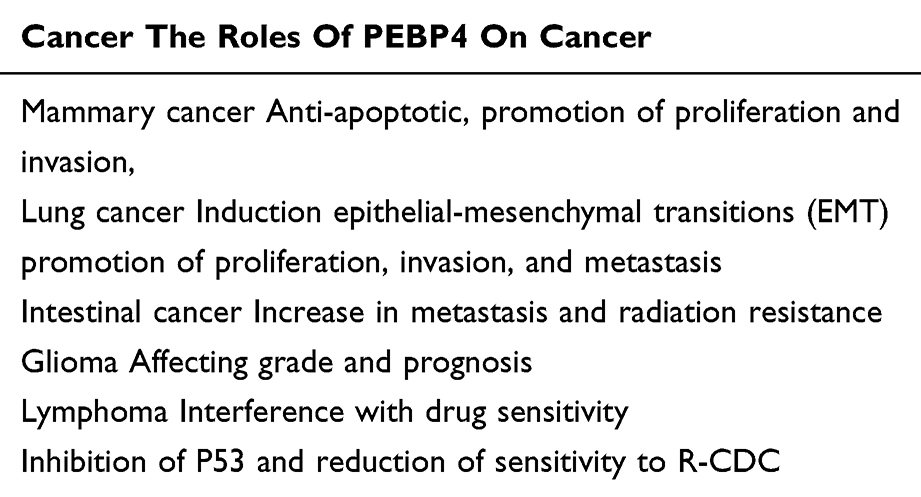 |
Table 1 Summary Of The Roles Of PEBP4 In Tumors. PEBP4 Was Found To Be Highly Expressed In All The Above Tumors. And PEBP4 Was Able To Promote The Development Of All Cancers |
The Signaling Pathways Affected By PEBP4 In Cancer
PEBP4 Enhanced The Resistance Of Cancer Cells Through The AKT Signaling Pathway
In a large number of treatments of malign cancers, activation of the PI3K/AKT signal pathway tended to render cells resistant to radiotherapy and chemotherapy.34–36 Other studies showed that PEBP4 participated in the activation of PI3K/AKT signal pathway, and PEBP4 seemed to be an important factor in the activation of AKT signaling pathway in cancer cells.9,37,38 Yu G. et al found that the overexpression of PEBP4 played activated roles in phosphorylation of AKT in HCC827 cells, and down-regulation of PEBP4 inhibited the phosphorylation of AKT.17 In addition, Zhang D. pointed out PEBP4 could interact with Akt and promote the phosphorylation of serine 473 of Akt.39 Zhang D. et al and Yu G. et al suggested that the promotion of PEBP4 for AKT activation in cancer cells may depend on phosphorylation of serine 473 of AKT.
PEBP4 can phosphorylate serine 473 of Akt; studies found that the roles of PEBP4 in AKT signaling pathway also seemed to be related to ROS. Li W. et al found knockdown PEBP4 could inhibit hypoxia-induced AKT activation, suggesting that PEBP4 participated in hypoxia-induced AKT activation37 and hypoxia could increase the accumulation of reactive oxygen species (ROS). Increasing accumulation of ROS was able to activate the AKT pathway.40 With the activation of the AKT pathway, hypermetabolism may induce an increase in oxygen consumption, further resulting in ROS-mediated AKT activation. One study indicated that ROS has two functions, on the one hand, it killed cancer cells directly, but on the other hand, it provided survival advantage for the activation of the stress system.40 PEBP4 seemed to enhance survival advantages of ROS for the activation of the stress system in cancer cells; Qiu, J et al.25 found (I) PEBP4 acted as a survival signal molecule in the activation of AKT mediated by ROS to help cancer cells resist killing. (II) During the course of radiation treatment of cancers, PEBP4 has been found to contribute to phosphorylation of ROS-mediated AKT, enhancing survival rates of cancer cells and their abilities to resist apoptosis and radiation. However, the activation of AKT by PEBP4 was not found in normal cells; studies by Garcia R. et al showed that PEBP4 had no effect on the phosphorylation of AKT in C2C12 muscle cells. These results suggested that the resistance to chemotherapy and radiotherapy of tumor cells activated by AKT signaling pathway is closely related to the overexpression of PEBP4.
The Expression Of PEBP4 Was Negatively Correlated With The Activation Of The ERK/MAKP Signaling Pathway
ERK is a key member of the MAPK family, and the MAPK/ERK signaling pathway is important for cytokines to regulate cellular pathways.41 Various growth factor receptors and nutrition-related factor receptors need ERK activation to complete the signal transduction process.42 PEBP4 has been found to be involved in MAPK signaling pathway. Garcia R. et al found that, in PEBP4-overexpressing COS1 cells, PEBP4 participated in the MEK/ERK signaling pathway, in which PEBP4 can enhance their activity as scaffolds for upstream proteins Raf-1 and MEK.4 The activation of EKR signaling pathway requires MEK to transfer to the nucleus and transmit signals.43 When PEBP4 formed ternary complexes with Raf-1 and MEK, the ability of MEK to transmit signals to the nucleus became blocked. However, this contradicted the fact that PEBP4 had been repeatedly reported to be involved in the occurrence and development of cancer. A large number of recent studies have explored the relationship between the ERK signaling pathway and tumors, which suggest that inhibition of ERK signaling pathway may be a promising cancer therapy.44–46 Activation of PEBP4 has been shown to inhibit the activation of ERK signaling pathway, but studies have also indicated that inhibiting the activation of ERK signaling pathway could enhance the resistance of cancer cells to TNF-α-induced apoptosis,10 and these results do not seem to contradict PEBP4’s role in promoting cancer development.
Wang X. et al found that when cancer cells overexpressing PEBP4 were stimulated by TNF-α, PEBP4 moved from the lysosome to the cell membrane, and then PEBP4 combined with Raf-1 and MEK-1 via the PE domain to inhibit the ERK signaling pathway. However, when PEBP4 did not have a PE domain, the transfer function of PEBP4 was blocked.10 Wang X. et al also found the same results in cancer cells overexpressing PEBP4 from stimulation of TNF-related apoptosis-inducing ligand (TRAIL).47 TNF-α and TRAIL have been found to act as stimulating signals for PEBP4’s transfer from lysosome to cell membrane, and Kai Wang et al found R-CDC could move PEBP4 to the cell membrane through PE in cells overexpressing PEBP4, allowing them to resist attacks from the membrane attack complex.11 These results suggested that the PE domain of PEBP4 may be an important structure because of its function, and PEBP4 can enhance the resistance of cancer cells to apoptosis by inhibiting the activation of ERK signaling pathways induced by different pathways.
PEBP4 Can Induce Tumorigenesis Through The SHH Signaling Pathway
The expression of Shh and Smo protein has been shown to be significantly higher in tumors, indicating that this signaling pathway is activated. Recently, it has been reported that PEBP4 was also involved in the SHH signaling pathway. Jian W. et al reported that when PEBP4 gene was silenced in tumor cells, the expression of Shh and Smo protein decreased markedly.16 This study indicated that PEBP4 was involved in this signaling pathway and could enhance Shh signaling pathway resulting in diseases and tumors, but much more research is needed.
Conclusion
Cancer is a major non-communicable disease with a significant death rate. Recently, more and more studies have shown that overexpression of PEBP4 is closely related to such advanced tumors as mammary cancer, lung cancer, and intestinal cancer. PEBP4 has a domain consisting of a big beta sheet connected to a small beta-sheet on both sides and two alpha-helixes in the C terminal. It also participates in some signaling pathways to promote or inhibit the activation of those signaling pathways such as AKT, MAPK, and SHH signaling pathways, thereby enhancing the anti-radiotherapy or anti-chemotherapy of tumors and promoting the proliferation, differentiation, and metastasis of cancer cells. We concluded the main functions of PEBP4 and its role in cancer development, and we attached great importance to its possible mechanisms prompting the development of cancer cells. Studies have suggested that PEBP4 can serve as a prognostic marker of cancer. However, PEBP4 is not currently used in clinical treatment, and the mechanism by which PEBP4 affects early cancer progression is not clear. More studies are needed to explore the feasibility of PEBP4 in clinical treatment and the roles of PEBP4 during the early stages of cancer.
Acknowledgments
This work was supported by the National Natural Science Foundation of China (Nos. 81760117 and 81460126) and the Natural Science Foundation of Jiangxi Province (No. 20181BAB205012).
We thank LetPub for its linguistic assistance during the preparation of this manuscript.
Disclosure
The authors report no conflicts of interest in this work.
References
1. Aribike O, Okafor I, Roberts A, Odugbemi T. Are Nigerian women pro-active about noncommunicable disease prevention? A quantitative survey. Ann Glob Health. 2019;85(1).
2. Siegel RL, Miller KD, Jemal A. Cancer statistics, 2017. CA Cancer J Clin. 2017;67(1):7–30. doi:10.3322/caac.21387
3. Bray F, Ferlay J, Soerjomataram I, Siegel RL, Torre LA, Jemal A. Global cancer statistics 2018: GLOBOCAN estimates of incidence and mortality worldwide for 36 cancers in 185 countries. CA Cancer J Clin. 2018;68(6):394–424. doi:10.3322/caac.21492
4. Garcia R, Grindlay J, Rath O, Fee F, Kolch W. Regulation of human myoblast differentiation by PEBP4. EMBO Rep. 2009;10(3):278–284. doi:10.1038/embor.2009.4
5. Al-Mulla F, Bitar MS, Taqi Z, Yeung KC. RKIP: much more than Raf kinase inhibitory protein. J Cell Physiol. 2013;228(8):1688–1702. doi:10.1002/jcp.24335
6. Fagerberg L, Hallström BM, Oksvold P, et al. Analysis of the human tissue-specific expression by genome-wide integration of transcriptomics and antibody-based proteomics. Mol Cell Proteomics. 2014;13(2):397–406. doi:10.1074/mcp.M113.035600
7. Yeung K, Janosch P, McFerran B, et al. Mechanism of suppression of the Raf/MEK/extracellular signal-regulated kinase pathway by the raf kinase inhibitor protein. Mol Cell Biol. 2000;20(9):3079–3085. doi:10.1128/mcb.20.9.3079-3085.2000
8. He H, Liu D, Lin H, et al. Phosphatidylethanolamine binding protein 4 (PEBP4) is a secreted protein and has multiple functions. Biochim Biophys Acta. 2016;1863(7 Pt A):1682–1689. doi:10.1016/j.bbamcr.2016.03.022
9. Wu Z, Liu B, Zheng X, Hou H, Li Y. Role of the PEBP4 protein in the development and metastasis of gastric cancer. Oncotarget. 2017;8(11):18177–18184. doi:10.18632/oncotarget.15255
10. Wang X, Li N, Liu B, et al. A novel human phosphatidylethanolamine-binding protein resists tumor necrosis factor alpha-induced apoptosis by inhibiting mitogen-activated protein kinase pathway activation and phosphatidylethanolamine externalization. J Biol Chem. 2004;279(44):45855–45864. doi:10.1074/jbc.M405147200
11. Wang K, Jiang Y, Zheng W, et al. Silencing of human phosphatidylethanolamine-binding protein 4 enhances rituximab-induced death and chemosensitization in B-cell lymphoma. PLoS One. 2013;8(2):e56829. doi:10.1371/journal.pone.0056829
12. Liu H, Qiu J, Li N, Chen T, Cao X. Human phosphatidylethanolamine-binding protein 4 promotes transactivation of estrogen receptor alpha (ERalpha) in human cancer cells by inhibiting proteasome-dependent ERalpha degradation via association with Src. J Biol Chem. 2010;285(29):21934–21942. doi:10.1074/jbc.M110.109876
13. Yang Y, Yang HH, Hu Y, et al. Immunocompetent mouse allograft models for development of therapies to target breast cancer metastasis. Oncotarget. 2017;8(19):30621–30643. doi:10.18632/oncotarget.15695
14. Wang X, Li N, Li H, et al. Silencing of human phosphatidylethanolamine-binding protein 4 sensitizes breast cancer cells to tumor necrosis factor-alpha-induced apoptosis and cell growth arrest. Clin Cancer Res. 2005;11(20):7545–7553. doi:10.1158/1078-0432.CCR-05-0879
15. Wang S-C, Zhou F, Zhou Z-Y, Hu Z, Chang L, Ma M-D. Knockdown of PEBP4 suppresses proliferation, migration and invasion of human breast cancer cells. Biomed Pharmacother. 2017;90:659–664. doi:10.1016/j.biopha.2017.03.098
16. Jian W, Bai Y, Li X, Kang J, Lei Y, Xue Y. Phosphatidylethanolamine-binding protein 4 promotes the epithelial-to-mesenchymal transition in non-small cell lung cancer cells by activating the sonic hedgehog signaling pathway. J Cell Biochem. 2018;120:5386–5395.
17. Yu G, Shen Z, Chen G, Teng X, Hu Y, Huang B. PEBP4 enhanced HCC827 cell proliferation and invasion ability and inhibited apoptosis. Tumour Biol. 2013;34(1):91–98. doi:10.1007/s13277-012-0514-0
18. Lelli D, Pedone C, Majeed M, Sahebkar A. Curcumin and lung cancer: the role of microRNAs. Curr Pharm Des. 2017;23(23):3440–3444. doi:10.2174/1381612823666170109144818
19. Del VV, Denti MA. microRNA and lung cancer. Adv Exp Med Biol. 2015;889:153–177. doi:10.1007/978-3-319-23730-5_9
20. Zhang Y, Yang Q, Wang S. MicroRNAs: a new key in lung cancer. Cancer Chemother Pharmacol. 2014;74(6):1105–1111. doi:10.1007/s00280-014-2559-9
21. Zhao Z, Zhang L, Yao Q, Tao Z. miR-15b regulates cisplatin resistance and metastasis by targeting PEBP4 in human lung adenocarcinoma cells. Cancer Gene Ther. 2015;22(3):108–114. doi:10.1038/cgt.2014.73
22. Yu G, Zhong N, Chen G, Huang B, Wu S. Downregulation of PEBP4, a target of miR-34a, sensitizes drug-resistant lung cancer cells. Tumour Biol. 2014;35(10):10341–10349. doi:10.1007/s13277-014-2284-3
23. Ma Z, Zhu J, Han G, et al. PEBP4 is upregulated in gastric cancer and promotes the growth and migration of cancer cells. Int J Clin Exp Med. 2016;9(7):12687–12695.
24. Liu H, Kong Q, Li B, He Y, Li P, Jia B. Expression of PEBP4 protein correlates with the invasion and metastasis of colorectal cancer. Tumour Biol. 2012;33(1):267–273. doi:10.1007/s13277-011-0279-x
25. Qiu J, Tao Y, Yang G, Xu K, Lin AL, Li L. Effect of a chemical inhibitor of human phosphatidylethanolamine-binding protein 4 on radiosensitivity of rectal cancer cells. World J Surg Oncol. 2016;14(1):221. doi:10.1186/s12957-016-0977-3
26. Qiu J, Yang G, Shen Z, Xie Y, Wang L. hPEBP4 as a predictive marker for the pathological response of rectal cancer to preoperative radiotherapy. Int J Colorectal Dis. 2013;28(2):241–246. doi:10.1007/s00384-012-1534-3
27. Olow A, Mueller S, Yang X, et al. BRAF status in personalizing treatment approaches for pediatric gliomas. Clin Cancer Res. 2016;22(21):5312–5321. doi:10.1158/1078-0432.CCR-15-1101
28. Berghoff AS, Kiesel B, Widhalm G, et al. Correlation of immune phenotype with IDH mutation in diffuse glioma. Neuro Oncol. 2017;19(11):1460–1468. doi:10.1093/neuonc/nox054
29. Huang R-Q, Shi D-L, Huang W, Chen F, Lu Y-C. Increased expression of phosphatidylethanolamine-binding protein 4 (PEBP4) strongly associates with human gliomas grade. J Neurooncol. 2016;127(2):235–242. doi:10.1007/s11060-015-2040-6
30. Huang RQ, Wang SQ, Zhu QB, et al. Knockdown of PEBP4 inhibits human glioma cell growth and invasive potential via ERK1/2 signaling pathway. Mol Carcinog. 2019;58(1):135–143. doi:10.1002/mc.22915
31. Batra R, Kaur H, Jindal S. Extranodal large B-cell type aggressive non-Hodgkin’s lymphoma. Indian J Dent. 2014;5(4):225–228. doi:10.4103/0975-962X.144742
32. Jacquemin G, Granci V, Gallouet AS, et al. Quercetin-mediated Mcl-1 and survivin downregulation restores TRAIL-induced apoptosis in non-Hodgkin’s lymphoma B cells. Haematologica. 2012;97(1):38–46. doi:10.3324/haematol.2011.046466
33. Stolz C, Schuler M. Molecular mechanisms of resistance to Rituximab and pharmacologic strategies for its circumvention. Leuk Lymphoma. 2009;50(6):873–885. doi:10.1080/10428190902878471
34. Wang Q, Chen X, Hay N. Akt as a target for cancer therapy: more is not always better (lessons from studies in mice). Br J Cancer. 2017;117(2):159–163. doi:10.1038/bjc.2017.153
35. Brown JS, Banerji U. Maximising the potential of AKT inhibitors as anti-cancer treatments. Pharmacol Ther. 2017;172:101–115. doi:10.1016/j.pharmthera.2016.12.001
36. Lien EC, Dibble CC, Toker A. PI3K signaling in cancer: beyond AKT. Curr Opin Cell Biol. 2017;45:62–71. doi:10.1016/j.ceb.2017.02.007
37. Li W, Dong Y, Zhang B, Kang Y, Yang X, Wang H. PEBP4 silencing inhibits hypoxia-induced epithelial-to-mesenchymal transition in prostate cancer cells. Biomed Pharmacother. 2016;81:1–6. doi:10.1016/j.biopha.2016.03.030
38. Yu G, Huang B, Chen G, Mi Y. Phosphatidylethanolamine-binding protein 4 promotes lung cancer cells proliferation and invasion via PI3K/Akt/mTOR axis. J Thorac Dis. 2015;7(10):1806–1816. doi:10.3978/j.issn.2072-1439.2015.10.17
39. Zhang D, Dai Y, Cai Y, et al. PEBP4 promoted the growth and migration of cancer cells in pancreatic ductal adenocarcinoma. Tumour Biol. 2016;37(2):1699–1705. doi:10.1007/s13277-015-3906-0
40. Zhao Y, Hu X, Liu Y, et al. ROS signaling under metabolic stress: cross-talk between AMPK and AKT pathway. Mol Cancer. 2017;16(1):79. doi:10.1186/s12943-017-0648-1
41. Burkhard K, Smith S, Deshmukh R, MacKerell AD, Shapiro P. Development of extracellular signal-regulated kinase inhibitors. Curr Top Med Chem. 2009;9(8):678–689. doi:10.2174/156802609789044416
42. Tanimura S, Takeda K. ERK signalling as a regulator of cell motility. J Biochem. 2017;162(3):145–154. doi:10.1093/jb/mvx048
43. Chen RH, Sarnecki C, Blenis J. Nuclear localization and regulation of erk- and rsk-encoded protein kinases. Mol Cell Biol. 1992;12(3):915–927. doi:10.1128/mcb.12.3.915
44. Caunt CJ, Sale MJ, Smith PD, Cook SJ. MEK1 and MEK2 inhibitors and cancer therapy: the long and winding road. Nat Rev Cancer. 2015;15(10):577–592. doi:10.1038/nrc4000
45. Karnoub AE, Weinberg RA. Ras oncogenes: split personalities. Nat Rev Mol Cell Biol. 2008;9(7):517–531. doi:10.1038/nrm2438
46. Wellbrock C, Karasarides M, Marais R. The RAF proteins take centre stage. Nat Rev Mol Cell Biol. 2004;5(11):875–885. doi:10.1038/nrm1498
47. Li H, Wang X, Li N, Qiu J, Zhang Y, Cao X. hPEBP4 resists TRAIL-induced apoptosis of human prostate cancer cells by activating Akt and deactivating ERK1/2 pathways. J Biol Chem. 2007;282(7):4943–4950. doi:10.1074/jbc.M609494200
 © 2019 The Author(s). This work is published and licensed by Dove Medical Press Limited. The
full terms of this license are available at https://www.dovepress.com/terms
and incorporate the Creative Commons Attribution
- Non Commercial (unported, 3.0) License.
By accessing the work you hereby accept the Terms. Non-commercial uses of the work are permitted
without any further permission from Dove Medical Press Limited, provided the work is properly
attributed. For permission for commercial use of this work, please see paragraphs 4.2 and 5 of our Terms.
© 2019 The Author(s). This work is published and licensed by Dove Medical Press Limited. The
full terms of this license are available at https://www.dovepress.com/terms
and incorporate the Creative Commons Attribution
- Non Commercial (unported, 3.0) License.
By accessing the work you hereby accept the Terms. Non-commercial uses of the work are permitted
without any further permission from Dove Medical Press Limited, provided the work is properly
attributed. For permission for commercial use of this work, please see paragraphs 4.2 and 5 of our Terms.