
Back to Journals » Journal of Inflammation Research » Volume 18
The Predictive Value of ADC Values, Degree of Edema and the Systemic Immune-Inflammation Index for Early Postoperative Recurrence in High-Grade Gliomas
Authors Wang M, Wang S, Li Q, Jiang X 
Received 11 April 2025
Accepted for publication 27 August 2025
Published 15 September 2025 Volume 2025:18 Pages 12739—12748
DOI https://doi.org/10.2147/JIR.S533915
Checked for plagiarism Yes
Review by Single anonymous peer review
Peer reviewer comments 2
Editor who approved publication: Dr Felix Marsh-Wakefield
Man Wang,1 Sisi Wang,2 Qian Li,1 Xili Jiang1
1Center for Radiation Medicine, The Second People’s Hospital of Hunan Province (Brain Hospital of Hunan Province), Changsha, 410007, People’s Republic of China; 2Laboratory Medicine Center, The Second People’s Hospital of Hunan Province (Brain Hospital of Hunan Province), Changsha, 410007, People’s Republic of China
Correspondence: Sisi Wang, Email [email protected]
Background: This study aimed to evaluate the predictive value of apparent diffusion coefficient (ADC) values, degree of edema and the systemic immune-inflammation index (SII) for early postoperative recurrence in high-grade gliomas (HGG).
Methods: This retrospective study analyzed data from patients with HGG who underwent surgery at the Second People’s Hospital of Hunan Province between May 2018 and June 2023. Patients were divided into early recurrence (within six months post-surgery) and non-early recurrence groups. Receiver operating characteristic (ROC) curves were used to develop predictive models for early recurrence of HGG based on ADC, degree of edema and SII.
Results: A total of 68 patients with HGG were included, of whom 20 (29.5%) had early recurrence. Significant differences were observed between the early recurrence and non-early recurrence groups in age, tumor size, degree of edema, and Pathologic grading (all P < 0.05). The area under the ROC curve (AUC) for tumor ADC was 0.623 (95% CI: 0.461– 0.786) with a sensitivity of 0.500 and a specificity of 0.854; for degree of edema, it was 0.652 (95% CI: 0.517– 0.787) with a sensitivity of 0.900 and a specificity of 0.458; and for SII, it was 0.781 (95% CI: 0.663– 0.900) with a sensitivity of 0.750 and a specificity of 0.729. The combined model yielded an AUC of 0.823 (95% CI: 0.715– 0.931) with a sensitivity of 0.800 and a specificity of 0.708.
Conclusion: The combined evaluation of tumor ADC values, SII, and the degree of edema offers predictive value for early recurrence in HGG.
Keywords: ADC value, SII, high grade gliomas, early recurrence, prognosis
Introduction
Gliomas are the most common primary tumors of the central nervous system (CNS) in adults, originating from glial cells.1 According to the World Health Organization (WHO) classification, high-grade gliomas (HGG), which include anaplastic astrocytomas (WHO Grade III) and glioblastomas (WHO Grade IV). Current first-line treatment for HGG involves maximal safe surgical resection followed by radiochemotherapy.2 However, due to their aggressive biological behavior, rapid growth, and strong invasiveness,3 HGGs are associated with poor prognosis, high recurrence rates, and high mortality. Despite advancements in treatment strategies, including surgical resection, radiotherapy, and chemotherapy, the median survival time for patients with Grade IV gliomas remains only 10–14 months, with a progression-free survival (PFS) of approximately 6 months.4 Studies have shown that timely resection at recurrence significantly prolongs the median survival of HGG patients.5 Therefore, predicting postoperative recurrence is crucial for HGG management. Early identification of patients at high risk of recurrence can facilitate timely intervention and personalized treatment strategies, potentially improving clinical outcomes and quality of life. However, reliable and non-invasive methods for predicting recurrence remain limited.
The apparent diffusion coefficient (ADC) value, derived from diffusion-weighted imaging (DWI), is a quantitative imaging biomarker that reflects the microstructural and functional properties of tissues.6 ADC values have shown promise in glioma grading, prognosis assessment, and treatment response evaluation. A meta-analysis has shown that ADC can serve as a non-invasive biomarker to differentiate tumor progression in glioma patients undergoing chemotherapy.7 The study by Liu et al8 provided novel insights into the use of ADC values to predict the survival outcomes of patients with recurrent glioblastoma treated with bevacizumab. The findings revealed a strong correlation between lower ADC cutoff values and better survival outcomes. Similarly, the systemic immune-inflammation index (SII), calculated from peripheral blood cell counts, is an emerging biomarker that reflects the systemic inflammatory response and immune status.9 SII has demonstrated value in predicting prognosis in various cancers, including gliomas.10 Clinical studies have shown that the SII is an independent risk factor for overall survival in patients with HGG, with higher SII values being closely associated with poorer prognosis.11 A recent study has also confirmed the critical prognostic value of inflammatory biomarkers such as SII in brain tumors: inflammatory factors, including SII, combined with MRI-enhancing volume, exhibited high accuracy in differentiating true tumor progression from pseudoprogression of HGG.12 Moreover, recent reviews and studies have also confirmed that inflammatory indexes, such as the neutrophil-to-lymphocyte ratio (NLR) and the SII, hold significant predictive advantages in the prognosis of gliomas.13 Meanwhile, a close association between brain edema and the prognosis as well as cognitive function in patients with gliomas has been observed.14 Peritumoral edema also plays a significant role in the progression of gliomas (GLI), meningiomas (MEN), and metastatic tumors (MET).15 A recent study has shown that the heterogeneity exhibited by Peritumoral edema is closely related to the recurrence sites of glioblastoma multiforme.16
Based on the aforementioned considerations, the combined application of ADC values, the degree of edema and SII may offer a novel, non-invasive approach for predicting postoperative recurrence in HGG. By integrating tumor microstructural information from ADC values with SII, this method provides a more comprehensive assessment of tumor biology, potentially enhancing predictive accuracy.17 This study aims to jointly evaluate tumor ADC values, SII, and the degree of edema, and to explore their predictive value for early postoperative recurrence in HGG and intended to provide a theoretical basis for personalized treatment and prognostic strategies.
Methods
Study Design and Patient
This retrospective study included 68 patients with HGG who were admitted to the Second People’s Hospital of Hunan Province between May 2018 and June 2023. The inclusion criteria was as follows: 1) Patients diagnosed with HGG by pathological examination after radical surgery; 2) Patients who had not received radiotherapy, chemotherapy, or immune-related treatments prior to surgery; 3) Patients without other concurrent tumors; 4) Patients with complete preoperative MRI data and clear diagnostic images; and 5) Patients with complete clinical follow-up data. Exclusion criteria included: 1) Patients with fever or infectious lesions (axillary temperature ≥37.2°C) within one week before surgery; 2) Patients with chronic inflammatory diseases; 3) Patients who had undergone prior tumor radiotherapy or chemotherapy; 4) Patients with poor cooperation or contraindications to MRI imaging; 5) Patients with incomplete follow-up data. The study was approved by the Medical Ethics Committee of the Second People’s Hospital of Hunan Province, and informed consent was exempted due to its retrospective nature.
Data Collection
Preoperative imaging data, routine blood test results, and clinical follow-up information were collected from the electronic medical records system for subsequent analysis.
All patients underwent preoperative magnetic resonance imaging (MRI), including routine sequences (T1-weighted imaging, T2-weighted imaging, and transverse T2-FLAIR scans) and DWI.
MRI Imaging
A 1.5T superconducting MRI scanner with an 8-channel head orthogonal coil was used. Routine MRI scans included T1-weighted imaging (T1WI), T2-weighted imaging (T2WI), transverse T2-FLAIR scans, and enhanced imaging. DWI was performed with a diffusion sensitivity coefficient (b-value) of 0 and 1000 s/mm². ADC values were calculated based on DWI images using postprocessing software.
ADC Measurement
Regions of interest (ROI) were placed independently by two radiologists with more than five years of diagnostic experience without knowledge of the clinical findings and the results were averaged. The ROIs were drawn on areas within the tumor exhibiting the lowest signal on ADC images and were cross-referenced with enhanced T1 images. Peritumoral edema was measured 10 mm away from the tumor center with an ROI of 20–45 mm. The average of three measurements was used for analysis, excluding cystic, necrotic, or liquefied areas. The radiological changes in two typical cases before and after surgery during follow-up were illustrated in Figures 1 and 2.
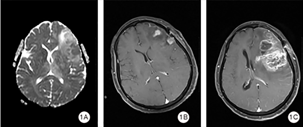 |
Figure 1 Radiological manifestations of Case 1. (A) The peritumoral ADC and tumor ADC values for the patient. (B) The enhanced T1-weighted image taken on the third day after surgery. (C) The follow-up enhanced T1-weighted image obtained on the 116th day after surgery. |
 |
Figure 2 Radiological manifestations of Case 2. (A) The peritumoral ADC and tumor ADC values for the patient. (B) The enhanced T1-weighted image taken on the fourth day post-surgery. (C) The follow-up enhanced T1-weighted image obtained on the 107th day after surgery. (D) The enhanced T1-weighted image on the 193rd day post-surgery. |
 |
Figure 3 ROC curve for determining critical values. |
Inflammatory Indexes
Peripheral blood samples were collected one week before surgery using a Mindray 7500CS fully automated blood analyzer. Hematological indicators included neutrophil, lymphocyte, monocyte, and platelet counts. Inflammatory indexes were calculated as follows: 1) SII: Platelet × neutrophil/lymphocyte count. 2) NLR: Neutrophil/lymphocyte count. 3) Platelet-to-Lymphocyte Ratio (PLR): Platelet/lymphocyte count. 4) Monocyte-to-Lymphocyte Ratio (MLR): Monocyte/lymphocyte count. 5) Systemic Inflammation Response Index (SIRI): Neutrophil × monocyte/lymphocyte count.
Outcome
Patients were divided into two groups based on early recurrence, defined as the appearance of new tumor tissue within six months after radical surgery:18 (1) Early Recurrence Group: recurrence within six months, and (2) Non-Early Recurrence Group: no recurrence within six months.
Statistical Analysis
Statistical analysis was conducted using SPSS 26.0 (IBM Corp., Armonk, NY, USA). The Kolmogorov–Smirnov test was used to assess normality, and the Levene test was applied to evaluate the homogeneity of variances. Continuous variables with a normal distribution were expressed as means ± standard deviations (SD) and were compared using the t-test, while non-normally distributed continuous variables were presented as medians with interquartile ranges (IQR) and were compared using the Wilcoxon rank-sum test. Categorical variables were expressed as frequencies and percentages and were compared using the chi-square test or Fisher’s exact test where appropriate. Inter-observer agreement was assessed by intraclass correlation coefficient (ICC) in 30 randomly selected cases, with ICC >0.85 indicating excellent consistency. Receiver operating characteristic (ROC) curve analysis was performed to convert continuous variables, such as hematological markers and tumor ADC values, into binary variables using optimal cutoff values. These binary variables were then compared between groups. To evaluate the predictive performance of ADC values, SII, and degree of edema for early recurrence, a predictive model was established using ROC curve analysis. The area under the curve (AUC) was calculated to assess diagnostic accuracy. A two-sided P < 0.05 was considered statistically significant in this study.
Results
A total of 68 patients with HGG were included in this study, among whom 20 patients (29.5%) were in the early recurrence group. Significant differences were observed between the two groups in terms of age (P = 0.029), tumor size (P = 0.047), degree of edema (P = 0.012), and pathological grade of the tumor (Table 1).
 |
Table 1 Comparison of Demographic Characteristics and Hematological Indicators Between Two Groups |
ICC analysis of a random sample of 30 cases showed excellent concordance, with ICC=0.91 (95% CI: 0.87–0.94) for the solid tumor component and ICC=0.88 (0.83–0.92) for peritumoral edema.
The optimal cutoff values for inflammatory indexes, tumor ADC values, and peritumor ADC values were determined using ROC curve analysis to convert these variables into binary categories. The results showed that the optimal cutoff values for the inflammatory indexes were as follows: NLR, 3.62; MLR, 0.275; PLR, 160.665; SII, 779.58; and SIRI, 1.345. The optimal cutoff values for tumor ADC values and peritumor ADC values were 1.136 and 1.783, respectively (Figure 1 and Table 2).
 |
Table 2 ROC Curve Parameter |
Further comparisons were made between the two groups regarding inflammatory indexes, tumor ADC values, and peritumor ADC values. The results showed that, compared with the non-early recurrence group, the early recurrence group had a higher proportion of cases with the following elevated values: NLR>3.62 (34:14 vs 6:14), MLR>0.275 (28:20 vs 6:14), PLR>160.665 (35:13 vs 5:15), SII>779.58 (35:13 vs 5:15), SIRI>1.345 (28:20 vs 5:15), tumor ADC values>1.136 (41:7 vs 10:10), and peritumor ADC values>1.783 (48:0 vs 17:3). The differences between the two groups in inflammatory indexes, tumor ADC values, and peritumor ADC values were statistically significant (all P < 0.05) (Table 1).
The AUC for tumor ADC values was 0.623, with a sensitivity of 0.500 and a specificity of 0.854. The AUC for the degree of edema was 0.652, with a sensitivity of 0.900 and a specificity of 0.458. The AUC for SII was 0.781, with a sensitivity of 0.750 and a specificity of 0.729. The combined model of these three indicators had an AUC of 0.823, with a sensitivity of 0.800 and a specificity of 0.708 (Figures 2–4 and Table 2).
 |
Figure 4 ROC curve for predicting early recurrence on the basis of the tumor ADC value, SII, and degree of edema. |
Discussion
This study demonstrates that the combination of the tumor ADC value, degree of edema, and SII provides acceptable predictive efficacy for early recurrence in HGG patients. The combined model achieved an AUC of 0.823, with sensitivity and specificity values of 80.0% and 70.8%, respectively, outperforming individual factors. These findings highlight the potential of integrating imaging and inflammatory markers as a noninvasive, cost-effective strategy for improving early recurrence prediction, enabling personalized treatment plans and better clinical outcomes.
The integration of imaging and inflammatory markers provides a more comprehensive assessment of tumor biology. Similar studies support this approach; for instance, He et al19 combined MRI-based radiomics with inflammatory markers to predict glioma recurrence, achieving AUCs over 0.90. The inclusion of multiple modalities allows for the evaluation of both tumor microstructure (via imaging biomarkers) and systemic tumor-host interactions (via inflammatory markers), leading to improved predictive performance.
This study found that SII was significantly higher in the early recurrence group than in the non-early recurrence group, with an AUC of 0.781 (sensitivity 0.075, specificity 0.729). These results were consistent with findings in other cancers. For example, a meta-analysis20 demonstrated that SII was an independent predictor of overall survival in glioblastoma. Similarly, studies showed that SII and other inflammatory markers, such as the NLR and PLR, were significantly associated with worse prognosis in gliomas.21,22 These studies emphasize the critical role of systemic inflammation in tumor progression and recurrence. Mechanistically, the SII reflects the balance between pro-tumor inflammatory responses and anti-tumor immune activity. Elevated neutrophils promote tumor growth by secreting cytokines, proteases, and reactive oxygen species that facilitate invasion and angiogenesis.23 Platelets protect circulating tumor cells from immune clearance and contribute to metastasis,24 while lymphocytes, key mediators of anti-tumor immunity, are often suppressed in patients with high SII values, leading to reduced immune resistance against tumor cells.25 Although neutrophils and lymphocytes are usually recruited synchronously, the imbalanced ratio after regulation can be partially reflected by SII. These mechanisms explain the association between high SII values and early postoperative recurrence observed in this study. Compared with NLR and PLR, SII combines the predictive strengths of both markers, making it a more comprehensive and reliable indicator of glioma outcomes.26,27 Interestingly, the critical SII value for predicting recurrence in this study was 779.58, which is consistent with values reported in some malignancies but higher than those in others (eg, gastric cancer or renal cell carcinoma).28 These differences may reflect the unique inflammatory and immune microenvironments of different tumor types, underscoring the need for tumor-specific research to establish optimal cutoff values for clinical application.
This study also demonstrated that imaging biomarkers, particularly tumor ADC values, provide valuable insights into tumor biology. In the early recurrence group, ten patients had tumor ADC values above the cutoff, whereas seven patients in the non-early recurrence group had tumor ADC values exceeding the cutoff. However, these findings conflict with prior research, which has demonstrated that lower ADC values are associated with higher tumor cell density and greater malignancy.29 For example, studies reported that patients with recurrent glioblastoma exhibited significantly lower ADC values due to increased tumor cell density and restricted water diffusion.30,31 Similarly, Kathleen M Schmainda reported that ADC values inversely correlated with tumor aggressiveness and were predictive of progression-free survival in glioblastoma patients.32 The reason for this difference may be related to the high heterogeneity of HGG. Although we tried to avoid the liquefaction and necrosis areas of the tumor as much as possible during delineation, due to the limitations of manual delineation, the recurrence group may still contain more necrotic areas, which usually have higher ADC values.33 The setting of scanning parameters may also have an impact on the measurement of ADC values. Research has shown that high b-value parameters are more valuable in predicting histological grading of gliomas and achieve higher consistency. Compared to high b-value, low b-value DWI images measure higher DWI images.34 In addition, We speculated that the observed discrepancy may be attributed to the small sample size of the study and the lack of adequate balance between groups. Further validation with larger sample sizes and more standardized ROI outlining strategies is needed to confirm these findings.
The moderate sensitivity of tumor ADC values in this study might be explained by the heterogeneity of gliomas. Factors such as necrosis, edema, and variations in tumor microstructure can influence ADC measurements, reducing their predictive power when used alone.35 However, the higher specificity (0.854) observed here suggested that ADC values are reliable indicators of aggressive tumor behavior, consistent with previous findings.
While both SII and tumor ADC values demonstrated predictive value individually, their combination with degree of edema significantly improved overall accuracy. Although prior studies have emphasized the significant prognostic value of peritumoral edema in gliomas,14,15 our research focuses on tumor edema and demonstrates its good predictive performance for early recurrence of HGG when combined with tumor ADC values and SII. Future studies could further optimize predictive models by incorporating additional biomarkers into the model-building process and employing machine learning techniques to explore more efficient and convenient predictive methods.
Limitations and Future Directions
This study has several limitations. First, as a retrospective, single-center study, there is potential for selection bias, and the results may not be generalizable to other populations. Future multicenter, prospective studies with larger sample sizes are needed to validate these findings. Second, this study focused on the SII, ADC value, and degree of edema, but other potentially valuable biomarkers (for example: other inflammation index such as NLR, MLR, PLR, SIRI) were not included. Future studies are adviced to explore the integration of additional biomarkers to further improve predictive models. Finally, the follow-up period in this study was limited to six months for assessing early recurrence, which may not capture late recurrences. Long-term follow-up studies are needed to provide a more comprehensive understanding of prognostic factors in HGG patients.
Conclusion
In conclusion, this study demonstrates that the combination of SII, tumor ADC value, and degree of edema is a valuable tool for predicting early recurrence in HGG patients. The integration of SII, tumor ADC value, and degree of edema offers a promising strategy for improving the prognostic accuracy in patients with gliomas. However, it is important to note that large samples and multicenter studies and multivariate analyses need to be validated before broad conclusions can be drawn. What’s more, further studies are required to explore additional biomarkers to refine the predictive models and optimize clinical decision-making.
Data Sharing Statement
All data generated or analysed during this study are included in this published article.
Ethics Approval and Patient Data Confidentiality
This work has been carried out in accordance with the Declaration of Helsinki (2000) of the World Medical Association. I confirm that all methods were performed in accordance with the relevant guidelines. The study was approved by the Medical Ethics Committee of the Second People’s Hospital of Hunan Province, and informed consent was exempted due to its retrospective nature. The researcher strictly adheres to China’s Personal Information Protection Law and ensures that the data is used only for research purposes. The researchers did not disclose any identifiable information. The original medical records remain sealed in the hospital archives and are not accessible to the research team.
Author Contributions
All authors made a significant contribution to the work reported, whether that is in the conception, study design, execution, acquisition of data, analysis and interpretation, or in all these areas; took part in drafting, revising or critically reviewing the article; gave final approval of the version to be published; have agreed on the journal to which the article has been submitted; and agree to be accountable for all aspects of the work.
Funding
This work was supported by Health research project of Hunan Provincial Health Commission 2023.
Disclosure
The authors declare that they have no competing interests.
References
1. Mohammadzadeh I, Hajikarimloo B, Niroomand B, et al. Application of artificial intelligence in forecasting survival in high-grade glioma: systematic review and meta-analysis involving 79,638 participants. Neurosurg Rev. 2025;48(1):240. doi:10.1007/s10143-025-03419-y
2. Koay JM, Michaelides L, Moniz-Garcia DP, et al. Repeated surgical resections for management of high-grade glioma and its impact on quality of life. J Neurooncol. 2024;167(2):267–273. doi:10.1007/s11060-024-04600-x
3. Chen H, Li C, Zheng L, Lu W, Li Y, Wei Q. A machine learning-based survival prediction model of high grade glioma by integration of clinical and dose-volume histogram parameters. Cancer Med. 2021;10(8):2774–2786. doi:10.1002/cam4.3838
4. Du P, Chen H, Lv K, Geng D. A survey of radiomics in precision diagnosis and treatment of adult gliomas. J Clin Med. 2022;11(13):3802. doi:10.3390/jcm11133802
5. Chaichana KL, Zadnik P, Weingart JD, et al. Multiple resections for patients with glioblastoma: prolonging survival. J Neurosurg. 2013;118(4):812–820. doi:10.3171/2012.9.JNS1277
6. Surov A, Pech M, Omari J, et al. Diffusion-weighted imaging reflects tumor grading and microvascular invasion in hepatocellular carcinoma. Liver Cancer. 2021;10(1):10–24. doi:10.1159/000511384
7. van den Elshout R, Scheenen TWJ, Driessen CML, Smeenk RJ, Meijer FJA, Henssen D. Diffusion imaging could aid to differentiate between glioma progression and treatment-related abnormalities: a meta-analysis. Insights Imaging. 2022;13(1):158. doi:10.1186/s13244-022-01295-4
8. Liu D, Li Z. Survival prediction using apparent diffusion coefficient values in recurrent glioblastoma under bevacizumab treatment: an updated systematic review and meta-analysis. Diagn Interv Radiol. 2024;30(4):270–274. doi:10.4274/dir.2024.232550
9. Kim SJ, Khadka D, Seo JH. Interplay between solid tumors and tumor microenvironment. Front Immunol. 2022;13:882718. doi:10.3389/fimmu.2022.882718
10. Kou J, Huang J, Li J, Wu Z, Ni L. Systemic immune-inflammation index predicts prognosis and responsiveness to immunotherapy in cancer patients: a systematic review and meta‑analysis. Clin Exp Med. 2023;23(7):3895–3905. doi:10.1007/s10238-023-01035-y
11. Liang R, Li J, Tang X, Liu Y. The prognostic role of preoperative systemic immune-inflammation index and albumin/globulin ratio in patients with newly diagnosed high-grade glioma. Clin Neurol Neurosurg. 2019;184:105397. doi:10.1016/j.clineuro.2019.105397
12. Satragno C, Schiavetti I, Cella E, et al. Systemic inflammatory markers and volume of enhancing tissue on post-contrast T1w MRI images in differentiating true tumor progression from pseudoprogression in high-grade glioma. Clin Transl Radiat Oncol. 2024;49:100849. doi:10.1016/j.ctro.2024.100849
13. Stepanenko AA, Sosnovtseva AO, Valikhov MP, et al. Systemic and local immunosuppression in glioblastoma and its prognostic significance. Front Immunol. 2024;15:1326753. doi:10.3389/fimmu.2024.1326753
14. Wang X, Chen D, Qiu J, Li S, Zheng X. The relationship between the degree of brain edema regression and changes in cognitive function in patients with recurrent glioma treated with bevacizumab and temozolomide. Quant Imaging Med Surg. 2021;11(11):4556–4568. doi:10.21037/qims-20-1084
15. Azemi G, Di Ieva A. Radiomic fingerprinting of the peritumoral edema in brain tumors. Cancers. 2025;17(3):478.
16. Long H, Zhang P, Bi Y, et al. MRI radiomic features of peritumoral edema may predict the recurrence sites of glioblastoma multiforme. Front Oncol. 2022;12:1042498. doi:10.3389/fonc.2022.1042498
17. Li J, Shu X, Xu J, et al. S100A9-CXCL12 activation in BRCA1-mutant breast cancer promotes an immunosuppressive microenvironment associated with resistance to immunotherapy. Nat Commun. 2022;13(1):1481. doi:10.1038/s41467-022-29151-5
18. Pignotti F, Ius T, Russo R, et al. Development and validation of a MRI-radiomics-based machine learning approach in high grade glioma to detect early recurrence. Front Oncol. 2024;14:1449235. doi:10.3389/fonc.2024.1449235
19. Guo J, Ren J, Shen J, Cheng R, He Y. Do the combination of multiparametric MRI-based radiomics and selected blood inflammatory markers predict the grade and proliferation in glioma patients? Diagn Interv Radiol. 2021;27(3):440–449. doi:10.5152/dir.2021.20154
20. Yang C, Hu BW, Tang F, et al. Prognostic value of Systemic Immune-Inflammation Index (SII) in patients with glioblastoma: a comprehensive study based on meta-analysis and retrospective single-center analysis. J Clin Med. 2022;11(24):7514. doi:10.3390/jcm11247514
21. Diem S, Schmid S, Krapf M, et al. Neutrophil-to-Lymphocyte ratio (NLR) and Platelet-to-Lymphocyte ratio (PLR) as prognostic markers in patients with non-small cell lung cancer (NSCLC) treated with nivolumab. Lung Cancer. 2017;111:176–181. doi:10.1016/j.lungcan.2017.07.024
22. Liu J, Li S, Zhang S, et al. Systemic immune-inflammation index, neutrophil-to-lymphocyte ratio, platelet-to-lymphocyte ratio can predict clinical outcomes in patients with metastatic non-small-cell lung cancer treated with nivolumab. J Clin Lab Anal. 2019;33(8):e22964. doi:10.1002/jcla.22964
23. Greten FR, Grivennikov SI. Inflammation and cancer: triggers, mechanisms, and consequences. Immunity. 2019;51(1):27–41. doi:10.1016/j.immuni.2019.06.025
24. Zhou L, Zhang Z, Tian Y, Li Z, Liu Z, Zhu S. The critical role of platelet in cancer progression and metastasis. Eur J Med Res. 2023;28(1):385. doi:10.1186/s40001-023-01342-w
25. Mao KY, Cao YC, Si MY, et al. Advances in systemic immune inflammatory indices in non-small cell lung cancer: a review. Medicine. 2024;103(18):e37967. doi:10.1097/MD.0000000000037967
26. Zhang Y, Sun Y, Zhang Q. Prognostic value of the systemic immune-inflammation index in patients with breast cancer: a meta-analysis. Cancer Cell Int. 2020;20:224. doi:10.1186/s12935-020-01308-6
27. Hu B, Yang XR, Xu Y, et al. Systemic immune-inflammation index predicts prognosis of patients after curative resection for hepatocellular carcinoma. Clin Cancer Res. 2014;20(23):6212–6222. doi:10.1158/1078-0432.CCR-14-0442
28. Lu Z, Zou J, Hu Y, et al. Serological markers associated with response to immune checkpoint blockade in metastatic gastrointestinal tract cancer. JAMA Network Open. 2019;2(7):e197621. doi:10.1001/jamanetworkopen.2019.7621
29. Li Z, Wang D, Sun J, Zhang G, Hu J. Diagnostic value of magnetic resonance diffusion-weighted imaging combined with routine scanning in body tumors. Contrast Media Mol Imaging. 2022;2022:5799815. doi:10.1155/2022/5799815
30. Kim BS, Kim ST, Kim JH, et al. Apparent diffusion coefficient as a predictive biomarker for survival in patients with treatment-naive glioblastoma using quantitative multiparametric magnetic resonance profiling. World Neurosurg. 2019;122:e812–e820. doi:10.1016/j.wneu.2018.10.151
31. Eoli M, Cuccarini V, Aquino D, et al. Apparent diffusion coefficient (ADC) decrease to predict longer survival in glioblastoma patients treated by dendritic cell immunotherapy plus standard of care. J Clin Oncol. 2017;35:2065. doi:10.1200/JCO.2017.35.15_suppl.2065
32. Schmainda KM. Diffusion-weighted MRI as a biomarker for treatment response in glioma. CNS Oncol. 2012;1(2):169–180. doi:10.2217/cns.12.25
33. Jajodia A, Goel V, Goyal J, et al. Combined diagnostic accuracy of diffusion and perfusion mr imaging to differentiate radiation-induced necrosis from recurrence in glioblastoma. Diagnostics. 2022;12(3):718. doi:10.3390/diagnostics12030718
34. Al-Agha M, Abushab K, Quffa K, Al-Agha S, Alajerami Y, Tabash M. Efficiency of high and standard b value diffusion-weighted magnetic resonance imaging in grading of gliomas. J Oncol. 2020;2020:6942406. doi:10.1155/2020/6942406
35. De Las Cuevas C, Peñate W. Psychometric properties of the eight-item Morisky Medication Adherence Scale (MMAS-8) in a psychiatric outpatient setting. Int J Clin Health Psychol. 2015;15(2):121–129. doi:10.1016/j.ijchp.2014.11.003
 © 2025 The Author(s). This work is published and licensed by Dove Medical Press Limited. The
full terms of this license are available at https://www.dovepress.com/terms
and incorporate the Creative Commons Attribution
- Non Commercial (unported, 4.0) License.
By accessing the work you hereby accept the Terms. Non-commercial uses of the work are permitted
without any further permission from Dove Medical Press Limited, provided the work is properly
attributed. For permission for commercial use of this work, please see paragraphs 4.2 and 5 of our Terms.
© 2025 The Author(s). This work is published and licensed by Dove Medical Press Limited. The
full terms of this license are available at https://www.dovepress.com/terms
and incorporate the Creative Commons Attribution
- Non Commercial (unported, 4.0) License.
By accessing the work you hereby accept the Terms. Non-commercial uses of the work are permitted
without any further permission from Dove Medical Press Limited, provided the work is properly
attributed. For permission for commercial use of this work, please see paragraphs 4.2 and 5 of our Terms.