Back to Journals » Drug Design, Development and Therapy » Volume 14
The NK-1R Antagonist Aprepitant Prevents LPS-Induced Oxidative Stress and Inflammation in RAW264.7 Macrophages
Authors Zhao X, Bai Z, Li C, Sheng C, Li H
Received 29 December 2019
Accepted for publication 7 April 2020
Published 20 May 2020 Volume 2020:14 Pages 1943—1952
DOI https://doi.org/10.2147/DDDT.S244099
Checked for plagiarism Yes
Review by Single anonymous peer review
Peer reviewer comments 2
Editor who approved publication: Dr Jianbo Sun
Xiao-nan Zhao,* Zhen-zi Bai,* Cheng-hua Li, Chuan-lun Sheng, Hong-yan Li
Department of Infectious, The Third Hospital of Jilin University, Changchun City, Jilin Province 130033, People’s Republic of China
*These authors contributed equally to this work
Correspondence: Hong-yan Li
Department of Infectious, The Third Hospital of Jilin University, No. 126, Sendai Avenue, Changchun City, Jilin Province 130033, People’s Republic of China
Tel +86-431-84995690
Email [email protected]
Background: The macrophage is one of the most important types of immune cells that protect against harmful stimuli. Macrophage activation plays a pivotal role in the progression and development of various inflammatory diseases. The neurokinin 1 receptor (NK-1R) is a G protein-coupled receptor that plays an important role in inflammatory diseases. Aprepitant is a kind of NK-1R antagonist. The purpose of this study is to determine the protective effect of aprepitant in lipopolysaccharide (LPS)-induced inflammatory responses in macrophages.
Methods: We examined the anti-inflammatory and anti-oxidant effects of aprepitant in LPS-treated RAW264.7 macrophages by using real-time PCR, ELISA, and Western blot analysis. We also assessed cellular oxidative stress signaling by measuring the levels of cellular MDA, total ROS, and NADPH oxidase expression. Cellular NO production was measured by DAF-FM DA staining. The inhibitory effect of aprepitant against NF-κB signaling was evaluated by luciferase assay and Western blot analysis.
Results: The expression of NK-1R is increased in LPS-induced macrophages, suggesting a potential role of the receptor in the inflammatory response. We show that aprepitant protects macrophages against oxidative stress by reducing the generation of ROS and the expression of NOX-4. Furthermore, aprepitant inhibits the secretion of pro-inflammatory cytokines and chemotactic factors by mediating the NF-κB signaling pathway.
Conclusion: The NK-1R receptor antagonist aprepitant acts as an anti-inflammatory agent, indicating that the blockage of the NK-1R pathway in macrophages has the potential to suppress inflammation.
Keywords: aprepitant, NK-1R, inflammation, NF-κB, oxidative stress
Introduction
Inflammatory response is a pathophysiological process against harmful stimuli called by pathogen infection or tissue damage.1 And it is well known that inflammation plays a vital role in various diseases such as cardiovascular disease, diabetes, and even cancers.2 Macrophages, which comprise a major component of the immune cell population, take an important part in innate immunity. Recent studies have corroborated macrophages participate in anti-inflammatory processes.3 Lipopolysaccharide (LPS) is a main component of the membrane from Gram-negative bacteria. It is well known that LPS can induce macrophages to differentiate into two kinds of phenotype – M1 and M2, which play different roles in the inflammation responses.4 Activated macrophages release various pro-inflammatory cytokines and chemotactic factors, such as tumor necrosis factor-α (TNF-α), interleukins (ILs),5 and matrix metalloproteinases (MMPs). And this process is upregulated by activating nuclear factor-κB (NF-κB) and mitogen-activated protein kinases (MAPKs) signaling pathways.6 Blockage of aberrant macrophage activation may become a promising therapeutic strategy in treating inflammatory disorders.
Neurokinin 1 receptor (NK-1R), a seven-transmembrane domain G-protein-coupled receptor, is an important member of three receptor hypotypes for tachykinins, regulates the function of the neuropeptide substance P (SP).7 NK-1R spreads widely in the nervous system and immune cells of respiratory and digestive tracts.8 The previous studies have corroborated that NK1R participates in important pathological and physiological processes including pain, inflammation, smooth muscle contraction, osteoblast differentiation, and intestinal fibrosis of colitis.9,10 Furthermore, the upregulation of NK1R has been found in malignant tumors such as malignant glioma,11 pancreatic cancer,12 and thyroid cancer. Aprepitant is a kind of neurokinin 1 receptor (NK-1R) antagonist permitted to impede vomiting and nausea caused by chemical therapy.13 As an NK-1R antagonist, aprepitant is safe and well tolerated for clinical use in most of the population.14 Furthermore, aprepitant has displayed a powerful anti-inflammatory capacity by suppressing the expression of chemokines and cytokines.15 However, the pharmacological function of aprepitant in LPS-induced macrophage activation has been less reported. The purposes of our study are to determine the anti-inflammatory effects of aprepitant in LPS-induced inflammatory responses in RAW264.7 macrophages.
Materials and Methods
Cell Culture and Treatment
RAW264.7 macrophages, acquired from the American Type Culture Collection (Manassas, USA), were maintained in Dulbecco’s Modified Eagle’s Medium (DMEM) containing 100 U/mL penicillin and 100 μg/mL streptomycin at 37°C in a 5% CO2 atmosphere. Cells were treated with 1 μg/mL LPS in the presence or absence of aprepitant (5, 10 μM) for 24 h.
Real-Time PCR Analysis
Total RNA was separated from R264.7 macrophages using the Qiazol (Qiagen, USA). cDNA was synthesized from total RNA using RT-PCR using a commercial cDNA synthesis kit (Bio-Rad, USA). One microgram of cDNA was used to measure the expression of target genes using a SYBR Green Master mix (Bio-Rad, USA). The relative levels of target gene were quantified by normalized to GAPDH using a comparative 2−ΔΔCT method and expressed as the fold induction. Primer sequences used for real-time PCR are shown in Table 1.
 |
Table 1 The Primers Sequences |
Western Blot Analysis
Cells cultured on 6-well plate were lysed with protein lysis buffer and broken by ultrasound. The supernatant was taken and the total protein concentration was measured by a BCA method. Separated by 100 g/L polyacrylamide gel electrophoresis, the supernatant was transferred to the nitrocellulose membrane with the 50 g/L skim milk powder chamber for 1 h. The membrane was washed and sequentially loaded with primary overnight at 4°C and secondary antibodies for 2 h at room temperature. The reaction was followed by 5 min after the addition of the chemiluminescence reagent, then exposed to X film, and the KODAK image was used to take analysis.16
Intracellular ROS Detection
Total cellular reactive oxygen (ROS) was measured to reflect the overall status of oxidative stress. In brief, the intracellular ROS level was determined by staining RAW264.7 cells with the ROS probe dihydroethidium (DHE). After washing 3 times, the cells were incubated with serum-free medium containing 5 µM DHE (Thermo Fisher Scientific, USA) for 30 min at a temperature of 37°C in darkness.16 The cells were then washed 3 times with PBS buffer, and the resulting fluorescent images were visualized using a fluorescence microscope. Image J software was used to analyze the fluorescence intensity to determine the concentration of ROS.
Determination of Malondialdehyde (MDA)
Levels of intracellular MDA in RAW264.7 cells were assessed to index lipid peroxidation based on a modification of the method described before.17 Cells were homogenized, followed by centrifugation at 1500 ×g for 10 min. Then, the cell lysis was added to a reaction mixture. After boiling for 1 h at 95°C and centrifugation at 3000 ×g for 10 min, OD value at 550 nm was recorded to index intracellular MDA.
Measurement of Nitric Oxide (NO)
Intracellular levels of NO were measured using DAF-FM staining approach as reported before.18 After treatment, cells were washed and loaded with 5 μM DAF-FM and incubated at 37°C for 20 min. After incubation, cells were washed three times by PBS at pH7.4. A confocal microscope equipped was used to asses the stained cells.
ELISA
Cells were treated with 1 μg/mL LPS in the presence or absence of aprepitant (5, 10 μM) for 24 h. We adopted the procedure of ELISA as reported before with a slight modification.18 In brief, cultured media were harvested to measure secreted levels of TNF-α, MCP-1, and PGE2. Cell lysates were used to measure MMP-2 and MMP-9 using commercial ELISA kits (R&D Systems, Beijing, China) according to the manufacturers’ instructions.
Luciferase Activity of NF-κB
RAW264.7 macrophage cells were cultured in a 12-well plate for 18 h. Cells were then transfected with plasmids containing NF-κB using Lipofectamine 2000 (Invitrogen, Carlsbad, CA, USA). After 24 h, cells were treated with 1 μg/mL LPS in the presence or absence of aprepitant (5, 10 μM) for 6 h. The luciferase activity of NF-κB was carried out using the commercial kit from Promega, USA according to the manufacturer’s instruction.19
Statistical Analysis
All results were presented as the mean ±SD. The differences were performed by the one-way ANOVA. Significance was defined as P-values<0.05.
Results
LPS Increases the Expression of NK-1R in Macrophages
To explore the function of aprepitant, we first treated macrophages with LPS and then measured the expression of NK-1R. As shown in Figure 1A, the mRNA levels increased to roughly 2.3-,3.1-, and 4.2-fold in response to stimulation with LPS (0.5, 1, 2 μg/mL) for 24 h. Meanwhile, the three doses of LPS increased the protein levels of NK-1R to 1.9-, 2.7-, and 3.6-fold (Figure 1B). Therefore, we confirmed that LPS increased both protein and mRNA levels of NK-1R in macrophages.
Aprepitant Protects Macrophages Against LPS-Induced Oxidative Stress
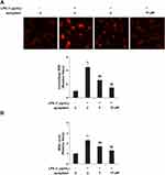
Intracellular ROS and MDA are typical indicators of oxidative stress. To examine the effects of aprepitant on oxidative stress, we measured on intracellular ROS and MDA levels. As shown in Figure 2A, intracellular ROS was increased by treatment with 1 μg/mL LPS to approximately 4.5-fold, which was significantly rescued by the two doses of aprepitant to only 2.6- and 1.5-fold, respectively. The MDA level increased to 2.3-fold by exposure to LPS. However, two doses of aprepitant decreased the MDA levels to 1.7- and 1.3-fold, respectively (Figure 2B). Macrophages treated with LPS alone resulted in high expression of NOX-2 and NOX-4 at both the mRNA (Figure 3A) and protein levels (Figure 3B), which were suppressed by aprepitant in a dose-dependent manner. Taken together, our data indicated that aprepitant suppressed LPS-induced oxidative stress.
Aprepitant Inhibits Expression of COX-2 and Secretion of PGE2 Induced by LPS
To investigate the effects of aprepitant on the expression of COX-2 and secretion of PGE2 induced by LPS, macrophages were pretreated with 5 and 10 μM aprepitant for 24 h, followed by stimulation with LPS. As shown in Figure 4A and B, the mRNA expression and protein secretion of COX-2 reduced to 4.3-and 3.4-fold. However, the two doses of aprepitant decreased the mRNA expression of COX-2 to 2.7- and 1.5- fold, while the secretion of protein was reduced to 2.2- and 1.4-fold, respectively. Concordantly, exposure to LPS alone increased the production of PGE2 to 1533.7 pg/mL, which was ameliorated by treatment with the two doses of aprepitant to concentrations of only 885.3 and 566.4 pg/mL (Figure 4C).
Aprepitant Prevents Expression of iNOS Induced by LPS in Macrophages
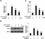
To determine the effects of aprepitant on the production of iNOS and NO induced by LPS, real-time PCR and Western blot analysis were employed. As shown in Figure 5A, the mRNA expression of iNOS was increased to 3.8-fold by exposure to LPS, which was decreased by treatment with 5 and 10 μM aprepitant to only 2.4- and 1.6-fold. At the protein level, LPS increased iNOS expression to 3.1-fold, which was decreased to 1.9- and 1.4-fold by aprepitant (Figure 5B). As shown in Figure 5C, NO production increased to 3.7-fold upon exposure to LPS, which was reduced to only 2.4 and 1.3-fold by the two doses of aprepitant.
Aprepitant Suppresses LPS-Induced Expression of Pro-Inflammatory Cytokines
When exposed to LPS alone, the mRNA expression of TNF-α was upregulated approximately 5.3-fold, while the protein secretion of TNF-α increased from 267.8 to 2098.9 pg/mL. Meanwhile, the mRNA of MCP-1 increased to 7.5-fold, protein secretion of MCP-1 was increased from 336.9 to 3058.7 pg/mL. However, these upregulations were significantly ameliorated by treatment with the two doses of aprepitant. The results in Figure 6A showed that mRNA levels of TNF-α were decreased to 3.2- and 1.9- fold, while MCP-1 expression was reduced to 4.5- and 2.3-fold, respectively. The results of Figure 6B showed that treatment with the two doses of aprepitant ameliorated protein secretion of TNF-α and MCP-1, reducing the protein concentration of TNF-α to 1566.5 and 1045.5, and that of MCP-1 to only 1877.2 and 1366.7 pg/mL, respectively. Thus, aprepitant is considered to have a potent anti-inflammatory effect.
Aprepitant Reduces LPS-Induced Expression of MMPs
To determine whether aprepitant treatment affects the expression of MMPs in macrophages induced by LPS, we investigated the expression of MMP2 and MMP9. The results in Figure 7A showed that the mRNA levels of MMP-2 and MMP-9 upregulated to 4.1- and 4.7-fold. Respectively, upon exposure to LPS alone. However, these levels were reduced to 2.9-, and 3.2-fold by 5 µM of aprepitant, respectively, while the dose of 10 µM further regulated these enzymes to only 2.3-, and 2.1-fold. The results in Figure 7B showed that the protein levels of MMP-2 and MMP-9 increased from 103.5 and 153.6 pg/mL to 1366.4 and 1988.3 pg/mL, respectively, upon exposure to LPS. The 5 µM aprepitant decreased these numbers to 923.6 and 1544.5 pg/mL, while the dose of 10 µM further reduced the productions to only 782.9 and 1222.6 pg/mL, respectively.
Aprepitant Mitigates Activation of NF-κB
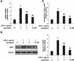
To determine the effects of aprepitant treatment on LPS-induced activation of NF-κB, we measured nuclear translocation of p65 and luciferase activity of NF-κB. As shown in Figure 8A, nuclear translocation of NF-κB p65 was increased to approximately 4.2-fold by treatment with LPS, which was significantly rescued by the two doses of aprepitant to only 2.7- and 1.6-fold, respectively. Then, we investigated the luciferase activity of NF-κB. Treatment with LPS increased it to 533.8-fold, which was rescued by the two doses of aprepitant to 366.8- and 125.6-fold, respectively (Figure 8B). The results indicated that aprepitant treatment can mitigate the activation of NF-κB.
Discussion
Chronic inflammation is testified to associate with the generation and development of cancer.20 For example, ulcerative colitis, which is a kind of chronic inflammation caused by intestinal flora, is clearly bound with the generation and development of colon cancer.21 Moreover, overexpression of NK-1R has been reported in the rectum and colon of patients with inflammatory bowel diseases,22 as well as in malignant glioma,11 pancreatic cancer,12 and thyroid cancer. These established cases built a relationship between NK-1R and inflammation. In our experiment, we used aprepitant to determine whether NK-1R antagonists affect the inflammatory response. As macrophages have been corroborated to play a key role in anti-inflammatory processes,3 we chose RAW264.7 macrophages for this experimentation.
Oxidative stress refers to the imbalance between oxidation and anti-oxidation in vivo. It has been confirmed that oxidative stress can lead to cell apoptosis,23 and is common in inflammation. Aprepitant treatment decreased the levels of ROS and MDA obviously. These findings suggested that aprepitant could protect macrophages by inhibiting oxidative stress. Furthermore, activated macrophages induced by LPS released various pro-inflammatory cytokines and chemokines24 such as MCP-1 and TNF-α. Excessive production of these factors takes an important part in affecting the development of inflammation. TNF-α is described as a primary inflammatory regulator in the pathogenesis of inflammation. For example, TNF-α is consumedly increased in synovial tissue in rheumatoid arthritis (RA).25 Interestingly, overexpression of NOX-4 has also been associated with the upregulation of TNF-α.26 In this study, the expression of these pro-inflammatory factors was decreased significantly by aprepitant. In addition, LPS stimulation activates COX-2 and iNOS transcription which lead to the overexpression of PGE2 and NO in macrophages, respectively.27 These inflammatory mediators are highly increased in inflammation.28 The results in this study show that aprepitant reduced PGE2 and NO production induced by LPS, due to its inhibition on the production of COX-2 and iNOS.
NF-κB-dependent signaling pathway is a key modulator in the development of inflammation. The LPS stimulation triggered the activation of NF-κB pathway. The phosphorylation of IκBα by upstream kinases is a critical process for the activation of NF-κB.29 NF-κB activation upregulated the expression of pro-inflammatory cytokines including TNF-α and MCP-1.30 Furthermore, NF-κB modulated the production of PGE2 and NO via regulating the expression of regulatory enzymes COX-2 and iNOS. NF-κB pathway has been considered as an important target for the treatment of inflammation. Our data showed that aprepitant inhibited the activation of NF-κB via modulating phosphorylation of IκBα.
Aprepitant treatment reversed substance P- and CCL5-mediated monocyte chemotaxis by interacting with a specific NK-1R isoform,31 suggesting that substance-dependent NK-1R signaling regulates monocyte function. Our study shows that LPS treatment directly induces the expression of NK-1R, indicating that LPS stimuli could activate NK-1R signaling in macrophages. Furthermore, we show that aprepitant-mediated blockage of NK-1R suppresses LPS-induced inflammation. These data indicate that aprepitant-mediated NK-1R inhibition in myeloid cells could occur through a substance P-dependent or -independent mechanism. Recently, NK-1R has been shown to influence the early fever response induced by LPS in mice.32 The blockage of the NK-1R pathway significantly attenuated LPS-induced systematic inflammation in vivo.33 Thus, NK-1R signaling is an important regulatory pathway of LPS stimuli.
In conclusion, this study investigates the effect of aprepitant in LPS-induced macrophages. The results show that aprepitant inhibits the oxidative stress via suppressing ROS and MDA. Furthermore, aprepitant decreases the production of pro-inflammatory cytokines and chemokines, reduces the expression of regulatory enzymes. In addition, aprepitant inhibits the activation of NF-κB pathway, which regulates the production of inflammatory factors mentioned above. Our data indicate that NK-1R antagonist such as aprepitant is a potent anti-inflammatory agent in vitro cultured macrophages.
Ethical Statement
RAW264.7 macrophages were acquired from the American Type Culture Collection (Manassas, USA). Experimental protocols were approved by Jilin University.
Disclosure
The authors report no conflicts of interest in this work.
References
1. Nathan C, Ding A. Nonresolving inflammation. Cell. 2010;140(6):871–882. doi:10.1016/j.cell.2010.02.029
2. Reuter S, Gupta SC, Chaturvedi MM, Aggarwal BB. Oxidative stress, inflammation, and cancer: how are they linked? Free Radic Biol Med. 2010;49(11):1603–1616. doi:10.1016/j.freeradbiomed.2010.09.006
3. Lee HT, Kim SK, Kim SH, et al. Transcription-related element gene expression pattern differs between microglia and macrophages during inflammation. Inflamm Res. 2014;63(5):389–397. doi:10.1007/s00011-014-0711-y
4. Mosser DM, Edwards JP. Exploring the full spectrum of macrophage activation. Nat Rev Immunol. 2008;8(12):958–969. doi:10.1038/nri2448
5. Shin JS, Hong Y, Lee HH, et al. Fulgidic Acid isolated from the rhizomes of cyperus rotundus suppresses LPS-Induced iNOS, COX-2, TNF-α, and IL-6 expression by AP-1 inactivation in RAW264.7 macrophages. Biol Pharm Bull. 2015;38(7):1081–1086. doi:10.1248/bpb.b15-00186
6. Jeong YH, Oh YC, Cho WK, Yim NH, Ma JY. Anti-Inflammatory effect of rhapontici radix ethanol extract via inhibition of NF-κB and MAPK and induction of HO-1 in macrophages. Mediators Inflamm. 2016;2016:7216912. doi:10.1155/2016/7216912
7. Schank JR, Heilig M. Substance P and the neurokinin-1 receptor: the New CRF. Int Rev Neurobiol. 2017;136:151–175.
8. Kook YA, Lee SK, Son DH, et al. Effects of substance P on osteoblastic differentiation and heme oxygenase-1 in human periodontal ligament cells. Cell Biol Int. 2009;33(3):424–428. doi:10.1016/j.cellbi.2008.12.007
9. Koon HW, Shih D, Karagiannides I, et al. Substance P modulates colitis-associated fibrosis. Am J Pathol. 2010;177(5):2300–2309. doi:10.2353/ajpath.2010.100314
10. Ebner K, Muigg P, Singewald G, Singewald N. Substance P in stress and anxiety: NK-1 receptor antagonism interacts with key brain areas of the stress circuitry. Ann N Y Acad Sci. 2008;1144(1):61–73. doi:10.1196/annals.1418.018
11. Mou L, Kang Y, Zhou Y, Zeng Q, Song H, Wang R. Neurokinin-1 receptor directly mediates glioma cell migration by up-regulation of matrix metalloproteinase-2 (MMP-2) and membrane type 1-matrix metalloproteinase (MT1-MMP). J Biol Chem. 2013;288(1):306–318. doi:10.1074/jbc.M112.389783
12. Friess H, Zhu Z, Liard V, et al. Neurokinin-1 receptor expression and its potential effects on tumor growth in human pancreatic cancer. Lab Invest. 2003;83(5):731–742. doi:10.1097/01.LAB.0000067499.57309.F6
13. Patel L, Lindley C. Aprepitant-a novel NK1-receptor antagonist. Expert Opin Pharmacother. 2003;4(12):2279–2296. doi:10.1517/14656566.4.12.2279
14. Quartara L, Altamura M. Tachykinin receptors antagonists: from research to clinic. Curr Drug Targets. 2006;7(8):975–992. doi:10.2174/138945006778019381
15. Liu X, Zhu Y, Zheng W, Qian T, Wang H, Hou X. Antagonism of NK-1R using aprepitant suppresses inflammatory response in rheumatoid arthritis fibroblast-like synoviocytes. Artif Cells Nanomed Biotechnol. 2019;47(1):1628–1634. doi:10.1080/21691401.2019.1573177
16. Wang C, Shao L, Pan C, et al. Elevated level of mitochondrial reactive oxygen species via fatty acid β-oxidation in cancer stem cells promotes cancer metastasis by inducing epithelial-mesenchymal transition. Stem Cell Res Ther. 2019;10(1):175–186. doi:10.1186/s13287-019-1265-2
17. Lee SI, Kang KS, Kang KS. Omega-3 fatty acids modulate cyclophosphamide induced markers of immunosuppression and oxidative stress in pigs. Sci Rep. 2019;9(1):2684–2691. doi:10.1038/s41598-019-39458-x
18. Ashley JW, Hancock WD, Nelson AJ, et al. Polarization of macrophages toward M2 phenotype is favored by reduction in iPLA2β (group VIA phospholipase A2). J Biol Chem. 2016;291(44):23268–23281. doi:10.1074/jbc.M116.754945
19. Zheng W, Pan H, Wei L, Gao F, Lin X. Dulaglutide mitigates inflammatory response in fibroblast-like synoviocytes. Int Immunopharmacol. 2019;74:105649. doi:10.1016/j.intimp.2019.05.034
20. Weitzman SA, Gordon LI. Inflammation and cancer: role of phagocyte-generated oxidants in carcinogenesis. Blood. 1990;76(4):655–663. doi:10.1182/blood.V76.4.655.655
21. Rosso M, Muñoz M, Berger M. The role of neurokinin-1 receptor in the microenvironment of inflammation and cancer. ScientificWorldJournal. 2012;2012:381434.
22. O’Connor TM, O’Connell J, O’Brien DI, Goode T, Bredin CP, Shanahan F. The role of substance P in inflammatory disease. J Cell Physiol. 2004;201(2):167–180. doi:10.1002/jcp.20061
23. Zhou X, Zhang HL, Gu GF, et al. Investigation of the relationship between chromobox homolog 8 and nucleus pulposus cells degeneration in rat intervertebral disc. In Vitro Cell Dev Biol Anim. 2013;49(4):279–286. doi:10.1007/s11626-013-9596-2
24. Guha M, Mackman N. LPS induction of gene expression in human monocytes. Cell Signal. 2001;13(2):85–94. doi:10.1016/S0898-6568(00)00149-2
25. Gu X, Gu B, Lv X, et al. 1, 25-dihydroxy-vitamin D3 with tumor necrosis factor-alpha protects against rheumatoid arthritis by promoting p53 acetylation-mediated apoptosis via Sirt1 in synoviocytes. Cell Death Dis. 2016;7(10):e2423. doi:10.1038/cddis.2016.300
26. Phull AR, Nasir B, Haq IU, Kim SJ. Oxidative stress, consequences and ROS mediated cellular signaling in rheumatoid arthritis. Chem Biol Interact. 2018;281:121–136. doi:10.1016/j.cbi.2017.12.024
27. Kiemer AK, Hartung T, Huber C, Vollmar AM. Phyllanthusamarus has anti-inflammatory potential by inhibition of iNOS, COX-2, and cytokines via the NF-κB pathway. J Hepatol. 2003;38(3):289–297. doi:10.1016/S0168-8278(02)00417-8
28. Pan MH, Lai CS, Wang YJ, Ho CT. Acacetin suppressed LPS-induced up-expression of iNOS and COX-2 in murine macrophages and TPA-induced tumor promotion in mice. Biochem Pharmacol. 2006;72(10):1293–1303. doi:10.1016/j.bcp.2006.07.039
29. Johnson GL, Lapadat R. Mitogen-activated protein kinase pathways mediated by ERK, JNK, and p38 protein kinases. Science. 2002;298(5600):1911–1912. doi:10.1126/science.1072682
30. Tak PP, Firestein GS. NF-κB: a key role in inflammatory diseases. J Clin Invest. 2001;107(1):7–11. doi:10.1172/JCI11830
31. Chernova I, Lai JP, Li H, et al. (SP) enhances CCL5-induced chemotaxis and intracellular signaling in human monocytes, which express the truncated neurokinin-1 receptor (NK1R). J Leukoc Biol. 2009;85(1):154–164. doi:10.1189/jlb.0408260
32. Pakai E, Tekus V, Zsiboras C, et al. The neurokinin-1 receptor contributes to the early phase of lipopolysaccharide-induced fever via stimulation of peripheral cyclooxygenase-2 protein expression in mice. Front Immunol. 2018;5(9):166. doi:10.3389/fimmu.2018.00166
33. Fulenwider HD, Smith BM, Nichenko AS, et al. Cellular and behavioral effects of lipopolysaccharide treatment are dependent upon neurokinin-1 receptor activation. J Neuroinflammation. 2018;15(1):60. doi:10.1186/s12974-018-1098-4
 © 2020 The Author(s). This work is published and licensed by Dove Medical Press Limited. The full terms of this license are available at https://www.dovepress.com/terms.php and incorporate the Creative Commons Attribution - Non Commercial (unported, v3.0) License.
By accessing the work you hereby accept the Terms. Non-commercial uses of the work are permitted without any further permission from Dove Medical Press Limited, provided the work is properly attributed. For permission for commercial use of this work, please see paragraphs 4.2 and 5 of our Terms.


