
Back to Journals » Journal of Inflammation Research » Volume 19
The Interaction Between Ferroptosis and Necroptosis in Acute and Chronic Kidney Diseases
Authors Song C, Chen F, Fang Y, Ding X, Han Q, Duan Y, Ren Q, Zhang D, Zhu H
Received 17 August 2025
Accepted for publication 24 February 2026
Published 23 March 2026 Volume 2026:19 559198
DOI https://doi.org/10.2147/JIR.S559198
Checked for plagiarism Yes
Review by Single anonymous peer review
Peer reviewer comments 2
Editor who approved publication: Professor Ning Quan
Chenwen Song,1,2,* Fei Chen,1,2,* Yi Fang,3,* Xiaonan Ding,1,2,* Qiuxia Han,4 Yingjie Duan,1,2 Qinqin Ren,1,2 Dong Zhang,1,2 Hanyu Zhu1,2
1Department of Nephrology, First Medical Center of Chinese PLA General Hospital, State Key Laboratory of Kidney Diseases, National Clinical Research Center for Kidney Diseases, Beijing Key Laboratory of Medical Devices and Integrated Traditional Chinese and Western Drug Development for Severe Kidney Diseases, Beijing, 100853, People’s Republic of China; 2Innovation Team and Talents Cultivation Program of National Administration of Traditional Chinese Medicine. (No: ZYYCXTD-D-202402), Beijing, 100853, People’s Republic of China; 3College of Medical, Nankai University, Tianjin, 300071, People’s Republic of China; 4Department of Nephrology, Beijing Chao-Yang Hospital, Capital Medical University, Beijing, 100020, People’s Republic of China
*These authors contributed equally to this work
Correspondence: Hanyu Zhu; Dong Zhang, Email [email protected]; [email protected]
Abstract: Kidney disease represents a major non-communicable disease characterized by complex pathogenesis and limited therapeutic options. Current research has revealed multiple underlying mechanisms, including ferroptosis and necroptosis, which play important roles in acute kidney injury (AKI) and chronic kidney disease (CKD). Ferroptosis is an iron-dependent form of programmed cell death caused by the accumulation of lipid reactive oxygen species and is characterized by iron ion aggregation, lipid peroxidation, and excessive oxidative stress. Necroptosis is a regulated form of necrosis mediated by RIPK1–RIPK3 and is characterized by the recruitment and phosphorylation of the pseudokinase mixed lineage kinase domain-like protein (MLKL). Ferroptosis and necroptosis play important roles in various diseases such as tissue injury, cancer, and neurodegenerative diseases. Due to the intricate architecture of the kidney, the convergence of multiple systemic pathogenic factors, and the interactive regulation of intercellular signaling pathways, renal diseases exhibit complex pathogenesis and present limited therapeutic interventions. Exploring cell death and the interactions between different forms of cell death is highly important for understanding the occurrence and development of kidney diseases and for finding new treatment strategies. Ferroptosis and necroptosis influence renal cell viability and contribute to the exacerbation of kidney injury via inflammatory responses and additional mechanisms. They share common initiating factors and intersecting signaling pathways in the context of kidney diseases, thereby synergistically intensifying the pathological progression of renal damage. This article describes the pathogenesis and pathophysiological roles of ferroptosis, necroptosis, and their interactions in kidney diseases such as AKI and CKD and elucidates the potential of inhibiting ferroptosis and necroptosis, or their combined inhibition, in the prevention and treatment of kidney diseases such as AKI and CKD.
Keywords: ferroptosis, necroptosis, lipid peroxidation, kidney disease, cell death
Molecular Mechanism of Ferroptosis and Its Role in Nephropathy
Molecular Mechanism
Iron metabolism imbalance includes iron accumulation and iron deficiency, both of which have significant implications for renal function.1 Excessive iron accumulation results in the presence of “free iron”, which acts as a catalyst for lipid peroxidation within renal cells, thereby inducing ferroptosis.2 Conversely, iron deficiency impairs erythropoiesis, leading to renal anemia and exacerbating hypoxic conditions within the kidney.3 Ferritinophagy and lipid peroxidation respectively represent the initiation and culmination of ferroptosis. Ferritin, the principal intracellular iron storage protein, releases iron through ferritinophagy in response to kidney pathology, elevating free iron concentrations and triggering ferroptotic pathways.4 Lipid peroxidation, the terminal event in ferroptosis, involves the iron-catalyzed oxidation of polyunsaturated fatty acids in renal cells, generating lipid peroxides that compromise cellular membrane integrity, culminating in cell lysis, tubular necrosis, and impaired renal function.1 In ischemia-reperfusion injury, ischemic insult leads to ferritin release from damaged renal cells, and subsequent reperfusion introduces oxygen that accelerates iron-mediated lipid peroxidation.5 Ferritinophagy and lipid peroxidation are implicated in drug-induced nephrotoxicity, sepsis-associated AKI, and chronic glomerulonephritis.6,7 In diabetic nephropathy, hyperglycemia promotes the generation of reactive oxygen species, thereby intensifying lipid peroxidation and advancing renal fibrosis.8 The glutathione peroxidase 4 (GPX4)/nuclear factor erythroid 2–related factor 2 (Nrf2) signaling axis constitutes a pivotal molecular link between renal injury mechanisms and therapeutic strategies.6,9,10 Targeting these pathways offers a promising approach to inhibit ferroptosis and ameliorate the progression of kidney diseases.(Table 1)
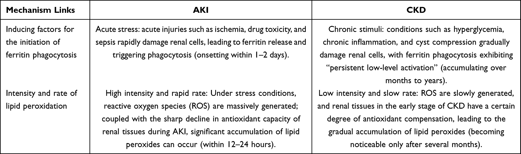 |
Table 1 The Mechanisms of Ferroptosis in Acute Kidney Injury and Chronic Kidney Diseases |
Iron Metabolism Imbalance
The body’s iron metabolism homeostasis is maintained by the continuous and stable expression of various proteins. For instance, ferritin reduces free iron levels by storing iron through its light (FTL) and heavy (FTH) chains. Ferroportin 1 (FPN1) is the only known protein responsible for exporting intracellular iron in mammals. Hepcidin, a liver-secreted homologous receptor, tightly controls serum iron levels through the hepcidin-FPN axis.11 When this balance is disturbed, ferroptosis can be triggered. Under normal conditions, Fe2⁺ generated from internal absorption and red blood cell breakdown is oxidized to Fe3⁺. Fe3⁺ enters cells by binding to transferrin (TF), forming a “TF-Fe3⁺-TFR1” complex with transferrin receptor 1 (TFR1). Inside the cell, Fe3⁺ is converted back to Fe2⁺ and stored in the labile iron pool (LIP). Most intracellular iron resides in the LIP, which supports functions like mitochondrial oxygen transport, energy metabolism, and iron–sulfur protein synthesis.12–14 Under pathological conditions, Fe3⁺ tends to undergo single electron transfer reactions, converting into Fe2⁺. The buildup of Fe2⁺ can drive the Fenton reaction, producing excessive reactive oxygen species (ROS) such as hydrogen peroxide and hydroxyl radicals. This leads to lipid peroxidation, tissue damage, and ultimately, ferroptosis.15
Ferritinophagy
Nuclear receptor coactivator 4 (NCOA4)-mediated ferritinophagy is a type of autophagy used for the lysosomal degradation of ferritin, and ferritin can release iron into the cytoplasm through autophagy.16 A study identified the proteins involved in autophagosomes through quantitative proteomics and revealed that NCOA4 is highly expressed in autophagosomes.17 In addition, as a specific receptor involved in autophagy, NCOA4 binds to ferritin and delivers it to the lysosome for degradation, leading to the release of free iron and an increase in the intracellular LIP level. Under conditions of cystine deprivation, ferritinophagy is activated, promoting the degradation of ferritin regulated by NCOA4. When ferritin is degraded, ROS accumulate, the content of unstable iron in the cells increases, and then ferroptosis is triggered.18 Therefore, the ferritinophagy pathway is one of the targets for enhancing the sensitivity of tumor cells to ferroptosis.
Lipid Peroxidation
Polyunsaturated fatty acids (PUFAs) are important components of the cell membrane and play important roles in signal transduction. This signaling often occurs through oxygenation reactions. Phospholipids containing polyunsaturated fatty acids (PUFA-PLs), it can drive the occurrence of ferroptosis.1 Unregulated and excessive activation of lipid peroxidation is one of the hallmarks of ferroptosis. The diallyl groups between the carbon-carbon double bonds in the PUFA acyl groups have a low bond dissociation energy, which is conducive to hydrogen extraction (ie, the removal of hydrogen ions), forming carbon-centered radicals. These radicals subsequently react with molecular oxygen to generate peroxyl radicals.19 When the peroxidation products cannot be converted into lipid hydroperoxides and reduced to the corresponding alcohols, the radical-mediated reaction leads to the formation of numerous secondary products, which disrupt the integrity of the membrane and ultimately cause the rupture of organelle membranes and the cell membrane.20 Some studies suggest that the critical level of lipid peroxides is a possible threshold for ferroptosis in different cell lines, and exogenous supplementation of lipid hydroperoxides to this threshold can increase the sensitivity of cells to RSL3-induced ferroptosis.21 Therefore, it is speculated that biological membranes with higher contents of PUFA-PLs are more susceptible to peroxidation, thereby causing cell damage. Through genome-wide screening and CRISPR/Cas9 technology screening, some studies have identified acyl-CoA synthetase long-chain family member 4 (ACSL4) and lysophosphatidylcholine acyltransferase 3 (LPCAT3) as important drivers of ferroptosis.22–25 In the context of ferroptosis, lipid peroxidation mainly affects esterified PUFAs and does not affect free PUFAs. Therefore, the activation of ACSL4 preferentially catalyzes the conversion of long-chain PUFAs into acyl-CoA esters and, to a certain extent, promotes the reacylation of AA into LPCAT3, which produces phospholipid peroxide.26 Lipid peroxidation is also mediated by lipoxygenase (LOX). An increase in the production of lipid hydroperoxides regulated by LOX can increase the sensitivity to ferroptosis.27 The excessive activation of ACSL4, LPCAT, and LOX results in the production of large amounts of phospholipid peroxides, thereby triggering ferroptosis. Some studies have used mitochondrial-targeted antioxidants SkQ1 and MitoTEMPO to prove that they may prevent erasing-induced ferroptosis by inhibiting mitochondrial lipid peroxidation, suggesting that mitochondrial lipid peroxidation may be an important cause of ferroptosis.28
GSH/System Xc−/ GPX4 Pathway
Glutathione (GSH) is a tripeptide composed of glutamic acid, cysteine, and glycine and is present in all mammalian tissues, especially the liver. GSH is the most abundant antioxidant in organisms, with most being in the cytoplasm and a small part being in the mitochondria and the endoplasmic reticulum. In the human body, GSH exists in two forms: reduced GSH and oxidized GSSH. Under normal circumstances, the content of GSSH in the human body is very low, and GSH mainly plays an antioxidant role.11 Notably, mitochondrial GSH (2–3mM) exists in a more oxidized state, uniquely adapted to buffer superoxide from Complex III without disrupting matrix redox signaling. Due to the limited concentration of cysteine in cells, cysteine is considered the rate-limiting precursor for GSH synthesis. Intracellular glutamate and extracellular cystine are exchanged at equal ratios. Cystine enters the cell through the Xc− system and is reduced to cysteine through a thioredoxin reductase 1 (TXNRD1)-dependent pathway, which then promotes the production of GSH.15,29,30
KEAP1 - Nrf2 Pathway
NRF2 is a stress-induced transcription factor. Under physiological conditions, the cytoplasmic protein kelch-like ECH-associated protein 1 (keap1) binds to NRF2 and inhibits its activation by mediating its ubiquitination and degradation. After exposure to electrophilic or oxidative stress, the cysteine residues of keap1, especially C151, C226, C273, and C288, are oxidized and modified, causing a conformational change that inhibits keap1-mediated NRF2 degradation.10 NRF2 is released from keap1, forms a complex with keap1, and translocates to the nucleus, where it binds to the antioxidant response elements in the promoters of target genes. Numerous studies have identified many target genes of NRF2 and revealed the pleiotropic functions of NRF2 beyond its redox regulation function. In addition, as an adaptor protein involved in selective autophagy and a target of NRF2, p62/SQSTM1 competitively binds to keap1, subsequently promoting the activation of NRF2. The phosphorylation of P62 significantly increases the binding affinity of p62 to keap1. Therefore, the p62 - keap1 - NRF2 axis forms a feedback loop that positively regulates the cytoprotective effect of NRF2.31 NRF2 is considered the main regulator of the antioxidant response because many of its downstream target genes are involved in preventing or correcting redox imbalances in cells. Appropriate NRF2 function is crucial for cell survival, especially in cells under conditions of oxidative stress or iron homeostasis imbalance. Therefore, NRF2 plays an important role in maintaining the cellular redox balance and preventing ferroptosis. The pathways by which NRF2 activity inhibits ferroptosis can be divided into the following three categories: regulation of iron/heme metabolism, GSH synthesis/metabolism, and the production pathways of intermediate metabolites.32–34 NRF2 upregulates the transcription of a group of genes related to the regulation of heme synthesis and transformation, such as heme oxygenase 1 (HO-1), ferrochelatase (FECH), and SLC48A1. These studies indicate that NRF2 is very important for maintaining iron/heme homeostasis. In addition to iron and heme, many genes related to GSH synthesis and metabolism are controlled by NRF2. GPX4, the genes involved in GSH synthesis such as γ-glutamylcysteine synthetase, GCLC, GCLM, and SLC7A11, are known targets of NRF2. Therefore, the activation of NRF2 is expected to protect cells from ferroptosis. In addition, NRF2 can also regulate the transcriptional expression of genes involved in intermediate metabolites, some of which play an important role in the regeneration of NADPH, a key electron donor required for reducing oxidized substrates. However, some research results suggest that NRF2 positively regulates other pathways to induce ferroptosis. For example, HO-1 exhibits dual abilities to promote or inhibit ferroptosis. Excessive activation of NRF2 will promote HO-1-mediated and iron-catalyzed ROS generation and induce ferroptosis.35 Therefore, a detailed understanding of the transcriptional mechanisms by which NRF2 regulates the expression of different target genes under different conditions and in different tissues is needed to fully utilize the ability of NRF2 to defend against ferroptosis and apply it to the treatment of human diseases.
Pathological Effects
Synchronous Ferroptosis of Renal Tubular Cells in AKI (PAF/PAF-LPL Signaling)
In 2014, Skouta et al demonstrated that the classic ferroptosis inhibitor ferrostatin-1 (Fer-1) has a protective effect on AKI of the proximal tubules in the kidneys of mice induced by rhabdomyolysis.36 In the same year, Friedmann Angeli et al caused a large number of deaths of renal tubular epithelial cells in mice by inducing the depletion of GPX4.9 In 2017, a study found that inhibiting ferroptosis could prevent kidney injury caused by folic acid-induced AKI (FA-AKI), while targeting apoptosis or necroptosis had no protective effect on FA-AKI. These findings suggest that ferroptosis is the main mechanism of cell death in the FA-AKI model, possibly the first activated cell death pathway in FA-AKI, and may also be a driving factor for other forms of cell death such as necroptosis.37 In 2024, new research has shown that synchronized iron apoptosis mediated by platelet-activating factor (PAF) and PAF-like phospholipids (PAF-LPLs) leads to nephron loss, which causes acute kidney injury. The presence of PAF and PAF-LPLs destabilizes the biofilm and signals the death of adjacent cells. This chain reaction can be blocked by PAF-acetylhydrolase (II) (PAFAH 2) or by administering antibodies against PAF. Knockout or pharmacological inhibition of PAFAH 2 increases PAF production, enhances synchronous iron apoptosis, and aggravates ischemia/reperfusion (I/R) -induced AKI.38
Fibrosis and Ferroptosis-Related Inflammation in CKD Progression
In 2023, the interferon gene-stimulating factor STING was shown to be regulated by ACSL 4-dependent ferroptosis, inflammation, and fibrosis in human kidney cells (HK-2 cell line), which in turn led to CKD and induced immune cell infiltration, tubular atrophy, and collagen accumulation.7 In 2024, an abnormally high expression of the key marker of ferroptosis, ACSL 4, was detected in the renal tubules of CKD patients (IgAN, MN, FSGS, LN, and DN) and adenine or UUO-induced CKD mice, as well as in damaged TCMK-1 cells.39 Moreover, a study showed that UUO in unilateral ureteral obstruction and unilateral ischemia-reperfusion induced iron apoptosis in renal tubular epithelial cells by increasing glutathione peroxidase 4 (GPX 4) protein levels and inhibiting lipid peroxidation in mouse kidneys. In vitro, vitexin treatment inhibited erasing-induced iron apoptosis in HK-2 cells and induced the expression of type I collagen and α-smooth muscle actin (α-SMA) in NRK-49 F cells. This is achieved by activating the NRF 2/heme oxygenase-1 (HO-1) pathway, inhibiting KEAP 1 and ubiquitination-mediated NRF 2 degradation, thereby increasing the expression of GPX4 and further inhibiting lipid peroxidation and iron-mediated apoptosis. In addition, knockout of NRF 2 strongly inhibited the antiapoptotic effects of vitexin.40
Molecular Mechanisms of Necroptosis and Its Role in AKI and CKD
Vital Pathways
Necroptosis exerts a dual detrimental effect on renal pathology. On the one hand, it directly compromises the structural integrity of renal tubules and glomeruli, resulting in clinical manifestations such as glycosuria, proteinuria, and hematuria, which collectively contribute to impaired renal function.41 On the other hand, necroptosis initiates a self-perpetuating inflammatory cascade characterized by the sequence: cell necrosis inflammation further necrosis. The activation of inflammasomes serves as the primary trigger for necroptosis; early necroptotic signals stimulate inflammasomes within renal cells, facilitating the processing of pro-inflammatory cytokines and thereby establishing a pro-inflammatory milieu.42 Subsequently, the release of inflammatory cytokines acts as an amplifier of tissue damage, directly targeting adjacent healthy renal cells, recruiting systemic immune cells to the kidney, and promoting the accumulation of inflammatory mediators and ROS, which exacerbate renal injury.43 Under ischemic conditions, renal tubular cells undergo necroptosis, releasing adenosine triphosphate (ATP) and high-mobility group box 1 (HMGB1), which activate the NOD-like receptor pyrin domain-containing 3 (NLRP3) inflammasome. During reperfusion, the abrupt reintroduction of oxygen enhances the secretion of inflammatory cytokines such as interleukin-1β (IL-1β) and tumor necrosis factor-α (TNF-α), thereby intensifying renal tubular necrosis.44 In the contexts of drug-induced and sepsis-associated AKI, both inflammasome activation and inflammatory cytokine release are pivotal processes mediating necroptosis. Moreover, these mechanisms are also implicated in CKD, including diabetic nephropathy, chronic glomerulonephritis, and lupus nephritis. The receptor-interacting serine/threonine-protein kinase 1 (RIPK1)/receptor-interacting serine/threonine-protein kinase 3 (RIPK3)/mixed lineage kinase domain-like pseudokinase (MLKL) signaling axis constitutes the central regulatory pathway of necroptosis.45 Targeting this core signaling cascade holds promise for elucidating the precise molecular mechanisms underlying renal cell necrosis, identifying novel therapeutic targets for precision medicine, and discovering potential biomarkers to guide disease prognosis. (Table 2)
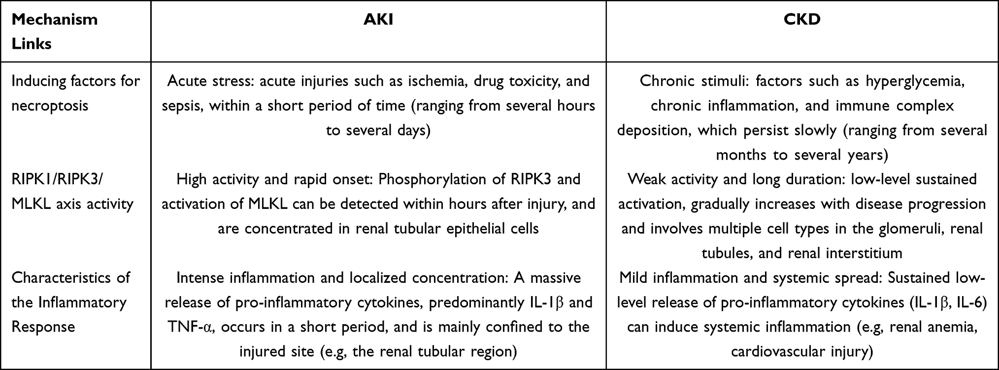 |
Table 2 The Mechanisms of Necroptosis in Acute Kidney Injury and Chronic Kidney Diseases |
Association Between Necroptosis and the Release of Inflammatory Factors
Necroptosis is a “double-edged sword” in both physiology and pathology. On the one hand, necroptosis can trigger an inflammatory cascade, leading to severe tissue damage, disease chronicization, and even tumor progression. On the other hand, as a host defense mechanism, necroptosis plays an antipathogenic and antitumor role through its powerful pro-inflammatory properties.46 Extracellular stress events, such as pathogen infection, ischemia-reperfusion injury, calcium overload, ionizing radiation, osmotic stress, heat stress, and anti-tumor drugs, can induce necroptosis through a variety of signaling pathways involving ligand-receptor binding, including TNF-α/tumor necrosis factor receptor (TNFR), Fas ligand/Fas, interferon-γ (IFN-γ)/interferon-α/β receptor 1 (IFNAR1), double-stranded RNA/Toll-like receptor 3 (TLR3), and double-stranded DNA/Z-DNA binding protein 1 (ZBP1).47 Among them, the molecular mechanism of TNF-α/TNFR-mediated necroptosis mediated by TNFR1 has been the most extensively studied.46 When TNF binds to TNFR against the background of exogenous or endogenous stimuli, different situations can occur depending on the cell type and enzyme activity. 1) In most cell types, the main result of TNF stimulation is not cell death but rather the production of a broad spectrum of cytokines and chemokines, that is, TNF induces an inflammatory response by activating pro-inflammatory genes through the nuclear factor kappa-light-chain-enhancer of activated B cells (NF-κB) signaling pathway.48 TNF-TNFR1 recruits TNFR1-associated death domain protein (TRADD), TNF receptor-associated factor 2 (TRAF2), RIPK1, cellular inhibitor of apoptosis protein (cIAP or cIAP2), and linear ubiquitin chain assembly complex (LUBAC) to assemble into complex I, activating the NF-κB pathway and promoting the production of inflammatory factors without inducing cell death.49 2) When the synthesis of NF-κB target proteins is disrupted or when the tumor suppressor protein (CYLD) causes the deubiquitination of RIPK1 and other proteins, TRADD and RIPK1 dissociate from complex I and assemble with Fas-associated protein with death domain (FADD) and caspase-8 to form complex IIa, leading to apoptosis.50 When the ubiquitination of RIPK1 is inhibited or the phosphorylation induced by cytotoxicity is inhibited, RIPK1, FADD, and caspase-8 assemble into complex IIb, mediating apoptosis.51 3) Some studies have proposed that necroptosis occurs only when two conditions are met. First, the cell must express RIPK3, and second, pharmacological or virus-derived caspase inhibitors need to be used to inhibit the activation of caspase-8.48 Therefore, when caspase-8 is absent or inactivated, RIPK1 recruits and phosphorylates RIPK3 through the receptor-interacting protein homotypic interaction motif (RHIM) domain to form a necrosome complex, which in turn leads to the cascade activation of MLKL phosphorylation and initiates the oligomerization of MLKL. The oligomerized MLKL subsequently binds to phosphatidylinositol and cardiolipin, causing the entire necrosome to be transferred from the cytoplasm to the cell membrane or organelle membrane, where it forms permeable pores, thus disrupting membrane integrity and mediating necroptosis.46,47,51,52 Therefore, studies have proposed that the transfer of MLKL and its accumulation on the plasma membrane are key points of necroptosis.53
The RIPK1/RIPK3/MLKL Signaling Axis and Inflammatory Body Activation
In addition, there is also a RIPK1-independent necroptosis pathway. After sensing viral RNA or intracellular endogenous RNA, the RHIM-containing protein ZBP1 can recruit RIPK3 through RHIM–RHIM homotypic interactions to induce necroptosis. Necroptosis plays an important role in acute injury to various organs. Some studies have suggested that RIPK3 is highly expressed in the ischemic myocardial cells of mice with myocardial infarction and that knocking out RIPK3 can alleviate unfavorable myocardial remodeling, dysfunctional myocardial hypertrophy, and inflammatory response after myocardial infarction.54 Other studies have suggested that necroptosis is involved in all stages of the progression of acute lung injury/acute respiratory distress syndrome.55 Some studies have found that necroptosis mediated by RIPK3 and MLKL plays a protective role in acute pancreatitis, and necroptosis inhibitors are not recommended for the treatment of acute pancreatitis.56
Pathological Effects
Association Between Ischemia-Reperfusion Injury and Necrotic Apoptosis in AKI
Necroptosis can also occur in various forms of AKI, including renal ischemia-reperfusion injury (IRI-AKI), sepsis and critical illness-induced AKI, crystal nephropathy, drug-induced AKI, etc.41 Common causes of IRI-AKI include kidney transplantation, circulatory arrest, and hypotensive shock. The use of the small-molecule inhibitor of RIPK1, necrostatin-1 (Nec-1), can alleviate AKI.47 A prospective study found that in critically injured patients, the plasma RIPK3 level at 48 hours was related to the AKI stage during resuscitation, indicating that necroptosis may be an important pathogenic factor of AKI in critically injured patients.57 In a mouse model of AKI induced by sepsis, high permeability of the intestinal barrier led to a sharp increase in 4-hydroxyphenylacetic acid (4-HPA) in the kidneys. 4-HPA inhibits the necroptosis of renal tubular epithelial cells by increasing the protein expression of the apoptosis inhibitor with a caspase recruitment domain (ARC) and enhancing the interaction between ARC and RIPK1.58 Cisplatin nephrotoxicity can affect 20–40% of treated patients, and its most common manifestation is acute kidney injury.59 Some studies suggest that inhibiting any core component of the necroptosis pathway, including RIPK1, RIPK3, and MLKL, by knockout or chemical inhibitors can reduce the injury of proximal tubules in mice induced by cisplatin.60 The use of novel RIPK1 inhibitors such as Cpd-71 and Cpd-2 or the novel RIPK3 inhibitor Cpd-42 can alleviate AKI induced by cisplatin (CP).61–63 In addition, an experiment in which cisplatin was repeatedly injected into mice revealed that continuous activation of the necroptosis pathway may promote the progression of cisplatin-induced acute kidney injury to chronic kidney disease.64 Some studies have verified that treatment with the RIPK1 inhibitor NEC-1 or the RIPK3 inhibitor Cpd-42 reduced the cytotoxicity of gentamicin-induced renal tubular cells.65 Another experiment found that low-intensity pulsed ultrasound significantly inhibited the expression of MI macrophage-related genes and significantly promoted the expression of M2 macrophage-related genes, inhibited necroptosis, and reversed the renal changes induced by gentamicin.66
Promotion of Persistent Inflammatory Response and Fibrosis in CKD
Nephrocalcinosis is related to the progressive loss of nephrons, fibrosis, and the rapid progression of CKD to end-stage renal failure. Many articles have proven that necroptosis plays an important role in the development of crystal nephropathy. Transforming growth factor-β (TGF-β) plays a core role in numerous cases of fibrosis.67 Experiments using a model of chronic oxalate nephropathy and a unilateral ureteral obstruction (UUO) model have demonstrated that TGF-β exposure induces the translocation of the RIPK3-MLKL necrosome to mitochondria, induces the production of mitochondrial ROS and activates CaMKII, thereby enhancing TGF-β-induced phosphorylation of Smad2/3 and the production of extracellular matrix (ECM) proteins, further promoting the proliferation of the extracellular matrix and exacerbating fibrosis. Inhibitors of necroptosis can improve the production of the intrarenal extracellular matrix during CKD.68
Interaction Between Ferroptosis and Necrotic Apoptosis
As two distinct modalities of programmed cell death, ferroptosis and necroptosis exhibit intricate crosstalk in the pathological progression of acute and chronic kidney diseases. This interactive relationship is not merely reflected in the convergence of shared initiators and intersecting signaling cascades, but more importantly, exacerbates renal injury through synergistic amplification of pathogenic processes. Drawing upon the latest advancements in the field, the following is a review of four dimensions: molecular triggers, regulatory network interactions, clinicopathological correlations, and potential therapeutic interventions. The main interactions between ferroptosis and necrotic apoptosis are shown in Figure 1.
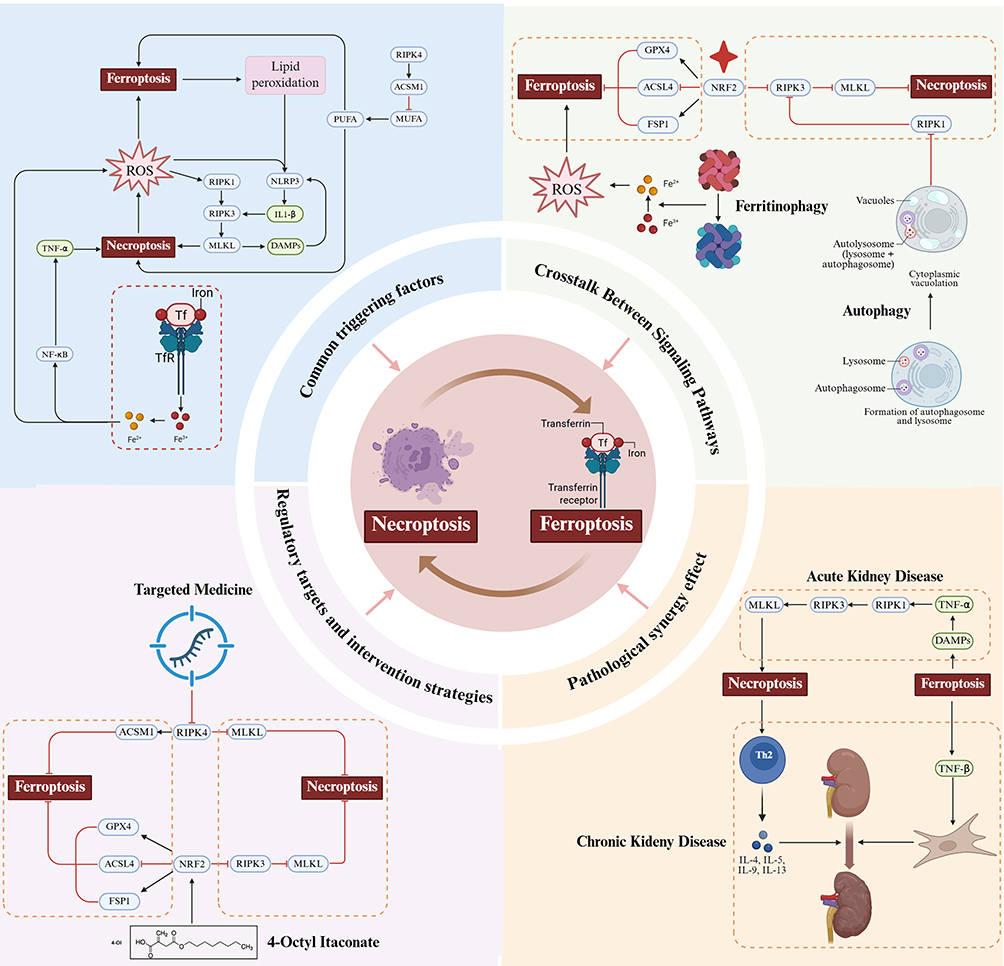 |
Figure 1 The main interactions between ferroptosis and necrotic apoptosis. Abbreviations: ROS, Reactive Oxygen Species; RIPK1, Receptor-Interacting serine/threonine - Protein Kinase 1; RIPK3, Receptor-Interacting serine/threonine - Protein Kinase 3; RIPK4, Receptor-Interacting serine/threonine - Protein Kinase 4; NLRP3, Nucleotide - binding oligomerization domain - like receptor protein 3; IL1-β, Interleukin – 1 beta; MLKL, Mixed Lineage Kinase Domain – Like; PUFA, Polyunsaturated Fatty Acid; MUFA, Monounsaturated Fatty Acid; ACSM1, Acyl - Coenzyme A Synthetase Medium Chain Family, Member 1; THF-α, Tumor Necrosis Factor – alpha; NF-κB, Nuclear Factor – kapa B; Tf, Transferrin; TfR, Transferrin Receptor; GPX4, Glutathione Peroxidase 4; ACSL4, acyl - CoA synthetase long - chain family member 4; NRF2, Nuclear factor erythroid 2 - related factor 2; FSP1, Ferroptosis Suppressor Protein 1. |
Common Triggers
Oxidative Stress and Lipid Peroxidation
The most prominent feature of ferroptosis is iron-dependent lipid peroxidation, while the process of necroptosis is also regulated by reactive oxygen species (ROS). For example, in cisplatin-induced acute kidney injury (AKI) models, iron overload generates ROS through the Fenton reaction, which on one hand triggers ferroptosis and the other hand activates the RIPK1-RIPK3-MLKL pathway of necroptosis, forming a “double-edged sword” effect of oxidative stress.69,70 Additionally, studies have found that lipid peroxidation products generated by ferroptosis not only directly damage cell membranes but also activate the NLRP3 inflammasome and promote the release of necroptosis-related inflammatory cytokine IL-1β, creating a vicious cycle.71,72
Dual Role of Abnormal Iron Metabolism
When iron homeostasis is disrupted, transferrin receptor TfR1 is upregulated, which not only promotes ferroptosis by increasing free iron but also enhances the expression of pro-inflammatory cytokines such as TNF-α through activating the NF-κB pathway, thereby indirectly activating necroptosis.73 Meanwhile, a recent study has revealed that RIPK4 downregulates acyl-CoA synthetase ACSM1, reducing the synthesis of monounsaturated fatty acids (MUFA) and leading to the accumulation of polyunsaturated fatty acids (PUFA). This process exacerbates both ferroptosis and oxidative stress necrosis, indicating that cross-regulation of iron metabolism and lipid remodeling is central to their interaction.74
Signaling Crosstalk
The Pivotal Role of NRF2
NRF2, a key regulator of the antioxidant response, inhibits ferroptosis by activating GPX4 and FSP1 while blocking necroptosis via suppressing RIPK3 phosphorylation. For instance, in ischemia-reperfusion kidney injury, NRF2 activation reduces the expression of both the ferroptosis marker (ACSL4) and the necroptosis marker (MLKL).32–35,75 Additionally, sex-differences studies indicate that increase NRF2 activity in female kidneys may explain their stronger increased resistance to ferroptosis and necroptosis, whereas testosterone-suppressed NRF2 signaling in males predisposes them to dual cell death patterns.76
Bidirectional Regulation of Autophagy
Autophagy has a dual role. On the one hand, ferritin autophagy can release free iron to promote ferroptosis, but at the same time, it can inhibit necrotic apoptosis by degrading RIPK1.16,77 For example, in chronic kidney disease (CKD) models, autophagic deficiency triggers co-activation of ferroptosis and necroptosis, thereby accelerating tubulointerstitial fibrosis.78 A study further showed that FOXO1-NCOA4 axis-mediated ferritinophagy can induce both ferroptosis and necroptosis simultaneously, highlighting the bridging role of autophagy-related proteins in their crosstalk.79
Pathological Synergy Effect
“Double Whammy” Model in AKI
In cisplatin- or ischemia-reperfusion-induced acute kidney injury (AKI), ferroptosis initiates first, triggering renal tubular epithelial cell injury and releasing damage-associated molecular patterns (DAMPs). This sequence then activates TNF-α signaling in macrophages, which in turn triggers necroptosis via the RIPK1-RIPK3-MLKL pathway, amplifying inflammatory responses and secondary injury.80,81 A previous animal study demonstrated that the combined use of ferroptosis and necroptosis inhibitors provided significantly superior renal function protection than monotherapy.82
Fibrosis Positive Feedback Loop in CKD
In chronic kidney disease (CKD), ferroptosis contributes to pathological remodeling by activating renal fibroblasts through the release of profibrotic cytokines (notably TGF-β), which drive their differentiation into myofibroblasts and mediate maladaptive repair characterized by excessive extracellular matrix deposition. Concurrently, necroptosis exacerbates the inflammatory microenvironment by releasing IL-33, a danger-associated molecular pattern (DAMP) that promotes Th2 lymphocyte polarization and subsequent secretion of pro-inflammatory cytokines (eg, IL-4, IL-13), thereby sustaining chronic inflammation. The combined actions of iron-dependent lipid peroxidation (ferroptosis) and RIPK1/RIPK3-driven necroptosis create a feedforward loop that synergistically accelerates renal interstitial fibrosis, a hallmark of CKD progression.83,84
Regulatory Targets and Intervention Strategies
Dual Pathway Inhibitors
Targeting the RIPK4‒ACSM1 axis through RIPK4 inhibition achieves dual suppression of ferroptosis and necroptosis by simultaneously upregulating ACSM1—thereby reducing the accumulation of polyunsaturated fatty acid (PUFA), a key substrate for lipid peroxidation in ferroptosis—and blocking MLKL phosphorylation, a critical step in necroptotic membrane rupture.64 Mechanistically, ACSM1-mediated synthesis of monounsaturated fatty acids (MUFAs) counteracts PUFA-driven oxidative stress, while MLKL phosphorylation triggers its translocation to the plasma membrane, inducing cell lysis. Additionally, 4-Octyl Itaconate, an NRF2 activator, has demonstrated therapeutic efficacy in preclinical kidney injury models by coordinately enhancing GPX4 (a pivotal ferroptosis suppressor that neutralizes lipid peroxides) and inhibiting RIPK3 (a necroptotic kinase essential for downstream signaling), thereby interrupting both cell death pathways.85–87
Metabolic Intervention
Research has shown that 7-dehydrocholesterol (7-DHC) inhibits lipid peroxidation and blocks ferroptosis via its 5,7-unsaturated double bond, while simultaneously downregulating RIPK3 expression, thus providing a novel target for metabolic intervention.88 Additionally, combined use of iron chelators (such as DFO) and lipid antioxidants (such as liproxstatin-1) can disrupt the cross-amplifying effect between ferroptosis and necroptosis.89
Potential Therapeutic Strategies for Targeted Regulation
Current research has focused mainly on inhibiting ferroptosis, and the inhibition of necrotizing apoptosis is mainly Necrostatin-1. However, there is limited research on combined targeting strategies, and the specific mechanisms require further investigation. The following mainly introduces the related studies on the inhibition of ferroptosis and necrotizing apoptosis.
Inhibition of Ferroptosis
Previous studies have shown that iron chelators and small-molecule inhibitors of ferroptosis have protective effects on various AKI models. Some studies have designed analogs that are more stable than Fer-1. Among them, Cqd-A1 can improve renal function in a dose-dependent manner and reduce tubular injury and inflammatory response in AKI induced by ischemia-reperfusion injury (IRI-AKI) and cecal ligation and puncture (CLP-induced AKI).90 Curcumin, which has strong antioxidant properties, can reduce AKI caused by rhabdomyolysis by decreasing ferroptosis-mediated cell death.91 Irisin can activate the SIRT1/Nrf2 signaling pathway both in vivo and in vitro, reduce the accumulation of ROS and iron, and improve mitochondrial dysfunction, thereby alleviating sepsis-associated acute kidney injury.92 In a study that established an AKI model related to crush syndrome, the combined use of rosmarinic acid and deferasirox inhibited ferroptosis through the Nrf2/Keap1 pathway to relieve AKI.93 Other studies have focused on improving the function of intracellular ultrastructure to inhibit ferroptosis. The ubiquitination and degradation of sulfide: quinone oxidoreductase (SQOR) may exacerbate ferroptosis by inducing mitochondrial dysfunction, thus promoting the occurrence and development of AKI induced by cisplatin, IRI, and FA. SYVN1 can mediate the degradation of ubiquitinated SQOR, suggesting that targeting the SYVN1-SQOR axis may be a potential therapeutic strategy for AKI.94 Leonurine can effectively restore abnormal mitochondrial ultrastructures, reduce the levels of ACSL4 and 4-HNE, scavenge reactive oxygen species, and increase the levels of GPX4 and GSH. Moreover, leonurine can also inhibit endoplasmic reticulum stress-related ferroptosis by regulating the ATF4/CHOP/ACSL4 signaling pathway, resulting in a significant protective effect against AKI.95 In vivo, vitexin treatment significantly improved renal tubular injury, interstitial fibrosis, and inflammation in UUO and UIR mouse kidneys. It effectively reduces ferroptosis induced by UUO and UIR in renal tubular epithelial cells by increasing the levels of glutathione peroxidase 4 (GPX 4) and decreasing lipid peroxidation in mouse kidneys. In vitro, vitexin treatment inhibited erastin-induced ferroptosis in HK 2 cells, which activated the NRF 2/heme oxygenase-1 (HO-1) pathway, increased the expression of GPX-4 by inhibiting KEAP-1 and ubiquitination-mediated NRF-2 degradation, and further inhibited lipid peroxidation and iron apoptosis. Moreover, it could reduce the expression of type I collagen and α-smooth muscle actin (α-SMA) in NRK-49 F cells induced by supernatant of HK 2 cells. In addition, knockout of NRF-2 strongly inhibited the anti-iron apoptotic effect of erastin.40 In an experimental model of adenine diet and UUO-induced CKD mice, as well as adenine and TGF-β 1-stimulated renal tubular epithelial cells, fisetin significantly improved renal insufficiency by inhibiting ACSL4-mediated renal tubular iron apoptosis in CKD, resulting in a decrease in serum creatinine and biomarkers of kidney injury, inhibition of pro-inflammatory cytokines (IL-1β, IL-6, TNF-α, MCP-1) levels, and a delay in the development of fibrotic phenotypes (α-SMA, FN, Col I, Col VI) for anti-fibrotic nephropathy.39
Inhibition of Necroptosis
Nec-1 can effectively protect against renal ischemia-reperfusion injury by inhibiting necrotizing apoptosis, oxidative stress, and inflammation, potentially through the HIF-1α/miR-26a/TRPC6/PARP1 signaling pathway.96 Moreover, during kidney transplantation, Necrostatin-1 protects non-heartbeat donor rats from ischemic acute kidney injury by blocking RIPK1 and inhibiting PANoptosis. Therefore, Necrostatin-1 may provide a new opportunity for end-stage nephropathy patients requiring kidney transplantation to protect donor kidneys from renal ischemia-reperfusion injury during transplantation.97 When the condition that apoptosis was blocked by benzyloxy-carbonyl-Val-Ala-Asp-fluoromethyl ketone (z-VAD-fetamine), the use of NEC-1 completely reversed cell viability, indicating that NEC-1 affects nonapoptotic cell death (necrotic apoptosis) and can inhibit renal tubular cell death.98
Challenges and Future Directions
According to the existing studies, the interaction between ferroptosis and necrotizing apoptosis is characterized by multi-level and dynamic networked regulation in acute and chronic kidney disease. However, the current research has focused mainly on ferroptosis and necroptosis, and the field of interaction research still needs to be further expanded. The existing challenges lie in the gaps on the research of related mechanisms and the difficulties in clinical translation, which are manifested in the differences between animal models and human diseases, and the development of sex-specific therapies. Future specific research directions may lie in exploring the role of mitochondrial dysfunction in bridging the two death modes, conducting immunofluorescence co-localization experiments, completing intervention studies in animal models, and using single-cell sequencing and spatial omics technologies to analyze the spatiotemporal specificity of the two in different kidney cell types. Moreover, the development of drugs that target dual regulatory nodes (such as NRF2 and RIPK4) is expected to break through existing therapeutic bottlenecks. Ferroptosis is no longer limited to the investigation of a single disease. In breast cancer and diverse other pathological states, ferroptosis has been found to be tightly linked to the regulation of cellular metabolism, lipid peroxidation, and the immune microenvironment.99 These interdisciplinary research efforts further imply that ferroptosis may act as a core pathological mechanism broadly existing across multiple systemic diseases, providing fresh viewpoints for understanding its role in kidney disorders.100,101
Funding
1.The National Natural Science Foundation of China (No. 62271506). Recipient: Hanyu Zhu 2.Innovation Team and Talents Cultivation Program of National Administration of Traditional Chinese Medicine (No: ZYYCXTD-D-202402). Recipient: Hanyu Zhu.
Disclosure
The authors report no conflicts of interest in this work.
References
1. Bayır H, Dixon SJ, Tyurina YY, Kellum JA, Kagan VE. Ferroptotic mechanisms and therapeutic targeting of iron metabolism and lipid peroxidation in the kidney. Nat Rev Nephrol. 2023;19(5):315–15. doi:10.1038/s41581-023-00689-x
2. Cheng X, Li Y, Chen L, et al. Excess iron accumulation mediated senescence in diabetic kidney injury. J Biochem Mol Toxicol. 2024;38(4):e23683. doi:10.1002/jbt.23683
3. Gafter-Gvili A, Schechter A, Rozen-Zvi B. Iron deficiency anemia in chronic kidney disease. Acta Haematol. 2019;142(1):44–50. doi:10.1159/000496492
4. Jin L, Yu B, Wang H, et al. STING promotes ferroptosis through NCOA4-dependent ferritinophagy in acute kidney injury - sciencedirect. Free Radic Biol Med. 2023;208:348–60.
5. Deng Y, Zeng L, Liu H, et al. Silibinin attenuates ferroptosis in acute kidney injury by targeting FTH1. Redox Biol. 2024;77:103360. doi:10.1016/j.redox.2024.103360
6. Zheng Q, Xing J, Li X, Tang X, Zhang D. PRDM16 suppresses ferroptosis to protect against sepsis-associated acute kidney injury by targeting the NRF2/GPX4 axis. Redox Biol. 2024;78:103417. doi:10.1016/j.redox.2024.103417
7. Gao L, Zhang J, Yang T, et al. STING/ACSL4 axis-dependent ferroptosis and inflammation promote hypertension-associated chronic kidney disease. Mol Ther. 2023;31(10):3084–3103. doi:10.1016/j.ymthe.2023.07.026
8. Lu Q, Yang L, Xiao JJ, et al. Empagliflozin attenuates the renal tubular ferroptosis in diabetic kidney disease through AMPK/NRF2 pathway. Free Radic Biol Med. 2023;195:89–102. doi:10.1016/j.freeradbiomed.2022.12.088
9. Friedmann Angeli JP, Schneider M, Proneth B, et al. Inactivation of the ferroptosis regulator Gpx4 triggers acute renal failure in mice. Nat Cell Biol. 2014;16(12):1180–1191. doi:10.1038/ncb3064
10. Baird L, Yamamoto M. The molecular mechanisms regulating the KEAP1-NRF2 pathway. Mol Cell Biol. 2020;40(13):e00099–20. doi:10.1128/MCB.00099-20
11. Zhang XD, Liu ZY, Wang MS, et al. Mechanisms and regulations of ferroptosis. Front Immunol. 2023;14:1269451. doi:10.3389/fimmu.2023.1269451
12. Hadzhieva M, Kirches E, Mawrin C. Review: iron metabolism and the role of iron in neurodegenerative disorders. Neuropathol Appl Neurobiol. 2014;40(3):240–257. doi:10.1111/nan.12096
13. Kruszewski M. Labile iron pool: the main determinant of cellular response to oxidative stress. Mutat Res. 2003;531(1–2):81–92. doi:10.1016/j.mrfmmm.2003.08.004
14. Philpott CC, Patel SJ, Protchenko O. Management versus miscues in the cytosolic labile iron pool: the varied functions of iron chaperones. Biochim Biophys Acta Mol Cell Res. 2020;1867(11):118830. doi:10.1016/j.bbamcr.2020.118830
15. Li J, Zheng S, Fan Y, Tan K. Emerging significance and therapeutic targets of ferroptosis: a potential avenue for human kidney diseases. Cell Death Dis. 2023;14(9):628. doi:10.1038/s41419-023-06144-w
16. Tang D, Chen X, Kang R, Kroemer G. Ferroptosis: molecular mechanisms and health implications. Cell Res. 2021;31(2):107–125. doi:10.1038/s41422-020-00441-1
17. Hou W, Xie Y, Song X, et al. Autophagy promotes ferroptosis by degradation of ferritin. Autophagy. 2016;12(8):1425–1428. doi:10.1080/15548627.2016.1187366
18. Sun K, Li C, Liao S, et al. Ferritinophagy, a form of autophagic ferroptosis: new insights into cancer treatment. Front Pharmacol. 2022;13:1043344. doi:10.3389/fphar.2022.1043344
19. Shchepinov MS. Polyunsaturated fatty acid deuteration against neurodegeneration. Trends Pharmacol Sci. 2020;41(4):236–248. doi:10.1016/j.tips.2020.01.010
20. Jiang X, Stockwell BR, Conrad M. Ferroptosis: mechanisms, biology and role in disease. Nat Rev Mol Cell Biol. 2021;22(4):266–282. doi:10.1038/s41580-020-00324-8
21. Shah R, Shchepinov MS, Pratt DA. Resolving the role of lipoxygenases in the initiation and execution of ferroptosis. ACS Cent Sci. 2018;4(3):387–396. doi:10.1021/acscentsci.7b00589
22. Yuan H, Li X, Zhang X, Kang R, Tang D. Identification of ACSL4 as a biomarker and contributor of ferroptosis. Biochem Biophys Res Commun. 2016;478(3):1338–1343. doi:10.1016/j.bbrc.2016.08.124
23. Zou Y, Palte MJ, Deik AA, et al. A GPX4-dependent cancer cell state underlies the clear-cell morphology and confers sensitivity to ferroptosis. Nat Commun. 2019;10(1):1617. doi:10.1038/s41467-019-09277-9
24. Doll S, Proneth B, Tyurina YY, et al. ACSL4 dictates ferroptosis sensitivity by shaping cellular lipid composition. Nat Chem Biol. 2017;13(1):91–98. doi:10.1038/nchembio.2239
25. Dixon SJ, Winter GE, Musavi LS, et al. Human haploid cell genetics reveals roles for lipid metabolism genes in nonapoptotic cell death. ACS Chem Biol. 2015;10(7):1604–1609. doi:10.1021/acschembio.5b00245
26. Zheng J, Conrad M. The metabolic underpinnings of ferroptosis. Cell Metab. 2020;32(6):920–937. doi:10.1016/j.cmet.2020.10.011
27. Do Q, Xu L. How do different lipid peroxidation mechanisms contribute to ferroptosis? Cell Rep Phys Sci. 2023;4(12). doi:10.1016/j.xcrp.2023.101683
28. Lyamzaev KG, Panteleeva AA, Simonyan RA, Avetisyan AV, Chernyak BV. Mitochondrial lipid peroxidation is responsible for ferroptosis. Cells. 2023;12(4):611. doi:10.3390/cells12040611
29. Chen X, Li J, Kang R, Klionsky DJ, Tang D. Ferroptosis: machinery and regulation. Autophagy. 2021;17(9):2054–2081. doi:10.1080/15548627.2020.1810918
30. Bannai S. Exchange of cystine and glutamate across plasma membrane of human fibroblasts. J Biol Chem. 1986;261(5):2256–2263. doi:10.1016/S0021-9258(17)35926-4
31. Sun X, Ou Z, Chen R, et al. Activation of the p62-Keap1-NRF2 pathway protects against ferroptosis in hepatocellular carcinoma cells. Hepatology. 2016;63(1):173–184. doi:10.1002/hep.28251
32. Dong H, Qiang Z, Chai D, et al. Nrf2 inhibits ferroptosis and protects against acute lung injury due to intestinal ischemia reperfusion via regulating SLC7A11 and HO-1. Aging. 2020;12(13):12943–12959. doi:10.18632/aging.103378
33. Anandhan A, Dodson M, Shakya A, et al. NRF2 controls iron homeostasis and ferroptosis through HERC2 and VAMP8. Sci Adv. 2023;9(5):eade9585. doi:10.1126/sciadv.ade9585
34. Dong H, Xia Y, Jin S, et al. Nrf2 attenuates ferroptosis-mediated IIR-ALI by modulating TERT and SLC7A11. Cell Death Dis. 2021;12(11):1027. doi:10.1038/s41419-021-04307-1
35. Chen C, Yang K, He D, et al. Induction of ferroptosis by HO-1 contributes to retinal degeneration in mice with defective clearance of all-trans-retinal. Free Radic Biol Med. 2023;194:245–254. doi:10.1016/j.freeradbiomed.2022.12.008
36. Skouta R, Dixon SJ, Wang J, et al. Ferrostatins inhibit oxidative lipid damage and cell death in diverse disease models. J Am Chem Soc. 2014;136(12):4551–4556. doi:10.1021/ja411006a
37. Martin-Sanchez D, Ruiz-Andres O, Poveda J, et al. Ferroptosis, but not necroptosis, is important in nephrotoxic folic acid-induced AKI. J Am Soc Nephrol. 2017;28(1):218–229. doi:10.1681/ASN.2015121376
38. Zhang Q, Sun T, Yu F, et al. PAFAH2 suppresses synchronized ferroptosis to ameliorate acute kidney injury. Nat Chem Biol. 2024;20(7):835–846. doi:10.1038/s41589-023-01528-7
39. Wang B, Yang LN, Yang LT, et al. Fisetin ameliorates fibrotic kidney disease in mice via inhibiting ACSL4-mediated tubular ferroptosis. Acta Pharmacol Sin. 2024;45(1):150–165. doi:10.1038/s41401-023-01156-w
40. Song J, Wang H, Sheng J, et al. Vitexin attenuates chronic kidney disease by inhibiting renal tubular epithelial cell ferroptosis via NRF2 activation. Mol Med. 2023;29:147. doi:10.1186/s10020-023-00735-1
41. Kolbrink B, von Samson-Himmelstjerna FA, Murphy JM, Krautwald S. Role of necroptosis in kidney health and disease. Nat Rev Nephrol. 2023;19:300–14.
42. Komada T, Muruve DA. The role of inflammasomes in kidney disease. Nat Rev Nephrol. 2019;15(8):501–520. doi:10.1038/s41581-019-0158-z
43. Anders HJ. Of inflammasomes and alarmins: IL-1β and IL-1α in kidney disease. J Am Soc Nephrol. 2016;27(9):2564–2575. doi:10.1681/ASN.2016020177
44. Pefanis A, Bongoni AK, McRae JL, et al. Dynamics of necroptosis in kidney ischemia-reperfusion injury. Front Immunol. 2023;14:1251452. doi:10.3389/fimmu.2023.1251452
45. Anders HJ. Necroptosis in acute kidney injury. Nephron. 2018;139(4):342–348. doi:10.1159/000489940
46. Ye K, Chen Z, Xu Y. The double-edged functions of necroptosis. Cell Death Dis. 2023;14(2):163. doi:10.1038/s41419-023-05691-6
47. Vanlangenakker N, Vanden Berghe T, Vandenabeele P. Many stimuli pull the necrotic trigger, an overview. Cell Death Differ. 2012;19(1):75–86. doi:10.1038/cdd.2011.164
48. Kearney CJ, Martin SJ. An inflammatory perspective on necroptosis. Mol Cell. 2017;65(6):965–973. doi:10.1016/j.molcel.2017.02.024
49. Annibaldi A, Meier P. Checkpoints in TNF-induced cell death: implications in inflammation and cancer. Trends Mol Med. 2018;24(1):49–65. doi:10.1016/j.molmed.2017.11.002
50. Grootjans S, Vanden Berghe T, Vandenabeele P. Initiation and execution mechanisms of necroptosis: an overview. Cell Death Differ. 2017;24(7):1184–1195. doi:10.1038/cdd.2017.65
51. Seo J, Nam YW, Kim S, Oh DB, Song J. Necroptosis molecular mechanisms: recent findings regarding novel necroptosis regulators. Exp Mol Med. 2021;53(6):1007–1017. doi:10.1038/s12276-021-00634-7
52. Liu Z, Dagley LF, Shield-Artin K, et al. Oligomerization-driven MLKL ubiquitylation antagonizes necroptosis. EMBO j. 2021;40(23):e103718. doi:10.15252/embj.2019103718
53. Samson AL, Zhang Y, Geoghegan ND, et al. MLKL trafficking and accumulation at the plasma membrane control the kinetics and threshold for necroptosis. Nat Commun. 2020;11(1):3151. doi:10.1038/s41467-020-16887-1
54. Guo X, Chen Y, Liu Q. Necroptosis in heart disease: molecular mechanisms and therapeutic implications. J Mol Cell Cardiol. 2022;169:74–83. doi:10.1016/j.yjmcc.2022.05.006
55. Dong J, Liu W, Liu W, et al. Acute lung injury: a view from the perspective of necroptosis. Inflamm Res. 2024;73(6):997–1018. doi:10.1007/s00011-024-01879-4
56. Boonchan M, Arimochi H, Otsuka K, et al. Necroptosis protects against exacerbation of acute pancreatitis. Cell Death Dis. 2021;12(6):601. doi:10.1038/s41419-021-03847-w
57. Shashaty MG, Reilly JP, Sims CA, et al. Plasma levels of receptor interacting protein kinase-3 (RIP3), an essential mediator of necroptosis, are associated with acute kidney injury in critically ill trauma patients. Shock. 2016;46(2):139–143. doi:10.1097/SHK.0000000000000596
58. An S, Yao Y, Wu J, et al. Gut-derived 4-hydroxyphenylacetic acid attenuates sepsis-induced acute kidney injury by upregulating ARC to inhibit necroptosis. Biochim Biophys Acta Mol Basis Dis. 2024;1870(1):166876. doi:10.1016/j.bbadis.2023.166876
59. Hamroun A, Lenain R, Bigna JJ, et al. Prevention of cisplatin-induced acute kidney injury: a systematic review and meta-analysis. Drugs. 2019;79(14):1567–1582. doi:10.1007/s40265-019-01182-1
60. Xu Y, Ma H, Shao J, et al. A role for tubular necroptosis in cisplatin-induced AKI. J Am Soc Nephrol. 2015;26(11):2647–2658. doi:10.1681/ASN.2014080741
61. He XY, Wang F, Suo XG, et al. Compound-42 alleviates acute kidney injury by targeting RIPK3-mediated necroptosis. Br J Pharmacol. 2023;180(20):2641–2660. doi:10.1111/bph.16152
62. Wang JN, Liu MM, Wang F, et al. RIPK1 inhibitor cpd-71 attenuates renal dysfunction in cisplatin-treated mice via attenuating necroptosis, inflammation and oxidative stress. Clin Sci. 2019;133(14):1609–1627. doi:10.1042/CS20190599
63. Li C, Chen QY, He Y, Liu YH, Meng XM, Liu MM. Discovery of a chalcone derivative as potent necroptosis inhibitor for the treatment of acute kidney injury. Clin Exp Pharmacol Physiol. 2022;49(8):824–835. doi:10.1111/1440-1681.13670
64. Landau SI, Guo X, Velazquez H, et al. Regulated necrosis and failed repair in cisplatin-induced chronic kidney disease. Kidney Int. 2019;95(4):797–814. doi:10.1016/j.kint.2018.11.042
65. Hu BF, Gong Q, Chen SQ, et al. Protective effect of inhibiting necroptosis on gentamicin-induced nephrotoxicity. FASEB j. 2022;36(9):e22487. doi:10.1096/fj.202200163R
66. Gouda SAA, Aboulhoda BE, Abdelwahed OM, et al. Low-intensity pulsed ultrasound (LIPUS) switched macrophage into M2 phenotype and mitigated necroptosis and increased HSP 70 in gentamicin-induced nephrotoxicity. Life Sci. 2023;314:121338. doi:10.1016/j.lfs.2022.121338
67. Hwang SH, Lee YM, Choi Y, et al. Role of human primary renal fibroblast in TGF-β1-mediated fibrosis-mimicking devices. Int J Mol Sci. 2021;22(19):10758. doi:10.3390/ijms221910758
68. Srivastava A, Tomar B, Sharma P, et al. RIPK3-MLKL signaling activates mitochondrial CaMKII and drives intrarenal extracellular matrix production during CKD. Matrix Biol. 2022;112:72–89. doi:10.1016/j.matbio.2022.08.005
69. Xie T, Yao L, Li X. Advance in iron metabolism, oxidative stress and cellular dysfunction in experimental and human kidney diseases. Antioxidants. 2024;13(6):659. doi:10.3390/antiox13060659
70. Wu WF, Wang JN, Li Z, et al. 7-hydroxycoumarin protects against cisplatin-induced acute kidney injury by inhibiting necroptosis and promoting Sox9-mediated tubular epithelial cell proliferation. Phytomedicine. 2020;69:153202. doi:10.1016/j.phymed.2020.153202
71. Chen Y, Fang ZM, Yi X, Wei X, Jiang DS. The interaction between ferroptosis and inflammatory signaling pathways. Cell Death Dis. 2023;14(3):205. doi:10.1038/s41419-023-05716-0
72. Orecchioni M, Kobiyama K, Winkels H, et al. Olfactory receptor 2 in vascular macrophages drives atherosclerosis by NLRP3-dependent IL-1 production. Science. 2022;375(6577):214–221. doi:10.1126/science.abg3067
73. Kenneth NS, Mudie S, Naron S, Rocha S. TfR1 interacts with the IKK complex and is involved in IKK-NF-κB signalling. Biochem J. 2013;449(1):275–284. doi:10.1042/BJ20120625
74. Zhang J, Wei Y, Yue Y, et al. RIPK4 promotes oxidative stress and ferroptotic death through the downregulation of ACSM1. Proc Natl Acad Sci U S A. 2024;121(40):e2410628121. doi:10.1073/pnas.2410628121
75. Fan Y, Lu J, Yu Z, Qu X, Guan S. 1,3-dichloro-2-propanol-induced renal tubular cell necroptosis through the ROS/RIPK3/MLKL pathway. J Agric Food Chem. 2022;70(35):10847–10857. doi:10.1021/acs.jafc.2c02619
76. Ide S, Ide K, Abe K, et al. Sex differences in resilience to ferroptosis underlie sexual dimorphism in kidney injury and repair. Cell Rep. 2022;41(6):111610. doi:10.1016/j.celrep.2022.111610
77. Rojas-Rivera D, Beltrán S, Muñoz-Carvajal F, et al. The autophagy protein RUBCNL/PACER represses RIPK1 kinase-dependent apoptosis and necroptosis. Autophagy. 2024;20(11):2444–2459. doi:10.1080/15548627.2024.2367923
78. Doke T, Susztak K. The multifaceted role of kidney tubule mitochondrial dysfunction in kidney disease development. Trends Cell Biol. 2022;32(10):841–853. doi:10.1016/j.tcb.2022.03.012
79. Wang X, Xu L, Meng Y, et al. FOXO1-NCOA4 axis contributes to cisplatin-induced cochlea spiral ganglion neuron ferroptosis via ferritinophagy. Adv Sci. 2024;11(40):e2402671. doi:10.1002/advs.202402671
80. Li S, Zhang Y, Lu R, et al. Peroxiredoxin 1 aggravates acute kidney injury by promoting inflammation through mincle/syk/NF-κB signaling. Kidney Int. 2023;104(2):305–323. doi:10.1016/j.kint.2023.04.013
81. Lin Z, Long F, Kang R, Klionsky DJ, Yang M, Tang D. The lipid basis of cell death and autophagy. Autophagy. 2024;20(3):469–488. doi:10.1080/15548627.2023.2259732
82. Linkermann A, Skouta R, Himmerkus N, et al. Synchronized renal tubular cell death involves ferroptosis. Proc Natl Acad Sci U S A. 2014;111(47):16836–16841. doi:10.1073/pnas.1415518111
83. Kim S, Kang SW, Joo J, et al. Characterization of ferroptosis in kidney tubular cell death under diabetic conditions. Cell Death Dis. 2021;12(2):160. doi:10.1038/s41419-021-03452-x
84. Liu C, Liu L, Huang Y, Shi R, Wu Y, Hakimah Binti Ismail I. Contribution of IL-33/ILC2-mediated TH2 cytokines during the progression of minimal change disease. Int Immunopharmacol. 2023;114:109493. doi:10.1016/j.intimp.2022.109493
85. Xu L, Cai J, Li C, et al. 4-octyl itaconate attenuates LPS-induced acute kidney injury by activating Nrf2 and inhibiting STAT3 signaling. Mol Med. 2023;29(1):58. doi:10.1186/s10020-023-00631-8
86. Wang Y, Zhou M, Jiang RY, Zhu CL. Pharmacological inhibition of STING-mediated GPX4 autophagic degradation by 4-octyl itaconate ameliorates sepsis-induced acute kidney injury. Apoptosis. 2025;30(5–6):1410–1423. doi:10.1007/s10495-025-02099-9
87. Ni ST, Li Q, Chen Y, et al. Anti-necroptotic effects of itaconate and its derivatives. Inflammation. 2024;47(1):285–306. doi:10.1007/s10753-023-01909-z
88. Li Y, Ran Q, Duan Q, et al. 7-dehydrocholesterol dictates ferroptosis sensitivity. Nature. 2024;626(7998):411–418. doi:10.1038/s41586-023-06983-9
89. Kim M, Bae JY, Yoo S, et al. 2-deoxy-d-ribose induces ferroptosis in renal tubular epithelial cells via ubiquitin-proteasome system-mediated xCT protein degradation. Free Radic Biol Med. 2023;208:384–393. doi:10.1016/j.freeradbiomed.2023.08.027
90. Chen Y, Wu MF, Xie MM, et al. Cpd-A1 alleviates acute kidney injury by inhibiting ferroptosis. Acta Pharmacol Sin. 2024;45:1673–85.
91. Guerrero-Hue M, García-Caballero C, Palomino-Antolín A, et al. Curcumin reduces renal damage associated with rhabdomyolysis by decreasing ferroptosis-mediated cell death. FASEB j. 2019;33(8):8961–8975. doi:10.1096/fj.201900077R
92. Qiongyue Z, Xin Y, Meng P, et al. Post-treatment with irisin attenuates acute kidney injury in sepsis mice through anti-ferroptosis via the SIRT1/Nrf2 pathway. Front Pharmacol. 2022;13:857067. doi:10.3389/fphar.2022.857067
93. Qiao O, Zhang L, Han L, et al. Rosmarinic acid plus deferasirox inhibits ferroptosis to alleviate crush syndrome-related AKI via Nrf2/Keap1 pathway. Phytomedicine. 2024;129:155700. doi:10.1016/j.phymed.2024.155700
94. Cai F, Li D, Xie Y, et al. Sulfide: Quinone oxidoreductase alleviates ferroptosis in acute kidney injury via ameliorating mitochondrial dysfunction of renal tubular epithelial cells. Redox Biol. 2024;69:102973. doi:10.1016/j.redox.2023.102973
95. Cheng R, Wang X, Huang L, et al. Novel insights into the protective effects of leonurine against acute kidney injury: inhibition of ER stress-associated ferroptosis via regulating ATF4/CHOP/ACSL4 pathway. Chem Biol Interact. 2024;395:111016. doi:10.1016/j.cbi.2024.111016
96. Shen B, Mei M, Pu Y, et al. Necrostatin-1 attenuates renal ischemia and reperfusion injury via meditation of HIF-1α/mir-26a/TRPC6/PARP1 signaling. Mol Ther Nucleic Acids. 2019;17:701–713. doi:10.1016/j.omtn.2019.06.025
97. Dokur M, Uysal E, Kucukdurmaz F, et al. Targeting the PANoptosome using necrostatin-1 reduces PANoptosis and protects the kidney against ischemia-reperfusion injury in a rat model of controlled experimental nonheart-beating donor. Transplant Proc. 2024;56(10):2268–2279. doi:10.1016/j.transproceed.2024.10.047
98. Tristão VR, Gonçalves PF, Dalboni MA, Batista MC, Durão MS, Monte JCM. Nec-1 protects against nonapoptotic cell death in cisplatin-induced kidney injury. Ren Fail. 2012;34(3):373–377. doi:10.3109/0886022X.2011.647343
99. Ge A, Xiang W, Li Y, et al. Broadening horizons: the multifaceted role of ferroptosis in breast cancer. Front Immunol. 2024;15:1455741. doi:10.3389/fimmu.2024.1455741
100. Zeng L, Yang K, Yu G, et al. Advances in research on immunocyte iron metabolism, ferroptosis, and their regulatory roles in autoimmune and autoinflammatory diseases. Cell Death Dis. 2024;15(7):481. doi:10.1038/s41419-024-06807-2
101. Ubellacker JM, Dixon SJ. Prospects for ferroptosis therapies in cancer. Nat Cancer. 2025;6(8):1326–1336. doi:10.1038/s43018-025-01037-7
 © 2026 The Author(s). This work is published and licensed by Dove Medical Press Limited. The
full terms of this license are available at https://www.dovepress.com/terms
and incorporate the Creative Commons Attribution
- Non Commercial (unported, 4.0) License.
By accessing the work you hereby accept the Terms. Non-commercial uses of the work are permitted
without any further permission from Dove Medical Press Limited, provided the work is properly
attributed. For permission for commercial use of this work, please see paragraphs 4.2 and 5 of our Terms.
© 2026 The Author(s). This work is published and licensed by Dove Medical Press Limited. The
full terms of this license are available at https://www.dovepress.com/terms
and incorporate the Creative Commons Attribution
- Non Commercial (unported, 4.0) License.
By accessing the work you hereby accept the Terms. Non-commercial uses of the work are permitted
without any further permission from Dove Medical Press Limited, provided the work is properly
attributed. For permission for commercial use of this work, please see paragraphs 4.2 and 5 of our Terms.