
Back to Journals » Journal of Inflammation Research » Volume 18

The Emerging Roles and Therapeutic Potential of Unconventional T Cells in Sepsis
Authors Gu S  , Zhang P, Zhang C , Tang T, Chang T, Dong L, Gao W, Tang Z
Received 10 June 2025
Accepted for publication 9 September 2025
Published 20 September 2025 Volume 2025:18 Pages 13139—13157
DOI https://doi.org/10.2147/JIR.S545532
Checked for plagiarism Yes
Review by Single anonymous peer review
Peer reviewer comments 2
Editor who approved publication: Dr Anh Ngo
Shuaipeng Gu,1 Peidong Zhang,1 Cong Zhang,1 Tingxuan Tang,2 Teding Chang,1 Liming Dong,1 Wei Gao,1,* Zhaohui Tang1,*
1Department of Trauma Surgery, Emergency Surgery & Surgical Critical, Tongji Trauma Center, Tongji Hospital, Tongji Medical College, Huazhong University of Science and Technology, Wuhan, 430030, People’s Republic of China; 2Department of Orthopedics, Tongji Hospital, Tongji Medical College, Huazhong University of Science and Technology, Wuhan, 430030, People’s Republic of China
*These authors contributed equally to this work
Correspondence: Wei Gao; Zhaohui Tang, Tongji Hospital, Tongji Medical College, Huazhong University of Science and Technology, No. 1095 Jiefang Avenue, Wuhan, Hubei, 430030, People’s Republic of China, Email [email protected]; [email protected]
Abstract: Sepsis represents a dynamic, dysregulated host immune response to infection in which unconventional T cells—γδ T cells, mucosal-associated invariant T (MAIT) cells, natural killer T (NKT) cells, and double-negative T cells—actively shape the balance between early hyperinflammation and subsequent immune paralysis across time and tissues. These cells employ unique antigen recognition mechanisms to trigger rapid immune responses. γδ T cells facilitate early pathogen elimination and immune regulation, whereas MAIT cells detect microbial metabolites and modulate the systemic inflammation. NKT cells balance immune homeostasis through dual pro- and anti-inflammatory cytokine production. This review classifies these subsets and examines their sepsis-related functions alongside immunotherapies targeting them, such as cytokine manipulation, immunomodulators, and checkpoint inhibitors. Elucidating the precise mechanisms underlying sepsis could advance therapies that restore immune equilibrium and potentially improve clinical outcomes. Future studies should unravel the interactions between unconventional T cells and broader immune networks while translating the findings into practical treatments. Understanding the dynamic roles of these cells provides pathways for tailored interventions in sepsis management.
Keywords: sepsis, γδ T cells, invariant natural killer t cells, mucosal-associated invariant t cells, double-negative t cells, immunotherapy
Introduction
Sepsis is characterized by organ dysfunction that poses a threat to life, resulting from an abnormal host response to infection.1 The key components of its pathophysiology include cytokine storm, characterized by the excessive release of pro-inflammatory cytokines, endothelial dysfunction, and immunosuppression.2–4 A major health problem that is often underappreciated, sepsis causes a significant number of deaths around the world each year.5 In 2017, globally, there were an estimated 48.9 million new cases of sepsis and 11.0 million deaths due to sepsis, which accounted for 19.7% of all deaths worldwide.6
Despite advances in supportive care, therapies targeting the underlying immunopathology remain elusive. Conventional T cells, which are central to adaptive immunity, have dominated sepsis research; however, their responses alone may not fully explain the rapid immunologic transitions of sepsis, which can be driven by innate-like activation and tissue residency typical of unconventional T cells.7,8 Unconventional T (UTC) cells consist of γδ T cells, mucosal-associated invariant T (MAIT) cells, and natural killer T (NKT) cells, and other emerging subpopulations such as double negative T (DNT) cells, whose precise definitions and functional roles are still under investigation.9–11 Unconventional T cells can directly recognize antigens (including non-peptide antigens). Hence, unconventional T cells combine elements of both innate and adaptive immune functions, with their enhanced “innate-like” properties, can rapidly detect and modulate pathogens and inflammatory responses, playing a pivotal role in early or localized immune defense.12 Crucially, UTCs exhibit functional plasticity: pro-inflammatory in early sepsis (eg, interleukin-17 [IL-17] and interferon-γ [IFN-γ]) yet regulatory or exhausted in late phases.13,14
While many studies have thoroughly explored the essential function of conventional T cells in sepsis, there is a lack of research and detailed reviews on how unconventional T cells connect pathophysiological processes with therapeutic possibilities. This review focuses on the primary classifications and functions of unconventional T cells, systematically delving into their potential mechanisms of action in sepsis pathogenesis and exploring their viability as novel therapeutic targets.
Unconventional T Cells: An Overview
Unconventional T cells exhibit non-classical antigen sensing and tissue-specific roles that set them apart from traditional CD4+ and CD8+ subsets.15 Conventional T cells are central players in the adaptive immune system, where they respond to peptide fragments displayed by MHC molecules on antigen-presenting cells. Rather than relying on peptide presentation via classical MHC molecules, unconventional T cells can detect alternative antigenic structures through non-traditional antigen presentation pathways.16 By participating in both early and antigen-specific immune processes, unconventional T cells help integrate innate and adaptive immune pathways.17–20
Unique Activation Pathways in Response to Infection
In contrast to the MHC-dependent recognition seen in αβ T cells, γδ T cells can engage diverse antigens—including non-peptide molecules—through an unconventional T-cell receptor (TCR)-based mechanism.21 This mode of antigen recognition by UTC is considered non-MHC restricted.22 In many cases, unconventional T cells recognize various non-peptide antigens. Non-peptide antigens consist of phosphoantigens, vitamins, and self- and non-self-lipids.23 In conclusion, it is noteworthy that recognition of non-peptide antigens by UTC forms the basis of their unique immunological functions.
Contribution to Innate and Adaptive Immunity
Characterized by rapid activation, pro-inflammatory cytokine release, and cytotoxic potential, unconventional T cells exhibit properties typically associated with innate immunity. Although they have innate-like qualities, unconventional T cells are also vital to adaptive immune responses. Unconventional T cells significantly contribute to immune regulation by exhibiting antigen-specific memory, modulating adaptive immune responses through cytokine secretion, and maintaining immune homeostasis.14 Unconventional T cells contribute to adaptive immunity by managing cytokine levels. These cells exert immunoregulatory effects primarily through the secretion of cytokines such as IL-4, IL-10, and IFN-γ, which collectively shape the behavior of antigen-presenting cells and guide lymphocyte differentiation.19,20,24 Cytokine-driven interactions are essential for orchestrating immune responses, maintaining homeostasis, and preventing immune dysregulation. Moreover, certain unconventional T cell subsets, including type II NKT cells and specific γδ T cell populations, serve as immunoregulatory mediators.25 These cells help suppress excessive inflammatory responses and promote immune tolerance, thereby preventing pathological immune activation and contributing to the immune equilibrium.26 In the context of sepsis, unconventional T cells contribute to immune homeostasis by bridging innate detection pathways with adaptive effector responses. The primary roles of UTCs in sepsis are outlined in Table 1 and will be discussed in the following sections.
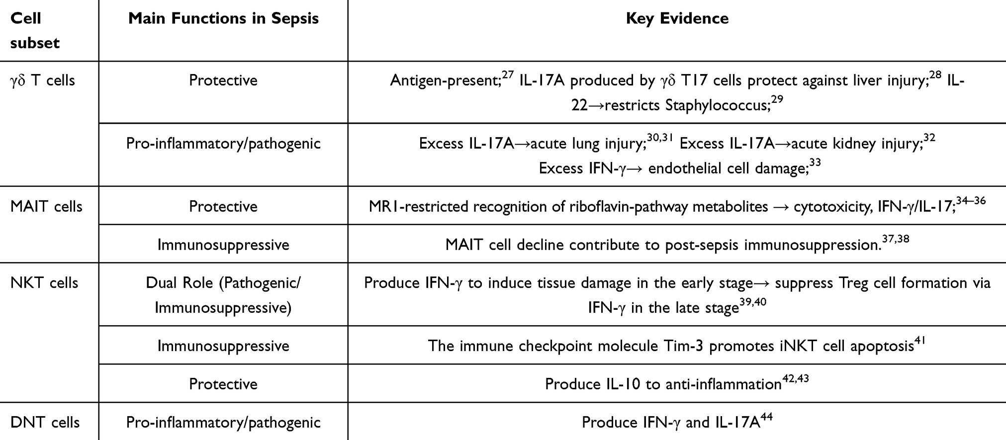 |
Table 1 Key Evidence on the Role of Unconventional T Cells in Sepsis |
γδ T Cells in Sepsis
Tissue-Specific Distribution of γδ T Cells
As key mediators of barrier immunity, γδ T cells preferentially localize to epithelial-rich environments such as the intestines, respiratory mucosa, and reproductive tissues. They form an essential part of intraepithelial lymphocytes (IEL) within the epidermis and mucosal tissues, representing only 1–5% of T cells found in peripheral blood.45
In contrast to αβ T cells composed of α and β chains, γδ T cells utilize γ and δ chains46 and encompass a diverse range of subsets, characterized by distinct tissue distributions and effector functions. They can be categorized into three groups according to the specific TRDV genes expressed in the δ chain: the Vδ1+, Vδ2+, and Vδ3+ subsets.47 Different subsets of γδ T cells exhibit significant variations in their function and tissue distribution.48,49 Notably, Vγ9Vδ2 T cells, a subset within the Vδ2 lineage, represent the most numerically dominant γδ T cells in human peripheral circulation. This subgroup is currently recognized as the most potent class of innate-like γδ T cells.50
Mechanisms of γδ T Cells Activation During Sepsis
When an infection occurs, microorganisms invade the human body and serve as antigens that activate γδ T cells, as summarized in Figure 1. Antigen-presenting γδ T cells (γδ T-APCs) can directly detect conserved molecular patterns, including lipopolysaccharide (LPS) and lipoteichoic acid (LTA), in various microbes through pattern recognition receptors.51 γδ T-APCs use their T cell receptors (TCRs) to directly detect phosphoantigens,52 thereby initiating a rapid immune response against pathogens. γδ T cells can specifically recognize the phosphoantigens (E)-4-hydroxy-3-methyl-but-2-enyl pyrophosphate (HMB-PP) and isopentenyl pyrophosphate (IPP),53 as summarized in Figure 1. Vγ9Vδ2 TCR activation requires Butyrophilin 3A1–Butyrophilin 2A1 interaction (BTN3A1-BTN2A1) complex formation upon phosphoantigen binding, which is a microbial sensing mechanism distinct from peptide-MHC recognition.54 The activation of Vγ9Vδ2 T cells in microbial contexts is heavily dependent on HMB-PP, despite its lack of direct interaction with the T cell receptor. Cytokines like IL-12, IL-18, and type I interferons may fine-tune the activation status of γδ T cells, thereby reinforcing their antiviral capabilities.55,56 γδ T cells also exhibit memory-like responses during certain infections.57 For example, in *Staphylococcus aureus* infection, memory γδ T cells undergo expansion after initial infection and provide enhanced protection upon reinfection.58
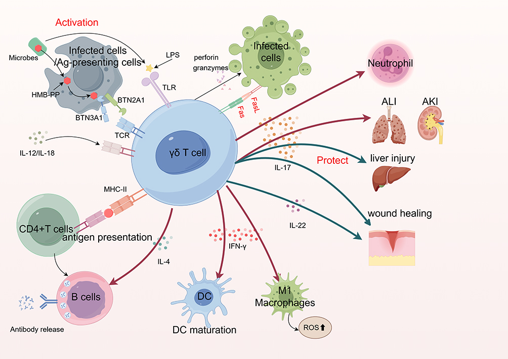 |
Figure 1 Schematic illustration of γδ T cell activation pathways and effector functions during sepsis.The diagram illustrates key pathways involved in γδ T cell activation during infectious challenges, along with their subsequent immune effector functions. Red arrows indicate pro-inflammatory or pathogenic effects, whereas green arrows represent protective or homeostatic functions. Created by Figdraw Activation pathways: (1) Phosphoantigens act as direct ligands for γδ T-cell receptors, triggering immediate immune responses.(2) Specific phosphoantigens, including (E)-4-hydroxy-3-methyl-but-2-enyl pyrophosphate (HMB-PP) and isopentenyl pyrophosphate (IPP), induce γδ T cell activation via a butyrophilin-dependent mechanism. HMB-PP/IPP first attaches intracellularly to BTN3A1, which induces a shape change that permits BTN2A1 to engage with the Vγ9 chain of Vγ9Vδ2 T-cells.(3) γδ T cells can be activated independently of TCR signaling, with inflammatory mediators such as IL-12 and IL-18 playing a pivotal role in this process.Effector functions:Cytotoxicity: Activated γδ T cells eliminate infected cells via the Fas-FasL pathway or by releasing perforin and granzyme. IL-17 production: IL-17 secreted by γδ T cells promotes neutrophil activation, granulopoiesis, and accumulation, contributing to acute kidney and lung injury during sepsis. However, IL-17 produced by liver-infiltrating γδ T17 cells plays a protective role against sepsis-induced liver injury. IL-17 and IL-22 also facilitate tissue and wound healing.Antigen presentation and bridging innate–adaptive immunity: By acquiring and processing soluble antigens, γδ T cells may facilitate the priming of αβ T cells, contributing to the initiation of adaptive immune responses. Activated CD4 + T cells subsequently provide costimulatory signals to B cells.Support of humoral immunity: IL-4 produced by γδ T cells facilitates early B cell activation and guides their maturation into antibody-secreting cells.IFN-γ production: IFN-γ enhances macrophage activation and bactericidal activity, promotes oxidative stress, contributes to endothelial cell damage, and supports dendritic cell maturation. Abbreviations: Ag, Antigen; HMB–PP, (E)-4-hydroxy-3-methyl-but-2-enyl pyrophosphate; BTN3A1, Butyrophilin3A1;BTN2A1, Butyrophilin2A1; LPS, Lipopolysaccharide; TLR, Toll -like receptor;IL, Interleukin; TCR, T-cell receptor; MHC – II, Major Histocompatibility Complex class II; IFN-γ, Interferon–gamma; DC, Dendritic cell; ALI, Acute Lung Injury; AKI, Acute Kidney Injury; ROS, Reactive Oxygen Species. |
Involvement in Modulating Early Immune Response During Sepsis
The cytotoxic response of γδ T cells involves the activation of apoptosis-inducing cascades in infected cells, mediated by molecules such as perforin and granzyme released upon stimulation.59 This cytotoxic response has been implicated in the host defense against pathogens that evade extracellular detection, including many viruses and intracellular bacteria.60 γδ T cells have been shown to fulfill antigen-presenting roles under certain conditions, highlighting their immunological versatility beyond innate-like cytotoxicity. This function is particularly evident when they interact with opsonized target cells, which can license them for antigen presentation.61 Through their antigen-presenting capabilities, γδ T cells help facilitate communication between the innate and adaptive arms of the immune system. Subsequently, CD4+ T cells activated through this pathway can activate B cells via co-stimulatory signals.24 However, during the immunosuppressive phase in patients with sepsis, γδ T-APCs exhibit significant impairment in their antigen-presenting function.27 Moreover, γδ T cells can influence dendritic cell (DC) function during infection by promoting DC maturation through IFN-γ and other mechanisms.62 Recent studies have demonstrated that unlike conventional dendritic cells, patients with sepsis have γδ T cells that show high expression of HLA-DR molecules.27 These findings highlight the therapeutic relevance of γδ T cells and support their potential exploitation in future immunotherapy strategies.
By producing cytokines such as IFN-γ, IL-17, IL-4, and IL-22, γδ T cells actively participate in shaping immune responses against infections.24 IL-17 is central to the immune response, as these cytokines are important for recruiting and activating other immune cells to eliminate pathogens. It can drive neutrophil activation, granulopoiesis, and neutrophil accumulation.63 During infection, IL-17 derived from γδ T cells supports host protection by coordinating innate immune responses, including neutrophil mobilization, antimicrobial peptide induction, and improved barrier resistance to pathogens. However, in sepsis patients, the overproduction of IL-17 also results in excessive inflammation and is a significant contributor to acute lung injury.30,31 In the pathogenesis of sepsis, in addition to lung-resident γδ T17 cells,64 γδ T17 cells originating from the gut are also major producers of IL-17.64,65 Xue-Wei Yang et al discovered that memory γδ T17 cells formed in the small intestine are capable of homing to the lungs and mediating immune-driven inflammation. The increase in these cells is correlated with the severity of lung injury.66,67 And within the kidney, IL-17A produced by γδ T cells is also a key factor contributing to sepsis-associated acute kidney injury.32 However, IL-17A, produced by γδ T17 cells that infiltrate the liver, is crucial for protection against liver injury resulting from sepsis.28 γδ T cell-derived IL-17 exacerbates lung/kidney injury but protects the liver during sepsis, highlighting the organ-specific pathophysiology. Therapeutic IL-17A blockade improves survival in murine lung injury models68 yet requires cautious organ-targeted delivery to avoid compromising hepatic defense. IFN-γ produced by γδ T cells can activate macrophages, enhance bactericidal activity, induce oxidative stress, and cause endothelial cell damage.69,70 Mechanical disruption of the skin triggers IL-22 secretion, primarily from γδ T cells, which contributes to wound healing and epithelial regeneration. γδ T cells have been demonstrated to enhance IL-22 expression in CD4+ T cells in the intestinal environment.71 This activity contributes to epithelial homeostasis and supports mucosal immune surveillance by maintaining barrier integrity and promoting local immune signaling. γδ T cell–derived IL-22 is instrumental in mounting an effective immune response against Staphylococcus aureus, particularly by promoting barrier integrity and host defense.29 And IL-4 produced by γδ T cells facilitates early B cell activation and guides their maturation into antibody-secreting cells.24,72
Furthermore, γδ T cells modulate immune responses partly by engaging αβ T cells and NK cells through cytokine-mediated signaling and physical interactions,24 as summarized in Figure 1. These interactions create an intricate immune network that strengthens both innate and adaptive responses, enabling rapid and effective elimination of pathogens.
γδ T Cell-Based Immunotherapy for Sepsis
Zoledronate, a clinically approved phosphatase activator, is extensively used to treat both malignant and nonmalignant bone resorption disorders.73 In the presence of γδ T cells, zoledronate stimulates human CD14+ monocytes in peripheral blood mononuclear cells to express inflammatory cytokines.74,75 Loïc Raffray demonstrated that zoledronate could reverse monocyte immunosuppression during acute sepsis, potentially improving clinical outcomes in severe infections.76 Furthermore, IL-7 administration can enhance IL-17 production by γδ T cells, aided by CXCR3 expression on these cells.77 Regrettably, our analysis revealed a paucity of current therapeutic research targeting γδ T cells for sepsis management. Notably, future investigations may explore adoptive transfer strategies and precise modulation (activation/inhibition) of γδ T cell functionality as innovative therapeutic approaches for this critical condition.
Mucosal-Associated Invariant T (MAIT) Cells
Tissue-Specific Distribution of MAIT Cells
MAIT cells are a distinct T cell lineage defined by recognition of riboflavin-derived antigens presented by the MHC class I-related molecule MR1. Upon activation by microbial metabolites, they mount innate-like immune responses. These antigens are produced from intermediate metabolites formed during the biosynthesis of riboflavin (vitamin B2).78 MAIT cells are a diverse and functionally rich group of innate-like T cells whose classification can be further refined based on dimensions such as surface markers, functional status, tissue distribution, antigen reactivity, and disease association.79 MAIT cells preferentially localize to sites such as the liver and gut, where constant antigen exposure necessitates rapid immune surveillance. They typically comprise a minor fraction of peripheral blood T cells (1–10%) but are far more abundant in intestinal (up to 10%) and liver tissues (up to 40%).80–82
Recognition of Bacterial Metabolites and Activation
MAIT cells detect microbial infections through MR1-presented 5-OP-RU,83–86 a riboflavin (vitamin B2) biosynthesis derivative synthesized by bacteria and fungi, but not humans.87–89 These vitamin B2-derived antigens (VitBAgs) serve as conserved microbial signatures, enabling MR1-dependent MAIT activation via TCR recognition (Figure 2).90 Antigen-presenting cells (APCs) bind intracellular VitBAgs to MR1, triggering MAIT effector responses such as direct cytolysis and inflammatory release (eg, IFN-γ and IL-17).91–93 While MAIT cell activation primarily relies on MR1-TCR interactions, it can also be partially triggered by cytokine signaling pathways, especially IL-12 and IL-18,94 as summarized in Figure 2.
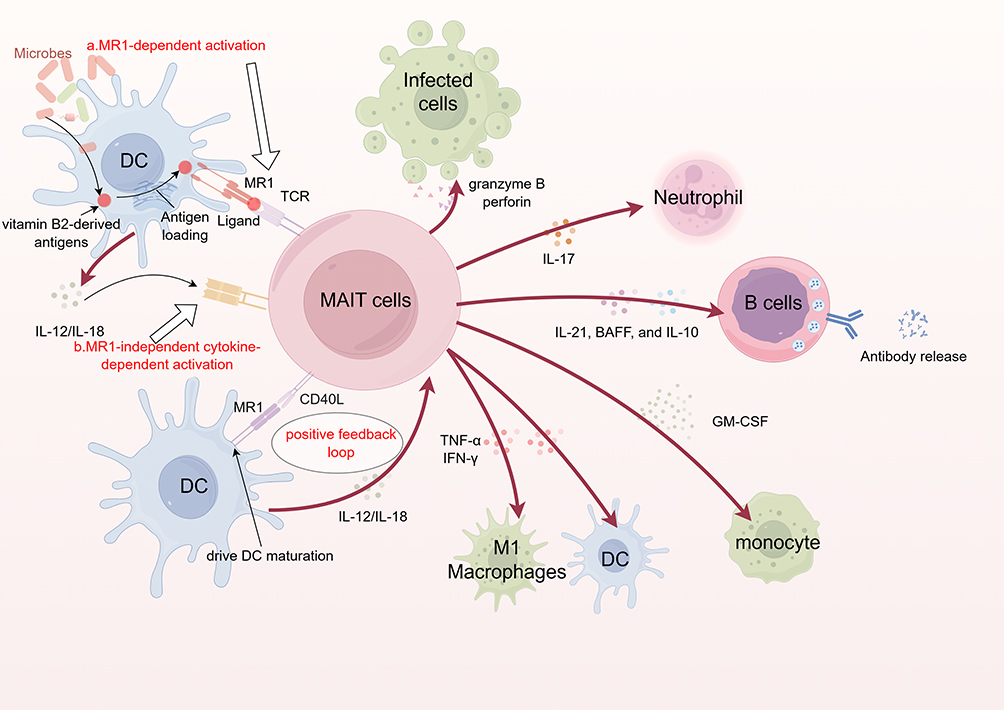 |
Figure 2 Schematic illustration of mucosal-associated invariant T (MAIT) cell activation pathways and effector functions during sepsis.The chart depicts the activation processes and immune functions of MAIT cells when responding to microbial infections in sepsis. Red arrows indicate immunostimulatory or pro-inflammatory effects, while green arrows represent protective or regulatory roles. Created by Figdraw. Activation pathways: (1) MR1-dependent activation: MAIT cells detect microbial vitamin B2 (riboflavin) metabolites through their semi-invariant T cell receptor (TCR) when presented by MHC class I-related molecules (MR1).(2) MR1-independent activation: Infected cells secrete IL-12 and IL-18, which engage cytokine receptors on MAIT cells and initiate activation through a TCR-independent signaling cascade.Effector functions: Cytotoxicity: Once MAIT cells are activated, they attack infected cells or pathogens by releasing granzyme B and perforin. Neutrophil recruitment: IL-17 secreted by MAIT cells promotes neutrophil chemotaxis to sites of infection. Support of humoral immunity: MAIT cells facilitate plasmablast differentiation and antibody secretion by producing B cell-activating cytokines such as IL-21, BAFF, and IL-10, thereby promoting B cell proliferation and immunoglobulin class switching. Activation of antigen-presenting cells and macrophages: The secretion of TNF-α and IFN-γ by MAIT cells is thought to facilitate dendritic cell maturation and support M1-type macrophage responses during inflammation. Monocyte recruitment: Activated MAIT cells secrete granulocyte-macrophage colony-stimulating factor (GM-CSF), promoting monocyte recruitment and activation. Feedback amplification via the CD40L-MR1 axis: Upon stimulation with the cognate antigen 5-A-RU/MG, MAIT cells upregulate CD40L expression, promoting DC maturation through the CD40L-MR1 costimulatory axis. Mature DCs, in turn, enhance IL-12 production, establishing a positive feedback loop that amplifies MAIT activation and supports cytotoxic T lymphocyte (CTL) priming. Abbreviations: DC, Dendritic cell, MR1,MHC class I-related molecules; TCR, T-cell receptor; IL: Interleukin; TNF-α, Tumor Necrosis Factor; IFN-γ, Interferon–γ; BAFF, B-cell–activating factor; GM-CSF, granulocyte-macrophage colony-stimulating factor. |
Pro-Inflammatory and Antimicrobial Functions
Although their role complements conventional adaptive immunity, MAIT cells may become indispensable in immunocompromised hosts or during overwhelming infections, where mobilizing every available defense mechanism is essential for survival. Notably, in critically ill patients, a reduced frequency of circulating MAIT cells correlates with an increased severity of acquired infections, further emphasizing their importance in host immunity.95
Activated MAIT cells can directly target and kill infected cells or pathogens by releasing granzyme B and perforin.34 Upon activation by superantigens such as streptococcal superantigens, human MAIT cells elicit a potent pro-inflammatory response by producing high levels of IFN-γ, IL-17, and tumor necrosis factor-alpha (TNF-α),96 as summarized in Figure 2. MAIT cells facilitate neutrophil recruitment via IL-17 production,97 and the proportion of MAIT17 cells correlates with how severe sepsis is.98 The secretion of TNF-α and IFN-γ by MAIT cells contributes to the immunological priming and functional maturation of dendritic cells and concurrently facilitate the activation of M1 macrophages, thereby orchestrating key processes in the innate immune response and bridging it with adaptive immunity.99 Excessive cytokine secretion has been implicated in the development of hyperinflammatory syndromes that threaten host viability.35,36 Human MAIT cells respond to 5-A-RU/MG by upregulating CD40L, engaging a CD40L-MR1 axis that supports the functional maturation of dendritic cells.100 Mature DCs subsequently enhance IL-12 production, thereby establishing a positive feedback loop that amplifies MAIT cell activation while synergistically facilitating cytotoxic T lymphocyte (CTL) priming.101 Human MAIT cells enhance humoral immune responses by secreting IL-21, B-cell–activating factor (BAFF), and IL-10. These cytokines not only promote plasmablast differentiation and antibody production in vitro but also directly facilitate B cell proliferation and immunoglobulin class switching, particularly following microbial activation.100,102 Furthermore, the cytokine granulocyte-macrophage colony-stimulating factor (GM-CSF), secreted by activated MAIT cells, plays a key role in promoting monocyte activation and facilitating their migration to sites of inflammation.103
Potential Role in Post-Sepsis Exhaustion
Despite growing interest in sepsis research, studies on the role of MAIT cells in sepsis remain relatively scarce. According to Trivedi et al, the absence of MAIT cells causes a rise in bacterial burden and mortality in sepsis experiments. Furthermore, during clinical sepsis, MAIT cells exhibit heightened activation but ultimately become dysfunctional. However, they also mediate tissue-specific cytokine responses that help mitigate sepsis-related mortalities.37 Similarly, Choi et al described a distinct post-sepsis immunosuppressive pattern involving a prolonged decline in MAIT cell functionality and abundance, even though their phenotypic characteristics remained largely unchanged. This dysfunction makes individuals more susceptible to secondary infections. These findings suggest that MAIT cell impairment is essential in post-sepsis immune suppression.38 Future research on the specific mechanisms of MAIT cells in sepsis is expected to enhance our understanding of the disease and facilitate the development of effective therapeutic strategies.
MAIT Cell-Targeted Therapies: Promises and Challenges in Sepsis
Evidence from Barber et al indicates that MAIT cell activity may delay the activation of CD4+ T cells in tuberculosis, highlighting the potential relevance of inhibitory ligands in modulating MAIT cell function during infection.104 Similarly, in the context of Helicobacter pylori infection, pathological outcomes like gastritis and mucosal damage—partly driven by MAIT cell-derived IL-17A—may be attenuated through targeted IL-17A neutralization.105 In sepsis, where MAIT cells may contribute to immune pathology, interventions aimed at modulating their cytokine release or cytolytic activity could represent a novel treatment option, as observed in other infections like TB and H. pylori. Bacterial superantigens have been shown to hyperactivate MAIT cells, potentially triggering a dysregulated cytokine response that contributes to systemic inflammatory damage.35 Therapeutic strategies targeting MAIT cell activity have explored inhibitory ligands like 6-FP106 and Ac-6-FP,107 particularly in contexts requiring immune suppression. This approach has been researched for treating tumors,108 inflammatory bowel disease,109 and other conditions. However, more research is required to explore the application of this therapy in cases of sepsis.
Invariant Natural Killer T (iNKT) Cells
Tissue-Specific Distribution of NKT Cells
NKT cells uniquely co-express T cell receptors and markers commonly associated with NK cells, reflecting their hybrid functional identity. Type I, type II, and NKT-like cells are the three main subsets of NKT cells, categorized by TCR usage and lipid antigen specificity.110 CD1d-restricted NKT cells encompass two distinct populations—type I and type II. Type I NKT cells, commonly referred to as invariant NKT (iNKT) cells, are characterized by the use of a conserved TCRα chain—Vα24-Jα18 in humans—together with a narrowly restricted set of TCRβ chains that shape their antigen specificity.19 And iNKT cells, a subset of NKT cells, are primarily responsible for immune functions and are generally seen as the most functionally and clinically relevant. Type II NKT cells have unique TCRα and TCRβ chains, indicating that their TCR shows increased ligand specificity.111 Some cells do not exhibit CD1d dependency and are referred to as NKT-like cells.110 In the livers of adult mice, NKT cells constitute the majority, making up approximately 12–30% of hepatic lymphocytes.112 In adult humans, the peripheral distribution of NKT cells appears to closely parallel that observed in adult mice. NKT cells are predominantly located in the liver, accounting for 20–30% of hepatic T cells; however, they remain rare in the peripheral blood, constituting only approximately 0.2% of circulating T cells.112,113
Activation of Lipid Antigen Sensing in Sepsis
iNKT cells, as CD1d-restricted T cells,114,115 recognize lipid antigens, including endogenous glycosphingolipids (GSLs), that regulate immune homeostasis and pathogen responses.116,117 Their activation primarily occurs through two distinct pathways: (1) TCR-dependent mechanisms, where CD1d-antigen complexes trigger broad cytokine secretion (eg, IFN-γ and IL-4), activating diverse immune effectors, such as NK cells and macrophages,19 and (2) LPS-induced IL-12 and IL-18 signaling can activate immune cells independently of TCR stimulation, leading to a cytokine-mediated surge in IFN-γ production.118 Certain gram-positive pathogens, including Staphylococcus and Streptococcus, are capable of activating iNKT cells, likely via superantigen exposure or direct interaction with microbial-associated molecular patterns (PAMPs). Sepsis-induced tissue injury may promote iNKT cell activation through CD1d-mediated presentation of self-glycolipid antigens. Once activated, they eliminate infected targets through direct cytotoxic mechanisms, including Fas-FasL interaction and secretion of perforin and granzymes.119
Staphylococcus and Streptococcus, as major contributors to Gram-positive bacterial sepsis, can produce superantigens that engage iNKT cells and potentially amplify systemic inflammation.120 It is also plausible that bacterial PAMPs are directly recognized by iNKT cells. Additionally, through iTCR engagement, iNKT cells can be activated by CD1d-presented endogenous glycolipids, which are often released or structurally modified following septic tissue damage. After activation, iNKT cells can kill infected cells through the Fas-FasL pathway or by releasing perforin/granzymes.
Cytokine Networks and Cellular Crosstalk in Sepsis
Different subsets of mouse iNKT cells can produce distinct types of cytokines upon activation, contributing to immune processes.121 For instance, NKT1 cells resemble Th1 cells and group 1 ILCs (ILC1s) due to their elevated levels of the transcription factor T-bet (encoded by TBX21) and their capacity to produce IFN-γ and TNF-α when activated.122 Notably, iNKT cell-induced damage is primarily mediated by IFN-γ. Compared with other iNKT subsets, NKT1 cells exhibited stronger cytotoxic activity. Th1-Type pro-inflammatory cytokines enhance M1-macrophage activation, neutrophil recruitment and improve bacterial clearance. NKT2 cells, on the other hand, primarily secrete IL-4 and IL-13, aligning them functionally with Th2 cells.123 Th2-Type anti-inflammatory cytokines regulate immune responses, prevent excessive inflammation, and enhance B cell activation for antibody production. Meanwhile, NKT17 cells share similarities with Th17 cells in their cytokine secretion profile, particularly in their production of IL-17.124 Th17-Type inflammatory cytokines enhance inflammatory responses, promote neutrophil recruitment, and improve immunity against bacterial infections. Additionally, the anti-inflammatory role of iNKT cells is partly mediated through IL-10, which limits the activity of APCs and NK cells.42,125–127However, in humans, the classification of iNKT subsets is less clearly defined than in mice. Upon activation, human DN and CD8+ iNKT cells demonstrate enhanced IFN-γ output and potent cytotoxic responses, aligning functionally with murine NKT1 cells.128 Human iNKT cells are capable of secreting both pro- and anti-inflammatory cytokines—such as IL-17 and IL-10—reflecting their dual role in modulating immune responses during infection.42,129
Beyond cytokine secretion, iNKT cells modulate immune function by engaging in direct cellular interactions that shape the behavior of other immune populations. Through their interactions with dendritic cells and B lymphocytes, these cells facilitate communication across the innate and adaptive immune systems. A crucial aspect of their role in adaptive immunity is facilitation of DC maturation into APCs. Upon recognizing CD1d-presented lipid antigens on DCs, iNKT cells upregulate CD40L and secrete cytokines, triggering DC maturation into potent APCs.125 This process enhances MHC class II expression, co-stimulatory markers (CD80/CD86), and IL-12 production, thereby optimizing T cell priming.130,131 For example, in vivo administration of α-GalCer activates iNKT cells, which subsequently stimulate dendritic cells to promote robust activation of CD4+ and CD8+ T cells in an antigen-specific manner.132 Furthermore, iNKT cells support cytotoxic immunity by licensing dendritic cells, thereby enhancing CD8+ T cell activity, and also shape the CD4+ T cell response through promoting Th1/Th2 differentiation pathways.133,134
Notably, iNKT cells balance the immune activation and homeostasis. They eliminate pro-inflammatory APCs via CD1d-dependent cytotoxic granule release,135,136 while supporting humoral immunity through dual mechanisms. Cognate interactions with CD1d+ B cells drive rapid antibody responses against lipid antigens, whereas non-cognate assistance via follicular helper T (TFH) cell activation sustains long-term memory against antigens.137 Additionally, iNKT cells regulate broader immune networks through cytokine signaling.
Dual Roles in Sepsis: Exacerbation and Protection
iNKT cells play dual roles in sepsis pathogenesis, acting as both amplifiers of early immunopathology and mediators of late immunosuppression. Their functional outcomes depend on the infection stage (early hyperinflammation vs late immunosuppression), pro- versus anti-inflammatory balance, and crosstalk with the immune cells (Figure 3).
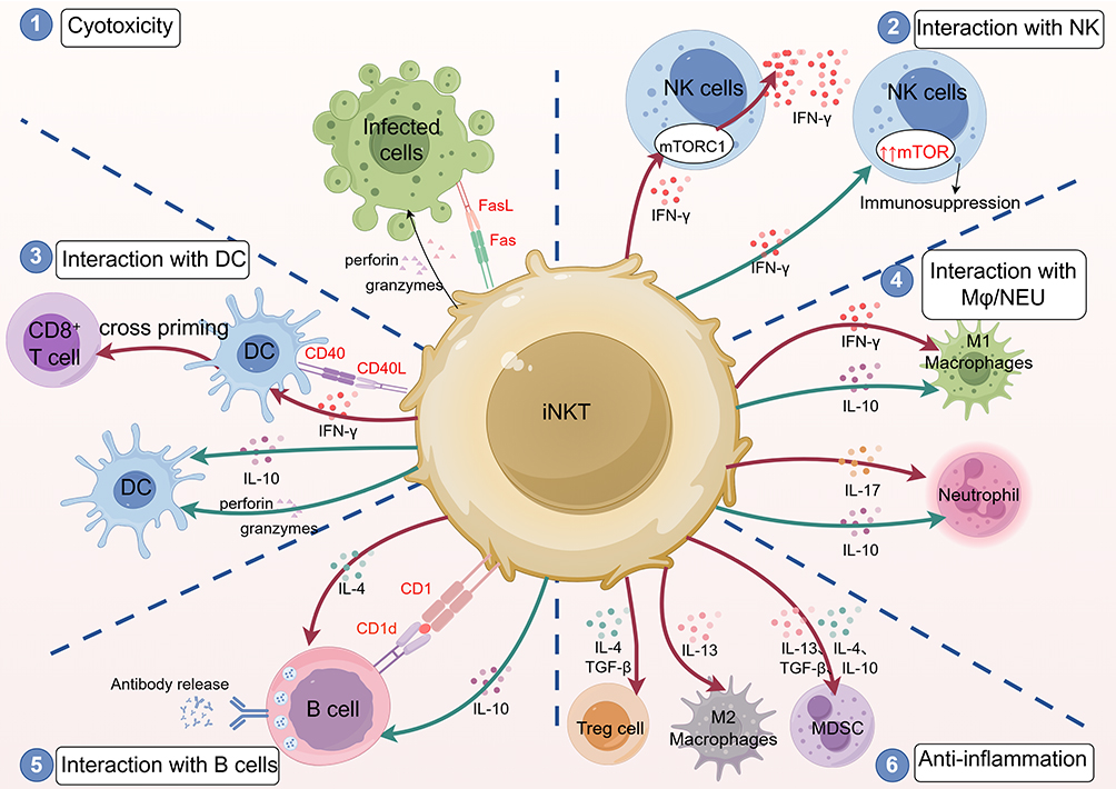 |
Figure 3 Schematic illustration of the major functions of invariant natural killer T (iNKT) cells following activation during sepsis.Upon activation, iNKT cells engage in complex interactions with various immune cells, exerting both pro-inflammatory and anti-inflammatory effects. Red arrows indicate stimulatory functions, while green arrows denote inhibitory functions. Created by Figdraw. 1.Cytotoxicity: iNKT cells directly kill infected cells via the Fas-FasL pathway or by releasing perforin and granzymes. 2.Interaction with NK cells: Through mTORC1 signaling, iNKT cells modulate NK cell function, leading to increased IFN-γ production. However, iNKT-derived cytokines may also suppress NK cell function during sepsis via the upregulation of the mTOR pathway. 3.Interaction with dendritic cells (DCs): iNKT cells rapidly produce cytokines and upregulate CD40L, which binds to CD40 on DCs and enhances their maturation. This process induces the upregulation of IL-12, along with key antigen-presenting components including MHC class II and the co-stimulatory molecules CD80 and CD86, thereby promoting antigen presentation and T-cell priming. iNKT cells contribute to CD8+ T cell activation via cross-priming mechanisms and shape CD4+ T cell responses toward Th1 and Th2 lineages. Additionally, iNKT cells regulate DC function through cytokines: IFN-γ enhances DC activity, IL-10 inhibits it, and cytolytic killing of inflammatory DCs helps restore immune homeostasis. 4.Interaction between macrophages and neutrophils: IFN-γ enhances M1 macrophage activation, while IL-17 promotes inflammation and neutrophil recruitment. Conversely, IL-10 from iNKT cells suppresses the pro-inflammatory activities of both M1 macrophages and neutrophils. 5.Interaction with B cells: iNKT cells support humoral immunity through both cognate (CD1d-mediated) and non-cognate (via TFH activation) B cell help. IL-4 enhances B cell activation and antibody production, while IL-10 inhibits B cell function to limit inflammation. 6.Anti-inflammatory regulation: iNKT cells contribute to immune suppression by secreting Th2-associated cytokines (IL-4 and IL-13) and regulatory cytokines (IL-10 and TGF-β) as well as by activating anti-inflammatory cell types, including M2 macrophages, myeloid-derived suppressor cells (MDSCs), and regulatory T cells (Tregs). Abbreviations: DC, Dendritic cell; IL, Interleukin; IFN-γ, Interferon–γ; mTORC1, mammalian target of rapamycin complex 1; Mφ, Macrophages; NEU, Neutrophil; TGF-β, Transforming growth factor – beta; Treg cell, Regulatory T cell; MDSC, Myeloid - derived suppressor cells. |
During the hyperinflammatory phase, iNKT cells exacerbate tissue damage through excessive IFN-γ production,39 which drives cytokine storms and hyperactivates NK cells via mTORC1 (Mammalian Target of Rapamycin complex 1) signaling,40 correlating with increased mortality in preclinical and clinical studies.138 Across various experimental models, iNKT cell-driven damage in sepsis has consistently been associated with excessive IFN-γ production.139,140 In the early stages of viral infection, NK cells contribute to host defense by eliminating infected cells and secreting immune-regulatory cytokines that influence downstream responses.141,142 Notably, following NKT cell activation, NK cells emerge as the predominant producers of IFN-γ. Conversely, iNKT cells mitigate inflammation through two mechanisms: (1) The secretion of IL-4, IL-10, and TGF-β helps to polarize anti-inflammatory Th2/M2 responses and recruit regulatory cells, including M2-polarized macrophages, myeloid-derived suppressor cells (MDSCs), and regulatory T cells (Tregs); and (2) elimination of hyperactivated APCs via cytolysis, restoring immune balance.43,135
In late sepsis, prolonged activation depletes iNKT cells and upregulates exhaustion markers (eg, Tim-3), leading to apoptosis and impaired bacterial clearance—a hallmark of immunosuppression.41,143,144 Upregulation of the mTOR (Mammalian Target of Rapamycin) signaling cascade, driven by cytokines released from NKT cells, has been implicated in the suppression of NK cell-mediated immune responses during sepsis.40 Therapeutic strategies targeting Tim-3 to inhibit iNKT cell apoptosis or modulating mTORC1 signaling may reverse late-phase immune paralysis. Thus, iNKT cells embody a double-edged sword: their early pro-inflammatory responses enhance pathogen clearance but risk collateral damage, while their late exhaustion perpetuates immunosuppression. Harnessing their phase-specific rolesvia timed cytokine blockade or checkpoint modulation could optimize sepsis immunotherapy.
Modulation of iNKT Cells to Prevent Cytokine Storms and Immunosuppression
Anti-Inflammatory Targeting (Early Phase)
iNKT cells are pivotal therapeutic targets in sepsis because of their dual role in driving hyperinflammation and immunosuppression. IL-30 therapy suppresses IFN-γ and TNF-α via the iNKT/IL-10 axis, thereby reducing mortality in LPS- and CLP-induced sepsis models.145 Treatment with IL-30 can slow down the inflammatory response through the NKT cell pathway in sepsis. Moreover, TIM-3 blockade using α-lactose was found to prevent iNKT cell apoptosis and attenuate cytokine storms in septic mice, thus providing a novel therapeutic approach.146 Furthermore, graphene oxide also holds potential for positive effects in the treatment of sepsis. Graphene oxide was found to polarize iNKT cells toward an anti-inflammatory phenotype by enhancing TGF-β production, reducing cytokine storms in sepsis models.147
Immune Restoration (Late Phase)
During the period of immunosuppression, the treatment targeting iNKT cells lies in activating the activity of iNKT cells. Among them, α-GalCer, as an effective activator for iNKT cells, is widely used in experiments.148 However, its repeated use can lead to immunological unresponsiveness of iNKT cells.140 Because activated iNKT cells mainly lean towards the Th1 type, which inhibits Treg cell differentiation and reduces effector function by decreasing Nr4a1 expression in the late stage of sepsis. The biphasic role of Treg cells in sepsis progression underscores the therapeutic relevance of iNKT cell regulation, particularly for mitigating late-stage immune suppression.39 Joshua Choi’s two-step treatment regimen, involving the sequential administration of various glycolipid agonists, successfully prevented iNKT cell anergy. Restoration of iNKT cell plasticity during polymicrobial sepsis has been achieved through a two-step regimen involving [(C2S,3S,4R)-1-O-(α-d-galactopyranosyl)-N-tetracosanoyl-2-amino-1,3,4-nonanetriol] (OCH), a TH2-biased ligand, administered prior to α-galactosylceramide, thereby promoting IFN-γ-driven immune recovery.149 IFN-γ is a significant factor in the initial immunopathology of sepsis, but its managed release during the immunosuppressive phase could help in immune recovery.138 Tailoring iNKT cell-based interventions to distinct disease stages may represent an effective strategy against sepsis, meriting continued exploration in future studies.
Double-Negative T (DNT) Cells
An Emerging Player in Inflammation and Sepsis
Double-negative T (DNT) cells are mature CD3+ T lymphocytes that lack both CD4 and CD8 co-receptors and constitute a heterogeneous, functionally active subset of the T-cell compartment.150,151 By phenotype, the DNT gate can include both TCRαβ+ and TCRγδ+ cells.152 Notably, most γδ T cells are naturally CD4−CD8− and would fall within this gate; nevertheless, γδ T cells are generally treated as a distinct unconventional T-cell lineage discussed earlier in this review. To maintain conceptual clarity, we focus on describing TCRαβ+ DNT cells in the subsequent several sections regarding DNT cells. In peripheral blood, DNT cells comprise 1–3% of total T lymphocytes.150,153 Beyond the circulation, they are present in secondary lymphoid organs and multiple tissues (eg, intestine, liver, lung, skin, and reproductive tract), where tissue-resident DNT populations have been described.150,154,155 Their contribution to sepsis is less well defined than that of other unconventional T cells; however, emerging evidence implicates DNT cells as important mediators of inflammatory responses in sepsis.18,156
Activation and Pro-Inflammatory Function
During infection, TCRαβ+ double-negative T (DNT) cells are activated through two principal routes. First, TCR-dependent signals derive from peptide–MHC (pMHC) complexes displayed by professional antigen-presenting cells (APCs);157 DNT cells can also acquire pMHC via trogocytosis, enabling antigen-specific recognition and effector function outside the APC niche.158 Second, an inflammatory cytokine milieu—classically IL-12 with IL-18 and type I/II interferons—drives cytokine-mediated bystander activation that heightens DNT responsiveness in the relative absence of TCR ligation.156,159 Human DNT cells express activating receptors such as NKG2D; when ligands are up-regulated on infected or stressed tissues, their regulatory or cytotoxic activity is augmented and can be associated with the emergence of induced DNT subsets.160 In mucosal infections of the respiratory tract, DNT cells may constitute prominent responders that produce IFN-γ and IL-17A and contribute to pathogen control (eg, Francisella tularensis, influenza).44 Leishmania-reactive TCRαβ+ DNT cells can activate macrophages, thereby enhancing anti-parasite effector functions.161 Depending on the pathogen and tissue context, DNT cell frequency and activation state correlate with disease severity, positioning these cells as sentinels of inflammatory activity.
Clinical Correlation with Sepsis
A 2025 clinical study reported a direct association between circulating DNT-cell frequency and sepsis severity within a defined infectious context.156 Expansion of DNT cells was strongly associated with the development of septic shock, and their frequencies tracked clinical severity. These data suggest that DNT cells are not passive bystanders but active participants in the immunopathology of severe sepsis. Their pronounced cytokine output likely fuels the cytokine storm and systemic inflammation that characterize septic shock8. Given their emerging links to severe disease,11,162 DNT cells warrant focused investigation as both biomarkers for early identification of patients at risk of shock and therapeutic targets to attenuate hyperinflammation.
Prospects, Challenges and Opportunities
The Sepsis-3 criteria, while distinguishing sepsis from uncomplicated infections,163 inadequately address patient heterogeneityranging from hyperinflammatory to immunosuppressive endotypes.164 Precision medicine165 frameworks integrating genomic, transcriptomic, and proteomic profiling of peripheral blood leukocytes have begun to stratify patients into immune subtypes with distinct outcomes and therapeutic responses.166–168 Categorizing sepsis based on distinct immune characteristics can enhance the effectiveness of tailored immunotherapeutic approaches for different patient subtypes.169 However, a consensus classification system remains elusive, hindering the development of tailored immunotherapy.170
Owing to their innate-like characteristics, unconventional T cells can initiate immune responses more rapidly than their conventional counterparts during infection. In the early stages of sepsis, when the infection progresses swiftly, this rapid response could be particularly beneficial, as timely intervention is critical for patient survival.171 These T cells might be integrated into combination therapies, alongside antibiotics, anti-inflammatory agents, and immune modulators, to enhance overall therapeutic efficacy. Their distinct mechanisms of action could complement traditional sepsis treatments, potentially improving patient recovery. Therefore, future research should focus on interventions during the early stages of infection to enhance the immunologic capacity of unconventional T cells.
Several innovative interventions for sepsis are being evaluated, including hemoadsorption,172,173 gut microbiota modulation,174,175 and immunoadjuvant therapy.176,177 Hemadsorption mitigates excessive immune-mediated injury in sepsis by modulating the systemic inflammatory response via the clearance of endotoxin (Polymyxin B) and excess inflammatory mediators, particularly cytokines (HA330/HA380).172,178,179 The removal of endotoxin (LPS) could reduce the direct activation of γδ T cells and attenuate the indirect,51 cytokine-driven activation of iNKT cells.118 Concurrently, the broad adsorption of inflammatory cytokines like IL-12 and IL-18 could dampen the TCR-independent activation of γδ T cells, MAIT cells, and iNKT cells, thereby mitigating the cytokine storm and potentially preventing their functional exhaustion.20,110 Therefore, future research on hemadsorption techniques should include immunophenotyping of unconventional T cells to elucidate these interaction mechanisms. Therapeutic modulation of the gut microbiota reduces microbial translocation and dampens systemic inflammation, leading to less organ injury in sepsis.180,181 And the function of protective, liver-infiltrating γδ T cells in sepsis is regulated by gut commensal microbes.28 This link is even more direct for MAIT cells, whose development is “imprinted” by the microbiota and whose activation is directly triggered by metabolites produced by bacteria and fungi.87–89 Therefore, modulating the gut microbiota through therapies like probiotics or fecal microbiota transplantation offers an indirect yet powerful pathway to “tune” the function of UTCs in sepsis, potentially mitigating their pathogenic hyperactivation or bolstering their protective roles. Immunoadjuvant therapies encompass pharmacological modulation to enhance unconventional T-cell responses, including cytokine adjuvants or adoptive transfer of ex vivo expanded T-cell populations. Harnessing their early effector functions may accelerate pathogen clearance and avert progression to sepsis-associated organ failure. During the subsequent immunosuppressive phase, rapidly restoring UTC function is a priority for investigation.13 Reactivation or replenishment of these cells may interrupt disease trajectories and improve clinical outcomes.
A significant challenge in utilizing unconventional T cells for sepsis therapy is the risk of immune exhaustion. Unconventional T cells may also become exhausted, diminishing their ability to manage the infection or modulate inflammation effectively. Moreover, given their scarcity in peripheral blood, obtaining sufficient numbers of these cells remains a practical challenge for therapeutic applications. Expanding these cells in vitro for therapeutic applications is both technically challenging and costly.182 To improve the efficiency and specificity of generating unconventional T cells, researchers have explored stem cell engineering techniques—including gene editing of HSPCs and iPSCs—followed by lineage-directed differentiation.183,184 While these strategies can enhance yield and purity, their clinical translation remains limited.
Although unconventional T cells show promise, their precise mechanisms of action in sepsis remain unknown. Unlike the well-understood developmental pathways and markers of αβ T cells, UTC development is complex and not fully understood.14,185 Grasping how these cells engage with other immune elements, manage pro-inflammatory and anti-inflammatory reactions, and control their function during sepsis is vital for maximizing their therapeutic benefits.
The application of integrative omics and data-driven systems approaches is anticipated to enhance our capacity to resolve immune heterogeneity in sepsis with greater resolution. This perspective is particularly crucial for individualized and precise administration of immunotherapy. Multi-omics technologies offer novel perspectives and tools for sepsis research, enhancing our understanding of its complex pathophysiology and providing a solid scientific basis for the advancement of novel diagnostic and therapeutic approaches.186,187
Conclusion
Among the key players in the immune landscape of sepsis are γδ T cells, MAIT cells, and NKT cells—several types of unconventional T cells that collectively contribute to both antimicrobial defense and immunoregulation. Their ability to recognize non-peptide antigens and rapidly respond to infections highlights their potential as key therapeutic targets. Current research underscores the dual nature of their functions, as these cells can either enhance immune defense or contribute to immune dysregulation, depending on the stage and severity of sepsis.
Modulating the activity of unconventional T cells (UTCs) presents a novel immunotherapeutic approach for sepsis. Specifically, this involves targeting γδ T cells for their antimicrobial and tissue-reparative functions, MAIT cells for their capacity to regulate systemic inflammation, and NKT cells for their cytokine-modulatory roles. The clinical application of these concepts may be realized through strategies such as immunomodulation, immune checkpoint inhibition, and cytokine-based therapies, which aim to potentiate the beneficial functions of these cells while counteracting their detrimental effects. Effective translation of these approaches into clinical practice is contingent upon the development of biomarkers to stratify patients into distinct immune endotypes, thereby enabling personalized interventions. Accordingly, early and sequential immune profiling, including the assessment of UTC subsets and their activation or exhaustion markers at baseline and again at days three to five post-admission, is advisable.
Future clinical trials should be strategically designed to target specific phases of the septic response. For instance, during the initial hyperinflammatory phase, interventions could focus on neutralizing pathogenic cytokines like IL-17A and IFN-γ. Conversely, during the subsequent immunosuppressive phase, the objective would shift to restoring host immune competence. Promising therapeutic candidates for investigation include immune checkpoint inhibitors targeting molecules such as Tim-3 to prevent NKT cell apoptosis, or agents like zoledronate to reverse immune paralysis. Ultimately, the integration of precise patient stratification with temporally targeted immunomodulatory interventions holds the greatest potential for improving survival outcomes in patients with sepsis.
However, significant challenges remain, including the need for a deeper mechanistic understanding of their activation pathways, functional plasticity, and their interactions with other immune components. Future research should focus on elucidating the optimal therapeutic windows for modulating these cells in sepsis as well as developing personalized immunotherapy approaches based on patient-specific immune profiles. By advancing our knowledge of unconventional T cells in sepsis, we may unlock novel therapeutic strategies that restore the immune balance and improve patient survival.
Author Contributions
All authors made a significant contribution to the work reported, whether that is in the conception, study design, execution, acquisition of data, analysis and interpretation, or in all these areas; took part in drafting, revising or critically reviewing the article; gave final approval of the version to be published; have agreed on the journal to which the article has been submitted; and agree to be accountable for all aspects of the work.
Funding
There is no funding to report.
Disclosure
The authors declare no conflicts of interest in this work.
References
1. Evans L, Rhodes A, Alhazzani W, et al. Surviving sepsis campaign: international guidelines for management of sepsis and septic shock 2021. Intensive Care Med. 2021;47(11):1181–1247. doi:10.1007/s00134-021-06506-y
2. Engelmann B, Massberg S. Thrombosis as an intravascular effector of innate immunity. Nat Rev Immunol. 2013;13(1):34–45. doi:10.1038/nri3345
3. Hotchkiss RS, Monneret G, Payen D. Sepsis-induced immunosuppression: from cellular dysfunctions to immunotherapy. Nat Rev Immunol. 2013;13(12):862–874. doi:10.1038/nri3552
4. Cao M, Wang G, Xie J. Immune dysregulation in sepsis: experiences, lessons and perspectives. Cell Death Discov. 2023;9(1):465. doi:10.1038/s41420-023-01766-7
5. Deutschman CS, Tracey KJ. Sepsis: current dogma and new perspectives. Immunity. 2014;40(4):463–475. doi:10.1016/j.immuni.2014.04.001
6. Rudd KE, Johnson SC, Agesa KM, et al. Global, regional, and national sepsis incidence and mortality, 1990–2017: analysis for the global burden of disease study. Lancet. 2020;395(10219):200–211. doi:10.1016/S0140-6736(19)32989-7
7. Jin H, Aziz M, Murao A, et al. Antigen-presenting aged neutrophils induce CD4+ T cells to exacerbate inflammation in sepsis. J Clin Investig. 2023;133(14):e164585. doi:10.1172/JCI164585
8. Jensen IJ, Sjaastad FV, Griffith TS, Badovinac VP. Sepsis-induced T cell immunoparalysis: the ins and outs of impaired T cell immunity. J Immunol. 2018;200(5):1543–1553. doi:10.4049/jimmunol.1701618
9. Darrigues J, Almeida V, Conti E, Ribot JC. The multisensory regulation of unconventional T cell homeostasis. Semin Immunol. 2022;61-64:101657. doi:10.1016/j.smim.2022.101657
10. Kurioka A, Klenerman P. Aging unconventionally: γδ T cells, iNKT cells, and MAIT cells in aging. Semin Immunol. 2023;69:101816. doi:10.1016/j.smim.2023.101816
11. Dossybayeva K, Zhubanova G, Mussayeva A, et al. Nonspecific increase of αβTCR+ double-negative T cells in pediatric rheumatic diseases. World J Pediatr. 2024;20(12):1283–1292. doi:10.1007/s12519-024-00854-7
12. Lin Q, Kuypers M, Philpott DJ, Mallevaey T. The dialogue between unconventional T cells and the microbiota. Mucosal Immunol. 2020;13(6):867–876. doi:10.1038/s41385-020-0326-2
13. Burton RJ, Raffray L, Moet LM, et al. Conventional and unconventional T-cell responses contribute to the prediction of clinical outcome and causative bacterial pathogen in sepsis patients. Clin Exp Immunol. 2024;216(3):293–306. doi:10.1093/cei/uxae019
14. Pellicci DG, Koay HF, Berzins SP. Thymic development of unconventional T cells: how NKT cells, MAIT cells and γδ T cells emerge. Nat Rev Immunol. 2020;20(12):756–770. doi:10.1038/s41577-020-0345-y
15. Harly C, Robert J, Legoux F. NKT, and MAIT cells during evolution: redundancy or specialized functions? J Immunol. 2022;209(2):217–225. doi:10.4049/jimmunol.2200105
16. Godfrey DI, Uldrich AP, McCluskey J, Rossjohn J, Moody DB. The burgeoning family of unconventional T cells. Nat Immunol. 2015;16(11):1114–1123. doi:10.1038/ni.3298
17. Groh V, Steinle A, Bauer S, Spies T. Recognition of stress-induced MHC molecules by intestinal epithelial gammadelta T cells. Science. 1998;279(5357):1737–1740. doi:10.1126/science.279.5357.1737
18. Velikkakam T, Gollob KJ, Dutra WO. Double-negative T cells: setting the stage for disease control or progression. Immunology. 2022;165(4):371–385. doi:10.1111/imm.13441
19. Crosby CM, Kronenberg M. Tissue-specific functions of invariant natural killer T cells. Nat Rev Immunol. 2018;18(9):559–574. doi:10.1038/s41577-018-0034-2
20. Godfrey DI, Koay HF, McCluskey J, Gherardin NA. The biology and functional importance of MAIT cells. Nat Immunol. 2019;20(9):1110–1128. doi:10.1038/s41590-019-0444-8
21. Benveniste PM, Roy S, Nakatsugawa M, et al. Generation and molecular recognition of melanoma-associated antigen-specific human γδ T cells. Sci Immunol. 2018;3(30):eaav4036. doi:10.1126/sciimmunol.aav4036
22. Vermijlen D, Gatti D, Kouzeli A, Rus T, Eberl M. γδ T cell responses: how many ligands will it take till we know? Semin Cell Dev Biol. 2018;84:75–86. doi:10.1016/j.semcdb.2017.10.009
23. Le Nours J, Shahine A, Gras S. Molecular features of lipid-based antigen presentation by group 1 CD1 molecules. Semin Cell Dev Biol. 2018;84:48–57. doi:10.1016/j.semcdb.2017.11.002
24. Qiu L, Zhang Y, Zeng X. The function of γδ T cells in humoral immune responses. Inflamm Res. 2023;72(4):747–755. doi:10.1007/s00011-023-01704-4
25. Dasgupta S, Kumar V. Type II NKT cells: a distinct CD1d-restricted immune regulatory NKT cell subset. Immunogenetics. 2016;68(8):665–676. doi:10.1007/s00251-016-0930-1
26. Torina A, Guggino G, La Manna MP, Sireci G. The janus face of NKT cell function in autoimmunity and infectious diseases. Int J Mol Sci. 2018;19(2):440. doi:10.3390/ijms19020440
27. Yang XW, Li H, Feng T, et al. Impairment of antigen-presenting function of peripheral γδ T cells in patients with sepsis. Clin Exp Immunol. 2022;207(1):104–112. doi:10.1093/cei/uxab029
28. W J, Z Q, H Y, et al. Infiltrated IL-17A-producing gamma delta T cells play a protective role in sepsis-induced liver injury and are regulated by CCR6 and gut commensal microbes. Front Cell Infect Microbiol. 2023:13. doi:10.3389/fcimb.2023.1149506.
29. Malhotra N, Yoon J, Leyva-Castillo JM, et al. IL-22 derived from γδ T cells restricts Staphylococcus aureus infection of mechanically injured skin. J Allergy Clin Immunol. 2016;138(4):1098–1107.e3. doi:10.1016/j.jaci.2016.07.001
30. Pacha O, Sallman MA, Evans SE. COVID-19: a case for inhibiting IL-17? Nat Rev Immunol. 2020;20(6):345–346. doi:10.1038/s41577-020-0328-z
31. Bai H, Gao X, Zhao L, et al. Respective IL-17A production by γδ T and Th17 cells and its implication in host defense against chlamydial lung infection. Cell Mol Immunol. 2017;14(10):850–861. doi:10.1038/cmi.2016.53
32. Turner JE, Krebs C, Tittel AP, et al. IL-17A production by renal γδ T cells promotes kidney injury in crescentic GN. J Am Soc Nephrol. 2012;23(9):1486–1495. doi:10.1681/ASN.2012010040
33. Tschöp J, Martignoni A, Goetzman HS, et al. γδ T cells mitigate the organ injury and mortality of sepsis. J Leukoc Biol. 2008;83(3):581–588. doi:10.1189/jlb.0707507
34. Le Bourhis L, Martin E, Péguillet I, et al. Antimicrobial activity of mucosal-associated invariant T cells. Nat Immunol. 2010;11(8):701–708. doi:10.1038/ni.1890
35. Emgård J, Bergsten H, McCormick JK, et al. MAIT cells are major contributors to the cytokine response in group a streptococcal toxic shock syndrome. Proc Natl Acad Sci USA. 2019;116(51):25923–25931. doi:10.1073/pnas.1910883116
36. Shaler CR, Choi J, Rudak PT, et al. MAIT cells launch a rapid, robust and distinct hyperinflammatory response to bacterial superantigens and quickly acquire an anergic phenotype that impedes their cognate antimicrobial function: defining a novel mechanism of superantigen-induced immunopathology and immunosuppression. PLoS Biol. 2017;15(6):e2001930. doi:10.1371/journal.pbio.2001930
37. Trivedi S, Labuz D, Anderson CP, et al. Mucosal-associated invariant T (MAIT) cells mediate protective host responses in sepsis. eLife. 2020;9:e55615. doi:10.7554/eLife.55615
38. Choi J, Schmerk CL, Mele TS, et al. Longitudinal analysis of mucosa-associated invariant T cells in sepsis reveals their early numerical decline with prognostic implications and a progressive loss of antimicrobial functions. Immunol cell biol. 2023;101(3):249–261. doi:10.1111/imcb.12619
39. Qin Y, Qian Y, Liu S, Chen R. A double-edged sword role of IFN-γ-producing iNKT cells in sepsis: persistent suppression of treg cell formation in an Nr4a1-dependent manner. iScience. 2024;27(12):111462. doi:10.1016/j.isci.2024.111462
40. Kim EY, Ner-Gaon H, Varon J, et al. Post-sepsis immunosuppression depends on NKT cell regulation of mTOR/IFN-γ in NK cells. J Clin Invest. 2020;130(6):3238–3252. doi:10.1172/JCI128075
41. Wu H, Tang T, Deng H, et al. Immune checkpoint molecule tim-3 promotes NKT cell apoptosis and predicts poorer prognosis in sepsis. Clin Immunol. 2023;254:109249. doi:10.1016/j.clim.2023.109249
42. Sag D, Krause P, Hedrick CC, Kronenberg M, Wingender G. IL-10–producing NKT10 cells are a distinct regulatory invariant NKT cell subset. J Clin Invest. 2014;124(9):3725–3740. doi:10.1172/JCI72308
43. Park YH, Lee SW, Kim TC, Park HJ, Van Kaer L, Hong S. The iNKT cell ligand α-GalCer prevents murine septic shock by inducing IL10-producing iNKT and B cells. Front Immunol. 2024;15:1457690. doi:10.3389/fimmu.2024.1457690
44. Cowley SC, Meierovics AI, Frelinger JA, Iwakura Y, Elkins KL. Lung CD4-CD8- double-negative T cells are prominent producers of IL-17A and IFN-gamma during primary respiratory murine infection with francisella tularensis live vaccine strain. J Immunol. 2010;184(10):5791–5801. doi:10.4049/jimmunol.1000362
45. Nielsen MM, Witherden DA, Havran. γδ T cells in homeostasis and host defence of epithelial barrier tissues. Nat Rev Immunol. 2017;17(12):733–745.
46. Perriman L, Tavakolinia N, Jalali S, et al. A three-stage developmental pathway for human Vγ9Vδ2 T cells within the postnatal thymus. Sci Immunol. 2023;8(85):eabo4365. doi:10.1126/sciimmunol.abo4365
47. Narayan K, Sylvia KE, Malhotra N, et al. Intrathymic programming of effector fates in three molecularly distinct γδ T cell subtypes. Nat Immunol. 2012;13(5):511–518. doi:10.1038/ni.2247
48. Johnson MD, Witherden DA, Havran WL. The role of tissue-resident γδ T cells in stress surveillance and tissue maintenance. Cells. 2020;9(3):686. doi:10.3390/cells9030686
49. Marshall AS, Silva JR, Bannerman CA, Gilron I, Ghasemlou N. Skin-resident γδ T cells exhibit site-specific morphology and activation states. J Immunol Res. 2019;2019:1–8. doi:10.1155/2019/9020234
50. Willcox CR, Mohammed F, Willcox BE. The distinct MHC-unrestricted immunobiology of innate-like and adaptive-like human γδ T cell subsets-Nature’s CAR-T cells. Immunol Rev. 2020;298(1):25–46. doi:10.1111/imr.12928
51. Wesch D, Peters C, Oberg HH, Pietschmann K, Kabelitz D. Modulation of γδ T cell responses by TLR ligands. Cell Mol Life Sci. 2011;68(14):2357–2370. doi:10.1007/s00018-011-0699-1
52. Brandes M, Willimann K, Moser B. Professional antigen-presentation function by human γδ T Cells. Science. 2005;309(5732):264–268. doi:10.1126/science.1110267
53. Karunakaran MM, Subramanian H, Jin Y, et al. A distinct topology of BTN3A IgV and B30.2 domains controlled by juxtamembrane regions favors optimal human γδ T cell phosphoantigen sensing. Nat Commun. 2023;14(1):7617. doi:10.1038/s41467-023-41938-8
54. Peigné CM, Léger A, Gesnel MC, et al. The juxtamembrane domain of butyrophilin BTN3A1 controls phosphoantigen-mediated activation of human Vγ9Vδ2 T Cells. J Immunol. 2017;198(11):4228–4234. doi:10.4049/jimmunol.1601910
55. Tsai CY, Liong KH, Gunalan MG, et al. Type I IFNs and IL-18 regulate the antiviral response of primary human γδ T cells against dendritic cells infected with dengue virus. J Immunol. 2015;194(8):3890–3900. doi:10.4049/jimmunol.1303343
56. Song Y, Teo HY, Liu Y, et al. Reviving human γδT cells from apoptosis induced by IL-12/18 via p-JNK inhibition. J Leukoc Biol. 2022;112(6):1701–1716. doi:10.1002/JLB.5MA0622-741R
57. Lalor SJ, McLoughlin RM. Memory γδ T Cells–Newly Appreciated Protagonists in Infection and Immunity. Trends Immunol. 2016;37(10):690–702. doi:10.1016/j.it.2016.07.006
58. Murphy AG, O’Keeffe KM, Lalor SJ, Maher BM, Mills KHG, McLoughlin RM. Staphylococcus aureus infection of mice expands a population of memory γδ T cells that are protective against subsequent infection. J Immunol. 2014;192(8):3697–3708. doi:10.4049/jimmunol.1303420
59. Zhu Y, Zhang S, Li Z, et al. miR-125b-5p and miR-99a-5p downregulate human γδ T-cell activation and cytotoxicity. Cell Mol Immunol. 2019;16(2):112–125. doi:10.1038/cmi.2017.164
60. Bessoles S, Ni M, Garcia-Jimenez S, Sanchez F, Lafont V. Role of NKG2D and its ligands in the anti-infectious activity of Vγ9Vδ2 T cells against intracellular bacteria. Eur J Immunol. 2011;41(6):1619–1628. doi:10.1002/eji.201041230
61. Anderson J, Gustafsson K, Himoudi N, Yan M, Heuijerjans J. Licensing of γδT cells for professional antigen presentation: a new role for antibodies in regulation of antitumor immune responses. Oncoimmunology. 2012;1(9):1652–1654. doi:10.4161/onci.21971
62. Cooper AJR, Lalor SJ, McLoughlin RM. Activation of human Vδ2+ γδ T cells by staphylococcus aureus promotes enhanced anti-staphylococcal adaptive immunity. J Immunol. 2020;205(4):1039–1049. doi:10.4049/jimmunol.2000143
63. Mills KHG. IL-17 and IL-17-producing cells in protection versus pathology. Nat Rev Immunol. 2023;23(1):38–54. doi:10.1038/s41577-022-00746-9
64. Chen W, Lai D, Li Y, et al. Neuronal-activated ILC2s Promote IL-17A production in lung γδ T Cells During Sepsis. Front Immunol. 2021;12:670676. doi:10.3389/fimmu.2021.670676
65. Lt W, Kg G, Ka K. Streptococcus agalactiae and Escherichia coli induce distinct effector γδ T cell responses during neonatal sepsis. iScience. 2024;27(5). doi:10.1016/j.isci.2024.109669
66. Xie B, Wang M, Zhang X, et al. Gut-derived memory γδ T17 cells exacerbate sepsis-induced acute lung injury in mice. Nat Commun. 2024;15(1):6737. doi:10.1038/s41467-024-51209-9
67. Xie B, Zhang Y, Han M, et al. Reversal of the detrimental effects of social isolation on ischemic cerebral injury and stroke-associated pneumonia by inhibiting small intestinal γδ T-cell migration into the brain and lung. J Cereb Blood Flow Metab. 2023;43(8):1267–1284. doi:10.1177/0271678X231167946
68. Li J, Zhang Y, Lou J, et al. Neutralisation of peritoneal IL-17A markedly improves the prognosis of severe septic mice by decreasing neutrophil infiltration and proinflammatory cytokines. PLoS One. 2012;7(10):e46506. doi:10.1371/journal.pone.0046506
69. Pamplona A, Silva‐Santos B. γδ T cells in malaria: a double‐edged sword. FEBS J. 2021;288(4):1118–1129. doi:10.1111/febs.15494
70. Casanova JL, MacMicking JD, Nathan CF. Interferon- γ and infectious diseases: lessons and prospects. Science. 2024;384(6693):eadl2016. doi:10.1126/science.adl2016
71. Tyler CJ, McCarthy NE, Lindsay JO, Stagg AJ, Moser B, Eberl M. Antigen-presenting human γδ T cells promote intestinal CD4+ T cell expression of IL-22 and mucosal release of calprotectin. J Immunol. 2017;198(9):3417–3425. doi:10.4049/jimmunol.1700003
72. Rampoldi F, Donato E, Ullrich L, et al. γδ T cells license immature B cells to produce a broad range of polyreactive antibodies. Cell Rep. 2022;39(8):110854. doi:10.1016/j.celrep.2022.110854
73. Silva-Santos B, Mensurado S, Coffelt. γδ T cells: pleiotropic immune effectors with therapeutic potential in cancer. Nat Rev Cancer. 2019;19(7):392–404. doi:10.1038/s41568-019-0153-5
74. Takimoto R, Suzawa T, Yamada A, et al. Zoledronate promotes inflammatory cytokine expression in human CD14-positive monocytes among peripheral mononuclear cells in the presence of γδ T cells. Immunology. 2021;162(3):306–313. doi:10.1111/imm.13283
75. Roelofs AJ, Jauhiainen M, Mönkkönen H, Rogers MJ, Mönkkönen J, Thompson K. Peripheral blood monocytes are responsible for γδ T cell activation induced by zoledronic acid through accumulation of IPP/DMAPP. Br J Haematol. 2009;144(2):245–250. doi:10.1111/j.1365-2141.2008.07435.x
76. R L, Rj B, Se B, Mp M, E M. Zoledronate rescues immunosuppressed monocytes in sepsis patients. Immunology. 2020;159(1). doi:10.1111/imm.13132
77. Kasten KR, Prakash PS, Unsinger J, et al. Interleukin-7 (IL-7) treatment accelerates neutrophil recruitment through γδ T-cell IL-17 production in a murine model of sepsis. Infect Immun. 2010;78(11):4714–4722. doi:10.1128/IAI.00456-10
78. Legoux F, Salou M, Lantz O. MAIT cell development and functions: the microbial connection. Immunity. 2020;53(4):710–723. doi:10.1016/j.immuni.2020.09.009
79. Constantinides MG, Link VM, Tamoutounour S, et al. MAIT cells are imprinted by the microbiota in early life and promote tissue repair. Science. 2019;366(6464):eaax6624. doi:10.1126/science.aax6624
80. Martin E, Treiner E, Duban L, et al. Stepwise development of MAIT cells in mouse and human. PLoS Biol. 2009;7(3):e1000054. doi:10.1371/journal.pbio.1000054
81. Lee OJ, Cho YN, Kee SJ, et al. Circulating mucosal-associated invariant T cell levels and their cytokine levels in healthy adults. Exp Gerontology. 2014;49:47–54. doi:10.1016/j.exger.2013.11.003
82. Gherardin NA, Souter MN, Koay H, et al. Human blood MAIT cell subsets defined using MR1 tetramers. IImmunol Cell Biol. 2018;96(5):507–525. doi:10.1111/imcb.12021
83. McWilliam HEG, Villadangos JA. MR1 antigen presentation to MAIT cells and other MR1-restricted T cells. Nat Rev Immunol. 2024;24(3):178–192. doi:10.1038/s41577-023-00934-1
84. Aj C, Sb E, Rw B, et al. T-cell activation by transitory neo-antigens derived from distinct microbial pathways. Nature. 2014;509(7500). doi:10.1038/nature13160
85. Jyw M, Rjd R, Hn H, et al. Potent immunomodulators developed from an unstable bacterial metabolite of vitamin B2 biosynthesis. Angew Chem. 2024;63(31). doi:10.1002/anie.202400632
86. Ar L, Je M, Da P, E M. Early innate responses to pathogens: pattern recognition by unconventional human T-cells. Curr Opin Immunol. 2015;36. doi:10.1016/j.coi.2015.06.002.
87. Koay HF, Gherardin NA, Enders A, et al. A three-stage intrathymic development pathway for the mucosal-associated invariant T cell lineage. Nat Immunol. 2016;17(11):1300–1311. doi:10.1038/ni.3565
88. Treiner E, Duban L, Bahram S, et al. Selection of evolutionarily conserved mucosal-associated invariant T cells by MR1. Nature. 2003;422(6928):164–169. doi:10.1038/nature01433
89. García-Angulo VA. Overlapping riboflavin supply pathways in bacteria. Crit Rev Microbiol. 2017;43(2):196–209. doi:10.1080/1040841X.2016.1192578
90. Tastan C, Karhan E, Zhou W, et al. Tuning of human MAIT cell activation by commensal bacteria species and MR1-dependent T-cell presentation. Mucosal Immunol. 2018;11(6):1591–1605. doi:10.1038/s41385-018-0072-x
91. Li YR, Zhou K, Wilson M, et al. Mucosal-associated invariant T cells for cancer immunotherapy. Mol Ther. 2023;31(3):631–646. doi:10.1016/j.ymthe.2022.11.019
92. Gold MC, Cerri S, Smyk-Pearson S, et al. Human mucosal associated invariant t cells detect bacterially infected cells. PLoS Biol. 2010;8(6):e1000407. doi:10.1371/journal.pbio.1000407
93. Le Bourhis L, Dusseaux M, Bohineust A, et al. MAIT cells detect and efficiently lyse bacterially-infected epithelial cells. PLoS Pathog. 2013;9(10):e1003681. doi:10.1371/journal.ppat.1003681
94. Wang H, Kjer-Nielsen L, Shi M, et al. IL-23 costimulates antigen-specific MAIT cell activation and enables vaccination against bacterial infection. Sci Immunol. 2019;4(41):eaaw0402. doi:10.1126/sciimmunol.aaw0402
95. Grimaldi D, Le Bourhis L, Sauneuf B, et al. Specific MAIT cell behaviour among innate-like T lymphocytes in critically ill patients with severe infections. Intensive Care Med. 2014;40(2):192–201. doi:10.1007/s00134-013-3163-x
96. Salio M. Unconventional MAIT cell responses to bacterial infections. Semin Immunopathol. 2022;61-64:101663. doi:10.1016/j.smim.2022.101663
97. Coulter F, Parrish A, Manning D, et al. IL-17 production from T helper 17, mucosal-associated invariant T, and γδ cells in tuberculosis infection and disease. Front Immunol. 2017;8:1252. doi:10.3389/fimmu.2017.01252
98. Li X, Fu S, Cheng H, et al. Differentiation of type 17 mucosal-associated invariant T cells in circulation contributes to the severity of sepsis. Am J Pathol. 2024;194(7):1248–1261. doi:10.1016/j.ajpath.2024.03.010
99. Hartmann N, Harriff MJ, McMurtrey CP, Hildebrand WH, Lewinsohn DM, Kronenberg M. Role of MAIT cells in pulmonary bacterial infection. Mol Immunol. 2018;101:155–159. doi:10.1016/j.molimm.2018.06.270
100. Wu S, Yang X, Lou Y, Xiao X. MAIT cells in bacterial infectious diseases: heroes, villains, or both? Clin Exp Immunol. 2023;214(2):144–153. doi:10.1093/cei/uxad102
101. Salio M, Gasser O, Gonzalez-Lopez C, et al. Activation of human mucosal-associated invariant T cells induces CD40L-dependent maturation of monocyte-derived and primary dendritic cells. J Immunol. 2017;199(8):2631–2638. doi:10.4049/jimmunol.1700615
102. Jensen O, Trivedi S, Meier JD, Fairfax KC, Hale JS, Leung DT. A subset of follicular helper-like MAIT cells can provide B cell help and support antibody production in the mucosa. Sci Immunol. 2022;7(67):eabe8931. doi:10.1126/sciimmunol.abe8931
103. Meierovics AI, Cowley SC. MAIT cells promote inflammatory monocyte differentiation into dendritic cells during pulmonary intracellular infection. J Exp Med. 2016;213(12):2793–2809. doi:10.1084/jem.20160637
104. Sakai S, Kauffman KD, Oh S, Nelson CE, Barry CE, Barber DL. MAIT cell-directed therapy of Mycobacterium tuberculosis infection. Mucosal Immunol. 2021;14(1):199–208. doi:10.1038/s41385-020-0332-4
105. D’Souza C, Pediongco T, Wang H, et al. Mucosal-associated invariant t cells augment immunopathology and gastritis in chronic helicobacter pylori infection. J Immunol. 2018;200(5):1901–1916. doi:10.4049/jimmunol.1701512
106. Patel O, Kjer-Nielsen L, Le Nours J, et al. Recognition of vitamin B metabolites by mucosal-associated invariant T cells. Nat Commun. 2013;4(1):2142. doi:10.1038/ncomms3142
107. Eckle SBG, Birkinshaw RW, Kostenko L, et al. A molecular basis underpinning the T cell receptor heterogeneity of mucosal-associated invariant T cells. J Exp Med. 2014;211(8):1585–1600. doi:10.1084/jem.20140484
108. Yan J, Allen S, McDonald E, et al. MAIT cells promote tumor initiation, growth, and metastases via tumor MR1. Cancer Discovery. 2020;10(1):124–141. doi:10.1158/2159-8290.CD-19-0569
109. Yasutomi Y, Chiba A, Haga K, et al. Activated mucosal-associated invariant T cells have a pathogenic role in a murine model of inflammatory bowel disease. CMGH. 2022;13(1):81–93. doi:10.1016/j.jcmgh.2021.08.018
110. Godfrey DI, MacDonald HR, Kronenberg M, Smyth MJ, Kaer LV. NKT cells: what’s in a name? Nat Rev Immunol. 2004;4(3):231–237. doi:10.1038/nri1309
111. Dg P, Ap U. Unappreciated diversity within the pool of CD1d-restricted T cells. Semin Cell Dev Biol. 2018;84. doi:10.1016/j.semcdb.2017.11.031
112. Zhao W, Li M, Song S, Zhi Y, Huan C, Lv G. The role of natural killer T cells in liver transplantation. Front Cell Dev Biol. 2024;11:1274361. doi:10.3389/fcell.2023.1274361
113. Poddighe D, Maulenkul T, Zhubanova G, Akhmaldtinova L, Dossybayeva K. Natural killer T (NKT) cells in autoimmune hepatitis: current evidence from basic and clinical research. Cells. 2023;12(24):2854. doi:10.3390/cells12242854
114. Rossjohn J, Pellicci DG, Patel O, Gapin L, Godfrey DI. Recognition of CD1d-restricted antigens by natural killer T cells. Nat Rev Immunol. 2012;12(12):845–857. doi:10.1038/nri3328
115. Le Nours J, Praveena T, Pellicci DG, et al. Atypical natural killer T-cell receptor recognition of CD1d-lipid antigens. Nat Commun. 2016;7:10570. doi:10.1038/ncomms10570
116. Adar T, Shankar Lankalapalli R, Bittman R, Ilan Y. The assembly of glycosphingolipid determines their immunomodulatory effect: a novel method for structure-based design of immunotherapy. Cell Immunol. 2020;355:104157. doi:10.1016/j.cellimm.2020.104157
117. Kim S, Cho S, Kim JH. CD1-mediated immune responses in mucosal tissues: molecular mechanisms underlying lipid antigen presentation system. Exp Mol Med. 2023;55(9):1858–1871. doi:10.1038/s12276-023-01053-6
118. Smithgall MD, Comeau MR, Park Yoon BR, Kaufman D, Armitage R, Smith DE. IL-33 amplifies both Th1-and Th2-type responses through its activity on human basophils, allergen-reactive Th2 cells, iNKT and NK cells. Int Immunol. 2008;20(8):1019–1030. doi:10.1093/intimm/dxn060
119. Díaz‐Basabe A, Burrello C, Lattanzi G, et al. Human intestinal and circulating invariant natural killer T cells are cytotoxic against colorectal cancer cells via the perforin–granzyme pathway. Mol oncol. 2021;15(12):3385–3403. doi:10.1002/1878-0261.13104
120. Genardi S, Visvabharathy L, Cao L, et al. Type II natural killer T cells contribute to protection against systemic methicillin-resistant staphylococcus aureus infection. Front Immunol. 2020:11. doi:10.3389/fimmu.2020.610010.
121. Hayakawa Y, Takeda K, Yagita H, Van Kaer L, Saiki I, Okumura K. Differential regulation of Th1 and TH2 functions of NKT cells by CD28 and CD40 costimulatory pathways. J Immunol. 2001;166(10):6012–6018. doi:10.4049/jimmunol.166.10.6012
122. Coquet JM, Chakravarti S, Kyparissoudis K, et al. Diverse cytokine production by NKT cell subsets and identification of an IL-17-producing CD4-NK1.1- NKT cell population. Proc Natl Acad Sci U S A. 2008;105(32):11287–11292. doi:10.1073/pnas.0801631105
123. Du Z, Huang L, Dai X, et al. Progranulin regulates the development and function of NKT2 cells through EZH2 and PLZF. Cell Death Differ. 2022;29(10):1901–1912. doi:10.1038/s41418-022-00973-6
124. Webster KE, Kim HO, Kyparissoudis K, et al. IL-17-producing NKT cells depend exclusively on IL-7 for homeostasis and survival. Mucosal Immunol. 2014;7(5):1058–1067. doi:10.1038/mi.2013.122
125. Joyce S, Girardi E, Zajonc DM. NKT cell ligand recognition logic: molecular basis for a synaptic duet and transmission of inflammatory effectors. J Immunol. 2011;187(3):1081–1089. doi:10.4049/jimmunol.1001910
126. Zhou F, Ciric B, Li H, et al. IL-10 deficiency blocks the ability of LPS to regulate expression of tolerance-related molecules on dendritic cells. Eur j immunol. 2012;42(6):1449. doi:10.1002/eji.201141733
127. Bedke T, Muscate F, Soukou S, Gagliani N, Huber S. Title: IL-10-producing T cells and their dual functions. Semin Immunol. 2019;44:101335. doi:10.1016/j.smim.2019.101335
128. Godfrey DI, Stankovic S, Baxter AG. Raising the NKT cell family. Nat Immunol. 2010;11(3):197–206. doi:10.1038/ni.1841
129. Moreira-Teixeira L, Resende M, Coffre M, et al. Proinflammatory environment dictates the IL-17–producing capacity of human invariant NKT cells. J Immunol. 2011;186(10):5758–5765. doi:10.4049/jimmunol.1003043
130. Hogan AE, O’Reilly V, Dunne MR, et al. Activation of human invariant natural killer T cells with a thioglycoside analogue of α-galactosylceramide. Clin Immunol. 2011;140(2):196–207. doi:10.1016/j.clim.2011.03.016
131. Feng H, Thasler W. Immune microenvironment modulation via the interaction of human invariant natural killer T cell (iNKT cells) and kupffer cells (KC), dendritic cells (DC) in colorectal liver metastasis. J Immunol. 2017;198(1_Supplement). doi:10.4049/jimmunol.198.Supp.208.20
132. Fujii S, Shimizu K, Smith C, Bonifaz L, Steinman RM. Activation of natural killer T cells by α-galactosylceramide rapidly induces the full maturation of dendritic cells in vivo and thereby acts as an adjuvant for combined CD4 and CD8 T cell immunity to a coadministered protein. J Exp Med. 2003;198(2):267–279. doi:10.1084/jem.20030324
133. Melo AM, Zhang L, Dockry ÉF, et al. Novel thioglycoside analogs of α-galactosylceramide stimulate cytotoxicity and preferential Th1 cytokine production by human invariant natural killer T cells. Glycobiology. 2018;28(7):512–521. doi:10.1093/glycob/cwy035
134. Qin Y, Bao X, Zheng M. CD8+ T-cell immunity orchestrated by iNKT cells. Front Immunol. 2023;13:1109347. doi:10.3389/fimmu.2022.1109347
135. Schmid H, Ribeiro EM, Secker KA, et al. Human invariant natural killer T cells promote tolerance by preferential apoptosis induction of conventional dendritic cells. haematol. 2021;107(2):427–436. doi:10.3324/haematol.2020.267583
136. Kok WL, Denney L, Benam K, et al. Pivotal Advance: invariant NKT cells reduce accumulation of inflammatory monocytes in the lungs and decrease immune-pathology during severe influenza A virus infection. J Leukoc Biol. 2011;91(3):357–368. doi:10.1189/jlb.0411184
137. Tonti E, Fedeli M, Napolitano A, et al. Follicular helper NKT cells induce limited B cell responses and germinal center formation in the absence of CD4+ T cell help. J Immunol. 2012;188(7):3217–3222. doi:10.4049/jimmunol.1103501
138. Szabo PA, Anantha RV, Shaler CR, McCormick JK, Haeryfar SMM. CD1d- and MR1-restricted t cells in sepsis. Front Immunol. 2015;6. doi:10.3389/fimmu.2015.00401
139. Hayworth JL, Mazzuca DM, Vareki SM, Welch I, McCormick JK, Haeryfar SM. CD1d‐independent activation of mouse and human i NKT cells by bacterial superantigens. Immunol Cell Biol. 2012;90(7):699–709. doi:10.1038/icb.2011.90
140. Parekh VV. Glycolipid antigen induces long-term natural killer T cell anergy in mice. J Clin Investig. 2005;115(9):2572–2583. doi:10.1172/JCI24762
141. Hoeksema MA, Scicluna BP, Boshuizen M, et al. IFN-γ priming of macrophages represses a part of the inflammatory program and attenuates neutrophil recruitment. J Immunol. 2015;194(8):3909–3916.
142. Riese P, Trittel S, May T, Cicin-Sain L, Chambers BJ, Guzmán CA. Activated NKT cells imprint NK-cell differentiation, functionality and education. Eur J Immunol. 2015;45(6):1794–1807. doi:10.1002/eji.201445209
143. Luo J, Zhang C, Chen D, et al. Tim-3 pathway dysregulation and targeting in sepsis-induced immunosuppression. Eur J Med Res. 2024;29(1):583. doi:10.1186/s40001-024-02203-w
144. Torres LK, Pickkers P, Van Der Poll T. Sepsis-induced immunosuppression. Annu Rev Physiol. 2022;84(1):157–181. doi:10.1146/annurev-physiol-061121-040214
145. Yan J, Mitra A, Hu J, et al. Interleukin-30 (IL27p28) alleviates experimental sepsis by modulating cytokine profile in NKT cells. J Hepatol. 2016;64(5):1128–1136. doi:10.1016/j.jhep.2015.12.020
146. Yao Y, Deng H, Li P, et al. a-lactose improves the survival of septic mice by blockade of TIM-3 signaling to prevent NKT cell apoptosis and attenuate cytokine storm. Shock. 2017;47(3):337–345. doi:10.1097/SHK.0000000000000717
147. Lee SW, Park HJ, Van Kaer L, Hong S. Graphene oxide polarizes iNKT cells for production of TGFβ and attenuates inflammation in an iNKT cell-mediated sepsis model. Sci Rep. 2018;8(1):10081. doi:10.1038/s41598-018-28396-9
148. Hung JT, Chiou SP, Yu A. Affinity and formulation of a phenyl glycolipid for stimulation of invariant natural killer T cells to inhibit tumor growth. J Immunol. 2024;212(1_Supplement):0744_7280–0744_7280. doi:10.4049/jimmunol.212.supp.0744.7280
149. Choi J, Mele TS, Porcelli SA, Savage PB, Haeryfar SMM. Harnessing the versatility of invariant NKT cells in a stepwise approach to sepsis immunotherapy. J Immunol. 2021;206(2):386–397. doi:10.4049/jimmunol.2000220
150. Zhang ZX, Yang L, Young KJ, DuTemple B, Zhang L. Identification of a previously unknown antigen-specific regulatory T cell and its mechanism of suppression. Nat Med. 2000;6(7):782–789. doi:10.1038/77513
151. Juvet SC, Zhang L. Double negative regulatory T cells in transplantation and autoimmunity: recent progress and future directions. J Mol Cell Biol. 2012;4(1):48–58. doi:10.1093/jmcb/mjr043
152. Chen X, Wang D, Zhu X. Application of double-negative T cells in haematological malignancies: recent progress and future directions. Biomark Res. 2022;10(1):11. doi:10.1186/s40364-022-00360-w
153. Fischer K, Voelkl S, Heymann J, et al. Isolation and characterization of human antigen-specific TCRαβ+ CD4-CD8-double-negative regulatory T cells. Blood. 2005;105(7):2828–2835.
154. Wu Z, Zheng Y, Sheng J, et al. CD3+CD4-CD8- (double-negative) T cells in inflammation, immune disorders and cancer. Front Immunol. 2022:13. doi:10.3389/fimmu.2022.816005.
155. D’Acquisto F, Crompton T. CD3+ CD4- CD8-(double negative) T cells: saviours or villains of the immune response? Biochem. Pharmacol. 2011;82(4):333–340.
156. Qiu PP, Cai SJ, Guo LL, et al. Elevated CD4- CD8- double-negative T cells in the peripheral blood of SFTS patients are associated with disease severity. Clin Chim Acta. 2025;574:120312. doi:10.1016/j.cca.2025.120312
157. De Libero G, Mori L. Recognition of lipid antigens by T cells. Nat Rev Immunol. 2005;5(6):485–496. doi:10.1038/nri1631
158. Ms FM, Young KJ, Gao J, Joe B, Zhang L. Cutting edge: in vivo trogocytosis as a mechanism of double negative regulatory T cell-mediated antigen-specific suppression. J Immunol. 2008;181(4):2271–2275. doi:10.4049/jimmunol.181.4.2271
159. Juvet SC, Han M, Vanama R, et al. Autocrine IFNγ controls the regulatory function of lymphoproliferative double negative T cells. PLoS One. 2012;7(10):e47732. doi:10.1371/journal.pone.0047732
160. Hu SH, Zhang LH, Gao J, et al. NKG2D enhances double-negative T cell regulation of B cells. Front Immunol. 2021;12:650788. doi:10.3389/fimmu.2021.650788
161. Mou Z, Liu D, Okwor I, Jia P, Orihara K, Uzonna JE. MHC class II restricted innate-like double negative T cells contribute to optimal primary and secondary immunity to leishmania major. PLoS Pathog. 2014;10(9):e1004396. doi:10.1371/journal.ppat.1004396
162. Poddighe D, Maulenkul T, Dossybayeva K, Zhubanova G, Mukusheva Z, Akhmaltdinova L. Double-negative T cells in pediatric rheumatic diseases. Clin Exp Pediatr. 2024;67(12):632–640. doi:10.3345/cep.2023.01760
163. Singer M, Deutschman CS, Seymour CW, et al. The third international consensus definitions for sepsis and septic shock (Sepsis-3). JAMA. 2016;315(8):801–810. doi:10.1001/jama.2016.0287
164. Van Der Poll T, Shankar-Hari M, Wiersinga WJ. The immunology of sepsis. Immunity. 2021;54(11):2450–2464. doi:10.1016/j.immuni.2021.10.012
165. König IR, Fuchs O, Hansen G, von Mutius E, Kopp MV. What is precision medicine? Eur Respir J. 2017;50(4):1700391. doi:10.1183/13993003.00391-2017
166. Scicluna BP, Van Vught LA, Zwinderman AH, et al. Classification of patients with sepsis according to blood genomic endotype: a prospective cohort study. Lancet Respir Med. 2017;5(10):816–826. doi:10.1016/S2213-2600(17)30294-1
167. Sweeney TE, Azad TD, Donato M, et al. Unsupervised analysis of transcriptomics in bacterial sepsis across multiple datasets reveals three robust clusters. Crit Care Med. 2018;46(6):915–925. doi:10.1097/CCM.0000000000003084
168. Davenport EE, Burnham KL, Radhakrishnan J, et al. Genomic landscape of the individual host response and outcomes in sepsis: a prospective cohort study. Lancet Respir Med. 2016;4(4):259–271. doi:10.1016/S2213-2600(16)00046-1
169. Stanski NL, Wong HR. Prognostic and predictive enrichment in sepsis. Nat Rev Nephrol. 2020;16(1):20–31. doi:10.1038/s41581-019-0199-3
170. Giamarellos-Bourboulis EJ, Aschenbrenner AC, Bauer M, et al. The pathophysiology of sepsis and precision-medicine-based immunotherapy. Nat Immunol. 2024;25(1):19–28. doi:10.1038/s41590-023-01660-5
171. Mayassi T, Barreiro LB, Rossjohn J, Jabri B. A multilayered immune system through the lens of unconventional T cells. Nature. 2021;595(7868):501–510. doi:10.1038/s41586-021-03578-0
172. Bottari G, Ranieri VM, Ince C, et al. Use of extracorporeal blood purification therapies in sepsis: the current paradigm, available evidence, and future perspectives. Crit Care. 2024;28(1):432. doi:10.1186/s13054-024-05220-7
173. Yildiz AB, Copur S, Tanriover C, et al. A potential approach toward the management of sepsis: the extracorporeal cytokine hemadsorption therapy. Semin Dial. 2024;37(2):117–121. doi:10.1111/sdi.13188
174. Tao YL, Wang JR, Liu M, et al. Progress in the study of the correlation between sepsis and intestinal microecology. Front Cell Infect Microbiol. 2024;14:1357178. doi:10.3389/fcimb.2024.1357178
175. Liang H, Song H, Zhang X, et al. Metformin attenuated sepsis-related liver injury by modulating gut microbiota. Emerg Microbes Infect. 2022;11(1):815–828. doi:10.1080/22221751.2022.2045876
176. Islam MM, Watanabe E, Salma U, et al. Immunoadjuvant therapy in the regulation of cell death in sepsis: recent advances and future directions. Front Immunol. 2024;15:1493214. doi:10.3389/fimmu.2024.1493214
177. Arapis A, Panagiotopoulos D, Giamarellos-Bourboulis EJ. Recent advances of precision immunotherapy in sepsis. Burns Trauma. 2025;13:tkaf001. doi:10.1093/burnst/tkaf001
178. Li Y, Han M, Yang M, Su B. Hemoperfusion with the HA330/HA380 cartridge in intensive care settings: a state-of-the-art review. Blood Purif. 2025;54(2):122–137. doi:10.1159/000542469
179. Osawa I, Goto T, Kudo D, et al. Targeted therapy using polymyxin B hemadsorption in patients with sepsis: a post-hoc analysis of the JSEPTIC-DIC study and the Euphrates trial. Crit Care. 2023;27:245. doi:10.1186/s13054-023-04533-3
180. Haak BW, Wiersinga WJ. The role of the gut microbiota in sepsis. Lancet Gastroenterol Hepatol. 2017;2(2):135–143. doi:10.1016/S2468-1253(16)30119-4
181. Marascio N, Scarlata GGM, Romeo F, et al. The role of gut microbiota in the clinical outcome of septic patients: state of the art and future perspectives. Int J Mol Sci. 2023;24(11):9307. doi:10.3390/ijms24119307
182. Xue D, Lu S, Zhang H, et al. Induced pluripotent stem cell-derived engineered T cells, natural killer cells, macrophages, and dendritic cells in immunotherapy. Trends Biotechnol. 2023;41(7):907–922. doi:10.1016/j.tibtech.2023.02.003
183. Li YR, Zhou Y, Yu J, et al. Generation of allogeneic CAR-NKT cells from hematopoietic stem and progenitor cells using a clinically guided culture method. Nat Biotechnol. 2024:1–16. doi:10.1038/s41587-024-02226-y
184. Li YR, Zhou Y, Kramer A, Yang L. Engineering stem cells for cancer immunotherapy. Trends Cancer. 2021;7(12):1059–1073. doi:10.1016/j.trecan.2021.08.004
185. Hu Y, Hu Q, Li Y, et al. γδ T cells: origin and fate, subsets, diseases and immunotherapy. Signal Transduct Target Ther. 2023;8(1):434. doi:10.1038/s41392-023-01653-8
186. Zhang Z, Chen L, Sun B, et al. Identifying septic shock subgroups to tailor fluid strategies through multi-omics integration. Nat Commun. 2024;15(1):9028. doi:10.1038/s41467-024-53239-9
187. Huang J, Chen Y, Guo Z, et al. Prospective study and validation of early warning marker discovery based on integrating multi-omics analysis in severe burn patients with sepsis. Burns Trauma. 2023:
 © 2025 The Author(s). This work is published and licensed by Dove Medical Press Limited. The
full terms of this license are available at https://www.dovepress.com/terms
and incorporate the Creative Commons Attribution
- Non Commercial (unported, 4.0) License.
By accessing the work you hereby accept the Terms. Non-commercial uses of the work are permitted
without any further permission from Dove Medical Press Limited, provided the work is properly
attributed. For permission for commercial use of this work, please see paragraphs 4.2 and 5 of our Terms.
© 2025 The Author(s). This work is published and licensed by Dove Medical Press Limited. The
full terms of this license are available at https://www.dovepress.com/terms
and incorporate the Creative Commons Attribution
- Non Commercial (unported, 4.0) License.
By accessing the work you hereby accept the Terms. Non-commercial uses of the work are permitted
without any further permission from Dove Medical Press Limited, provided the work is properly
attributed. For permission for commercial use of this work, please see paragraphs 4.2 and 5 of our Terms.