")
Back to Journals » Clinical, Cosmetic and Investigational Dermatology » Volume 15
The Efficacy of Noninvasive 1060-Nm Diode Lasers for Submental Lipolysis: A Pilot Study
Authors Wanitphakdeedecha R , Evangelista KER, Yan C , Apinuntham C, Techapichetvanich T, Eimpunth S, Lektrakul N, Manuskiatti W
Received 13 August 2022
Accepted for publication 8 November 2022
Published 19 December 2022 Volume 2022:15 Pages 2775—2783
DOI https://doi.org/10.2147/CCID.S382582
Checked for plagiarism Yes
Review by Single anonymous peer review
Peer reviewer comments 2
Editor who approved publication: Dr Jeffrey Weinberg
Rungsima Wanitphakdeedecha,1 Kristy Elleza R Evangelista,1,2 Chadakan Yan,1 Chalermkwan Apinuntham,1 Thanya Techapichetvanich,1 Sasima Eimpunth,1 Nittaya Lektrakul,3 Woraphong Manuskiatti1
1Department of Dermatology, Faculty of Medicine Siriraj Hospital, Mahidol University, Bangkok, Thailand; 2Department of Dermatology, Research Institute for Tropical Medicine, Manila, Philippines; 3Department of Radiology, Faculty of Medicine Siriraj Hospital, Mahidol University, Bangkok, Thailand
Correspondence: Rungsima Wanitphakdeedecha, Department of Dermatology, Faculty of Medicine Siriraj Hospital, Mahidol University, 2 Prannok Road, Bangkok Noi, 10700, Thailand, Tel +66 2 419 4333, Fax +66 2 411 9922, Email [email protected]
Background: Submental fat is a noticeable fat in the submental region that is of great concern aesthetically, especially to female patients. A 1060-nm diode laser is a clinically proven device for the laser lipolysis of subcutaneous fat cells. This study aimed to evaluate the safety and efficacy of a 1060-nm diode laser for submental fat reduction.
Methods: Twenty subjects with unwanted localized submental fat were treated with a single session of a 1060-nm diode laser with an energy setting between 0.95 and 1.40 W/cm2, depending on each patient’s tolerance. Submental fat thickness measurements were documented at baseline, and 1, 3, and 6 months after treatment. Clinical photographs, ultrasound images, and adverse events were evaluated at each follow-up visit. Subjects responded to a satisfaction questionnaire at the end of the study.
Results: The subjects had a mean age of 34.55 ± 6.19 years, a mean body weight of 70.66 ± 10.55 kilograms, and most (95%) were women. The average energy setting was 0.95– 1.40 W/cm2, with a pain score of 3.90 ± 1.30 on a 0-to-10 scale. A significant reduction in submental fat thickness measured by ultrasound was noted at post-treatment month 3 (falling to 0.46 ± 0.13; P = 0.013). However, there was a slight increase in the submental fat thickness at the 6-month follow-up (to 0.48 ± 0.12); the change in the thickness relative to the baseline was nonsignificant (P = 0.121). Most subjects reported an improvement 6 months after the treatment. No severe adverse events were observed throughout the study period.
Conclusion: Our study demonstrated the potential role of 1060-nm Diode laser for the treatment of localized submental subcutaneous adiposities. It is a promising alternative treatment modality for patients seeking an in-office, nonsurgical procedure for fat reduction without severe complications.
Keywords: 1060-nm, diode laser, lipolysis, submental fat
Introduction
Currently, people seek consultations to improve their facial appearance. Maintaining a good body physique increases individuals’ self-esteem and improves their emotional and social well-being.1 Facial features are considered indicators of the attractiveness of a person.1 The appearance of the chin and jawline or the submental area is a common facial concern that can profoundly impact a person’s feelings.1
According to the 2019 American Society for Dermatologic Surgery consumer survey, 73% of 3645 respondents were concerned about excess fat in their chin and neck.2 Excessive submental fat is either due to genetics or lifestyle factors, such as an unhealthy diet or a sedentary lifestyle.1 This excessive fat can be corrected through surgical procedures, including submental liposuction and submentoplasty.3,4 However, the procedures have pitfalls that can be frustrating to patients. They include nerve injury, bleeding, hematoma, seroma, postinflammatory hyperpigmentation, infection, and scarring.4,5
More and more people are actively seeking noninvasive fat reduction and rejuvenation procedures.5 Noninvasive treatment modalities have emerged that address unwanted fat in the submental area with less downtime and complications than surgical procedures. Among them are injection adipolysis,5–8 cryolipolysis,9–11,20–22 radiofrequency,12 high-intensity focus ultrasound,13,14 laser lipolysis15–18 and microwave technology.23
A recent study done by Salsi et al used microwaves technology for unwanted fat reduction and submental skin tightening.23 The technology utilizes selective microwave frequency producing localized heat and is absorbed by that fat through a biophysical process called “dielectric heating”.23 A total of 48 subjects underwent submental treatment for 6 sessions and showed significant decrease in submental fat and laxity at 12 weeks follow-up.23 No significant adverse effects were noted among the subjects.23
Studies have demonstrated the efficacy of a 1060-nm diode laser for fat reduction in some areas of the body.15–18 The laser has a high affinity for adipose tissue due to its long wavelength. It raises the temperature of adipose cells to between 42 ℃ and 47 ℃, damaging their structural integrity. Inflammatory responses are stimulated. The body naturally eliminates disrupted fat cells and cellular debris by attracting macrophages and removing the cells and debris through phagocytosis. Several investigations have established the safety of the laser in patients with skin of color.15–18
Decorato et al demonstrated that the optimal time for a 1060-nm diode laser to cause significant adipocyte injury is 20 to 25 minutes. Use for longer than 25 minutes can create palpable nodules in the subcutaneous fat.15,17 Work by Bass and associates and by Katz et al demonstrated a significant fat reduction in the abdomen and flanks, respectively, 12 weeks after treatment, measured by ultrasound.15,18 Neither study documented any severe complications.15,18
There was also an increasing demand for leg contouring among Asian patients. A recent study done in Thailand by Yan et al showed efficacy of 1060 nm Diode laser in reducing medial knee fat thickness.24 A total of 19 subjects with localized unwanted fat on the medial knees were enrolled into this study. Significant reduction in knee circumferences at 1-, 3-, and 6-month follow-up visits compared with baseline, and knee fat thickness measured by ultrasound in both axial and sagittal plane at 1 and 6 months after treatment were recorded.24 Minimal and transient side effects were noted among patients.24
In view of these scientific findings, our investigation aimed to evaluate the safety and efficacy of a 1060-nm diode laser for submental fat reduction.
Methods
This single-site, prospective, clinical pilot study was conducted at the Skin Laser Center of Siriraj Hospital, Mahidol University in Bangkok, Thailand. We enrolled 20 healthy women and men aged between 20 and 65 and weighing 60 to 80 kilograms. Written informed consent was obtained from all subjects before prior the start of the study. The study was approved by the Human Research Ethics Committee of Siriraj Hospital (approval reference: Si420/2018). The procedures used in the research study adhered to the tenets of the 1975 Declaration of Helsinki.
The investigators gathered all demographic data, clinical histories, and physical examination findings related to the submental area prior to its treatment with a 1060-nm Diode laser.
The inclusion criteria consisted of healthy adult volunteers of both sexes. All subjects agreed to neither receive any kind of dermatologic/surgical procedures nor other body treatments aiming at reducing body fat during the entire study period. Subjects were excluded from the study if any of the following were met: pregnant or lactating women; a history of liposuction, surgical procedures, laser procedures, gold therapy, neuropathic disorders, skin malignancies, hypertrophic scar or keloid formation, skin infections, skin hypersensitivity, photosensitivity reactions, or herpes virus infection; evidence of compromised healing; an immunodeficiency disorder (including HIV infection or AIDS); the use of immunosuppressive medications; impaired skin sensation; diabetic neuropathy; or underlying psychological conditions.
The investigators evaluated each subject’s unwanted localized fat in the submental area. The baseline fat thickness was measured in a supine position using an ultrasound imaging device (iU 22 xMatrix, Philips Healthcare, Eindhoven, the Netherlands). The primary endpoint of the study was a statistically significant reduction in submental fat layer thickness compared with the baseline at follow-up visits held 1, 3, and 6 months after treatment. The secondary endpoint was a subject satisfaction score of 3 or higher after the treatment, measured by improvement scale surveys conducted at the follow-ups.
A 1060-nm Diode laser device, Sculpsure (Cynosure, Westford, MA, USA), was used in the study. The investigator and all the subjects wore protective eyewear goggles specific to protect against the 1060-nm wavelength during the laser treatment. The submental treatment was performed using one applicator with head gear. The flat non-suction applicator was placed on the subject’s submental area. The applicator was inserted into the appropriate frame and was placed in contact with the skin.
The subject was treated with a single laser session of 1060-nm Diode laser. For each patient, the optimal power setting was identified to maximize treatment efficacy and at the same time, guarantee safety and patient comfort. The power setting was in the range between 0.95 and 1.40 W/cm2, depending on an individual subject’s pain tolerance. The spot size was 14.2cm2. The exposure time for all subjects was 25 minutes,15,18. The treatment of fat in the submental area started from 1.5 cm below the lower border of the mandible up to the hyoid bone. This is to avoid treating the area of the arteries, veins and marginal mandibular nerve. The Diode laser at 1060 nm delivers heat below the surface of the skin to safely and permanently destroy fat cells. It also uses a constant cooling technology to keep subjects comfortable and safe without the risks of any burns. After the treatment, the subjects rated their pain using a 0-to-10 scale (0 = none; 10 = worst possible).18
Clinical photographs and ultrasound images were taken at all follow-ups to assess the efficacy of the laser. All subjects answered an improvement scale survey, and adverse events were monitored at each follow-up visits. Submental fat thickness was also measured using ultrasound images, and the subjects’ body weights (in kilograms) were documented. The submental fat thickness of subjects showing weight gain and no weight gain at 1, 3, and 6 months after their treatment were compared with baseline values.
Descriptive analysis was used for the demographic data. The data were analyzed using a paired t-test (normality) for parametric distribution. The statistical analysis was performed using the statistical software PASW Statistics for Windows, version 18.0 (SPSS Inc, Chicago, IL, USA), with a P value < 0.05 indicating statistical significance.
Results
Twenty healthy subjects completed the study. Their demographic data are detailed in Table 1. The subjects’ mean age was 34.55 ± 6.19 years, their mean body weight was 70.61 ± 10.56 kilograms, and 95% (19/20) were women. They received a single treatment with a 1060-nm diode laser, with an average energy setting of 0.95 to 1.40 W/cm2 (Table 1). The documented body weight changes of the subjects at the follow-ups were nonsignificant (Table 2).
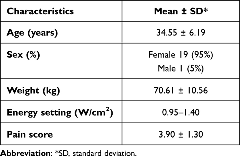 |
Table 1 Demographic Data of Patients Enrolled in the Study |
 |
Table 2 Evaluation Parameters of Whole Study |
The average submental fat thickness at baseline was 0.51 ± 0.09 cm (Table 2). There was a significant reduction in the fat thickness at post-treatment month 3 compared with the baseline, with a decrease to 0.46 ± 0.13 cm (P = 0.013). However, 6 months after the treatment, the submental thickness had increased (to 0.48 ± 0.12 cm). The change in the thickness relative to the baseline was nonsignificant (P = 0.121; Table 2).
Based on the dynamic subgroup analysis, the subjects who did not gain body weight responded better to treatment (Table 3). The mean baseline fat thickness was 0.52 ± 0.11 (Table 3). The fat thickness of the nonweight-gain group showed a significant reduction at post-treatment months 1 (0.46 ± 0.12 cm; P = 0.014) and 3 (0.46 ± 0.15; P = 0.042) compared with the baseline (Table 3). On the other hand, the mean fat thickness of the weight-gain group did not show a significant reduction at either post-treatment months 1 or 3 (P = 0.106 and 0.197, respectively; Table 3).
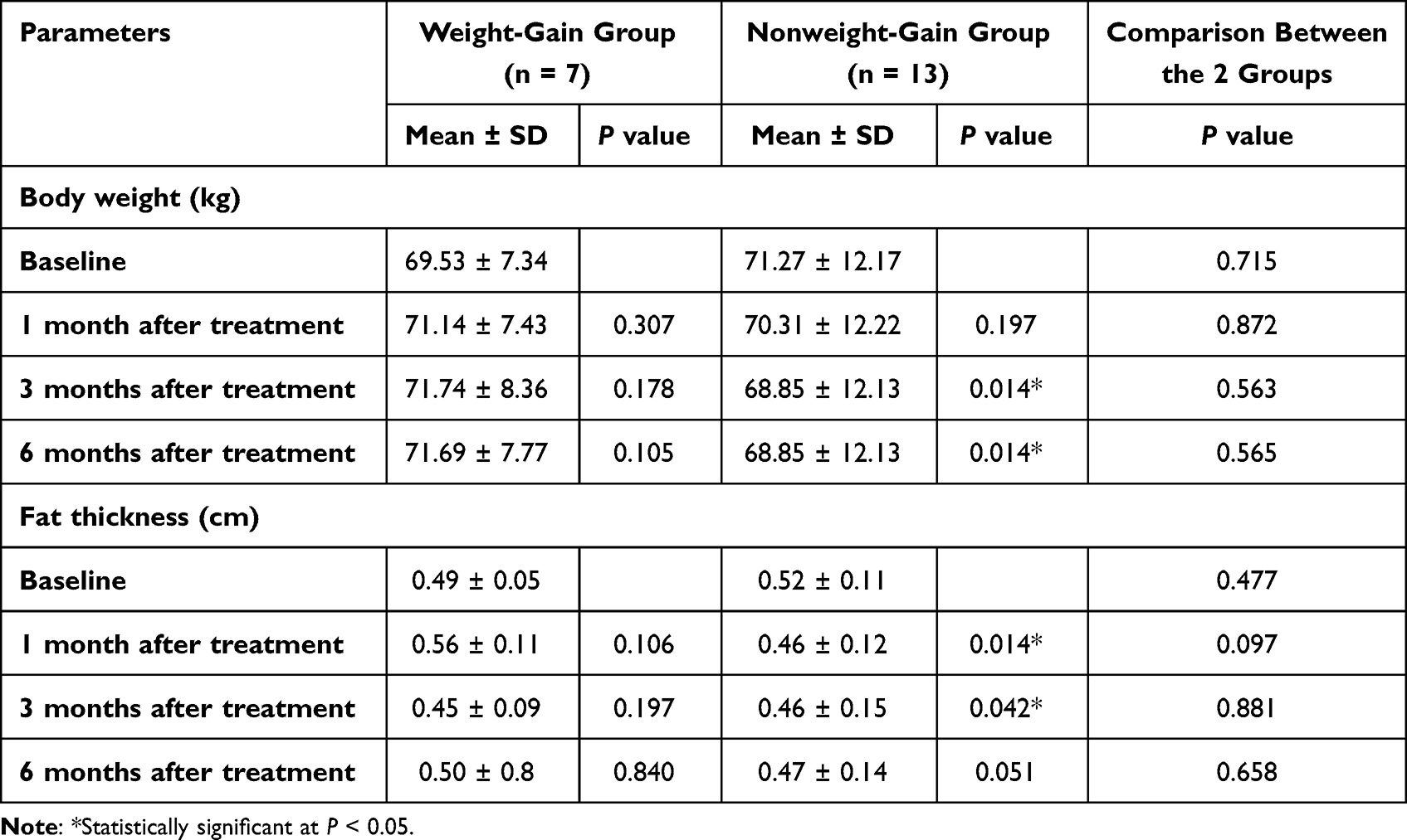 |
Table 3 Comparison of Evaluation Parameters of Weight-Gain and Nonweight-Gain Groups |
Most subjects were satisfied with their treatment outcomes 1 and 6 months after their treatment (Figure 1). The mean pain score was 3.90 ± 1.30 on a 0-to-10 scale (Table 1). All subjects reported mild erythema and mild tenderness immediately after treatment, but the side effects were transient. No severe side effects (hypopigmentation, hyperpigmentation, scarring, fat dystrophy, or infection) were found throughout the study period.
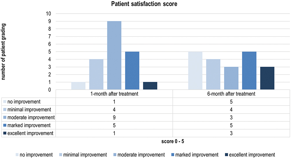 |
Figure 1 Subjects’ self-improvement scores for both groups at all follow-ups. |
Clinical photographs and ultrasound images of a representative subject are presented in Figures 2 and 3. They illustrate improvements 3 and 6 months after the treatment.
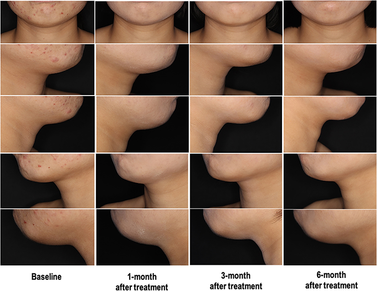 |
Figure 2 Clinical photographs of a representative patient. |
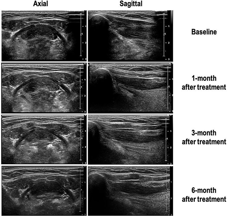 |
Figure 3 Ultrasound images (2 dimensions) of the representative patient. |
Discussion
Excessive submental fat can substantially impact people’s feelings of attractiveness.1 Work by Bauman et al validated that submental fat can negatively affect the psychological well-being of individuals. In their study analysis, the researchers documented that excessive submental fat can affect the emotional well-being of people. The data revealed that concerns about chin and neck appearance negatively impacted women’s feelings and emotional well-being more than men’s.1 In the present study, majority of subjects included were women. Behavioral changes occurred in as many as 75% of people with a considerable amount of chin fat.1 More female than male respondents avoided taking photographs (40% vs 31%). Additionally, more women than men (44% vs 34%) said posting photographs of themselves on social media sites was stressful due to their perception that their chin and neck area appeared unattractive.1 Because of these emotional sequelae, physicians must evaluate the submental area during aesthetic consultations.
Laser lipolysis generates controlled hyperthermia using a specific wavelength and optimal energy in the subcutaneous layer. Hyperthermia then causes photomechanical and photothermal reactions that destroy adipocytes and stimulate an inflammatory response.17,18
1060-nm Diode laser wavelength technology has a high affinity for adipose tissue. The laser raises the temperature of adipose cells between 42℃ and 47℃, damaging their structural integrity.24 After the treatment session, the body naturally eliminates the disrupted cells over the next few months.24 Disrupted fat cells are permanently eliminated from the body and will not regenerate. Previous studies on 1060-nm diode lasers showed significant reductions in localized subcutaneous fat on the abdomen15 and flanks.18
In the study by Bass et al, significant mean reductions in the fat layer thickness of the abdomen from baseline were observed 6 and 12 weeks after treatment.15 At 12 weeks, 91% (31/34) of the subjects were satisfied with the treatment.15 Using the same laser device, an earlier pilot investigation on the flanks by Katz et al demonstrated a significant mean fat reduction based on ultrasound images.18 Twelve weeks after the treatment, 96% (41/43) of the subjects reported being satisfied with their treatment outcomes.18
A recent study done in Thailand by Yan et al, used the same device, 1060 nm Diode laser, to evaluate its efficacy for medial knee fat reduction. This showed significant reduction in knee circumferences (p< 0.001) at 1-, 3-, and 6-month follow-up visits compared with baseline, and knee fat thickness measured by ultrasound in both axial and sagittal plane at 1 and 6 months after treatment (p = 0.036 and p <0.001, respectively) were recorded.24
In the present study on the submental area, we found a similar significant reduction in fat thickness at post-treatment month 3 (0.46 ± 0.13 cm) compared with the baseline (P = 0.013). However, at the 6-month follow-up, there was a slight increase in the submental thickness (to 0.48 ± 0.12 cm). The change in the thickness relative to the baseline was nonsignificant (P = 0.121; Table 2). Most of our subjects expressed satisfaction with their treatment outcomes 1 and 6 months after the treatments (Figure 1). This study shows that the 1060-nm diode laser is helpful for subcutaneous fat reduction in the submental area for at least a short period after treatment. The authors believe additional treatment sessions must be undertaken to maintain the fat reduction in the submental area.
As with other investigations, no severe adverse events were reported by the subjects in our study.9,15,17,18 In a Korean cryolipolysis study on the submental area, no serious adverse effects (eg, prolonged erythema and permanent hyperpigmentation) were reported.9,10 In a laser lipolysis study by Katz and associates, the majority of reported events (83%) were mild and temporary. For instance, edema resolved within 4 to 6 days, pain and bruising within 9 to 11 days, and subcutaneous nodules within 32 to 78 days.18 Bass et al documented posttreatment tenderness as the most frequent adverse event.15 Seventy-four percent (74%) of the events were reported as mild, 26% as moderate, and none as severe.15 In addition, Yan et al, all subjects reported mild tenderness and erythema immediately after the treatment for medial knee reduction. No hypopigmentation, hyperpigmentation, scarring, fat dystrophy, infection, or paradoxical adipose hyperplasia were documented.24
The subjects in the current investigation reported an average pain score of 3.90 ± 1.30 on a 0-to-10 scale (Table 1). Although all subjects reported mild erythema and mild tenderness immediately after treatment, these side effects were transient. No severe side effects (hypopigmentation, hyperpigmentation, scarring, fat dystrophy, or infection) were found throughout the study period. The patient satisfaction done in the present study is due to their observation of minimal complications. 1060-nm Diode laser has far fewer adverse effects than other treatment modalities, such as surgery, microneedling devices, and injections. Regarding the safety profile, submental laser lipolysis is comparable to submental cryolipolysis and has higher patient tolerability than the ATX-101 procedure, which is a form of injection adipolysis.10 Considering that the submental area is a visible facial region, it is prudent to avoid any side effects that could cause emotional burden among patients, such as scarring or permanent dyspigmentation.
The fat thickness of the nonweight-gain group at baseline was 0.52 ± 0.11 (Table 3). The fat thickness of this group showed significant reductions at post-treatment month 1 (0.46 ± 0.12 cm) and post-treatment month 3 (0.46 ± 0.15) compared with the baseline (P = 0.014 and 0.042, respectively; Table 3). On the other hand, the average fat thickness of the weight-gain group did not show a significant reduction at 1 or 3 months after treatment (P = 0.106 and 0.197, respectively; Table 3). Consequently, the authors believe that diet control and regular exercise must be undertaken by subjects to achieve and maintain satisfactory treatment results and patient satisfaction.
In conclusion, this study validated that using a 1060-nm diode laser safely and effectively reduces submental fat. This is the first study to report the clinical efficacy and safety of 1060-nm diode lasers for treating excessive subcutaneous fat in the submental areas of Asian subjects. The study’s limitations are a single treatment administration, a small sample size, the absence of a comparative group, and a short follow-up period. Further studies are encouraged to determine whether additional treatment sessions would yield more fat reduction and to identify if long-term complications arise from this treatment modality.
Conclusion
Our study demonstrated the potential role of 1060-nm Diode laser for the treatment of localized submental subcutaneous adiposities. 1060 nm Diode Lasers is a promising alternative treatment modality for patients seeking an in-office, nonsurgical procedure for fat reduction without severe complications.
Data Sharing Statement
The datasets generated during and/or analyzed during the current study are available from the corresponding author on reasonable request.
Compliance with Ethics Guidelines
The study was approved by the ethics committee of the Siriraj Institutional Review Board. Written informed consent was obtained for the publication and use of all patients’ images prior to their enrollment in the study. This study was performed in accordance with the Helsinki Declaration of 1964 and its subsequent amendments.
Acknowledgments
The authors thank Ms. Phonsuk Yamlexnoi, Ms. Chutikan Kiatphansodsai, and Ms. Apichaya Jutaphonrakul for their assistance in recruiting subjects and managing the database. The authors are also indebted to Mr David Park for the English-language editing of this paper. In addition, the authors thank the patients who participated in the study.
Author Contributions
All authors made a significant contribution to the work reported, whether that is in the conception, study design, execution, acquisition of data, analysis and interpretation, or in all these areas; took part in drafting, revising or critically reviewing the article; gave final approval of the version to be published; have agreed on the journal to which the article has been submitted; and agree to be accountable for all aspects of the work. All authors read and approved the final manuscript.
Funding
This research project was supported by the Faculty of Medicine Siriraj Hospital, Mahidol University.
Disclosure
All authors declare that they have no conflicts of interest in this work.
References
1. Baumann L, Shridharani SM, Humphrey S, Gallagher CJ. Personal (Self) perceptions of submental fat among adults in the United States. Dermatol Surg. 2019;45(1):124–130. doi:10.1097/DSS.0000000000001648
2. American Society for Dermatologic Surgery. 2019 consumer survey on cosmetic dermatologic procedures; 2019. Available from: https://www.asds.net/2019-consumer-survey/.
3. Ziccardi VB, Newwark NJ. Basic technique of submental liposuction. J Oral Maxillofac Surg. 2004;62:104–105. doi:10.1016/j.joms.2004.05.086
4. Koehler J. Complications of neck liposuction and submentoplasty. Oral Maxillofac Surg Clin North Am. 2009;21(1):
5. Shamban AT. Noninvasive submental fat compartment treatment. Plast Reconstr Surg Glob Open. 2016;4(12Suppl):e1155. doi:10.1097/GOX.0000000000001155
6. Dayan SH, Humphrey S, Jones DH, et al. Overview of ATX-101 (deoxycholic acid injection): a nonsurgical approach for reduction of submental fat. Dermatol Surg. 2016;42(Suppl 1):
7. Ascher B, Fellmann J, Monheit G. ATX-101 (deoxycholic acid injection) for reduction of submental fat. Expert Rev Clin Pharmacol. 2016;9(9):
8. Kamalpour S, Leblanc K
9. Suh DH, Park JH, Jung HK, Lee SJ, Kim HJ, Ryu HJ. Cryolipolysis for submental fat reduction in Asians. J Cosmet Laser Ther. 2018;20(1):
10. Kilmer SL, Burns AJ, Zelickson BD. Safety and efficacy of cryolipolysis for non-invasive reduction of submental fat. Lasers Surg Med. 2016;48(1):
11. Avram MM, Harry RS. Cryolipolysis for subcutaneous fat layer reduction. Lasers Surg Med. 2009;41(10):7038. doi:10.1002/lsm.20864
12. Park JH, Kim JI, Park HJ, et al. Evaluation of safety and efficacy of noninvasive radiofrequency technology for submental rejuvenation. Lasers Med Sci. 2016;31:1599–1605. doi:10.1007/s10103-016-2023-7
13. Fabi SG. Noninvasive skin tightening: focus on new ultrasound techniques. Clin Cosmet Investig Dermatol. 2015;8:
14. Oni G, Hoxworth R, Teotia S, Brown S, Kenkel JM. Evaluation of a microfocused ultrasound system for improving skin laxity and tightening in the lower face. Aesthet Surg J. 2014;34(7):
15. Bass LS, Doherty ST. Safety and efficacy of a noninvasive 1060-nm diode laser for fat reduction of the abdomen. J Drugs Dermatol. 2018;17(1):106–112.
16. Schilling L, Saedi N, Weiss R. 1060 nm diode hyperthermic laser lipolysis: the latest in noninvasive body contouring. J Drugs Dermatol. 2017;16(1):48–52.
17. Decorato JW, Chen B, Sierra R. Subcutaneous adipose tissue response to a non-invasive hyperthermic treatment using a 1060 nm laser. Lasers Surg Med. 2017;49(5):480–489. doi:10.1002/lsm.22625
18. Katz B, Doherty ST. Safety and efficacy of a noninvasive 1060-nm diode laser for fat reduction of the flanks. Dermatol Surg. 2018;44:388–396. doi:10.1097/DSS.0000000000001298
19. ZELTIQ® receives FDA clearance to treat submental fat with CoolSculpting® procedure: introduction of new CoolMini™ applicator uniquely designed non-invasively treat smaller pockets of fat; 2015. Available from: http://www.biospace.com.
20. Wanitphakdeedecha R, Sathaworawong A, Manuskiatti W. The efficacy of cryolipolysis treatment on arms and inner thighs. Lasers Med Sci. 2015;30(8):2165–2169. doi:10.1007/s10103-015-1781-y
21. Jalian HR, Avram MM. Cryolipolysis: a historical perspective and current clinical practice. Semin Cutan Med Surg. 2013;32(1):31–34.
22. Putra IB, Jusuf NK, Dewi NK. Utilisation of cryolipolysis among Asians: a review on efficacy and safety. Open Access Maced J Med Sci. 2019;7(9):1548–1554. doi:10.3889/oamjms.2019.318
23. Salsi B, Fusco I. Non-invasive system delivering microwaves energy for unwanted fat reduction and submental skin tightening: clinical evidence. J Cosmet Dermatol. 2022;00:1–8.
24. Yan C, Wanitphakdeedecha R, Evangelista KER, et al. Efficacy of noninvasive 1060-nm diode laser for medial knee fat reduction. Dermatol Ther. 2022;12:1253–1261. doi:10.1007/s13555-022-00730-0
 © 2022 The Author(s). This work is published and licensed by Dove Medical Press Limited. The
full terms of this license are available at https://www.dovepress.com/terms.php
and incorporate the Creative Commons Attribution
- Non Commercial (unported, v3.0) License.
By accessing the work you hereby accept the Terms. Non-commercial uses of the work are permitted
without any further permission from Dove Medical Press Limited, provided the work is properly
attributed. For permission for commercial use of this work, please see paragraphs 4.2 and 5 of our Terms.
© 2022 The Author(s). This work is published and licensed by Dove Medical Press Limited. The
full terms of this license are available at https://www.dovepress.com/terms.php
and incorporate the Creative Commons Attribution
- Non Commercial (unported, v3.0) License.
By accessing the work you hereby accept the Terms. Non-commercial uses of the work are permitted
without any further permission from Dove Medical Press Limited, provided the work is properly
attributed. For permission for commercial use of this work, please see paragraphs 4.2 and 5 of our Terms.