
Back to Journals » Cancer Management and Research » Volume 18

The Cuproptosis-Associated AL139023.1/miR-139-5p/ELOVL5 ceRNA Axis Regulates Proliferation, Migration, and Cell Cycle in Esophageal Squamous Cell Carcinoma
Authors Liang K, Gao Y, Zhang X, Yang J 
Received 28 December 2025
Accepted for publication 7 July 2026
Published 18 July 2026 Volume 2026:18 592300
DOI https://doi.org/10.2147/CMAR.S592300
Checked for plagiarism Yes
Review by Single anonymous peer review
Peer reviewer comments 2
Editor who approved publication: Professor Yong Teng
Kai Liang,1 Yun Gao,2 Xiangyu Zhang,1 Jin Yang1
1General Surgery Department, Sir Run Run Shaw Hospital, School of Medicine, Zhejiang University, Hangzhou, Zhejiang, 310016, People’s Republic of China; 2Gynecology Department, Women’s Hospital, Zhejiang University School of Medicine, Hangzhou, Zhejiang, 310006, People’s Republic of China
Correspondence: Jin Yang, General Surgery Department, Sir Run Run Shaw Hospital, School of Medicine, Zhejiang University, Hangzhou, Zhejiang, 310016, People’s Republic of China, Tel +13600512712, Email [email protected]
Purpose: To investigate the regulatory mechanism of cuproptosis-associated competing endogenous RNA (ceRNA) networks in esophageal squamous cell carcinoma (ESCC) and elucidate the role of the AL139023.1/miR-139-5p/ELOVL5 axis in ESCC progression.
Patients and Methods: Laboratory models of ESCC were used to quantify copper ions and reactive oxygen species (ROS) following ELOVL5 suppression. Functional assays assessed cell proliferation, cell cycle progression, and migration. Reporter gene assays evaluated ceRNA interactions between AL139023.1 and miR-139-5p, and rescue experiments validated the AL139023.1-ELOVL5 regulatory dependency.
Results: Suppression of ELOVL5 triggered cuproptosis in ESCC cells. The AL139023.1/miR-139-5p/ELOVL5 axis modulated ESCC cell proliferation, cell cycle progression, and migration. AL139023.1 acted as a competing endogenous RNA that sponged miR-139-5p to upregulate ELOVL5 expression. The protumorigenic effects of AL139023.1 on ESCC malignant progression were partially dependent on ELOVL5.
Conclusion: These findings reveal a novel cuproptosis-related ceRNA regulatory network in ESCC mediated by the AL139023.1/miR-139-5p/ELOVL5 axis, which holds promise as a diagnostic biomarker, a prognostic indicator, and a therapeutic target for ESCC, highlighting its clinical relevance.
Keywords: cuproptosis, ELOVL5, ESCC, cell cycle, cell proliferation, cell migration
Introduction
Esophageal squamous cell carcinoma (ESCC), a highly invasive cancer, is characterized by rapid progression, early-stage metastasis, and a dismal prognosis.1 Despite advancements in clinical treatment, the five-year survival rate of patients with ESCC remains poor.2 Current therapeutic modalities mainly include surgical resection, chemotherapy, radiotherapy, and, more recently, immunotherapy.3 However, these strategies are unsatisfactory. Surgery is often limited to early-stage disease; chemoradiotherapy frequently leads to severe toxicity and treatment resistance; and responses to immune checkpoint inhibitors only benefit a subset of patients due to tumor heterogeneity and immune evasion mechanisms. Considering these significant constraints, new molecular pathways and potential treatment targets merit investigation to enhance the treatment results of ESCC.
Non-coding RNAs (ncRNAs), particularly microRNAs (miRNAs) and long non-coding RNAs (lncRNAs), are important regulators of gene expression in cancer.4 MiRNAs typically suppress target gene expression by binding to complementary mRNAs, whereas lncRNAs can function as competing endogenous RNAs (ceRNAs) to modulate miRNA activity.5 Increasing evidence indicates that dysregulated ncRNA networks contribute to ESCC progression, including proliferation, metastasis, and therapeutic resistance.6 Among them, miR-139-5p has been reported to act as a tumor suppressor in multiple cancers, including ESCC, where it inhibits migration, invasion, and angiogenesis.7 However, its upstream regulatory mechanisms remain incompletely understood. In addition, the role of lncRNA AL139023.1 in ESCC has not yet been elucidated, and its potential involvement in ceRNA regulatory networks warrants further investigation.8
Regulated cell death (RCD) is essential for maintaining cellular homeostasis and plays a critical role in cancer development and therapeutic response.9 Recently, a novel copper-dependent form of RCD, termed cuproptosis, has been identified.10,11 This process is triggered by intracellular copper accumulation, leading to aggregation of lipoylated mitochondrial proteins, destabilization of iron–sulfur cluster proteins, and subsequent proteotoxic stress.12 Emerging evidence suggests that cuproptosis is closely associated with tumor metabolism and mitochondrial function, and may represent a potential therapeutic vulnerability in cancer.13 Although studies on cuproptosis in ESCC remain limited, preliminary findings indicate that dysregulation of cuproptosis-related genes may influence tumor progression and metabolic reprogramming, highlighting the need for further investigation.14,15 Understanding the mechanistic involvement of cuproptosis in ESCC can provide new insights into tumor biology and facilitate the identification of innovative molecular targets for future therapeutic development.
ELOVL5 belongs to the family of enzymes involved in the elongation of very long-chain fatty acids (ELOVL) and plays a pivotal role in lipid metabolism through the catalysis of polyunsaturated fatty acid elongation.16,17 Abnormal ELOVL5 regulation has been implicated in various cancers, where alterations in lipid composition affect membrane dynamics, signaling pathways, and cellular energy homeostasis.18 ELOVL5 is an essential regulator of cellular fatty-acid biosynthesis and metabolic reprogramming.19 Dysregulation of ELOVL5 may indirectly affect cellular redox balance and metabolic vulnerability because lipid metabolism is tightly linked to mitochondrial function and the oxidative respiratory chain.19,20 However, despite its metabolic importance, the relationship between ELOVL5 and cuproptosis has not yet been reported. This knowledge gap highlights the need to investigate whether ELOVL5-mediated lipid metabolic processes may intersect with copper-induced cell death pathways in ESCC.
In this study, we sought to address a critical knowledge gap regarding whether a cuproptosis-related ceRNA regulatory axis involving AL139023.1, miR-139-5p, and ELOVL5 exists in ESCC and whether it holds diagnostic, prognostic, or therapeutic relevance. To this end, we discovered a hitherto uncharacterized regulatory axis in ESCC. Within this axis, AL139023.1 served as a ceRNA sponging miR-139-5p, thereby regulating the levels of the cuproptosis-associated gene ELOVL5. We first demonstrated that inhibition of ELOVL5 increased intracellular copper accumulation and ROS levels, thereby promoting cuproptosis. Moreover, enrichment analyses and functional assays demonstrated that the AL139023.1/miR-139-5p/ELOVL5 axis regulated ESCC cell proliferation, cell-cycle progression, and migration. Mechanistically, luciferase reporter assays confirmed that AL139023.1 directly interacted with miR-139-5p, facilitating its regulation of ELOVL5 expression through a ceRNA-dependent mechanism. The role of AL139023.1 in ESCC and its potential link to cuproptosis has never been previously investigated. By constructing a cuproptosis-related ceRNA axis, we have filled a notable gap in the ESCC research field. Collectively, these findings suggest that this axis has potential as a diagnostic biomarker, a prognostic indicator, and a therapeutic target for ESCC, thereby supporting its clinical translatability.
Materials and Methods
Data Collection
Data and clinical information from 198 patients with esophageal carcinoma, including clinical data from 13 pairs of tumor tissues and adjacent non-tumor tissues (TCGA-ESCA), were collected from The Cancer Genome Atlas (TCGA). These data were used for differential expression and univariate Cox survival analyses.
Differential Expression Analysis
Differentially expressed miRNAs, mRNAs, and lncRNAs in ESCC were identified using limma v3.52.4 with the cutoff criteria of |log2FC| > 1 and adjusted p < 0.05, and volcano plots to visualize their relationship were generated using ggplot2 v3.4.0.
Univariate Cox Analysis
Univariate Cox analysis, a basic instrument for survival analysis, was employed to assess the influence of each variable on the survival or relapse rates of patients with ESCC. This method enabled the identification of variables significantly associated with survival outcomes (HR > 1, P < 0.05), offering insights into risk ratios, significance levels (P-values), and confidence intervals.
Consensus Clustering
The ConsensusClusterPlus package in R was used to divide ESCC samples into cuproptosis-linked subgroups. Following normalization, cuproptosis genes curated from databases were extracted; iterative clustering was run under preset maximum cluster number, sampling frequency, and algorithm. Robustness was assessed using consensus matrices and CDF curves, visualized as heatmaps and diagrams, and the resulting clusters were explored for unique signatures and biological relevance.
Construction of the ceRNA Network
To construct the competing endogenous RNA (ceRNA) network, we integrated the expression profiles of differentially expressed lncRNAs, miRNAs, and mRNAs (|log2FC| > 1, adjusted p < 0.05). The interaction pairs between miRNAs and target mRNAs, as well as between miRNAs and lncRNAs, were predicted using TargetScan (v7.2), miRanda (v3.3a), and miRTarBase (v9.0), retaining only interactions supported by at least two databases. Suitable lncRNAs were selected with their targeting miRNAs, and significant co-expression relationships among lncRNAs, miRNAs, and mRNAs were integrated to construct the network. Visualization of the network was performed using Cytoscape (v3.9.1).
Gene Set Enrichment Analysis (GSEA)
GSEA was performed to explore the biological significance of differential gene expression. The ClusterProfiler package in R was used to analyze hallmark gene sets. Differentially expressed genes were ordered according to their log2FC values to calculate the normalized enrichment score (NES). Significantly enriched pathways were identified when |NES| exceeded 1, and the p - value was below 0.05.
DAVID Enrichment Analysis
Differentially expressed genes related to ESCC were analyzed using the DAVID platform, focusing on Gene Ontology (GO) terms associated with ESCC. Significant enrichment was identified based on a p-value cut-off of < 0.05. Bubble plots were generated to highlight the key biological processes in ESCC, thus providing a visual summary of the enrichment results.
Assessing Lipid Peroxidation
ESCC cells cultured in six-well plates were treated with specific reagents, harvested, and rinsed. The cells were resuspended in phosphate-buffered solution (PBS) containing 4 µM DCFH-DA (D399, Thermo Fisher Scientific, USA) and incubated at 37 °C for 25 min. After two washes with PBS, the fluorescent signals were measured by flow cytometry with an excitation wavelength of 485 nm and an emission wavelength of 530 nm. At least 10,000 cells per sample were analyzed. The results were analyzed via Flowjo.
Cell Culture
ESCC cell lines KYSE-150 and TE-1 were cultured in RPMI-1640 medium supplemented with 10% fetal bovine serum, 100 µg/mL streptomycin, and 100 µg/mL penicillin. The cells were maintained in a moisture-controlled incubator with 5% CO2 at 37 °C. Our cell lines were obtained from the Cell Bank at the Chinese Academy of Sciences (Shanghai, China).
Transfection
Lipofectamine 3000 (L3000001, Invitrogen, USA) was used to transfect mimics, inhibitors, siRNAs, and ASOs purchased from relevant suppliers (Biomics, Nantong, China) into ESCC cells. Transfection was performed according to the manufacturer’s guidelines to achieve optimal efficiency. The sequence of siRNA was in Box S1.
CCK-8 Assay
ESCC cells were seeded at a density of 3 × 10³/well in 96-well plates, incubated overnight (37°C, 5% CO2), and treated with 10 µL CCK-8 reagent (ab228554, Abcam, USA) for 3 h. Absorbance at 450 nm was measured on a Molecular Devices microplate reader and recorded.
Cell Migration Assay
ESCC migratory capacity was assessed using Matrigel-free Boyden chambers. Briefly, 1×10⁵ cells in 150 µL of serum-free medium were placed in the upper well, and 500 µL of medium containing 15% fetal bovine serum, serving as a chemoattractant, was placed in the lower well. After incubation, non-migrated cells were wiped off; those on the lower surface were fixed (3.7% formaldehyde, 20 min), stained (0.5% toluidine blue, 20 min), washed, air-dried, and counted in ≥6 random fields under a light microscope.
Cell Cycle Analysis
ESCC lines were collected using trypsin digestion. ESCC cells were subjected to two rounds of washing using PBS supplemented with 10% FBS. Subsequently, the cells were fixed in 75% ethanol at - 20 °C for at least 4.5 h. Following centrifugation for alcohol removal, ESCC cells were washed with PBS once more. Subsequently, propidium Iodide (PI)/RNase staining solution (4087S, CST, USA) was added to the cells, and they were incubated for 12 min. Finally, the cells were investigated using flow cytometry, and the data were analyzed using the Flowjo software.
RNA Extraction and qRT-PCR
Total RNA was extracted from ESCC cells using the RNeasy Mini Kit (74104, Qiagen, Germany) following the manufacturer’s instructions. RNA quantity and quality were assessed with a NanoDrop spectrophotometer (A260/A280 and A260/A230). Reverse transcription was performed using the PrimeScript RT Reagent Kit (RR037A, Takara, China), and qRT-PCR was performed with SYBR Green PCR Master Mix (4364346, Thermo Scientific, USA) on a QuantStudio 5 system with reactions set in triplicate. Expression levels were normalized against those of GAPDH and analyzed using the 2^−ΔΔCt method. Primer sequences are listed in Box S2.
Measurement of the Intracellular Copper Concentration
ESCC cells were seeded in six-well plates, allowed to adhere for 24 h, and treated with CuCl2 and elesclomol. After removing the medium and washing with chilled PBS, cells were harvested, resuspended in PBS, sonicated, centrifuged, and the intracellular copper in the supernatant was quantified using a copper ion detection kit (E-BC-K775-M, Elabscience, USA).
Luciferase Reporter Gene Assay
Wild-type and mutant 3’ UTRs were cloned into the GP-miRGLO luciferase reporter vector (Genepharma, China). ESCC cells seeded in 24-well plates for 12 h were co-transfected with plasmids containing Renilla luciferase using Lipofectamine 3000 (L3000015, Invitrogen, USA). After 24 h, activities of firefly and Renilla were measured with the Dual-Luciferase Reporter Assay System (E1910, Promega, USA); firefly/Renilla ratios reflected 3’ UTR regulation.
Measurement of Cellular OCR via Seahorse Extracellular Flux Analysis
Mitochondrial respiratory capacity of ESCC cell lines (KYSE-150 and TE-1) was assessed by real-time oxygen consumption rate (OCR) measurement using the Agilent Seahorse XFe96 Extracellular Flux Analyzer. After 24 h of post-transfection recovery, cells were seeded into Seahorse 96-well plates and incubated overnight for adherence. Subsequent drug intervention with Elesclomol and the corresponding rescue inhibitor was performed 16 h before metabolic detection, while DMSO was used as the vehicle control. Prior to detection, the culture medium was replaced with pre-warmed Seahorse XF DMEM medium supplemented with glucose, glutamine, and sodium pyruvate, and plates were equilibrated in a CO2-free incubator. Sequential injection of mitochondrial modulators, including oligomycin, FCCP, and rotenone/antimycin A mixture, was performed to dynamically record real-time OCR values, covering basal respiration, ATP-coupled respiration, maximal respiration, and non-mitochondrial respiration status. All raw OCR data were normalized to the total protein content of each well determined by BCA assay to eliminate.
Western Blot
Cells or tissues were lysed using RIPA Lysis Buffer on ice for 30 min, followed by centrifugation to collect the supernatant. The supernatant was mixed with loading buffer and heated at 100 °C for 5 min. Proteins were separated by sodium dodecyl sulfate-polyacrylamide gel electrophoresis (SDS-PAGE) and transferred onto PVDF membranes. The membranes were blocked with 5% skimmed milk for 2 h at room temperature to prevent non-specific binding. Subsequently, the membranes were incubated overnight at 4 °C with primary antibodies: anti-DLAT (13426-1-AP, 1:2000), anti-DLST (ab177934, 1:2000), anti-lipoic acid (ab58724, 1:10,000), anti-GAPDH (10494-1-AP, 1:10,000). After washing, the membranes were incubated with horseradish peroxidase (HRP)-conjugated anti-rabbit IgG or anti-mouse IgG for 1 h at room temperature. Chemiluminescent reactions were performed using an ECL substrate, and images were captured using a multifunctional imager. Protein band intensities were quantified using ImageJ software (National Institutes of Health, Bethesda, USA).
Stable Cell Construction with Puromycin Selection
The coding sequence of ELOVL5 (ELOVL5) was cloned into a lentiviral vector carrying a puromycin resistance gene. Lentiviral particles were produced in HEK293T cells by co-transfecting the ELOVL5-overexpressing vector with psPAX2 and pMD2.G using Lipofectamine 3000. Viral supernatants were collected 48 h post-transfection, filtered (0.45 µm), and used to transduce KYSE-150 cells at an MOI of 0.15. At 48 h post-transduction, the medium was supplemented with puromycin at 1 µg/mL. After 72 h of selection, surviving cells were expanded as a stable polyclonal cell line for knockdown analysis.
In vivo Subcutaneous Xenograft Model
KYSE-150 cells (1 × 10⁶) were subcutaneously injected into immunodeficient mice. When tumor volumes reached approximately 100–150 mm3 (calculated as length × width2/2), mice received intratumoral injection of the lncRNA ASO targeting AL139023.1 at a dose of 5 nmol every three days. Control mice were injected with scrambled ASO. Tumor dimensions were measured every three days using digital calipers, and body weights were monitored. All animal handling, treatment and euthanasia procedures strictly complied with the AVMA Guidelines for the Euthanasia of Animals: 2020 Edition. After the final measurement of tumor size and body weight, all mice were humanely euthanized via intraperitoneal injection of sodium pentobarbital (100 mg/kg; Sigma-Aldrich, P3761). Subsequently, tumor tissues were harvested, photographed, and weighed for subsequent analyses.
Statistical Analysis
GraphPad Prism 10.1.0 was used for all ESCC data analyses, presented as mean ± SD. Pairwise comparisons were performed with the two-sample independent Student’s t-test; three or more groups were compared by one-way ANOVA. Two-tailed p < 0.05 was considered statistically significant. Linear relationships between continuous variables were evaluated with Pearson’s r (range −1 to +1); |r| ≈ 1 indicated a strong positive/negative linear linkage, r = 0 indicated no linear association.
Results
Screening lncRNAs, microRNAs, and mRNAs Associated with the Malignant Progression of ESCC
Biomarkers specific to tumors exhibit significant changes in expression during tumorigenesis and development, with some functioning as oncogenes or tumor suppressor genes to regulate malignant progression. Nonetheless, the function of the ceRNA networks constituted by these genes in ESCC is yet to be clarified. We analyzed differentially expressed mRNAs, lncRNAs, and miRNAs between 13 pairs of ESCC tumor tissues and their adjacent non-tumor counterparts. We identified 1010 upregulated and 913 downregulated mRNAs, along with 426 upregulated and 326 downregulated lncRNAs. Sixty-four miRNAs were upregulated, and 9 were downregulated (Figure 1A and C, and Supplementary Figure 1A). Through pathway enrichment analysis of differentially expressed mRNAs, we found that these differentially expressed genes were mainly enriched in pathways related to cell proliferation, cell cycle, and cell metabolism, affecting the progression of ESCC (Supplementary Figure 1B). Univariate Cox analysis was conducted for the mRNAs, lncRNAs, and miRNAs expressed differentially in the ESCC tumor specimens. Through this analysis, 92 oncogenic genes and 302 tumor-suppressive genes were identified among mRNAs, 98 oncogenic genes and 165 tumor-suppressive genes were found among lncRNAs, and 14 oncogenic genes and 2 tumor-suppressive genes were detected among miRNAs (Figure 1B).
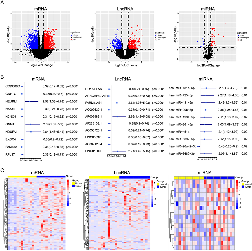 |
Figure 1 Screening of lncRNAs, microRNAs, and mRNAs associated with the malignant progression of ESCC. (A) Volcano plots of differential expression analysis in mRNAs, lncRNAs and miRNAs. The two vertical black dotted lines represent the threshold of log2(fold change) = ±1 (2-fold expression change), and the horizontal black dotted line denotes the cutoff of adjusted p-value = 0.05. Genes with log2FC > 1 and adjusted p < 0.05 were defined as significantly upregulated (red dots), those with log2FC < −1 and adjusted p < 0.05 as significantly downregulated (blue dots), and non-differentially expressed RNAs are shown as black dots. Screening criteria: log2FC > 1, adjusted p < 0.05. (B) The top ten results of univariate cox in mRNAs, lncRNAs and miRNAs. (C) Heatmaps of differential expression analysis in mRNAs and lncRNAs and miRNAs. |
Consensus Clustering Analysis for Cuproptosis-Related Genes
In ESCC, cuproptosis serves as both a crucial form of cell death and a key mechanism regulating tumor cell metabolism, proliferation, and the microenvironment, providing a theoretical foundation and novel targets for precision therapy. To categorize patients with ESCC according to cuproptosis levels, a consensus clustering analysis relying on the expression data of 13 cuproptosis-linked genes was performed. This analysis split 198 ESCC patients obtained from The Cancer Genome Atlas (TCGA) into k clusters (k=2-9). The best-fitting categorization was attained at k = 2 (Figure 2A, Supplementary Figure 2A and B). Kaplan–Meier analysis demonstrated a notable disparity in the overall survival between the two groups. Specifically, Group 1 exhibited a remarkably more favorable prognosis compared to Group 2 (Figure 2B). Differential expression analysis between Clusters 1 and 2 using the limma package identified 1794 upregulated and 1512 downregulated differentially expressed genes (Figure 2C and D, Supplementary Figure 2C). GSEA analysis of the above-mentioned DEGs showed that the differential genes were primarily enriched in cell cycle-related pathways (Figure 2E).
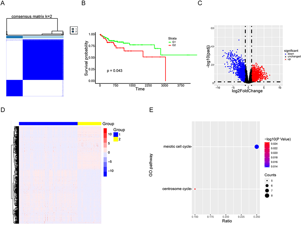 |
Figure 2 Consensus cluster analysis of cuproptosis related genes. (A) Consensus clustering heatmap of cuproptosis related genes with k=2. The clustering stability was evaluated for k values ranging from 1 to 9, and k=2 was determined as the optimal grouping. Label “1” corresponds to cluster 1, and label “2” corresponds to cluster 2. (B) Kaplan–Meier survival curve between cluster 1 and cluster 2. G1 refers to cluster 1 and G2 refers to cluster 2 obtained from the optimal consensus clustering (k=2) of cuproptosis-related genes. (C) The volcanic map showed the differential genes of the two clusters (log2FC > 1, adjusted p < 0.05). (D) The heatmap of differentially expressed genes. Label “1” denotes cluster 1, and label “2” denotes cluster 2 derived from the optimal consensus clustering (k=2) of cuproptosis-related genes. (E) GO enrichment analysis of cuproptosis related genes by DAVID database. |
Screening the Cuproptosis-Related ceRNA Network That Regulates the Malignant Progression of ESCC
In light of the aforementioned findings, we established a ceRNA network that modulated the malignant progression of ESCC. This network was built using mRNAs from the differential expression analysis of cuproptosis-related genes and lncRNAs/miRNAs from the differential expression analysis of the 13 paired ESCC samples. This network comprised 29 lncRNAs, 2 miRNAs, and 7 mRNAs, along with 107 interaction connections. (Figure 3A and Supplementary Figure 3A). We prioritized lncRNAs with higher expression abundance as core network nodes, as high-abundance lncRNAs are more likely to exert effective ceRNA sponge adsorption through a dose effect. To identify key ceRNA axes, we first selected the top six lncRNAs based on count values > 100: AC010275.1, AC090709.1, AL139023.1, FOXD2-AS1, HOXC-AS3, and TNFRSF10A-AS1. Differential expression and survival analyses were performed on these lncRNAs. Differential expression analysis showed that all six lncRNAs were highly expressed in ESCC (Supplementary Figure 3B). According to the survival analysis, high expression of lncRNA AL139023.1 was significantly associated with poorer survival (p = 0.043), while the expression of other lncRNAs showed no clear relationship with survival (Supplementary Figure 3C). The ROC curve indicated that lncRNA AL139023.1 had a higher AUC value compared with the other lncRNAs (AUC = 0.57). Therefore, we selected AL139023.1 as the key lncRNA (Supplementary Figure 3D). To sum up, we hypothesized that AL139023.1 might be the key lncRNA in the ceRNA network, and the AL139023.1/miR-139-5p axis might be the critical cuproptosis-related ceRNA axis regulating malignant progression of ESCC (Figure 3B).
 |
Figure 3 Screening for cuproptosis-related ceRNA network regulating the malignant progression of ESCC. (A) LncRNA-miRNA-mRNA regulatory axes extracted from the ceRNA network. (B) The LncRNA-miRNA-mRNA pattern diagram. |
ELOVL5 is the Key mRNA Regulated by the ceRNA Axis AL139023.1/miR-139-5p
We screened for key mRNAs within the ceRNA network. The mRNAs potentially regulated by AL139023.1 in the network included DCBLD2, ELOVL5, GPR37, and TBX1, all associated with tumors. ELOVL5 is an important protein that regulates cellular lipid metabolism. Metabolism and the oxidative respiratory chain are associated, so we hypothesized that ELOVL5 might be closely related to cuproptosis. To refine our results, we knocked down AL139023.1 using ASO and measured changes in DCBLD2, ELOVL5, GPR37, and TBX1 at the mRNA level. ELOVL5 expression was significantly regulated by AL139023.1, while no statistically significant differences were observed for the other mRNAs (Figure 4A). Elesclomol, a classic cuproptosis inducer, was used to further investigate the role of ELOVL5 in cuproptosis. The IC50 values of elesclomol in KYSE-150 and TE-1 cells were 19.56 nM and 24.23 nM, respectively (Figure 4B). Subsequently, cells from the control group and the ELOVL5 knockdown group were exposed to 10 nM and 20 nM concentrations of elesclomol. We examined the intracellular Cu2+ concentration and the expression of lipoylated proteins DLAT and DLST in KYSE-150 and TE-1 cells following ELOVL5 knockdown. The results showed that after the addition of a cuproptosis inducer, the Cu2+ concentration and the expression of lipoylated DLAT and DLST were increased in the control group. Compared with the control group, ELOVL5 knockdown followed by the addition of the cuproptosis inducer significantly upregulated the Cu2+ concentration and the expression of lipoylated DLAT and DLST. However, the addition of the cuproptosis inhibitor TTM (tetrathiomolybdate) reversed the effects induced by ELOVL5 knockdown (Figure 4C and D). Given that cuproptosis is closely associated with mitochondrial metabolism, we measured the oxygen consumption rate (OCR). The OCR assay results further indicated that, compared with the control group, ELOVL5 knockdown followed by the addition of the cuproptosis inducer markedly promoted OCR. On this basis, the further addition of the cuproptosis inhibitor attenuated this promoting effect on OCR (Figure 4E). Therefore, knocking down ELOVL5 increased the sensitivity of ESCC cells to the cuproptosis inducer. Furthermore, the expressions of miR-139-5p and ELOVL5 were all correlated with patient survival (Supplementary Figure 3E). ELOVL5 expression showed strong associations with tumor stage and lymph node metastasis, indicating good potential for clinical translation. In contrast, the clinical translation potential of AL139023.1 and miR-139-5p was found to be dependent on ELOVL5 (Box S3). We thus identified the cuproptosis-related ceRNA axis regulating the development of ESCC, namely AL139023.1/miR-139-5p/ELOVL5.
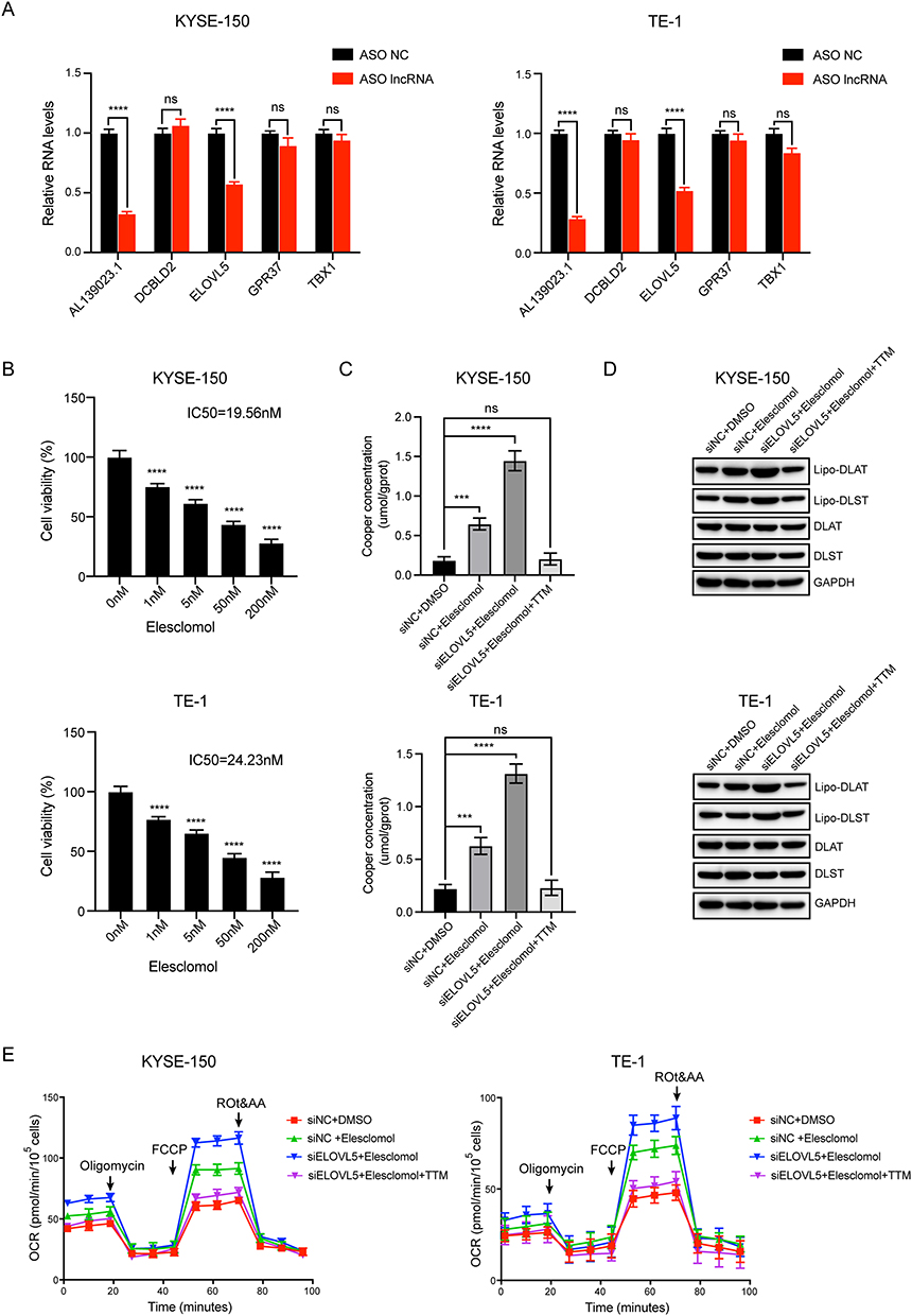 |
Figure 4 AL139023.1 regulates cuproptosis related gene ELOVL5. (A) RT-qPCR assays were used to detect the expression of potential mRNAs with AL139023.1 knockdown by ASO (n = 3). (B) CCK8 assays were used to measure cell viability 24 hours after exposure to increasing doses of Elesclomol (n = 3). (C) ELOVL5 knockdown cells and control groups cells were treated with increasing doses of Elesclomol and TTM (40 μM) for 24 hours, and the concentration of copper (Cu2+) was measured (n = 3). (D) Western Blot was used to detect the expression of lipoylated DLAT and DLST. (E) The OCR assay was used to detect the effects of ELOVL5 knockdown on mitochondrial metabolism (n = 3). The statistical methods are one-way ANOVA and two-way ANOVA. Values were expressed as the mean±SD from three experiments, and the asterisk indicates the statistical significance compared to the controls (NS, Not Significant, ***, p < 0.001, ****, p < 0.0001). |
AL139023.1, miR-139-5p, and ELOVL5 Regulate Proliferation, Migration, and Cell Cycle in ESCC
Based on these predictions, AL139023.1/miR-139-5p/ELOVL5 may constitute a ceRNA axis regulating ESCC development, but their specific roles in malignant progression remain unclear. Consequently, we categorized ESCC specimens into subgroups based on high and low expression levels of AL139023.1 (the same approach was applied to miR-139-5p and ELOVL5). We performed a differential gene expression analysis and GSEA. The findings indicated that the differentially expressed genes modulated by these three genes were mainly enriched in biological pathways associated with the cell cycle, cell proliferation, and cell migration (Figure 5A and Supplementary Figure 4). For further verification of the regulatory impacts of these three genes in terms of the cell cycle, cell proliferation, and cell migration in ESCC, the above experiments were performed following the individual inhibition of each gene. Inhibition of AL139023.1 or ELOVL5 expression suppressed ESCC cell proliferation and migration and induced cell cycle arrest. Besides, overexpressing miR-139-5p by mimic also suppressed ESCC cell proliferation and migration and induced cell cycle arrest. These findings corroborated the results of GSEA (Figure 5B and D). The above results suggest that AL139023.1, miR-139-5p, and ELOVL5 can each modulate cancerous development in ESCC.
 |
Figure 5 AL139023.1, miR-139-5p, and ELOVL5 regulate proliferation, migration, and cell cycle in ESCC. (A) The bubble diagrams of GO enrichment analysis in low expression group and high expression group among ELOVL5, AL139023.1 and miR-139-5p by DAVID database. (B) The CCK8 assays of ESCC cancer cells were performed when ELOVL5, AL139023.1 and miR-139-5p knockdown or over-expressed for 48h (n = 3). (C) The cell migration assays were used to detect the migratory ability of ESCC cells when ELOVL5, AL139023.1 and miR-139-5p knockdown or over-expressed for 48h (n = 3). (D) The cell cycle assays were performed to examine cell cycle arrest in ESCC cells when ELOVL5, AL139023.1 and miR-139-5p knockdown or over-expressed for 48h (n = 3). The statistical methods are one-way ANOVA and T-test. Values were expressed as the mean ± SD from three experiments, and the asterisk indicates the statistical significance compared to the controls (**, p < 0.01, ***, p < 0.001, ****, p < 0.0001). |
AL139023.1 and miR-139-5p Regulate ELOVL5 in ESCC
To assess the relationship between AL139023.1, miR-139-5p, and ELOVL5, we knocked down AL139023.1 and detected changes in the expression of miR-139-5p and ELOVL5. ELOVL5 was downregulated following AL139023.1 knockdown, while miR-139-5p was upregulated (Figure 6A). Subsequently, we employed mimics and inhibitors to overexpress and inhibit miR-139-5p, correspondingly, and monitored the alterations in the protein expression of AL139023.1 and ELOVL5. Both AL139023.1 and ELOVL5 were subject to negative regulation by miR-139-5p (Figure 6B and C). The findings indicate that in ESCC, lncRNA AL139023.1 and miR-139-5p can modulate ELOVL5, in line with the characteristics of the ceRNA axis.
 |
Figure 6 AL139023.1 and miR-139-5p regulate ELOVL5 in ESCC. (A) RT-qPCR assays were used to detect the expression of ELOVL5, AL139023.1 and miR-139-5p in ESCC cells treated with AL139023.1 ASO (n = 3). (B) RT-qPCR assays were used to detect the expression of ELOVL5, AL139023.1 and miR-139-5p in ESCC cells treated with miR-139-5p mimic for 48h (n = 3). (C) RT-qPCR assays were used to detect the expression of ELOVL5, AL139023.1 and miR-139-5p in ESCC cells treated with miR-139-5p inhibitor for 48h (n = 3). The statistical method is T-test. Values were expressed as the mean±SD from three experiments, and the asterisk indicates the statistical significance compared to the controls. (****, p < 0.0001). |
AL139023.1 Acts as a ceRNA for miR-139-5p to Regulate ELOVL5, Thereby Influencing the Proliferation of ESCC
To assess whether AL139023.1 functions as a ceRNA for miR-139-5p, we first performed a Pearson correlation analysis, which showed a high negative correlation between AL139023.1 and miR-139-5p (r = −0.7423, p < 0.0001) (Figure 7A). Subsequently, we performed complementary mutations on the predicted binding sites between AL139023.1 and miR-139-5p (Figure 7B). A dual-luciferase reporter assay was used to assess the impact of altering miR-139-5p levels on luciferase activity with wild-type versus mutant AL139023.1. Luciferase activity of wild-type AL139023.1 was modulated by miR-139-5p, whereas no statistically significant effect was detected for the mutant (Figure 7C). Subsequently, we assessed gene enrichment co-governed by AL139023.1 along with ELOVL5 in the biological pathways associated with the carcinogenic development of ESCC. The findings indicated marked gene enrichment, especially in pathways associated with cell proliferation (Figure 7D and E). To validate this finding, we performed ASO-mediated knockdown of AL139023.1 in both the control group and the ELOVL5-knockdown group, and examined subsequent changes in ESCC. The outcomes indicated that within the control cohort, knockdown of AL139023.1 inhibited the proliferation of tumor cells. In contrast, within the ELOVL5 knockdown cohort, this inhibitory effect was not detected (Figure 7F). Subsequently, we performed a rescue experiment by overexpressing ELOVL5 in AL139023.1-knockdown cells. Knockdown of AL139023.1 by ASO inhibited cell proliferation, whereas overexpression of ELOVL5 in AL139023.1-knockdown cells partially reversed this inhibitory effect. This result further validates that ELOVL5 is a key downstream mediator (Figure 7G). In addition, we further validated our conclusions through in vivo experiments. After knocking down AL139023.1 in the control group and the ELOVL5-overexpressing group, respectively, the results of subcutaneous tumor-bearing experiments in nude mice showed that knockdown of AL139023.1 did not suppress tumor cell growth or reduce tumor weight in the ELOVL5-overexpressing group, whereas in the control group, it significantly inhibited tumor growth and decreased tumor weight (Figure 8A and B). In summary, these findings suggest that AL139023.1 functions as a ceRNA of miR-139-5p to modulate ELOVL5, consequently affecting the malignant development of ESCC.
 |
Figure 7 AL139023.1 acts as a ceRNA for miR-139-5p to regulate ELOVL5, thereby influencing the proliferation of ESCC. (A) Pearson correlation coefficient analysis between AL139023.1 and miR-139-5p. (B) Predicted binding site between miR-139-5p and AL139023.1. (C) Luciferase assays were performed to test the effect of miR-139-5p on wild-type or mutant AL139023.1 after treating with miR-139-5p mimic for 48h (n = 3). (D) Venn diagrams represent the overlapped genes between AL139023.1 and ELOVL5. (E) The bubble diagram of GO enrichment analysis of AL139023.1 and ELOVL5 overlapped genes by DAVID database. (F) The CCK8 results of siNC or siELOVL5 ESCC cell lines transfected with AL139023.1 ASO (n = 3). (G) The CCK8 results of oeControl or oeELOVL5 ESCC cell lines transfected with AL139023.1 ASO (n = 3). The statistical methods are T-test. Values were expressed as the mean ± SD from three experiments, and the asterisk indicates the statistical significance compared to the controls (NS, Not Significant, ***, p < 0.001, ****, p < 0.0001). |
 |
Figure 8 AL139023.1 may regulate the growth of ESCC through ELOVL5 in vivo. (A) Representative images of subcutaneous tumors derived from KYSE-150 cells in nude mice. Mice were inoculated with either control cells or ELOVL5-overexpressing cells in combination with AL139023.1 ASO treatment. (B) Quantitative analysis of tumor weight at the end of the experiment. Values were expressed as the mean ± SD (n = 5 per group), and the asterisk indicates the statistical significance compared to the controls (NS, Not Significant, ****, p < 0.0001). |
Discussion
ESCC is a highly invasive malignancy with suboptimal clinical results, highlighting an immediate requirement for more potent diagnostic indicators and treatment approaches. Growing attention has been directed toward post-transcriptional regulatory mechanisms, particularly the intricate interactions among lncRNAs, miRNAs, and their downstream targets.21 The ceRNA hypothesis provides a conceptual framework to understand how these RNA species communicate through shared microRNA response elements and collectively shape cancer cell behavior.22 This multilayered regulatory mechanism has emerged as a crucial contributor to ESCC pathogenesis, and affects diverse biological processes, such as proliferation, invasion, chemoresistance, and metabolic adaptation. Several well-characterized ceRNA axes illustrate the functional relevance of this mechanism in ESCC. For instance, lncRNA HOTAIR promotes ESCC cell proliferation and metastasis by sponging miR-125/miR-143, subsequently enhancing the expression of oncogenic targets.23 These examples collectively emphasize that ceRNA-mediated regulation is not an isolated phenomenon but a recurrent theme contributing to cancer initiation and progression. Building upon these insights, our study expands the ceRNA regulatory landscape in ESCC by establishing a novel axis involving AL139023.1, miR-139-5p, and ELOVL5. Through integrated expression analyses and functional validation, we demonstrated that AL139023.1 could competitively bind to miR-139-5p, consequently modulating ELOVL5 expression and affecting malignant behaviors. By delineating this regulatory relationship, our findings substantially enrich the current ceRNA network in ESCC and provide new directions for understanding ncRNA-mediated metabolic and oncogenic regulation in ESCC.
Cuproptosis, a recently characterized form of RCD, represents a unique cytotoxic process triggered by intracellular copper accumulation and destabilization of mitochondrial protein homeostasis. Unlike apoptosis, ferroptosis, or pyroptosis, cuproptosis originates from copper-induced aggregation of lipoylated proteins within the TCA cycle, leading to proteotoxic stress and subsequent cell death. Because metabolic plasticity and mitochondrial dependence vary widely across malignancies, cuproptosis has emerged as an intriguing biological mechanism with potential relevance to tumor progression and therapeutic vulnerability. Copper dysregulation contributes to tumorigenesis by modulating oxidative stress, metabolic reprogramming, and redox balance, and tumors with heightened mitochondrial activity may be particularly susceptible to copper-induced toxicity.24 Although research specifically addressing cuproptosis in ESCC is in its infancy, the broader field of RCD has been well documented in this cancer type. For example, ferroptosis, another metal-dependent form of RCD, modulates ESCC cell proliferation and therapeutic response, while pyroptosis and apoptosis pathways also contribute to tumor suppression, collectively supporting the notion that multiple RCD modalities influence ESCC biology.25,26 These results offer a basis for uncovering cuproptosis as a new regulatory mechanism and a potential therapeutic target. In our study, we utilized consensus clustering relying on the expression signatures of 13 well-recognized cuproptosis-associated genes and effectively divided patients with ESCC into two subsets with disparate cuproptosis traits. Differential expression analysis between these clusters revealed a set of genes potentially associated with copper-induced cell death in ESCC. This integrative approach delineates the heterogeneity of cuproptosis within ESCC and uncovers molecular signatures that may reflect differential sensitivity to copper-dependent cytotoxicity. Our results thus provide new insights into the landscape of cuproptosis in ESCC, along with a foundation for identifying candidate genes that could serve as therapeutic targets for cuproptosis-oriented interventions. Importantly, although ceRNA regulatory networks and cuproptosis have each been implicated in cancer, their potential crosstalk remains largely unexplored, particularly in ESCC. Existing studies on cuproptosis are predominantly based on bioinformatic analyses, with limited mechanistic insight into its upstream regulation. In this context, our study provides evidence that a ceRNA network can directly modulate a cuproptosis-associated gene, thereby linking post-transcriptional regulation to copper-dependent cell death. The discovery of this AL139023.1/miR-139-5p/ELOVL5 ceRNA axis expands the functional scope of ceRNA networks and offers a new perspective on the regulatory mechanisms governing cuproptosis in ESCC.
ELOVL5, a key enzymatic player in the fatty acid elongation pathway, is responsible for generating long-chain polyunsaturated fatty acids essential for membrane structure, lipid signaling, and cellular energy homeostasis. As an important regulator of lipid biosynthesis, ELOVL5 participates in shaping the lipid composition of cellular membranes and organelles, thereby exerting considerable influence on metabolic state and cellular behavior. Dysregulated lipid metabolism contributes to malignant transformation, and ELOVL5 is aberrantly expressed in several cancers, whereby it promotes tumor proliferation, metastasis, or metabolic adaptation depending on the tumor context.27,28 ELOVL5 has been consistently highlighted central molecule linking lipid synthesis to broader metabolic pathways. Because fatty acid metabolism is intimately connected to mitochondrial activity and the oxidative respiratory chain, alterations in ELOVL5 may influence mitochondrial homeostasis, redox balance, and metabolic vulnerability. However, despite its well-defined metabolic functions, the participation of ELOVL5 in cuproptosis remains unreported. This underscores a significant knowledge gap regarding whether lipid elongation pathways may intersect with copper-induced cell death mechanisms. ELOVL5 function can be modulated through ceRNA-mediated post-transcriptional regulation.29 For instance, in hepatocellular carcinoma and breast cancer, specific lncRNA–miRNA pairs regulate ELOVL5 expression through competitive interactions, demonstrating that ELOVL5 is embedded within ceRNA-controlled metabolic networks.28,29 These findings support the plausibility of ncRNA-dependent regulation of ELOVL5 in other malignancies as well. Building upon this notion, our work establishes a novel regulatory network in ESCC, revealing that ELOVL5 expression is influenced by the AL139023.1/miR-139-5p axis. By delineating this interaction in ESCC, our study offers theoretical evidence for targeting ELOVL5 as a potential diagnostic or therapeutic strategy and broadens the mechanistic understanding of ncRNA–metabolic gene crosstalk in this disease. Our findings further suggest that lipid metabolism-related enzymes such as ELOVL5 may participate in regulating cellular susceptibility to cuproptosis, thereby establishing a potential link between metabolic reprogramming and copper-dependent cell death in ESCC.
Despite the strengths of this study, several limitations should be acknowledged. First, our findings are primarily derived from in vitro experiments and bioinformatic analyses, and lack validation in well-characterized clinical cohorts. As a result, the clinical applicability, including diagnostic and prognostic value, of the AL139023.1/miR-139-5p/ELOVL5 axis in ESCC remains to be determined. Second, although our data suggest that ELOVL5 participates in regulating cuproptosis, the precise molecular mechanisms connecting lipid metabolic pathways with copper-induced cell death are not yet fully elucidated. Future studies integrating multicenter clinical samples will be necessary to substantiate these findings and facilitate their potential translation into clinical applications.
In summary, our study reveals a previously unacknowledged regulatory axis in ESCC where AL139023.1 acts as a ceRNA against miR-139-5p to regulate the copper-dependent cell death-related gene ELOVL5. We demonstrated that suppression of ELOVL5 enhanced intracellular copper accumulation and ROS generation, thereby promoting cuproptosis. Functional and enrichment analyses revealed that the AL139023.1/miR-139-5p/ELOVL5 pathway orchestrates key malignant phenotypes, including ESCC cell proliferation, cell-cycle progression, and migration. Mechanistic validation using reporter assays further confirmed the ceRNA interaction between AL139023.1 and miR-139-5p governing ELOVL5 expression. These findings demonstrate a copper-death–associated ceRNA regulatory network in ESCC and broaden the current understanding of how metabolic vulnerability is integrated into ncRNA-mediated gene regulation. Future clinical cohort studies are warranted to validate the translational relevance of this ceRNA axis, and exploring therapeutic strategies that target the AL139023.1/miR‑139‑5p/ELOVL5 pathway could offer a promising avenue for ESCC intervention. Our work offers mechanistic perspectives on ESCC progression and provides promising molecular candidates for developing new-generation approaches for diagnosis and treatment.
Data Sharing Statement
The data generated in this study are available within the article and its supplementary data files.
Ethics Approval
This study used anonymized data from The Cancer Genome Atlas (TCGA) public database. All data are publicly available, anonymized, and cannot be traced back to any identifiable individual. According to Article 32 of the Measures for Ethical Review of Life Sciences and Medical Research Involving Human Subjects (2023), research conducted using lawfully obtained public data that does not cause harm to human subjects, does not involve sensitive personal information, or does not involve commercial interests may be exempt from ethical review. This study has been confirmed as exempt from ethical review by the Medical Ethics Committee of Sir Run Run Shaw Hospital, School of Medicine, Zhejiang University. All procedures were performed in strict accordance with the ethical principles of the Declaration of Helsinki. All animal experiments were conducted in accordance with the guidelines for the care and use of laboratory animals and approved by the Institutional Animal Care and Use Committee (IACUC) of Zhejiang Provincial Laboratory Animal Center (Approval No. ZJCLA-IACUC-20030185).
Author Contributions
All authors made a significant contribution to the work reported, whether that is in the conception, study design, execution, acquisition of data, analysis and interpretation, or in all these areas; took part in drafting, revising or critically reviewing the article; gave final approval of the version to be published; have agreed on the journal to which the article has been submitted; and agree to be accountable for all aspects of the work.
Funding
No funding was received for this research.
Disclosure
All the authors declare that they have no conflicts of interest in this work.
References
1. Zhao YX, Zhao HP, Zhao MY, et al. Latest insights into the global epidemiological features, screening, early diagnosis and prognosis prediction of esophageal squamous cell carcinoma. World J Gastroenterol. 2024;30(20):2638–18. doi:10.3748/wjg.v30.i20.2638
2. Jiang W, Zhang B, Xu J, Xue L, Wang L. Current status and perspectives of esophageal cancer: a comprehensive review. Cancer Commun. 2025;45(3):281–331. doi:10.1002/cac2.12645
3. Christodoulidis G, Agko SE, Koumarelas KE, Kouliou MN, Zacharoulis D. Advancements and challenges in the treatment of esophageal cancer: a comprehensive review. World J Clin Oncol. 2024;15(12):1463–1467. doi:10.5306/wjco.v15.i12.1463
4. George TP, Subramanian S, Supriya MH. A brief review of noncoding RNA. Egypt J Med Hum Genet. 2024;25(1):98. doi:10.1186/s43042-024-00553-y
5. Chiglintseva DA, Patutina OA, Zenkova MA. Endogenous ribonucleases: therapeutic targeting of the transcriptome through oligonucleotide-triggered RNA inactivation. Biomolecules. 2025;15(7):965. doi:10.3390/biom15070965
6. Wei QY, Jin F, Wang ZY, et al. MicroRNAs: a novel signature in the metastasis of esophageal squamous cell carcinoma. World J Gastroenterol. 2024;30(11):1497–1523. doi:10.3748/wjg.v30.i11.1497
7. Zhang J, Li Z, Han J, Tian Z, Meng Q, Niu W. KLF7 enhances the invasion and migration of colorectal cancer cells via the miR-139-5p/TPD52 axis. Cancer Biol Ther. 2024;25(1):2385172. doi:10.1080/15384047.2024.2385172
8. Zhang H, Yang X, Guo Y, Zhao H, Jiang P, Yu QQ. The regulatory role of lncRNA in tumor drug resistance: refracting light through a narrow aperture. Oncol Res. 2025;33(4):837–849. doi:10.32604/or.2024.053882
9. Guo Z, Liu Y, Chen D, et al. Targeting regulated cell death: apoptosis, necroptosis, pyroptosis, ferroptosis, and cuproptosis in anticancer immunity. J Transl Intern Med. 2025;13(1):10–32. doi:10.1515/jtim-2025-0004
10. Pan C, Ji Z, Wang Q, et al. Cuproptosis: mechanisms, biological significance, and advances in disease treatment—A systematic review. CNS Neurosci Ther. 2024;30(9):e70039. doi:10.1111/cns.70039
11. Lou Q-M, Lai F-F, Li J-W, Mao K-J, Wan H-T, He Y. Mechanisms of cuproptosis and its relevance to distinct diseases. Apoptosis. 2024;29(7):981–1006. doi:10.1007/s10495-024-01983-0
12. Tang D, Kroemer G, Kang R. Targeting cuproplasia and cuproptosis in cancer. Nat Rev Clin Oncol. 2024;21(5):370–388. doi:10.1038/s41571-024-00876-0
13. Zhang S, Huang Q, Ji T, Li Q, Hu C. Copper homeostasis and copper-induced cell death in tumor immunity: implications for therapeutic strategies in cancer immunotherapy. Biomarker Res. 2024;12(1):130. doi:10.1186/s40364-024-00677-8
14. Zhang R, Tan Y, Xu K, et al. Cuproplasia and cuproptosis in hepatocellular carcinoma: mechanisms, relationship and potential role in tumor microenvironment and treatment. Cancer Cell Inter. 2025;25(1):137. doi:10.1186/s12935-025-03683-4
15. Ma J, Liu D, Li Y, Tang L, Gao X, Zhu F. A risk prognostic signature basing on cuproptosis-related ferroptosis and immune genes for esophageal squamous cell carcinoma patients. Cell Biochem Biophys. 2025;83(4):4769–4779. doi:10.1007/s12013-025-01802-6
16. Ferrero E, Vaz FM, Cheillan D, Brusco A, Marelli C. The ELOVL proteins: very and ultra long-chain fatty acids at the crossroads between metabolic and neurodegenerative disorders. Mol Gene Metabol. 2025;144(3):109050. doi:10.1016/j.ymgme.2025.109050
17. Jiang C, Shi Y, Shi X, et al. ELOVL5 and VLDLR synergistically affect n-3 PUFA deposition in eggs of different chicken breeds. Poultr Sci. 2024;103(9):104016. doi:10.1016/j.psj.2024.104016
18. Klycheva KV, Razumovskaya AV, Shatsillo AD, et al. ELOVL5 regulates ferroptosis in breast cancer cells. Dokl Biochem Biophys. 2025;525:532–540. doi:10.1134/S1607672925601246
19. Vouilloz A, Bourgeois T, Diedisheim M, et al. Impaired unsaturated fatty acid elongation alters mitochondrial function and accelerates metabolic dysfunction-associated steatohepatitis progression. Metabolism. 2025;162:156051. doi:10.1016/j.metabol.2024.156051
20. Sun D, Wang L, Wu Y, et al. Lipid metabolism in ferroptosis: mechanistic insights and therapeutic potential. Review. Front Immunol. 2025. doi:10.3389/fimmu.2025.1545339
21. Pooresmaeil F, Azadi S, Hasannejad-Asl B, Takamoli S, Bolhassani A. Pivotal role of miRNA–lncRNA interactions in human diseases. Mol Biotechnol. 2025;67(12):4363–4385. doi:10.1007/s12033-024-01343-y
22. Yilmaz A, Ari yuka S. The role of ceRNAs in breast cancer microenvironmental regulation and therapeutic implications. J Mol Med. 2025;103(1):33–49. doi:10.1007/s00109-024-02503-y
23. Ghorbani V, Ghorbian S. Gene expression profiling of lncRNA-HOTAIR and lncRNA-MALAT1 in esophageal cancer: uncovering links to lifestyle factors and diagnostic significance. Discov Oncol. 2025;16(1):630. doi:10.1007/s12672-025-02465-8
24. Jomova K, Alomar SY, Valko R, Nepovimova E, Kuca K, Valko M. The role of redox-active iron, copper, manganese, and redox-inactive zinc in toxicity, oxidative stress, and human diseases. Excli J. 2025;24:880–954. doi:10.17179/excli2025-8449
25. Hu J, Li Y, Lian B, Mao Y, Zhao L. Mechanism and role of regulated cell death in tumor immunity and immunotherapy. Cancer Commun. 2025;45(11):1456–1495. doi:10.1002/cac2.70064
26. Tang M-X, Chen J-F, Zhao F-Z, Peng J. Ferroptosis: a critical link to treatment resistance in esophageal carcinoma. iScience. 2025;28(7):112901. doi:10.1016/j.isci.2025.112901
27. Pakiet A, Ciosek M, Lange O, et al. Very long-chain fatty acids accumulate in breast cancer tissue and serum. Cancer Cell Inter. 2025;25(1):296. doi:10.1186/s12935-025-03928-2
28. Wu G, Zhang Y, Liang B, et al. miR-218-5p promotes hepatic lipogenesis through targeting Elovl5 in non-alcoholic fatty liver disease. Biochem Pharmacol. 2024;226:116411. doi:10.1016/j.bcp.2024.116411
29. Shi Y, Wu X, Meng G, et al. Identification and analysis of circular RNAs in mammary gland from yaks between lactation and dry period. Animals. 2025;15(1):89. doi:10.3390/ani15010089
 © 2026 The Author(s). This work is published and licensed by Dove Medical Press Limited. The
full terms of this license are available at https://www.dovepress.com/terms
and incorporate the Creative Commons Attribution
- Non Commercial (unported, 4.0) License.
By accessing the work you hereby accept the Terms. Non-commercial uses of the work are permitted
without any further permission from Dove Medical Press Limited, provided the work is properly
attributed. For permission for commercial use of this work, please see paragraphs 4.2 and 5 of our Terms.
© 2026 The Author(s). This work is published and licensed by Dove Medical Press Limited. The
full terms of this license are available at https://www.dovepress.com/terms
and incorporate the Creative Commons Attribution
- Non Commercial (unported, 4.0) License.
By accessing the work you hereby accept the Terms. Non-commercial uses of the work are permitted
without any further permission from Dove Medical Press Limited, provided the work is properly
attributed. For permission for commercial use of this work, please see paragraphs 4.2 and 5 of our Terms.