
Back to Journals » International Journal of Nanomedicine » Volume 20
The Construction Strategy of Curcumin Nanomedicine Delivery System and Its Application in the Treatment of Ulcerative Colitis
Authors Ning H  , Huang X, Lin X , Sun Q, Zheng Y, Deng N, Xu Y
Received 13 October 2025
Accepted for publication 5 December 2025
Published 16 December 2025 Volume 2025:20 Pages 15135—15166
DOI https://doi.org/10.2147/IJN.S573966
Checked for plagiarism Yes
Review by Single anonymous peer review
Peer reviewer comments 2
Editor who approved publication: Dr RDK Misra
Hang Ning,1,* Xinyu Huang,1,* Xiaoyuan Lin,1,* Qifang Sun,1 Yuchen Zheng,1 Na Deng,2 Yin Xu1
1Gastroenterology department, The First Hospital of Hunan University of Chinese Medicine, Changsha, Hunan, 410007, People’s Republic of China; 2School of Traditional Chinese Medicine, Hunan University of Chinese Medicine, Changsha, Hunan, 410208, People’s Republic of China
*These authors contributed equally to this work
Correspondence: Na Deng, School of Traditional Chinese Medicine, Hunan University of Chinese Medicine, 300 Xue Shl Road, Sclence & Education Park, YueLu District, Changsha, Hunan, 410208, People’s Republic of China, Tel +86-13548615590, Email [email protected] Yin Xu, Gastroenterology department, The First Hospital of Hunan University of Chinese Medicine, No. 95 Shaoshan Middle Road, Yuhua District, Changsha, Hunan, 410007, People’s Republic of China, Tel +86-13787262655, Email [email protected]
Abstract: Ulcerative colitis (UC) is a chronic inflammatory bowel disease with a continuously increasing incidence worldwide. The existing treatment options are limited due to low drug bioavailability and systemic side effects. Natural products, such as curcumin, have emerged as potential effective drugs for UC treatment due to their multi-target and multi-mechanism therapeutic advantages. However, the clinical trial and experimental research results of curcumin show a contrast due to its own physicochemical limitations (low solubility, low bioavailability, etc). gastrointestinal digestion factors (pH, digestive enzymes, etc). and the combined limitations of the colonic intestinal barrier (intestinal flora, mucus barrier, intestinal epithelial barrier, etc). The clinical translation is thus hindered. The Nano-Drug Delivery System (NDDS) uses size control, surface functionalization, and intelligent stimulus-responsive design to transform the factors that limit curcumin absorption and utilization into delivery targets, constructing pH-dependent, gut flora-dependent, receptor-dependent, etc. NDDS, achieving improved drug solubility, enhanced absorption, controlled release, and targeted delivery, significantly enhancing the therapeutic effect of curcumin for UC. This review focuses on the pathophysiology of UC and uniquely systematically analyzes the construction logic of natural product NDDS from the perspective of the above biological barriers, clarifying the applicable scenarios and core advantages of various strategies. At the same time, this article also discusses the key challenges faced by the clinical translation of NDDS, including the toxicity risk caused by enhanced drug absorption, the safety of the carrier itself, and the transformation obstacles caused by species receptor spectrum differences etc. as well as an important point to recognize is that there is still a considerable distance to the clinical translation of NDDS. In summary, NDDS brings broad prospects for the clinical application of natural products, but the current research level is far from meeting the needs of clinical translation. Future design must deeply align with the pathological characteristics of UC to promote its transition from the laboratory to clinical application.
Keywords: ulcerative colitis, curcumin, nano-drug delivery system, natural products
Introduction
Ulcerative colitis (UC) is a lifelong inflammatory disease characterized by continuous and diffuse inflammatory responses in the mucosa of the colon and rectum. The exact mechanism of UC remains unclear to date, and it is generally believed to be related to genetic susceptibility, intestinal mucosal barrier defects, intestinal flora imbalance, and autoimmune dysregulation. From a global epidemiological perspective, the prevalence and incidence of UC are continuously increasing. Currently, the highest prevalence is found in Northern Europe (Norway: 505/100,000 people) and North America (the United States: 286/100,000 people), and the highest incidence is found in Northern Europe (Faroe Islands: 57.9/100,000 people per year), North America (Canada: 23.1/100,000 people per year), and Australia (Australia: 17.4/100,000 people per year).1 Studies have shown that the incidence of UC is closely related to the level of industrialization, and accordingly, emerging industrial countries in Asia have shown a rapidly increasing incidence of UC. One study showed that the incidence of UC in South Korea increased rapidly from 0.3/100,000 people per year from 1986–1990 to 3.1/100,000 people per year from 2000–2006.2 The incidence of UC in China was 10.04/100,000 people per year in 2016.3 Predictions indicate that the prevalence of UC in Asian countries will increase to four times the current level by 2035.1 Due to the alternating clinical characteristics of UC, which involves episodes, remissions, and relapses, any dietary impropriety and emotional stimulation can easily induce relapses of UC, significantly affecting the quality of life of patients. A study reported that the temporary disability rate of UC patients was 12.3%, the permanent disability rate was 1.6%,4 and the depression rate was 48.5%.5 Due to the imbalance between incidence and mortality, lifelong treatment imposes a huge economic burden on patients. Studies have shown that the lifetime incremental cost for patients diagnosed between the ages of 0–11 is $369,955, for those aged 70 and above is $132,396, and for all age groups is $230,102 on average. At the same time, due to the continuous accumulation of the prevalence, it imposes a heavy burden on the global healthcare system. The lifetime total cost of UC patients in the United States in 2016 is estimated to be $377 billion.6
The current treatment goal for UC is to induce and maintain endoscopic remission and normal biochemical markers. The treatment plan includes two steps: inducing remission and maintaining remission; the choice of drugs depends on the scope of the disease and the degree of clinical activity, including natural products, 5-aminosalicylic acid (5-ASA), glucocorticoids, and immunomodulators, immunosuppressants, and biologics.1 Oral administration is the most commonly selected administration method, which is considered a non-invasive and safe method that allows for flexible and highly compliant self-administration.7 However, due to the unique attributes of UC as a digestive system disease, many obstacles limit the efficacy of oral administration, such as pH gradients, digestive enzymes, intestinal mucus, and intestinal flora,8 resulting in drug degradation, poor absorption, and non-specific distribution. Curcumin is a “star product” derived from natural substances, which has unique therapeutic advantages of acting on multiple targets and multiple pathways through multiple components. Both basic and clinical studies have affirmed its good effect in the prevention and treatment of UC.9 At the same time, due to the limitations of the physical and chemical properties of curcumin itself, curcumin has problems such as poor solubility and low bioavailability. In addition, gastrointestinal digestion factors and the intestinal barrier of the colon also jointly limit the efficacy of natural products represented by curcumin. The Nano-Drug Delivery System (NDDS) as a novel technological approach in the field of pharmaceuticals, through strategies such as size control, charge design, shape optimization, functional group modification, and stimulus-responsive design, can transform the “bottleneck factors” that limit curcumin absorption into “delivery targets”, achieving improvements in drug solubility, enhancement of absorption efficiency, controllable release, and targeted delivery, providing a key solution to break through the dilemma of curcumin delivery in the body.10
This paper takes curcumin as a representative natural product, based on the latest knowledge of UC pathology and physiology, and from the perspective of diseases, reviews the construction strategies of natural product NDDS for treating UC, with the aim of providing references for the development of this field; to ensure the systematicness and criticality of the review, this study adopts a clear methodological framework: classifying strategies according to the specific biological barriers (such as mucus barrier, intestinal epithelial barrier) and key environmental factors of the gastrointestinal tract (such as pH, intestinal flora) targeted by NDDS, while analyzing the design logic and mechanism of action of various strategies, and critically evaluating their potential for clinical translation and inherent limitations (such as the potential toxicity risks of enhanced drug absorption, the potential safety hazard of the carrier itself, and the transformation barriers caused by receptor spectrum differences between species); To clearly present the research context, this review will be structured as follows: First, elaborating on the three major limiting factors faced when treating UC with oral curcumin (physical and chemical factors defects, interference of gastrointestinal digestion environment, and barrier obstruction of the colonic intestinal barrier), clarifying the core issues that NDDS needs to solve; then, based on the corresponding biological barriers or environmental characteristics of the above limiting factors, systematically classifying and in-depth analyzing various NDDS construction strategies (such as pH-dependent, ROS-dependent, mucus-dependent, receptor-dependent, etc)., clarifying the suitable scenarios, core advantages, and technical shortcomings of different strategies; finally, focusing on the key challenges in the clinical translation process of NDDS, clearly pointing out that there is still a huge gap between the current research progress and the clinical translation goals, and combining the current deficiencies in safety assessment, cross-species validation, and adaptability to large-scale production, proposing targeted future design directions, ultimately providing ideas for promoting natural product NDDS from laboratory research to clinical application.
Limiting Factors of Oral Curcumin Therapy for UC
Curcumin is the main active component of the rhizome of the plant Curcuma longa L., accounting for 2%-5% of the root and rhizome content. It also contains demethylated curcumin, di-demethylated curcumin, and other derivatives. In commercial curcumin, these three components account for approximately 82%, 15%, and 3% respectively.11 Curcumin has medicinal, flavoring, and coloring (E100) functions. Its chemical nature is 1,7-bis(4-hydroxy-3-methoxyphenyl)-1,6-heptadiene-3,5-dione (molecular formula C21H2₀O6).12 Experimental studies have confirmed that curcumin can regulate signaling pathways such as MAPK/NF-κB/Nrf2/NLRP3,13 SphK1/NF-κB,14 JAK/STAT,15 Treg cells,16 macrophages,17 mTh/mTfh cells,18 etc., immune cells, inflammatory factors such as TNF-α, IFN-γ, IL-8, IL-6,19 inflammatory mediators such as MIP-2,20 MMP-3,21 etc., and antioxidant stress,22 and regulate the intestinal microbiota,23 and improve the intestinal mucosal barrier,24 thereby exerting therapeutic effects on UC. However, a systematic review and meta-analysis of a randomized controlled trial (RCT) for UC indicates that in clinical practice, curcumin may have no effect on symptom relief in patients with mild to moderate UC; although some individual RCT evidence for patients with mild to moderate UC suggests that curcumin may be beneficial for the improvement of UC symptoms in this group, the aggregated data for patients with mild to moderate UC do not show significant positive effects.25 The contrasting results of curcumin in experimental studies and clinical trials limit its development in clinical and commercial applications.
Further research has clarified that the contrasting results of curcumin in experimental studies and clinical trials are caused by the physical and chemical factors of curcumin itself, gastrointestinal digestion factors, and intestinal barrier factors, all of which jointly limit the therapeutic effect of oral curcumin. Therefore, before discussing curcumin NDDS, we must take into account the influence of curcumin’s own physical and chemical factors, the differences in anatomy, physiology, and absorption characteristics of different parts of the gastrointestinal tract, as well as the differences between healthy and diseased gastrointestinal tracts. Only by deeply understanding the comprehensive impact of the interaction between curcumin and the gastrointestinal environment on the treatment of UC by oral curcumin, can we carry out promising drug design.
The Limitations of Curcumin’s Physicochemical Properties
In recent years, as research has progressed, the factors restricting the transformation of curcumin from the laboratory to clinical application have gradually become clear, including pre-absorption degradation, insufficient absorption, high systemic pre-metabolism, low bioavailability, rapid clearance, and potential toxicity. Curcumin consists of three structural fragments: a 7-carbon linker containing an α,β-unsaturated β-diketone group, and two aromatic ring systems with adjacent methoxyphenolic groups.12 It is hydrophobic, with an oil-water partition coefficient (logP) of approximately 3.2, and as a polyphenol, it has extremely low water solubility (only 30 nM) at acidic and neutral pH values26- this indicates that curcumin can tolerate the gastric pH and reach the colon without modification or encapsulation, and also means that it is almost insoluble in physiological fluids.27 Pre-absorption degradation refers to the metabolic action of intestinal bacteria on curcumin: for example, Escherichia coli (ATCC 8739) and Escherichia fergusonii (ATCC 35469) can metabolize curcumin into dihydrocurcumin, which then converts to tetrahydrocurcumin;28 Blautia sp. MRG-PMF1 undergoes demethylation reactions, converting curcumin into demethylcurcumin and bis-demethylcurcumin derivatives.29 Insufficient absorption is manifested as: hydrophobic curcumin can only be absorbed by a small amount by colonic epithelial cells,30 and the absorbed curcumin will bind to intracellular proteins in the intestinal epithelium and undergo structural changes, further reducing bioavailability.31 In addition, curcumin also has the problem of high systemic pre-metabolism: both the liver32 and the colon33 are important sites for the metabolism of curcumin in the human body, where curcumin undergoes extensive first- and second-level metabolism, ultimately converting into water-soluble metabolites such as curcumin glucuronide and curcumin sulfate conjugates, which are excreted through the gallbladder and kidneys, resulting in a half-life of only 6–7 hours in the human body.34 Due to the low solubility, poor absorption, low bioavailability, and rapid metabolism defects of curcumin, to achieve the expected therapeutic effect, it is necessary to increase the dosage or frequency of use, which will amplify potential toxicity and cause adverse reactions in patients. A clinical study showed that daily intake of 0.9–3.6g of curcumin for 1–4 months may cause adverse reactions such as nausea and vomiting, and may also be accompanied by an increase in serum lactate dehydrogenase levels.35 Experimental studies have provided more evidence of the potential toxicity of curcumin: first, curcumin and its metabolites can react with toxic enzymes such as glutathione S-transferase (GST) and cytochrome P450 (CYP450), inhibiting the activity of these enzymes and potentially leading to impaired detoxification function and potential drug toxicity;36 second, curcumin may act as an iron chelator in the body, affecting systemic iron metabolism, especially for individuals with poor iron nutrition status;37 third, there are also studies that have found that curcumin has cytotoxicity to normal human lymphocytes.38
Limitations of Gastrointestinal Digestion Factors
The gastrointestinal digestion factors that may limit the therapeutic effect of curcumin in UC mainly include colonic fluid volume, colonic transit time, pH, digestive enzymes, etc. The colon has a high water absorption capacity, capable of absorbing approximately 90% of the water entering the colon.39 Therefore, when the intestinal contents transfer to the descending colon, the viscosity of the intestinal contents increases, making it challenging for curcumin to dissolve from the formulation and may affect the local drug bioavailability. The colonic transit time is an important factor affecting the bioavailability of oral curcumin. Studies have shown that during normal colonic transit, the time ranges from 41.1 to 62.3 hours, with an average of approximately 51.7 hours,40 while the colonic propulsion activity of UC patients is significantly increased,41 resulting in a significantly shortened transit time, approximately 24.3 hours.40 The shortening of colonic transit time will reduce the contact between the UC lesion colon segment and curcumin, leading to a corresponding decrease in curcumin absorption. Additionally, compared to smaller particle size drugs, larger particle size drug formulations exhibit shorter transit times, meaning they are expelled more quickly.42 Moreover, the extreme changes in pH in the gastrointestinal tract43 pose a huge challenge to the stability of the drug. The pH of healthy adult gastric juice is acidic, ranging from 1.7 to 4.7. In the duodenum, due to the neutralization effect of carbonates and bile, the pH rises to 5.9–6.3, then to pH 7.4–7.8 in the distal ileum and the ileum, and the pH of the colon may fluctuate between 5 and 8, with significant individual differences.43 Extreme changes in pH may cause changes in the drug conformation, resulting in loss of efficacy. More importantly, the gastrointestinal digestion environment of UC patients is not completely similar to that of healthy adults. Studies have reported an overall trend of increased colonic pH in UC patients, which may drop to 2–3.44 Finally, various high-concentration digestive enzymes in the gastrointestinal tract have become important obstacles to destroying the structure of curcumin and hindering its effective absorption and utilization. Traditional pharmaceutical experience indicates that drugs can avoid the action of digestive enzymes through enteric-coated formulations. The design of the human gastrointestinal structure and function is largely based on dietary components and economic considerations, and natural selection has led to a roughly matched digestive capacity with food load and a moderate excess, which also explains the differences in digestive enzymes between different species and among different individuals of the same species.45 And UC patients often have a special diet.46 Based on this, we must recognize that UC patients may have digestive enzymes that are not exactly the same as those of healthy adults, and we must evaluate the impact of this difference on curcumin in subsequent studies (Table 1).
 |
Table 1 Comparison of Gastrointestinal Digestion Factors Between UC Patients and Healthy Individuals |
Limitations of the Intestinal Intestinal Barrier
The intestinal mucosal barrier is an important defense system of the intestine. Currently, it is classified into four major types: biological, chemical, mechanical, and immune barriers.8 The first three serve as natural barriers to protect the intestinal epithelial layer from various external stimuli, thereby regulating the balance between mucosal immunity and external stimuli, and during this process, they hinder the absorption and utilization of oral drugs.47 The intestine is a complex micro-ecosystem where the intestinal microbiota co-evolve with the host and establish mutual relationships, and a series of bacterial enzymes such as polysaccharide enzymes, glycosidase, protease, and peptidease first degrade large molecular polymers.48 This process will also affect the metabolism and absorption of oral drugs in the human body. The intestinal mucus layer is a powerful intestinal barrier, with mucin 2 (MUC2) being the most critical component. It is interconnected by disulfide bonds and linked to the glycan layer of the intestinal epithelium, forming a protein network in the mucus layer.49 The average pore size is approximately 200 nm, functioning as a size-exclusion filter, meaning that larger particles cannot enter the mucus layer or the intestinal epithelium.50 At the same time, the mucus layer interacts non-covalently with large molecules through hydrogen bonds, van der Waals forces, hydrophobic forces, and electrostatic forces, restricting the diffusion of most foreign substances including drugs.51 Moreover, the strong and continuous secretory ability of goblet cells gives the intestinal mucus layer a high turnover rate,52 making it difficult for drugs to reach the surface of the intestinal epithelial cells before being excreted, thereby limiting local drug penetration and absorption, resulting in poor therapeutic effects. For UC patients, there are reports of a thinning of the intestinal mucus layer, especially the reduction of the core component MUC2.8 Some viewpoints suggest that this pathological change may be conducive to the absorption of oral administration. The intestinal epithelial layer is composed of tight junctions, adherens junctions, desmosomes, and the associated intestinal epithelial cells.53 These structures give the intestinal epithelial layer selectivity, allowing it to exist as a mechanical barrier between intestinal contents and the internal environment, maintaining the balance between nutrition and immunity, and actually strictly limiting the transport of hydrophilic molecules including proteins, lipids, and microbial-derived peptides, as well as the transport of oral drugs.54 Similar to the intestinal mucus layer, UC patients are mostly accompanied by the disruption of the integrity of the mechanical barrier, with a large number of intestinal epithelial cells dying locally, increased M-cell phagocytosis, and disrupted intercellular connections,8 allowing more drugs to pass through through open transport channels, which is an attractive pharmacological target (Figure 1).
 |
Figure 1 The Mechanism by Which the Intestinal Barrier Limits the Absorption of Curcumin. ① Under normal circumstances, the mucus layer composed of MUC2 and the intestinal epithelial layer jointly prevent the absorption and utilization of drugs. ② Under pathological conditions, the pH becomes acidic, accompanied by intestinal flora imbalance. At the same time, the thinness of the intestinal mucus layer and the damage to the intestinal epithelial layer jointly created favorable conditions for drug delivery. |
Construction Strategies of NDDS
The physicochemical factors of curcumin, gastrointestinal digestion factors, and the intestinal barrier jointly limit the therapeutic effect of oral curcumin, presenting both challenges and opportunities for curcumin treatment in UC. The continuous progress of NDDS in recent years has given us the possibility to address these challenges and seize these opportunities. NDDS can have different surface and overall properties, the former including hydrophilicity/hydrophobicity, surface charge, and ligand modification, and the latter including particle size, shape, and elasticity, which determine the interaction of NDDS with the intestine,55 affecting its transport, absorption, and therapeutic effect.56 Based on a deep understanding of the physicochemical factors of curcumin, gastrointestinal digestion factors, and the intestinal barrier, by constructing NDDS with appropriate surface and overall properties, and dissolving, dispersing, adsorbing, or encapsulating curcumin in NDDS, its physicochemical properties can be modified, enhancing solubility and permeability, achieving controlled release, and even facilitating targeted delivery.
Time-Dependent
The construction strategy for time-dependent NDDS is to design the disintegration time based on the gastrointestinal transit time after oral administration, in order to minimize the premature release of the drug in the stomach and small intestine. Therefore, the type and thickness of the material are crucial factors.57 Due to the fact that the gastrointestinal transit time is affected by numerous factors, such as diet58 and disease,40 there are significant individual and intra-individual differences, resulting in suboptimal efficacy of time-dependent NDDS.59 This original NDDS construction strategy is rarely used.
Pressure-Dependent
The construction strategy for pressure-dependent NDDS is to utilize colonic osmotic pressure60 or colonic lumen pressure61 to achieve controlled release in the colon, while a protective device is needed to assist the drug pass through the stomach and small intestine. The protective structure of osmotic pressure-dependent NDDS exposes an internal push-pull unit composed of a semi-permeable membrane, a push-pull zone, and a drug zone after colonic disintegration. Water passes through the semi-permeable membrane into the push-pull zone, increasing the static water pressure within the push-pull unit, thereby pushing the drug out of the push-pull unit and achieving osmotic pressure-controlled drug release.62 The pressure-dependent NDDS that safely passes through the stomach and small intestine then disintegrates under colonic lumen pressure, releasing the internal drug.63 However, due to the significant differences in gastrointestinal parameters under physiological and pathological conditions, the efficacy of pressure-dependent NDDS is not stable and ideal.
pH-Dependent
The construction strategy for pH-dependent NDDS is to use pH-sensitive materials that preferentially disintegrate in colonic pH to encapsulate the drug, in order to achieve drug protection and delayed release under acidic pH conditions and target colonic pH.64 Acrylic derivatives and methacrylic acid methyl ester copolymers (Eudragit), cellulose acetate phthalate (CAP), polyvinyl acetate (PVAP), hydroxypropyl methylcellulose (HPMCP), and ethyl cellulose (EC) are the most common polymers.65 By changing the proportion of polymer combinations, the pH at which disintegration occurs can be adjusted. For example, Eudragit L100 and Eudragit S100 dissolve at pH=6 and pH=7 respectively, and are usually combined in various proportions to control the release of the drug within the pH range of 6–7.66 However, studies have shown that pH-sensitive materials may not resist bile salts and disintegrate prematurely.67 Moreover, NDDS triggered by a single pH factor may have various limitations and cannot effectively deliver the drug to the colon. pH-dependent NDDS mainly targets the entire colon rather than specific UC lesion sites, which may lead to insufficient drug accumulation at the UC lesion sites and cause adverse reactions. And due to the individual and intra-individual differences in gastrointestinal pH, especially whether there are pH differences between UC patients and healthy adults has not been conclusively determined, which may affect the design of pH-dependent NDDS.
Reactive Oxygen Species (ROS) Dependent
Normal physiological metabolism produces endogenous reactive oxygen species (ROS). In the colonic mucosa of UC patients, the concentration of ROS increases 10–100 times, limited to the UC lesion site, and is related to disease progression.68 Studies suggest that the increase in ROS in the UC mucosa is mainly caused by inflammatory cells in the UC colonic mucosa.69 The construction strategy of ROS-dependent NDDS utilizes the characteristic of increased ROS in the local colon mucosa of UC lesions. The focus of this strategy is the design of ROS-sensitive link groups. ROS-sensitive link groups include various connection modes and are used between drugs and NDDS, as well as between two or more molecules of the same or different drugs.57 They can disintegrate in response to ROS stimulation, achieving targeted delivery of curcumin to the UC lesion sites with high levels of ROS expression. The existing ROS-sensitive link groups include disulfide bonds, diselenide bonds, succinimide-sulfide bonds, and “trimethyl lock” benzoquinone, etc.70 Although ROS-dependent NDDS has shown good effects in the treatment of UC, the current understanding of the ROS levels in the active colonic mucosa of UC, the concentration range required for ROS response, and the differences in the effects of different ROS-sensitive link groups are still unclear, which hinders the further improvement of ROS-dependent NDDS.
Gut Microbiota Dependency
The extensive distribution of gut microbiota in the colon leads to the inevitable contact between curcumin and gut microbiota, and the interaction between the two is like a “double-edged sword”. On the one hand, it causes the enzymes secreted by gut microbiota to degrade curcumin, thereby reducing its bioavailability and efficacy;48 on the other hand, it provides an opportunity for targeted colon therapy. The various bacterial enzymes produced by gut microbiota can break down various macromolecular compounds, including curcumin, among which pectin, guar gum, and sodium alginate are not degraded in the stomach and small intestine.71 The construction strategy of gut microbiota-dependent NDDS utilizes the bacterial enzymes secreted by gut microbiota, and loading curcumin onto polysaccharides can help it resist the digestive environment of the stomach and small intestine, and be degraded by the bacterial enzymes secreted by gut microbiota in the colon, ultimately achieving colon-targeted drug delivery.72 In addition to loading and encapsulating curcumin, its prodrug design can also achieve gut microbiota-dependent colon targeting. Prodrug design allows curcumin to be temporarily protected in an inactive form and transformed into a pharmacologically active form when exposed to specific enzymes,73 and curcumin prodrug design targeting specific bacterial enzymes is also a possible construction strategy for gut microbiota-dependent NDDS.74 Moreover, the lipopolysaccharide (LPS) produced by Gram-negative bacteria in the gut microbiota in the colon also provides an additional pathway for the design of gut microbiota-dependent NDDS.75 PELNs also show gut microbiota-dependent colon targeting, and ginger-derived lipid nanoparticles (GDLPs) can specifically target Lactobacillus in a lipid-dependent manner.76
The complexity and richness of the composition and function of gut microbiota are beyond human cognition. Based on current research, human understanding of gut microbiota is still at an early stage, which greatly affects the construction of gut microbiota-dependent NDDS. Studies have shown that the characteristics of NDDS, such as particle size,77 shape,78 and charge,79 also affect its interaction with the gut microbiota. However, the specific mechanism and effect have not been clarified. In addition, PELNs, as an emerging research hotspot, has demonstrated many very promising characteristics, including gut microbiota-dependent colon targeting,76 but targeted research is still very limited.
Mucin Dependency
The mucin layer covering the surface of the colon mucosa is one of the main obstacles affecting the absorption and utilization of curcumin. According to the characteristics of the mucin layer, the construction strategies of mucin-dependent NDDS have two completely opposite ideas, mucin adhesion and mucin penetration. The mucin adhesion type NDDS strategy aims to prolong the time of the drug staying in the mucin layer of the colon to achieve continuous release and absorption utilization of the drug; the mucin penetration type NDDS strategy aims to accelerate the passage of curcumin through the mucin layer of the colon and reach the colonic epithelial layer for absorption utilization as soon as possible.80 The main factors affecting the interaction between NDDS and the mucin layer are its own properties and characteristics, including particle size, shape, charge, functional groups, etc.50
For mucin adhesion type NDDS, most nanoparticles can produce non-specific adhesion to the mucin layer, but only specific designs can significantly enhance the mucin adhesion ability of NDDS to meet the needs of clinical treatment. Because the mucin layer is negatively charged, designing NDDS with a positively charged surface will be beneficial for mucin adhesion.81 In addition, hydrophobic interaction is also an important strategy in the construction of mucin adhesion type NDDS. Studies have shown that the mucin adhesion ability of hydrophobic nanoparticles is 100 times that of hydrophilic nanoparticles.82 The shape design of NDDS is also extremely important. The square NDDS, due to its polygonal, multi-sided structure and larger contact area, exhibits a stronger adhesion ability to the colonic mucus layer.83 By modifying with mucin adhesion polymers, such as thiolation, the retention of NDDS in the mucus layer can be prolonged.84 Utilizing mucin adhesion materials such as hydrogels85 and chitosan86 to load curcumin is also a common construction strategy.
For mucin-permeable NDDS, designing NDDS with hydrophilic surfaces,87 neutral surface charges,88 and higher elasticity89 will be beneficial for mucin permeation. The shape design of mucin-permeable NDDS enhances its mucin permeation ability by adjusting micro-movements and reducing the contact area with the colonic mucus layer, such as rod-shaped,90 tubular,91 dendritic,92 spiral-shaped,93 and pseudo-viral-shaped nanoparticles,94 all of which show stronger mucin permeation ability. Additionally, the chiral design of NDDS also affects its mucin permeation ability, and different chiral designs exhibit different tendencies towards the intestinal tract.95 There are two opposite design ideas for the particle size of mucin-permeable NDDS. On one hand, the mainstream view holds that a smaller particle size design is beneficial for the mucin permeation of NDDS;88 on the other hand, some studies have reported that large NDDS with a particle size over 500nm may also have better mucin permeability while having a higher drug loading.96 Therefore, whether there is an optimal size for the particle size design of mucin-permeable NDDS remains to be confirmed. Functional group modification is also a common design strategy, such as modification with mannosamine, hydroxyl, polyethylene glycol (PEG), and amino groups. The order of permeability is mannosamine > PEG > carboxyl > amino.50,97 In the construction of mucin-permeable NDDS, in addition to designing particle size, shape, charge, and functional groups, another strategy is to use mucin solubilizers as an auxiliary to reduce the thickness of the mucin layer and disrupt its structure, such as Pulmozyme98 and Mucinex.99 However, based on current understanding, the structural weakening of the colonic mucus barrier is an early event in the pathogenesis of UC,8 and the non-specific destruction of the colonic mucus layer by mucin solubilizers may have potential hazards.
The choice between mucin-adhesion NDDS and mucin-permeable NDDS depends on the comprehensive judgment based on the drug characteristics and the delivery target. The key lies in whether the drug needs to be absorbed into the bloodstream or retained at the lesion site. For the treatment of UC, the strategy of keeping NDDS in the mucin layer for a slow and sustained release of curcumin may be safer and more effective. On the one hand, existing studies have already focused on the potential adverse effects of systemic exposure and deposition of nanoparticles;100 on the other hand, UC lesions often occur in the colonic mucosal layer, and targeted treatment of UC in the colonic mucosal layer has shown good results.101 However, non-specific mucin-dependent NDDS still cannot meet the needs of clinical precision medicine. The drug may be widely released into the entire mucin layer of the small intestine and large intestine, unable to precisely act on the UC lesion site. Therefore, it is necessary to combine mucin-dependent NDDS with specific targeting design based on specific markers of UC lesions (such as high-expression proteins or ROS in the UC lesion site) to achieve precise release and enrichment of the drug at the lesion site and achieve the desired therapeutic goal.
Intestinal Epithelium-Dependent
The construction strategy of intestinal epithelium-dependent NDDS is similar to that of mucin-dependent NDDS. The design of the surface properties and overall characteristics of NDDS occupies a dominant position in the construction strategy. Through effective targeted design of the surface properties and overall characteristics of NDDS to enhance its transport across the cell pathway and paracellular pathway, it achieves the crossing of the intestinal epithelial barrier. According to the physiological and pathological structure of the intestinal epithelial layer, the main drug delivery targets are concentrated on the intestinal epithelial cells, M cells, and tight junctions between cells. Among them, the drug delivery strategy for the intestinal epithelial layer is divided into enhancing adhesion and enhancing uptake.
For intestinal epithelial cells and M cells, the NDDS construction strategy with the path of enhancing adhesion mostly focuses on the design of the surface charge of NDDS. Although the influence of surface charge on NDDS shows different tendencies between intestinal epithelial cells and M cells, for intestinal epithelial cells, the negatively charged mucin layer on the surface of the UC intestinal epithelial layer is weakened, while certain positively charged proteins in intestinal epithelial cells, such as transferrin,102 are overexpressed, which is conducive to the adhesion of negatively charged NDDS through electrostatic interaction;103 however, studies have shown that the uptake of negatively charged nanoparticles by M cells is less than that of the same type of positively charged nanoparticles;104 in addition, it should be noted that the potential danger of NDDS charge design. Compared with neutral nanoparticles, positively charged or negatively charged nanoparticles may induce intestinal cell inflammation.105 For the NDDS construction strategy with the path of enhancing uptake, the particle size design of NDDS is very important. Smaller particle sizes often show higher uptake efficiency,106 but some literature reports that smaller nanoparticles have potential biological hazards compared to larger nanoparticles, such as activating the NLRP3/ASC inflammasome and inducing the secretion of IL-1β by macrophages, thereby aggravating the intestinal inflammation induced by DSS.107 Moreover, the design of lipophilic NDDS shows multiple advantages. Its hydrophobic surface can enter the hydrophobic region of the cell membrane,108 thereby changing the fluidity of the cell membrane and promoting its fusion with the cell membrane, ultimately enhancing the uptake of intestinal epithelial cells;109 and the lipophilic NDDS can enhance the uptake through the M cell pathway, through lymph circulation absorption and transportation, bypassing the first-pass metabolism effect of the liver, and improving the bioavailability of the drug.110 Regarding the shape construction of NDDS, compared with the traditional spherical design, non-spherical shapes such as rod-shaped,111 disc-shaped,112 dendritic-shaped,92 spiral-shaped,93 and worm-shaped113 have shown better intestinal epithelial cell uptake. Furthermore, different aspect ratios114 and surface roughness115 of similar shapes of NDDS will also affect the uptake of intestinal epithelial cells. Finally, the rigidity of NDDS is also considered to affect its uptake and endocytosis. When the rigidity is low, NDDS will exhibit obvious bending movement during uptake, which increases the contact area between NDDS and the cell, thereby promoting uptake and endocytosis.113 On the contrary, when the rigidity exceeds a certain limit, due to excessive rigidity, NDDS requires higher bending energy for the cell membrane during endocytosis, resulting in cell membrane rupture and membrane damage, which leads to a decrease in uptake and endocytosis efficiency.91 In addition to the above designs, modifying NDDS with specific groups to promote intestinal epithelial cell penetration and M cell uptake is also an effective construction strategy, such as cell penetrating peptides116 and invasins,117 etc. The design targeting M cells provides a potential strategy to bypass first-pass metabolism and enhance the bioavailability of curcumin.118 However, an existing concern is that drugs transported via the M cell pathway may cause abnormal immune responses.119 Finally, the increased expression and secretion of degradation enzymes such as esterases and matrix metalloproteinases (MMPs) are key features of the intestinal mucosa surface in UC, especially in erosive and ulcerated areas.120 Therefore, NDDS with enzyme instability characteristics are designed to selectively bind to the inflamed intestinal mucosa and exert their therapeutic effects under the enzymatic activity of UC colon mucosa.121
Under normal circumstances, the tight junctions between cells allow molecules with a size range of 0.3–1 nm to pass through, and even when fully opened, the diameter of the open channels of the tight junctions does not exceed 20 nm, which greatly limits the trans-biologic transport of NDDS via the cell bypass pathway.56 However, recent studies have found that the tight junctions between intestinal epithelial cells are dynamic and are affected by actin contraction and intracellular calcium ion concentration. For example, positively charged chitosan and polyacrylate122 can affect the Ca²+ complex in the structure of the tight junction through the interaction with the negative charge on the membrane surface. The increase in intracellular Ca²+ concentration will affect the phosphorylation of myosin, causing the contraction of the light chain surrounding the connection and opening the tight junction to enhance the permeability of the cell bypass pathway,123 thereby improving the trans-biologic transport of NDDS. In addition to positive charge surface modification, the selection of inorganic materials to construct NDDS can induce cytotoxicity by generating oxidative stress effects, thereby affecting the expression of γ-catenin and ZO-1, destroying the ultrastructure of intestinal epithelial cells, and opening the intercellular tight junctions,124 thereby increasing the trans-biologic transport of NDDS. And this effect is size-dependent (17nm > 53nm > 100nm)125 and morphology-dependent (linear > rod-shaped > spherical).126 The rigidity of NDDS also affects its interaction with tight junctions. Studies have shown that more rigid nanotubes have a stronger ability to instantly open the tight junctions.127 In addition to designing NDDS to achieve higher tight junction crossing efficiency, there are also strategies focusing on the combined use of absorption promoters to help open the tight junctions, such as chelating agents, surfactants, etc. Chelating agents include EDTA, citric acid, etc., which reduce the intracellular Ca²+ level through chelation, thereby opening the tight junctions to enhance cell bypass absorption.128 Surfactants are the most promising absorption promoters, including sodium stearate/dodecyl sulfate and its derivatives, endogenous bile salts, etc., which are believed to significantly enhance the trans-intestinal epithelial delivery of NDDS, although the related mechanisms are still not fully understood. Some studies suggest that it opens the tight junctions by reducing the trans-epithelial resistance (TEER) and increasing the permeability of mannitol.129 However, it should be noted that the disruption of tight junctions caused by intestinal barrier dysfunction is an important step in the occurrence and development of UC, although it is not clear whether it is a cause or a result, we need to be cautious about the safety of opening tight junctions130 (Figure 2).
 |
Figure 2 Construction Strategy of Natural Product NDDS. ① It presents the delivery targets, construction methods and characteristics of different NDDS. |
Receptor-Dependent
In the colonic mucosa of UC, intestinal epithelial cells and activated macrophages will overexpress specific antigens or receptors, such as the mannose receptor (Mannose receptor), folate receptor (Folate receptor), peptide transporter 1 (Peptide transporter 1, PEST1), etc. The specific receptors expressed in the lesion site provide targets for the targeted delivery of curcumin. By loading specific ligands on the surface of NDDS, the interaction between the receptor and the ligand is utilized to achieve specific targeted drug delivery.
The mannose receptor is a 175 kDa transmembrane protein belonging to the C-type lectin family and is expressed on the surface of macrophages,131 dendritic cells,132 and M cells.133 Studies have shown that glycosylated NDDS can bind to the mannose receptor with high affinity and trigger endocytosis rapidly, and the efficiency of this cellular uptake is related to the density of mannose on the surface of NDDS.131
CD44 is a multifunctional transmembrane receptor responsible for cell adhesion, cell uptake, and immune activation, etc.134 CD44 is present on almost all cells and is highly expressed on the surface of intestinal epithelial cells and macrophages in the colonic mucosa of UC,135 making CD44 an effective delivery target for NDDS.136 Based on this, ligands of CD44, such as hyaluronic acid (Hyaluronic Acid, HA),137 chondroitin sulfate (Chondroitin Sulfate, CS),138 etc., have been widely used for surface modification of NDDS.
CD98 is a transmembrane glycoprotein heterodimer responsible for cell adhesion, signal transduction, and protein transport, etc. It is expressed at a balanced level on the surface of normal cells except platelets.139 However, when the integrity of the colonic epithelial barrier is damaged and pathogenic microorganisms invade, CD98 is highly expressed on the apical side of the colonic epithelium,140 such as overexpressed CD98 on the surface of intestinal epithelial cells and macrophages in UC colonic mucosa141 and macrophages,142 which significantly promotes the occurrence, development, and carcinogenesis of UC.141 At the same time, surface modification of NDDS with CD98 Fab’143 and single-chain CD98 antibodies144 can enable them to target intestinal epithelial cells and macrophages in the UC colon and enhance the endocytosis of intestinal epithelial cells and macrophages by density-dependent manner.143 CD98 provides a new approach for targeted treatment of UC.
Peptide transporter 1 (Peptide transporter 1, PepT1) is a proton-dependent peptide transporter mainly expressed in the small intestinal tissue. The protein is digested in the intestinal lumen of the small intestine into free amino acids and oligopeptides, among which the transport of free amino acids is mediated by various amino acid transporters, while the transport of dipeptides and tripeptides is driven entirely by PepT1.145 Under healthy conditions, PepT1 does not exist in colonic tissue, but it is highly expressed in the intestinal epithelial cells and macrophages of UC colonic mucosa.146 The lysine-proline-valine (KPV) tripeptide has a high affinity for PepT1. Based on this, a novel peptide receptor-targeted fluorescent probe for real-time monitoring of ulcerative colitis KPV-PepT1 was constructed, which can directly and non-invasively observe the inflammatory area of the UC colon.147 The interaction of KPV-PepT1 also provides a pathway for the delivery of NDDS. KPV-modified NDDS can achieve targeted delivery to UC colonic epithelial cells and macrophages.148 It should be noted that studies have shown that 5-aminosalicylic acid (5-ASA), as a common UC treatment drug, has an inhibitory effect on PepT1 uptake, so 5-ASA and its derivatives should not be used together with delivery systems based on KPV-PepT1.149
The transferrin receptor (TfR) is a transmembrane glycoprotein that can efficiently take up transferrin through receptor-mediated endocytosis to maintain intracellular iron homeostasis.150 TfR is expressed at low levels in almost all normal cell types, such as colonic epithelial cells, but is overexpressed in UC colonic102 and colorectal cancer (CRC)151 epithelial cells. Due to the good affinity between ligands and receptors, specific TfR ligands such as transferrin,152 anti-transferrin receptor antibodies,153 and the (Histidine-Alanine-Isoleucine-Tyrosine-Alanine-Arginine-Histidine) heptapeptide154 are used for the functional modification of NDDS.
F4/80, also known as mouse EGF-like module-containing mucin-like hormone receptor-like 1 (EMR1) or adhesion G protein-coupled receptor E1 (ADGRE1), is a transmembrane glycoprotein consisting of 7 transmembrane domains and some extracellular domains similar to epidermal growth factor (EGF).155 F4/80 is only present in human eosinophils, but is widely distributed in mice, including monocytes, macrophages, myeloid dendritic cells, and eosinophils.156 For mice, F4/80 is overexpressed in UC colons,157 and F4/80 Ab Fab’ modified NDDS can target drugs to F4/80 and show good therapeutic effects.158 However, since F4/80 is only present in human eosinophils, although the key role of eosinophils in UC has been recognized,159 careful review is still required when applying delivery based on F4/80 to humans with UC.
Macrophage galactose lectin (MGL), also known as CD301, is a transmembrane type II C-type binding protein composed of an N-terminal cytoplasmic domain, transmembrane domain, extracellular extended domain, and C-type binding region, and can specifically bind galactose and N-acetylgalactosamine.160 MGL is highly expressed on the surface of activated macrophages and dendritic cells.161 Based on the efficient interaction between MGL and its ligands, compounds containing galactose residues have been widely used for surface modification of NDDS to improve the targeting delivery ability.162
The folate receptor (Folate Receptor, FR) can highly bind to folic acid (Folic acid, FA) and effectively transport it to the cytoplasm through receptor-mediated endocytosis. It includes four subtypes: FR-α, FR-β, FR-γ, and FR-δ.163 Among them, FR-β is present at extremely high levels in activated macrophages, but cannot be detected in quiescent/hibernating macrophages or any other normal cells.164 Studies have shown that during UC, a large number of FR-β-positive macrophages enter the inflammatory sites of the UC colon.165 Due to this accurate expression pattern, folate-modified NDDS can target the FR-β on macrophages in the UC colon through the oral route.166 Since different tissues of different organisms may simultaneously express different FR subtypes, and folic acid cannot distinguish these differences, it may lead to the failure of folate-modified NDDS to deliver all to the target site. With the deepening of research, some specific folic acid ligands have been synthesized, such as N5,N10-dimethyl tetrahydrofolic acid (DMTHF), a synthetic folic acid derivative, which can avoid the recognition of other FRs and specifically bind to FR-α.167 However, for FR-β, there are no reports of successful synthesis of related specific folic acid ligands. A meta-analysis indicates that UC is associated with severe serum folic acid deficiency,168 although the causal sequence of the two events is not determined. Further research shows that folic acid deficiency is related to the deterioration of UC to CRC.169 And appropriate folic acid supplementation is considered as an auxiliary therapy for UC.168 Therefore, folate-modified NDDS may have unique advantages in the treatment of UC (Table 2).
 |
Table 2 Main Receptors - Ligands Utilized for NDDS Functionalization |
Although receptor-dependent NDDS constructed based on the interaction between receptors and ligands have demonstrated efficient and specific targeted drug delivery and good therapeutic effects in model animals, considering the possible differences in receptor spectra between humans and model animals, such as differences in receptor expression levels,170 misalignment of receptor tissue distribution,171 receptor expression deficiency,172 and differences in receptor biological effects,173 there is still a considerable gap between the clinical trials and clinical translation of receptor-dependent NDDS. The case we must learn from is that due to the species differences in CD28 expression on CD4+ effector memory T cells, in a Phase I clinical trial in 2006, 6 healthy volunteers rapidly developed a life-threatening “cytokine storm” after using the CD28 hyper-agonist monoclonal antibody (mAb) TGN1412, and this reaction was not predicted in the preclinical safety tests.174 Therefore, before conducting clinical translation, we must thoroughly consider the safety and efficacy issues brought about by possible differences in receptor spectra between humans and model animals.
Construction of Curcumin NDDS
Curcumin, as a “star product” among natural substances, has received extensive attention from researchers. In the construction of NDDS, curcumin has become the most commonly selected therapeutic drug. Therefore, the development history of curcumin NDDS can well demonstrate the development of natural product NDDS in the treatment of UC.
The limitations of curcumin’s own physicochemical factors represent a common problem for natural products, including preabsorption degradation, insufficient absorption, high systemic pre-metabolism, low bioavailability, rapid clearance, and potential toxicity. The early strategies for improving curcumin using nanotechnology can be divided into two types. One is the nanization of curcumin itself, such as preparing curcumin nanoparticles (Curcumin Nanoparticles, CNPs).175 However, the various characteristics of CNPs cannot completely overcome the limitations of curcumin’s own physicochemical factors, and the improvement effect of this method is insufficient to meet the needs of experimental research and clinical treatment. The other is the nanization of curcumin from the direct extraction of plant-derived exosome-like nanovesicles (Plant-derived Exosome-like Nanovesicles, PELNs) to form curcumin-derived exosome-like nanovesicles (Curcumin-derived Exosome-like Nanovesicles, CELNs).176 Due to the excellent physicochemical properties of PELNs, such as small particle size, negative charge, lipid membrane, and hydrophilic surface, they possess good barrier penetration, mucosal adhesion, gastrointestinal stability, biocompatibility, and natural targeting properties, allowing CELNs to avoid the influence of most gastrointestinal physiological disorders and safely target UC lesions. Therefore, compared with CNPs, CELNs have become the main research path for the nanization strategy of curcumin itself. Another strategy is to use specific materials to coat curcumin, such as natural polymers,177 lipid compounds,178 inorganic materials,179 and synthetic polymers,180 to increase the solubility of curcumin, reduce preabsorption degradation, increase the absorption of curcumin by the colon, and prolong the elimination time181 (Table 3).
 |
Table 3 Research on the Self-Nanofusion of Curcumin NDDS and the Coating of Curcumin with Specific Materials |
With the widespread adoption of the “precision medicine” concept, simply coating curcumin with specific materials to improve its absorption and metabolism can no longer meet the modern medical needs.196 Further designing curcumin NDDS based on gastrointestinal digestion factors, colonic intestinal barrier and the pathological physiological characteristics of UC to achieve precise drug delivery has become the future development trend of curcumin NDDS for treating UC. The construction strategies can mainly be divided into passive response and active targeting. The passive response design relies on the interaction between the colonic pH,197 ROS198 and intestinal flora199 and the NDDS, while the active targeting utilizes the receptors highly expressed in the UC colon, and actively targets macrophages and colonic intestinal epithelial cells200 through receptor-ligand interactions. However, NDDS based on a single mechanism often cannot achieve a good drug delivery effect. For example, pH-dependent NDDS can release drugs in the entire colon in response to the colon pH. However, the pathological characteristics of UC are segmental distribution,201 which means only a part of the drugs can reach the lesion site of UC. Therefore, a composite design should be an important direction for the future construction strategy of NDDS.
The NDDS composite design refers to integrating two or more different levels of conditional response mechanisms within a system. Through step-by-step triggering of the drug release mode, it achieves precise drug delivery to the UC lesion site. For example, a pH-dependent and ROS-dependent composite NDDS can first target deliver the drug to the colonic region by recognizing pH, and then, with the high concentration of ROS in the UC lesion site, trigger a second precise drug release.138 This significantly enhances the drug’s enrichment efficiency at the lesion site. Another more typical case is that researchers used macrophage ligand D-Man, pH-sensitive material PBAE, and ROS-sensitive material cysteine acetic acid to construct an NDDS. This construction strategy integrates three conditional response mechanisms: pH-dependent, ROS-dependent, and macrophage targeting. It can trigger drug release step by step from organs, tissues, to cells, ultimately delivering curcumin precisely to macrophages, achieving highly precise in vivo targeted drug delivery202 (Table 4).
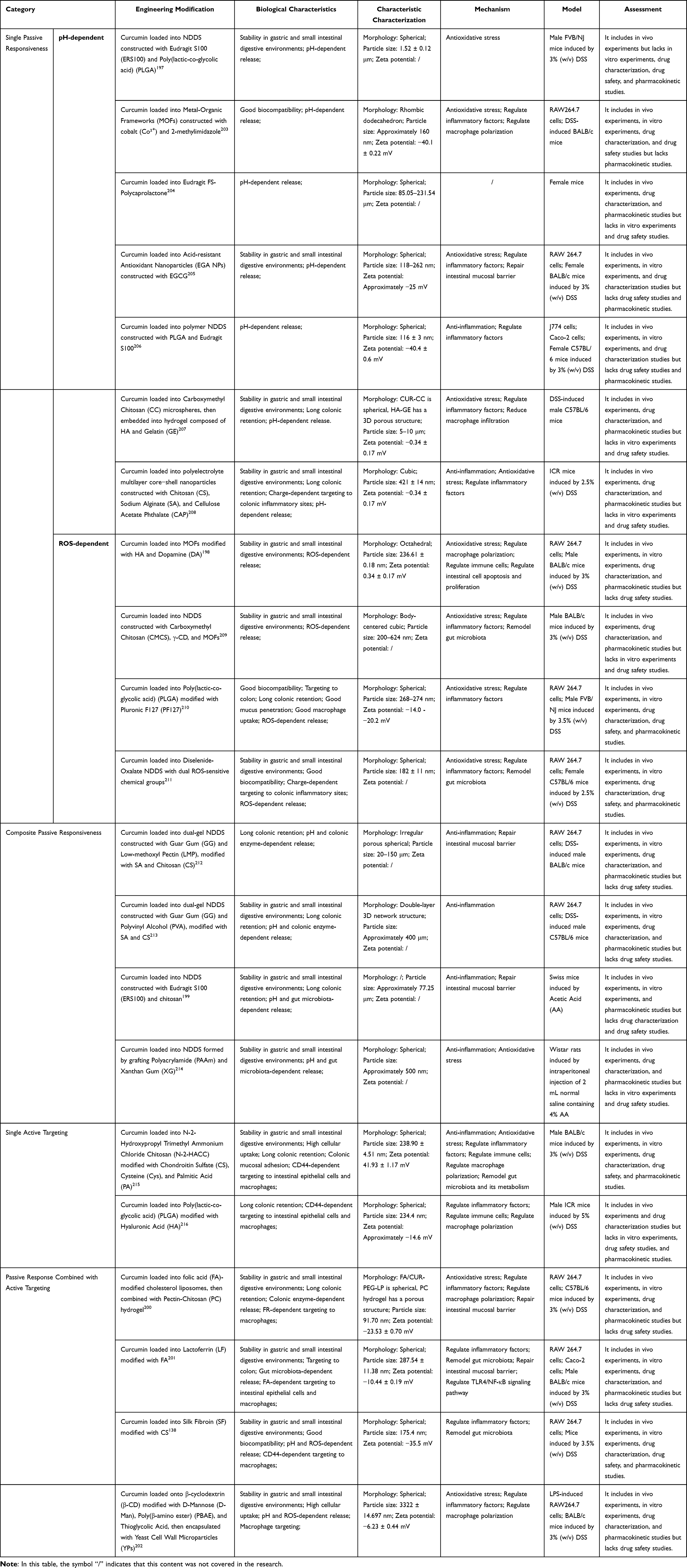 |
Table 4 Research on the Passive Response and Active Targeting of Curcumin NDDS |
Conclusion and Challenges
This study, taking curcumin as a representative natural product, systematically explored the construction logic and practical approaches of NDDS based on the pathological and physiological essence of UC. Curcumin, with its multi-target and multi-mechanism therapeutic advantages, is regarded as a potential candidate drug for the treatment of UC. However, due to the limitations of curcumin’s own physicochemical factors, it exhibits low solubility, poor absorption, low bioavailability, and rapid metabolism in the body; in addition, gastrointestinal digestion factors and the colonic intestinal barrier jointly limit the therapeutic effect of curcumin. Nevertheless, thanks to the rapid development of NDDS, NDDS has achieved improvements in drug solubility, promotion of absorption, controlled release, and targeted delivery through various strategies such as size control, charge design, shape design, functional group modification, and stimulus-responsive design. Factors that may damage the structure of curcumin (such as pH, digestive enzymes, and intestinal flora) and factors that may hinder the absorption of curcumin (such as the intestinal mucus layer and intestinal epithelial barrier) have become the targets of curcumin NDDS delivery.
From the existing research evidence, single-mechanism NDDS is unable to adapt to the complex intestinal microenvironment of UC and is difficult to meet clinical needs. Specifically: time-dependent and stress-dependent NDDS have been gradually eliminated in the early stage due to significant individual differences in gastrointestinal transit time and colonic lumen pressure; pH-dependent, intestinal flora-dependent, mucus-dependent, and intestinal epithelium-dependent single strategies have the “pan-colonic release” defect, that is, they cannot precisely focus on the segmental lesions of UC, and some designs also have safety hazards, such as mucus dissolving agents breaking the mucus barrier and absorption promoters opening tight junctions, which may aggravate intestinal barrier dysfunction; ROS-dependent NDDS can achieve targeted drug release by taking advantage of the increased ROS concentration in the UC lesion site, but the “critical response concentration of ROS in the active phase of UC” and “the effect differences of different ROS-sensitive link groups” have not been clarified yet, limiting its further optimization.
Based on this, the integration of two or more response mechanisms into a composite design NDDS has become the current core direction with the greatest clinical transformation potential. This strategy “stepwise targeting and precise drug release” adapts to the multiple pathological links of UC: for example, pH/ROS-dependent combined receptor targeting can achieve a three-level targeting from “organ (colon) - tissue (inflammatory area) - cell (macrophage)” - a typical case is the NDDS constructed by “pH-sensitive material PBAE + ROS-sensitive mercaptoacetic acid + D-Man macrophage targeting”, which can not only locate the colon through pH response, but also focus on the inflammatory site through ROS response, and finally target macrophages through the mannose receptor-mediated targeting, achieving highly precise targeted drug delivery, and demonstrating superior anti-inflammatory effects and mucosal repair capabilities in animal models.
Although the NDDS field has developed rapidly in recent years and has achieved significant breakthroughs in various aspects, there is still a long way to go before natural product NDDS is used in clinical practice. At least the following issues need to be carefully considered: ① Toxicity of enhanced absorption of natural products: Given the defects of low solubility, poor absorption, low bioavailability, and rapid metabolism of curcumin, some studies have increased the dosage and frequency of curcumin use to achieve the expected therapeutic effect. However, long-term high-dose intake has shown certain adverse reactions.35 Therefore, it can be assumed that the high tolerance of the human body for curcumin and the low incidence of adverse reactions over time may be related to absorption deficiency and low bioavailability. Essentially, improving the absorption and bioavailability of curcumin through NDDS is consistent with increasing the dosage and frequency of use, both of which will affect the tolerance of the human body to curcumin and lead to potential toxic reactions. Therefore, the clinical translation of NDDS should be cautiously considered, especially given that safety studies and pharmacokinetic studies related to NDDS are largely lacking in current animal experiments. Future research should focus on the potential safety issues and pharmacokinetic studies of NDDS in humans, and determine an appropriate dose based on the combined impact intensity of NDDS on natural products, so as to minimize the toxic side effects caused by enhanced absorption and improved bioavailability. ② Toxicity of NDDS itself: Research on the toxicity of NDDS originated from inhalation toxicology, initially due to concerns about human exposure to ultrafine pollutants in the air, and gradually extending to the toxicity of nanoparticles, including NDDS in biomedical technologies.217 Since inorganic NDDS belongs to inorganic substances, it is generally considered to have toxicity and can cause inflammation, structural damage, and functional disruption.218 Organic NDDS, due to its high biodegradability and bioavailability, is considered safe.219 However, recent studies have shown that the physical and chemical properties of NDDS determine their interaction with the human body and, to a certain extent, their toxicity.220 This indicates that organic NDDS may also have potential toxicity. Further research has shown that the long-term toxicity of inorganic NDDS is significantly affected by its particle size.218 Considering that all NDDS have a nanoscale particle size, an important question is whether all NDDS may have potential toxicity under the condition of slow excretion and decomposition. Although toxicological studies on NDDS are gradually being conducted, they are far less than studies on the efficacy of NDDS, and considering the long experimental period required for toxicological studies of NDDS, another important issue is that the current animal models for UC may not support feasible toxicological studies of NDDS. The DSS induction method is the most widely used method for establishing UC models. Since the UC mice subjected to this method have a certain tendency to heal themselves, the administration period for the experimentalists is mostly controlled within 7–14 days.221 In fact, UC has clinical characteristics of alternating episodes, remission, and recurrence. The treatment cycle for UC patients is long and repetitive. The long-term toxicity study period of NDDS should be much longer than 14 days. The UC animal model induced by DSS cannot meet the needs of NDDS toxicity research. Testing the toxicity of NDDS in normal animals is also not entirely feasible. The construction strategy of NDDS is based on the colonic environment of UC patients, which is quite different from that of normal humans. The toxicity results of NDDS tested in normal animals cannot be equivalent to its performance in UC patients. We need to attach extreme importance to the toxicity research of NDDS and the development of corresponding animal models. This determines the future of NDDS clinical transformation. ③ The endpoint of NDDS for UC treatment: For the NDDS treatment of UC, the question we must clarify is where we want the drug and NDDS to reach. Considering that the UC colon can directly come into contact with oral NDDS, perhaps we do not need the drug to enter the systemic circulation. A study compared the therapeutic effects of different functional NDDS loaded with curcumin, and found that retaining curcumin within the intestinal epithelial cells showed better efficacy than promoting its passage through the intestinal epithelial cells.178 Therefore, for the treatment of UC, only maintaining a high concentration of the drug in the local UC colon, without maintaining the drug concentration in the systemic circulation, is a possible choice. Another important issue is whether the loaded drug NDDS needs to be completely absorbed. Considering that most NDDS still lack studies on decomposition and excretion rates, as well as the acute toxicity and chronic systemic deposition after entering the systemic circulation, the best strategy at present may be for the loaded drug NDDS to release the drug after passing through the intestinal mucus layer, ensuring a high drug concentration in the local UC colon epithelium, and leaving the NDDS in the intestine to wait for excretion to avoid potential toxicity. Further, after curcumin enters the intestinal epithelial cells, only a small portion may be internalized and remain in the cells, some will be transported to lysosomes for decomposition, and the rest will be excreted from the apical and basal sides of the cell through exocytosis.56 Therefore, a more in-depth delivery strategy may involve the design of the drug itself to avoid lysosomal decomposition and exocytosis, maintaining the drug concentration in the cells.
In the construction of natural product NDDS, understanding the pathological physiological characteristics of UC as a digestive system disease remains the most important foundational work and is also the key point for NDDS to achieve breakthrough progress.
Abbreviations
UC, ulcerative colitis; NDDS, Nano-Drug Delivery System; 5-ASA, 5-aminosalicylic acid; ROS, reactive oxygen species; GALT, gut-associated lymphoid tissues; MMPs, matrix metalloproteinases; PELNs, Plant-derived Exosome-like Nanovesicles; CELNs, Curcumin-derived Exosome-like Nanovesicles; CNPs, Curcumin Nanoparticles; SCFA, short-chain fatty acids; MUC, mucoprotein; JAK/STAT, Janus kinase/signal transducer and activator of transcription; MAPK, mitogen-activated protein kinase; NF-κB, nuclear factor kappa-B; Nrf2, nuclear factor erythroid 2-related factor 2; NLRP3, NOD-like receptor pyrin domain-containing 3; SphK1, sphingosine kinase 1; TNF-α, tumor necrosis factor-alpha; IFN-γ, interferon-gamma; IL, interleukin; MIP-2, macrophage inflammatory protein-2; MMP-3, matrix metalloproteinase-3; GST, glutathione S-transferase; CYP450, cytochrome P450; IOIBD, International Organization for the Study of Inflammatory Bowel Diseases; CRC, colorectal cancer; PEG, polyethylene glycol; TEER, trans-epithelial electrical resistance; EDTA, ethylenediaminetetraacetic acid; MGL, macrophage galactose lectin; FR, Folate Receptor; DMTHF, N5,N10-dimethyl tetrahydrofolic acid; PepT1, Peptide transporter 1; KPV, lysine-proline-valine; TfR, transferrin receptor; EMR1, EGF-like module-containing mucin-like hormone receptor-like 1; ADGRE1, adhesion G protein-coupled receptor E1; HA, hyaluronic acid; CS, chondroitin sulfate; CAP, cellulose acetate phthalate; PVAP, polyvinyl acetate phthalate; HPMCP, hydroxypropyl methylcellulose phthalate; EC, ethyl cellulose; PLGA, poly(lactic-co-glycolic acid); PCL, polycaprolactone; MPN, Metal-Polyphenol Network; EGCG, epigallocatechin gallate; YM, Yeast Microcapsules; AA, acetic acid; DSS, dextran sulfate sodium; NaCas, sodium caseinate; γ-CD, γ-cyclodextrin; SA, succinic acid; EWDP, Egg White-Derived Peptides; HTCC, quaternary ammonium chitosan; EWP, Egg White Peptides; DEX, dexamethasone; HES, hydroxyethyl starch; MOFs, Metal-Organic Frameworks; ERS100, Eudragit S100; CC, carboxymethyl chitosan; GE, gelatin; ICR, Institute of Cancer Research (mouse strain); FVB/NJ, FVB/NJ mouse strain; BALB/c, BALB/c mouse strain; C57BL/6, C57BL/6 mouse strain; KM, Kunming mouse strain; N-2-HACC, N-2-Hydroxypropyl Trimethyl Ammonium Chloride Chitosan; PAAm, polyacrylamide; XG, xanthan gum; LF, lactoferrin; PC, pectin-chitosan; SF, silk fibroin; D-Man, D-Mannose; PBAE, poly(β-amino ester); YPs, Yeast Cell Wall Microparticles; TLR4, toll-like receptor 4; PF127, Pluronic F127; GG, Guar Gum; LMP, Low-methoxyl Pectin; PVA, Polyvinyl Alcohol; DPPG, Dipalmitoylphosphatidylglycerol; PBAs, Prussian Blue Analogs.
Author Contributions
All authors made a significant contribution to the work reported, whether that is in the conception, study design, execution, acquisition of data, analysis and interpretation, or in all these areas; took part in drafting, revising or critically reviewing the article; gave final approval of the version to be published; have agreed on the journal to which the article has been submitted; and agree to be accountable for all aspects of the work.
Funding
The author(s) declare financial support was received for the research, authorship, and/or publication of this article. This work was supported by [the Ministry of Education of the People’s Republic of China] under Grant [S202410541019]; [Hunan Provincial Department of Science and Technology] under Grant [2023JJ60044], [24A0274] and [Furong Project for Young Talents] under Grant [2025RC3189]; and [Hunan Provincial Administration of Traditional Chinese Medicine] under Grant [XPXHZ03], [2025790] and [the Domestic First-class Construction Discipline of Chinese Medicine in Hunan University of Chinese Medicine].
Disclosure
The authors report no conflicts of interest in this work.
References
1. Le Berre C, Honap S, Peyrin-Biroulet L. Ulcerative colitis. Lancet. 2023;402(10401):571–584. doi:10.1016/S0140-6736(23)00966-2
2. Ahn HS. Increased incidence of inflammatory bowel disease in Korea may not be explained by food additives. Inflamm Bowel Dis. 2015;21(8):E17. doi:10.1097/MIB.0000000000000480
3. Yu Q, Zhu C, Feng S, et al. Economic burden and health care access for patients with inflammatory bowel diseases in China: web-based survey study. J Med Internet Res. 2021;23(1):e20629. doi:10.2196/20629
4. Siebert U, Wurm J, Gothe RM, et al. Predictors of temporary and permanent work disability in patients with inflammatory bowel disease: results of the Swiss inflammatory bowel disease cohort study. Inflamm Bowel Dis. 2013;19(4):847–855. doi:10.1097/MIB.0b013e31827f278e
5. Williet N, Sarter H, Gower-Rousseau C, et al. Patient-reported outcomes in a french nationwide survey of inflammatory bowel disease patients. J Crohns Colitis. 2017;11(2):165–174. doi:10.1093/ecco-jcc/jjw145
6. Lichtenstein GR, Shahabi A, Seabury SA, et al. Lifetime economic burden of crohn’s disease and ulcerative colitis by age at diagnosis. Clin Gastroenterol Hepatol. 2020;18(4):889–897.e10. doi:10.1016/j.cgh.2019.07.022
7. Moroz E, Matoori S, Leroux JC. Oral delivery of macromolecular drugs: where we are after almost 100years of attempts. Adv Drug Deliv Rev. 2016;101:108–121. doi:10.1016/j.addr.2016.01.010
8. Ning H, Liu J, Tan J, et al. The role of the Notch signalling pathway in the pathogenesis of ulcerative colitis: from the perspective of intestinal mucosal barrier. Front Med Lausanne. 2024;10:1333531. doi:10.3389/fmed.2023.1333531
9. Sun H, Zhan M, Mignani S, et al. Modulation of macrophages using nanoformulations with curcumin to treat inflammatory diseases: a concise review. Pharmaceutics. 2022;14(10):2239. doi:10.3390/pharmaceutics14102239
10. Patra JK, Das G, Fraceto LF, et al. Nano based drug delivery systems: recent developments and future prospects. J Nanobiotechnology. 2018;16(1):71. doi:10.1186/s12951-018-0392-8
11. Pituch-Zdanowska A, Dembiński Ł, Banaszkiewicz A. Old but fancy: curcumin in ulcerative colitis-current overview. Nutrients. 2022;14(24):5249. doi:10.3390/nu14245249
12. Nelson KM, Dahlin JL, Bisson J, et al. The essential medicinal chemistry of curcumin. J Med Chem. 2017;60(5):1620–1637. doi:10.1021/acs.jmedchem.6b00975
13. Wang L, Zheng W, Men Q, et al. Curcumin-loaded polysaccharide microparticles alleviated DSS-induced ulcerative colitis by improving intestinal microecology and regulating MAPK/NF-κB/Nrf2/NLRP3 pathways. Int J Biol Macromol. 2024;281(Pt 4):136687. doi:10.1016/j.ijbiomac.2024.136687
14. Zhang X, Zhang H, Wang J, et al. Curcumin attenuates ulcerative colitis via regulation of Sphingosine kinases 1/NF-κB signaling pathway. Biofactors. 2025;51(1):e70001. doi:10.1002/biof.70001
15. Long D, Mao C, Zhang W, et al. Natural products for the treatment of ulcerative colitis: focus on the JAK/STAT pathway. Front Immunol. 2025;16:1538302. doi:10.3389/fimmu.2025.1538302
16. Cong Y, Wang L, Konrad A, et al. Curcumin induces the tolerogenic dendritic cell that promotes differentiation of intestine-protective regulatory T cells. Eur J Immunol. 2009;39(11):3134–3146. doi:10.1002/eji.200939052
17. Ghany LMAA, Beshay BY, Youssef Moustafa AM, et al. Design, synthesis, anti-inflammatory evaluation, and molecular modelling of new coumarin-based analogs combined curcumin and other heterocycles as potential TNF-α production inhibitors via upregulating Nrf2/HO-1, downregulating AKT/mTOR signalling pathways and downregulating NF-κB in LPS induced macrophages. J Enzyme Inhib Med Chem. 2023;38(1):2243551. doi:10.1080/14756366.2023.2243551
18. Zheng LX, Guo KE, Huang JQ, et al. Curcumin alleviated dextran sulfate sodium-induced colitis by recovering memory Th/Tfh subset balance. World J Gastroenterol. 2023;29(36):5226–5239. doi:10.3748/wjg.v29.i36.5226
19. Lin Y, Liu H, Bu L, Chen C, Ye X. Review of the effects and mechanism of curcumin in the treatment of inflammatory bowel disease. Front Pharmacol. 2022;13:908077. doi:10.3389/fphar.2022.908077
20. Larmonier CB, Midura-Kiela MT, Ramalingam R, et al. Modulation of neutrophil motility by curcumin: implications for inflammatory bowel disease. Inflamm Bowel Dis. 2011;17(2):503–515. doi:10.1002/ibd.21391
21. Epstein J, Docena G, MacDonald TT, et al. Curcumin suppresses p38 mitogen-activated protein kinase activation, reduces IL-1beta and matrix metalloproteinase-3 and enhances IL-10 in the mucosa of children and adults with inflammatory bowel disease. Br J Nutr. 2010;103(6):824–832. doi:10.1017/S0007114509992510
22. Cao S, Wang C, Yan J, et al. Curcumin ameliorates oxidative stress-induced intestinal barrier injury and mitochondrial damage by promoting Parkin dependent mitophagy through AMPK-TFEB signal pathway. Free Radic Biol Med. 2020;147:8–22. doi:10.1016/j.freeradbiomed.2019.12.004
23. Koh YC, Tsai YW, Lee PS, et al. Amination potentially augments the ameliorative effect of curcumin on inhibition of the IL-6/Stat3/c-Myc pathway and gut microbial modulation in colitis-associated tumorigenesis. J Agric Food Chem. 2022;70(46):14744–14754. doi:10.1021/acs.jafc.2c06645
24. Xu Z, Zhu W, Xu D, et al. Supplementation of curcumin promotes the intestinal structure, immune barrier function and cecal microbiota composition of laying hens in early laying period. Poult Sci. 2024;103(12):104355. doi:10.1016/j.psj.2024.104355
25. Grammatikopoulou MG, Gkiouras K, Theodoridis X, et al. Oral adjuvant curcumin therapy for attaining clinical remission in ulcerative colitis: a systematic review and meta-analysis of randomized controlled trials. Nutrients. 2018;10(11):1737. doi:10.3390/nu10111737
26. Kotha RR, Luthria DL. Curcumin: biological, pharmaceutical, nutraceutical, and analytical aspects. Molecules. 2019;24(16):2930. doi:10.3390/molecules24162930
27. Kharat M, Du Z, Zhang G, et al. Physical and chemical stability of curcumin in aqueous solutions and emulsions: impact of pH, temperature, and molecular environment. J Agric Food Chem. 2017;65(8):1525–1532. doi:10.1021/acs.jafc.6b04815
28. Tan S, Rupasinghe TW, Tull DL, et al. Degradation of curcuminoids by in vitro pure culture fermentation. J Agric Food Chem. 2014;62(45):11005–11015. doi:10.1021/jf5031168
29. Burapan S, Kim M, Han J. Curcuminoid demethylation as an alternative metabolism by human intestinal microbiota. J Agric Food Chem. 2017;65(16):3305–3310. doi:10.1021/acs.jafc.7b00943
30. Zhang S, Asghar S, Yu F, et al. The enhancement of N-acetylcysteine on intestinal absorption and oral bioavailability of hydrophobic curcumin. Eur J Pharm Sci. 2020;154:105506. doi:10.1016/j.ejps.2020.105506
31. Jacob S, Kather FS, Morsy MA, et al. Advances in nanocarrier systems for overcoming formulation challenges of curcumin: current insights. Nanomaterials (Basel). 2024;14(8):672. doi:10.3390/nano14080672
32. Garcea G, Jones DJ, Singh R, et al. Detection of curcumin and its metabolites in hepatic tissue and portal blood of patients following oral administration. Br J Cancer. 2004;90(5):1011–1015. doi:10.1038/sj.bjc.6601623
33. Ireson CR, Jones DJ, Orr S, et al. Metabolism of the cancer chemopreventive agent curcumin in human and rat intestine. Cancer Epidemiol Biomarkers Prev. 2002;11(1):105–111.
34. Jäger R, Lowery RP, Calvanese AV, et al. Comparative absorption of curcumin formulations. Nutr J. 2014;13(1):11. doi:10.1186/1475-2891-13-11
35. Burgos-Morón E, Calderón-Montaño JM, Salvador J, et al. The dark side of curcumin. Int, J, Cancer. 2010;126(7):1771–1775. doi:10.1002/ijc.24967
36. Oetari S, Sudibyo M, Commandeur JN, et al. Effects of curcumin on cytochrome P450 and glutathione S-transferase activities in rat liver. Biochem Pharmacol. 1996;51(1):39–45. doi:10.1016/0006-2952(95)02113-2
37. Chin D, Huebbe P, Frank J, et al. Curcumin may impair iron status when fed to mice for six months. Redox Biol. 2014;2:563–569. doi:10.1016/j.redox.2014.01.018
38. Kuttan R, Bhanumathy P, Nirmala K, et al. Potential anticancer activity of turmeric (Curcuma longa). Cancer Lett. 1985;29(2):197–202. doi:10.1016/0304-3835(85)90159-4
39. Sandle GI. Salt and water absorption in the human colon: a modern appraisal. Gut. 1998;43(2):294–299. doi:10.1136/gut.43.2.294
40. Hebden JM, Blackshaw PE, Perkins AC, et al. Limited exposure of the healthy distal colon to orally-dosed formulation is further exaggerated in active left-sided ulcerative colitis. Aliment Pharmacol Ther. 2000;14(2):155–161. doi:10.1046/j.1365-2036.2000.00697.x
41. Bassotti G, Antonelli E, Villanacci V, et al. Gastrointestinal motility disorders in inflammatory bowel diseases. World J Gastroenterol. 2014;20(1):37–44. doi:10.3748/wjg.v20.i1.37
42. Stubbs JB, Valenzuela GA, Stubbs CC, et al. A noninvasive scintigraphic assessment of the colonic transit of nondigestible solids in man. J Nucl Med. 1991;32(7):1375–1381.
43. Koziolek M, Grimm M, Becker D, et al. Investigation of pH and temperature profiles in the GI tract of fasted human subjects using the intellicap (®) system. J Pharm Sci. 2015;104(9):2855–2863. doi:10.1002/jps.24274
44. Fallingborg J, Christensen LA, Jacobsen BA, et al. Very low intraluminal colonic pH in patients with active ulcerative colitis. Dig Dis Sci. 1993;38(11):1989–1993. doi:10.1007/BF01297074
45. Karasov WH, Martínez Del Rio C, Caviedes-Vidal E. Ecological physiology of diet and digestive systems. Annu Rev Physiol. 2011;73(1):69–93. doi:10.1146/annurev-physiol-012110-142152
46. Limketkai BN, Godoy-Brewer G, Parian AM, et al. Dietary interventions for the treatment of inflammatory bowel diseases: an updated systematic review and meta-analysis. Clin Gastroenterol Hepatol. 2023;21(10):2508–2525.e10. doi:10.1016/j.cgh.2022.11.026
47. Wang M, Fu R, Xu D, et al. Traditional Chinese Medicine: a promising strategy to regulate the imbalance of bacterial flora, impaired intestinal barrier and immune function attributed to ulcerative colitis through intestinal microecology. J Ethnopharmacol. 2024;318(Pt A):116879. doi:10.1016/j.jep.2023.116879
48. Williams BA, Grant LJ, Gidley MJ, et al. Gut fermentation of dietary fibres: physico-chemistry of plant cell walls and implications for health. Int J Mol Sci. 2017;18(10):2203. doi:10.3390/ijms18102203
49. Wu L, Shan W, Zhang Z, et al. Engineering nanomaterials to overcome the mucosal barrier by modulating surface properties. Adv Drug Deliv Rev. 2018;124:150–163. doi:10.1016/j.addr.2017.10.001
50. Bhattacharjee S, Mahon E, Harrison SM, et al. Nanoparticle passage through porcine jejunal mucus: microfluidics and rheology. Nanomedicine. 2017;13(3):863–873. doi:10.1016/j.nano.2016.11.017
51. Zhu Q, Chen Z, Paul PK, et al. Oral delivery of proteins and peptides: challenges, status quo and future perspectives. Acta Pharm Sin B. 2021;11(8):2416–2448. doi:10.1016/j.apsb.2021.04.001
52. Lundquist P, Artursson P. Oral absorption of peptides and nanoparticles across the human intestine: opportunities, limitations and studies in human tissues. Adv Drug Deliv Rev. 2016;106(Pt B):256–276. doi:10.1016/j.addr.2016.07.007
53. Farquhar MG, Palade GE. Junctional complexes in various epithelia. J Cell Biol. 1963;17(2):375–412. doi:10.1083/jcb.17.2.375
54. Suzuki T. Regulation of intestinal epithelial permeability by tight junctions. Cell Mol Life Sci. 2013;70(4):631–659. doi:10.1007/s00018-012-1070-x
55. Zheng Y, Luo S, Xu M, et al. Transepithelial transport of nanoparticles in oral drug delivery: from the perspective of surface and holistic property modulation. Acta Pharm Sin B. 2024;14(9):3876–3900. doi:10.1016/j.apsb.2024.06.015
56. He Y, Cheng M, Yang R, et al. Research progress on the mechanism of nanoparticles crossing the intestinal epithelial cell membrane. Pharmaceutics. 2023;15(7):1816.
57. Zheng B, Wang L, Yi Y, et al. Design strategies, advances and future perspectives of colon-targeted delivery systems for the treatment of inflammatory bowel disease. Asian J Pharm Sci. 2024;19(4):100943. doi:10.1016/j.ajps.2024.100943
58. Fukui E, Miyamura N, Uemura K, et al. Preparation of enteric coated timed-release press-coated tablets and evaluation of their function by in vitro and in vivo tests for colon targeting. Int J Pharm. 2000;204(1–2):7–15. doi:10.1016/S0378-5173(00)00454-3
59. Worsøe J, Fynne L, Gregersen T, et al. Gastric transit and small intestinal transit time and motility assessed by a magnet tracking system. BMC Gastroenterol. 2011;11(1):145. doi:10.1186/1471-230X-11-145
60. Liu H, Yang XG, Nie SF, et al. Chitosan-based controlled porosity osmotic pump for colon-specific delivery system: screening of formulation variables and in vitro investigation. Int J Pharm. 2007;332(1–2):115–124. doi:10.1016/j.ijpharm.2006.09.038
61. Amidon S, Brown JE, Dave VS. Colon-targeted oral drug delivery systems: design trends and approaches. AAPS Pharm Sci Tech. 2015;16(4):731–741. doi:10.1208/s12249-015-0350-9
62. Almoshari Y. Osmotic pump drug delivery systems-a comprehensive review. Pharmaceuticals (Basel). 2022;15(11):1430. doi:10.3390/ph15111430
63. Takaya T, Niwa K, Muraoka M, et al. Importance of dissolution process on systemic availability of drugs delivered by colon delivery system. J Control Release. 1998;50(1–3):111–122. doi:10.1016/S0168-3659(97)00123-5
64. Yoshida T, Lai TC, Kwon GS, et al. pH- and ion-sensitive polymers for drug delivery. Expert Opin Drug Deliv. 2013;10(11):1497–1513. doi:10.1517/17425247.2013.821978
65. Hu S, Zhao R, Xu Y, et al. Orally-administered nanomedicine systems targeting colon inflammation for the treatment of inflammatory bowel disease: latest advances. J Mater Chem B. 2023;12(1):13–38. doi:10.1039/D3TB02302H
66. Hua S, Marks E, Schneider JJ, et al. Advances in oral nano-delivery systems for colon targeted drug delivery in inflammatory bowel disease: selective targeting to diseased versus healthy tissue. Nanomedicine. 2015;11(5):1117–1132. doi:10.1016/j.nano.2015.02.018
67. Barea MJ, Jenkins MJ, Gaber MH, et al. Evaluation of liposomes coated with a pH responsive polymer. Int J Pharm. 2010;402(1–2):89–94. doi:10.1016/j.ijpharm.2010.09.028
68. Chen L, You Q, Hu L, et al. The antioxidant procyanidin reduces reactive oxygen species signaling in macrophages and ameliorates experimental colitis in mice. Front Immunol. 2018;8:1910. doi:10.3389/fimmu.2017.01910
69. Grisham MB. Oxidants and free radicals in inflammatory bowel disease. Lancet. 1994;344(8926):859–861. doi:10.1016/S0140-6736(94)92831-2
70. Guo X, Cheng Y, Zhao X, et al. Advances in redox-responsive drug delivery systems of tumor microenvironment. J Nanobiotechnology. 2018;16(1):74. doi:10.1186/s12951-018-0398-2
71. Zhang J, Sun S, Chen H, et al. Advances in natural compound-based nanomedicine and the interaction with gut microbiota in ulcerative colitis therapy. Front Pharmacol. 2023;14:1197144. doi:10.3389/fphar.2023.1197144
72. Lee Y, Kamada N, Moon JJ. Oral nanomedicine for modulating immunity, intestinal barrier functions, and gut microbiome. Adv Drug Deliv Rev. 2021;179:114021. doi:10.1016/j.addr.2021.114021
73. Manda JN, Markovic M, Zimmermann EM, et al. Phospholipid cyclosporine prodrugs targeted at inflammatory bowel disease (IBD) treatment: design, synthesis, and in vitro validation. ChemMedChem. 2020;15(17):1639–1644. doi:10.1002/cmdc.202000317
74. Naeem M, Awan UA, Subhan F, et al. Advances in colon-targeted nano-drug delivery systems: challenges and solutions. Arch Pharm Res. 2020;43(1):153–169. doi:10.1007/s12272-020-01219-0
75. Cattani VB, Fiel LA, Jäger A, et al. Lipid-core nanocapsules restrained the indomethacin ethyl ester hydrolysis in the gastrointestinal lumen and wall acting as mucoadhesive reservoirs. Eur J Pharm Sci. 2010;39(1–3):116–124. doi:10.1016/j.ejps.2009.11.004
76. Teng Y, Ren Y, Sayed M, et al. Plant-derived exosomal MicroRNAs shape the gut microbiota. Cell Host Microbe. 2018;24(5):637–652.e8. doi:10.1016/j.chom.2018.10.001
77. Applerot G, Lipovsky A, Dror R, et al. Enhanced antibacterial activity of nanocrystalline ZnO due to increased ROS-mediated cell injury. Adv Funct Mater. 2009;19(6):842–852. doi:10.1002/adfm.200801081
78. Chen H, Wang B, Gao D, et al. Broad-spectrum antibacterial activity of carbon nanotubes to human gut bacteria. Small. 2013;9(16):2735–2746. doi:10.1002/smll.201202792
79. Zhu B, Xia X, Xia N, et al. Modification of Fatty acids in membranes of bacteria: implication for an adaptive mechanism to the toxicity of carbon nanotubes. Environ Sci Technol. 2014;48(7):4086–4095. doi:10.1021/es404359v
80. Ensign LM, Schneider C, Suk JS, et al. Mucus penetrating nanoparticles: biophysical tool and method of drug and gene delivery. Adv Mater. 2012;24(28):3887–3894. doi:10.1002/adma.201201800
81. Niebel W, Walkenbach K, Béduneau A, et al. Nanoparticle-based clodronate delivery mitigates murine experimental colitis. J Control Release. 2012;160(3):659–665. doi:10.1016/j.jconrel.2012.03.004
82. Griffiths PC, Cattoz B, Ibrahim MS, et al. Probing the interaction of nanoparticles with mucin for drug delivery applications using dynamic light scattering. Eur J Pharm Biopharm. 2015;97(Pt A):218–222. doi:10.1016/j.ejpb.2015.05.004
83. Christfort JF, Guillot AJ, Melero A, et al. Cubic microcontainers improve in situ colonic mucoadhesion and absorption of amoxicillin in rats. Pharmaceutics. 2020;12(4):355. doi:10.3390/pharmaceutics12040355
84. Bernkop-Schnürch A. Thiomers: a new generation of mucoadhesive polymers. Adv Drug Deliv Rev. 2005;57(11):1569–1582. doi:10.1016/j.addr.2005.07.002
85. Wang D, Wang W, Wang P, et al. Research progress of colon-targeted oral hydrogel system based on natural polysaccharides. Int J Pharm. 2023;643:123222. doi:10.1016/j.ijpharm.2023.123222
86. Chen F, Zhang ZR, Yuan F, et al. In vitro and in vivo study of N-trimethyl chitosan nanoparticles for oral protein delivery. Int J Pharm. 2008;349(1–2):226–233. doi:10.1016/j.ijpharm.2007.07.035
87. Huckaby JT, Lai SK. PEGylation for enhancing nanoparticle diffusion in mucus. Adv Drug Deliv Rev. 2018;124:125–139. doi:10.1016/j.addr.2017.08.010
88. Maisel K, Ensign L, Reddy M, et al. Effect of surface chemistry on nanoparticle interaction with gastrointestinal mucus and distribution in the gastrointestinal tract following oral and rectal administration in the mouse. J Control Release. 2015;197:48–57. doi:10.1016/j.jconrel.2014.10.026
89. Zheng Y, Xing L, Chen L, et al. Tailored elasticity combined with biomimetic surface promotes nanoparticle transcytosis to overcome mucosal epithelial barrier. Biomaterials. 2020;262:120323. doi:10.1016/j.biomaterials.2020.120323
90. Yu M, Wang J, Yang Y, et al. Rotation-facilitated rapid transport of nanorods in mucosal tissues. Nano Lett. 2016;16(11):7176–7182. doi:10.1021/acs.nanolett.6b03515
91. Bao C, Liu B, Li B, et al. Enhanced transport of shape and rigidity-tuned α-lactalbumin nanotubes across intestinal mucus and cellular barriers. Nano Lett. 2020;20(2):1352–1361. doi:10.1021/acs.nanolett.9b04841
92. Zhou W, Li B, Min R, et al. Mucus-penetrating dendritic mesoporous silica nanoparticle loading drug nanocrystal clusters to enhance permeation and intestinal absorption. Biomater Sci. 2023;11(3):1013–1030. doi:10.1039/D2BM01404A
93. Wang Y, Ke J, Guo X, et al. Chiral mesoporous silica nano-screws as an efficient biomimetic oral drug delivery platform through multiple topological mechanisms. Acta Pharm Sin B. 2022;12(3):1432–1446. doi:10.1016/j.apsb.2021.08.014
94. Sang Z, Xu L, Ding R, et al. Nanoparticles exhibiting virus-mimic surface topology for enhanced oral delivery. Nat Commun. 2023;14(1):7694. doi:10.1038/s41467-023-43465-y
95. Wang Y, Zhao L, Dai Y, et al. Enantioselective oral absorption of molecular chiral mesoporous silica nanoparticles. Adv Mater. 2023;35(49):e2307900. doi:10.1002/adma.202307900
96. Lai SK, O’Hanlon DE, Harrold S, et al. Rapid transport of large polymeric nanoparticles in fresh undiluted human mucus. Proc Natl Acad Sci U S A. 2007;104(5):1482–1487. doi:10.1073/pnas.0608611104
97. Iglesias T, López de Cerain A, Irache JM, et al. Evaluation of the cytotoxicity, genotoxicity and mucus permeation capacity of several surface modified poly(anhydride) nanoparticles designed for oral drug delivery. Int J Pharm. 2017;517(1–2):67–79. doi:10.1016/j.ijpharm.2016.11.059
98. Shah PL, Scott SF, Knight RA, et al. In vivo effects of recombinant human DNase I on sputum in patients with cystic fibrosis. Thorax. 1996;51(2):119–125. doi:10.1136/thx.51.2.119
99. Henke MO, Ratjen F. Mucolytics in cystic fibrosis. Paediatr Respir Rev. 2007;8(1):24–29. doi:10.1016/j.prrv.2007.02.009
100. Elsaesser A, Howard CV. Toxicology of nanoparticles. Adv Drug Deliv Rev. 2012;64(2):129–137. doi:10.1016/j.addr.2011.09.001
101. Yao D, Dai W, Dong M, et al. MUC2 and related bacterial factors: therapeutic targets for ulcerative colitis. EBioMedicine. 2021;74:103751.
102. Tirosh B, Khatib N, Barenholz Y, et al. Transferrin as a luminal target for negatively charged liposomes in the inflamed colonic mucosa. Mol Pharm. 2009;6(4):1083–1091. doi:10.1021/mp9000926
103. Xu S, Yang Q, Wang R, et al. Genetically engineered pH-responsive silk sericin nanospheres with efficient therapeutic effect on ulcerative colitis. Acta Biomater. 2022;144:81–95. doi:10.1016/j.actbio.2022.03.012
104. Ren T, Wang Q, Xu Y, et al. Enhanced oral absorption and anticancer efficacy of cabazitaxel by overcoming intestinal mucus and epithelium barriers using surface polyethylene oxide (PEO) decorated positively charged polymer-lipid hybrid nanoparticles. J Control Release. 2018;269:423–438. doi:10.1016/j.jconrel.2017.11.015
105. Tada-Oikawa S, Eguchi M, Yasuda M, et al. Functionalized surface-charged SiO nanoparticles induce pro-inflammatory responses, but are not Lethal to Caco-2 cells. Chem Res Toxicol. 2020;33(5):1226–1236. doi:10.1021/acs.chemrestox.9b00478
106. Schmidt C, Lautenschlaeger C, Collnot EM, et al. Nano- and microscaled particles for drug targeting to inflamed intestinal mucosa: a first in vivo study in human patients. J Control Release. 2013;165(2):139–145. doi:10.1016/j.jconrel.2012.10.019
107. Wang Y, Mo Y, Sun Y, et al. Intestinal nanoparticle delivery and cellular response: a review of the bidirectional nanoparticle-cell interplay in mucosa based on physiochemical properties. J Nanobiotechnology. 2024;22(1):669.
108. Li Y, Chen X, Gu N. Computational investigation of interaction between nanoparticles and membranes: hydrophobic/hydrophilic effect. J Phys Chem B. 2008;112(51):16647–16653. doi:10.1021/jp8051906
109. Griffin BT, Guo J, Presas E, et al. Pharmacokinetic, pharmacodynamic and biodistribution following oral administration of nanocarriers containing peptide and protein drugs. Adv Drug Deliv Rev. 2016;106(Pt B):367–380. doi:10.1016/j.addr.2016.06.006
110. Sanjula B, Shah FM, Javed A, et al. Effect of poloxamer 188 on lymphatic uptake of carvedilol-loaded solid lipid nanoparticles for bioavailability enhancement. J Drug Target. 2009;17(3):249–256. doi:10.1080/10611860902718672
111. Li D, Zhuang J, He H, et al. Influence of particle geometry on gastrointestinal transit and absorption following oral administration. ACS Appl Mater Interfaces. 2017;9(49):42492–42502. doi:10.1021/acsami.7b11821
112. Banerjee A, Qi J, Gogoi R, et al. Role of nanoparticle size, shape and surface chemistry in oral drug delivery. J Control Release. 2016;238:176–185. doi:10.1016/j.jconrel.2016.07.051
113. Liu N, Becton M, Zhang L, et al. Mechanism of coupling nanoparticle stiffness with shape for endocytosis: from rodlike penetration to wormlike wriggling. J Phys Chem B. 2020;124(49):11145–11156. doi:10.1021/acs.jpcb.0c08089
114. Li Y, Zhang M, Zhang Y, et al. A computational study of the influence of nanoparticle shape on clathrin-mediated endocytosis. J Mater Chem B. 2023;11(27):6319–6334. doi:10.1039/D3TB00322A
115. Cao Y, Janjua TI, Qu Z, et al. Virus-like silica nanoparticles enhance macromolecule permeation in vivo. Biomater Sci. 2023;11(13):4508–4521. doi:10.1039/D3BM00137G
116. Luo Z, Paunović N, Leroux JC. Physical methods for enhancing drug absorption from the gastrointestinal tract. Adv Drug Deliv Rev. 2021;175:113814. doi:10.1016/j.addr.2021.05.024
117. Wang D, Jiang Q, Dong Z, et al. Nanocarriers transport across the gastrointestinal barriers: the contribution to oral bioavailability via blood circulation and lymphatic pathway. Adv Drug Deliv Rev. 2023;203:115130. doi:10.1016/j.addr.2023.115130
118. Luo H, Zhang Y, Chi C, et al. Starch nanocarriers for enhanced M - cell transport and oral delivery of bioactive proteins. ACS Appl Nano Mater. 2023;6(6):4793–4802. doi:10.1021/acsanm.3c00309
119. Amin MK, Boateng J. Surface functionalization of PLGA nanoparticles for potential oral vaccine delivery targeting intestinal immune cells. Colloids Surf B Biointerfaces. 2023;222:113121. doi:10.1016/j.colsurfb.2022.113121
120. Jakubowska K, Pryczynicz A, Iwanowicz P, et al. Expressions of matrix metalloproteinases (MMP-2, MMP-7, and MMP-9) and their inhibitors (TIMP-1, TIMP-2) in inflammatory bowel diseases. Gastroenterol Res Pract. 2016;2016:2456179. doi:10.1155/2016/2456179
121. Peng Y, Luo X, Wang X, et al. Bioresponsive and transformable coacervate actuated by intestinal peristalsis for targeted treatment of intestinal bleeding and inflammation. Bioact Mater. 2024;41:627–639. doi:10.1016/j.bioactmat.2024.08.020
122. Dodane V, Amin Khan M, Merwin JR. Effect of chitosan on epithelial permeability and structure. Int J Pharm. 1999;182(1):21–32. doi:10.1016/S0378-5173(99)00030-7
123. Salama NN, Eddington ND, Fasano A. Tight junction modulation and its relationship to drug delivery. Adv Drug Deliv Rev. 2006;58(1):15–28. doi:10.1016/j.addr.2006.01.003
124. Ude VC, Brown DM, Viale L, et al. Impact of copper oxide nanomaterials on differentiated and undifferentiated Caco-2 intestinal epithelial cells; assessment of cytotoxicity, barrier integrity, cytokine production and nanomaterial penetration. Part Fibre Toxicol. 2017;14(1):31. doi:10.1186/s12989-017-0211-7
125. Kalive M, Zhang W, Chen Y, et al. Human intestinal epithelial cells exhibit a cellular response indicating a potential toxicity upon exposure to hematite nanoparticles. Cell Biol Toxicol. 2012;28(5):343–368. doi:10.1007/s10565-012-9229-7
126. García-Rodríguez A, Vila L, Cortés C, et al. Effects of differently shaped TiONPs (nanospheres, nanorods and nanowires) on the in vitro model (Caco-2/HT29) of the intestinal barrier. Part Fibre Toxicol. 2018;15(1):33. doi:10.1186/s12989-018-0269-x
127. Li X, Jafari SM, Zhou F, et al. The intracellular fate and transport mechanism of shape, size and rigidity varied nanocarriers for understanding their oral delivery efficiency. Biomaterials. 2023;294:121995. doi:10.1016/j.biomaterials.2023.121995
128. Welling SH, Hubálek F, Jacobsen J, et al. The role of citric acid in oral peptide and protein formulations: relationship between calcium chelation and proteolysis inhibition. Eur J Pharm Biopharm. 2014;86(3):544–551. doi:10.1016/j.ejpb.2013.12.017
129. Brayden D, Creed E, O’Connell A, et al. Heparin absorption across the intestine: effects of sodium N-[8-(2-hydroxybenzoyl)amino]caprylate in rat in situ intestinal instillations and in Caco-2 monolayers. Pharm Res. 1997;14(12):1772–1779. doi:10.1023/A:1012192115828
130. Nighot P, Al-Sadi R, Rawat M, et al. Matrix metalloproteinase 9-induced increase in intestinal epithelial tight junction permeability contributes to the severity of experimental DSS colitis. Am J Physiol Gastrointest Liver Physiol. 2015;309(12):G988–97. doi:10.1152/ajpgi.00256.2015
131. Chu S, Tang C, Yin C. Effects of mannose density on in vitro and in vivo cellular uptake and RNAi efficiency of polymeric nanoparticles. Biomaterials. 2015;52:229–239. doi:10.1016/j.biomaterials.2015.02.044
132. Coco R, Plapied L, Pourcelle V, et al. Drug delivery to inflamed colon by nanoparticles: comparison of different strategies. Int J Pharm. 2013;440(1):3–12. doi:10.1016/j.ijpharm.2012.07.017
133. Hua S. Advances in oral drug delivery for regional targeting in the gastrointestinal tract - influence of physiological, pathophysiological and pharmaceutical factors. Front Pharmacol. 2020;11:524. doi:10.3389/fphar.2020.00524
134. Chen C, Zhao S, Karnad A, et al. The biology and role of CD44 in cancer progression: therapeutic implications. J Hematol Oncol. 2018;11(1):64. doi:10.1186/s13045-018-0605-5
135. Rosenberg WM, Prince C, Kaklamanis L, et al. Increased expression of CD44v6 and CD44v3 in ulcerative colitis but not colonic Crohn’s disease. Lancet. 1995;345(8959):1205–1209. doi:10.1016/S0140-6736(95)91991-0
136. Chen S, Chen Z, Wang Y, et al. Targeted delivery of Chinese herb pair-based berberine/tannin acid self-assemblies for the treatment of ulcerative colitis. J Adv Res. 2022;40:263–276. doi:10.1016/j.jare.2021.11.017
137. Xiao B, Zhang Z, Viennois E, et al. Combination therapy for ulcerative colitis: orally targeted nanoparticles prevent mucosal damage and relieve inflammation. Theranostics. 2016;6(12):2250–2266. doi:10.7150/thno.15710
138. Gou S, Huang Y, Wan Y, et al. Multi-bioresponsive silk fibroin-based nanoparticles with on-demand cytoplasmic drug release capacity for CD44-targeted alleviation of ulcerative colitis. Biomaterials. 2019;212:39–54. doi:10.1016/j.biomaterials.2019.05.012
139. Yan Y, Vasudevan S, Nguyen HT, et al. Intestinal epithelial CD98: an oligomeric and multifunctional protein. Biochim Biophys Acta. 2008;1780(10):1087–1092. doi:10.1016/j.bbagen.2008.06.007
140. Charania MA, Laroui H, Liu H, et al. Intestinal epithelial CD98 directly modulates the innate host response to enteric bacterial pathogens. Infect Immun. 2013;81(3):923–934. doi:10.1128/IAI.01388-12
141. Nguyen HT, Dalmasso G, Torkvist L, et al. CD98 expression modulates intestinal homeostasis, inflammation, and colitis-associated cancer in mice. J Clin Invest. 2011;121(5):1733–1747. doi:10.1172/JCI44631
142. MacKinnon AC, Farnworth SL, Hodkinson PS, et al. Regulation of alternative macrophage activation by galectin-3. J Immunol. 2008;180(4):2650–2658. doi:10.4049/jimmunol.180.4.2650
143. Xiao B, Yang Y, Viennois E, et al. Glycoprotein CD98 as a receptor for colitis-targeted delivery of nanoparticle. J Mater Chem B. 2014;2(11):1499–1508. doi:10.1039/c3tb21564d
144. Xiao B, Laroui H, Viennois E, et al. Nanoparticles with surface antibody against CD98 and carrying CD98 small interfering RNA reduce colitis in mice. Gastroenterology. 2014;146(5):1289–300.e1–19. doi:10.1053/j.gastro.2014.01.056
145. Spanier B, Rohm F. Proton coupled oligopeptide transporter 1 (PepT1) function, regulation, and influence on the intestinal homeostasis. Compr Physiol. 2018;8(2):843–869. doi:10.1002/j.2040-4603.2018.tb00027.x
146. Merlin D, Si-Tahar M, Sitaraman SV, et al. Colonic epithelial hPepT1 expression occurs in inflammatory bowel disease: transport of bacterial peptides influences expression of MHC class 1 molecules. Gastroenterology. 2001;120(7):1666–1679. doi:10.1053/gast.2001.24845
147. Zeng M, Shao A, Li H, et al. Peptide receptor-targeted fluorescent probe: visualization and discrimination between chronic and acute ulcerative colitis. ACS Appl Mater Interfaces. 2017;9(15):13029–13036. doi:10.1021/acsami.7b00936
148. Wu Y, Sun M, Wang D, et al. A PepT1 mediated medicinal nano-system for targeted delivery of cyclosporine A to alleviate acute severe ulcerative colitis. Biomater Sci. 2019;7(10):4299–4309. doi:10.1039/C9BM00925F
149. Miyake M, Fujishima M, Nakai D. Inhibitory potency of marketed drugs for ulcerative colitis and Crohn’s disease on PEPT1. Biol Pharm Bull. 2017;40(9):1572–1575. doi:10.1248/bpb.b17-00181
150. Kawabata H. Transferrin and transferrin receptors update. Free Radic Biol Med. 2019;133:46–54. doi:10.1016/j.freeradbiomed.2018.06.037
151. Shen Y, Li X, Dong D, et al. Transferrin receptor 1 in cancer: a new sight for cancer therapy. Am J Cancer Res. 2018;8(6):916–931.
152. Yong JM, Mantaj J, Cheng Y, et al. Delivery of nanoparticles across the intestinal epithelium via the transferrin transport pathway. Pharmaceutics. 2019;11(7):298. doi:10.3390/pharmaceutics11070298
153. Harel E, Rubinstein A, Nissan A, et al. Enhanced transferrin receptor expression by proinflammatory cytokines in enterocytes as a means for local delivery of drugs to inflamed gut mucosa. PLoS One. 2011;6(9):e24202. doi:10.1371/journal.pone.0024202
154. Du W, Fan Y, Zheng N, et al. Transferrin receptor specific nanocarriers conjugated with functional 7peptide for oral drug delivery. Biomaterials. 2013;34(3):794–806. doi:10.1016/j.biomaterials.2012.10.003
155. Dos Anjos Cassado A. F4/80 as a major macrophage marker: the case of the peritoneum and spleen. Results Probl Cell Differ. 2017;62:161–179.
156. Gordon S, Hamann J, Lin -H-H, et al. F4/80 and the related adhesion-GPCRs. Eur J Immunol. 2011;41(9):2472–2476. doi:10.1002/eji.201141715
157. Schiechl G, Bauer B, Fuss I, et al. Tumor development in murine ulcerative colitis depends on MyD88 signaling of colonic F4/80+CD11b(high)Gr1(low) macrophages. J Clin Invest. 2011;121(5):1692–1708. doi:10.1172/JCI42540
158. Laroui H, Viennois E, Xiao B, et al. Fab’-bearing siRNA TNFα-loaded nanoparticles targeted to colonic macrophages offer an effective therapy for experimental colitis. J Control Release. 2014;186:41–53. doi:10.1016/j.jconrel.2014.04.046
159. Kim HJ, Jung Y. The emerging role of eosinophils as multifunctional leukocytes in health and disease. Immune Netw. 2020;20(3):e24. doi:10.4110/in.2020.20.e24
160. Zizzari IG, Napoletano C, Battisti F, et al. MGL receptor and immunity: when the ligand can make the difference. J Immunol Res. 2015;2015:450695. doi:10.1155/2015/450695
161. van Vliet SJ, Paessens LC, Broks-van den Berg VC, et al. The C-type lectin macrophage galactose-type lectin impedes migration of immature APCs. J Immunol. 2008;181(5):3148–3155. doi:10.4049/jimmunol.181.5.3148
162. Xiao B, Chen Q, Zhang Z, et al. TNFα gene silencing mediated by orally targeted nanoparticles combined with interleukin-22 for synergistic combination therapy of ulcerative colitis. J Control Release. 2018;287:235–246. doi:10.1016/j.jconrel.2018.08.021
163. Nawaz FZ, Kipreos ET. Emerging roles for folate receptor FOLR1 in signaling and cancer. Trends Endocrinol Metab. 2022;33(3):159–174. doi:10.1016/j.tem.2021.12.003
164. Poh S, Chelvam V, Ayala-López W, et al. Selective liposome targeting of folate receptor positive immune cells in inflammatory diseases. Nanomedicine. 2018;14(3):1033–1043. doi:10.1016/j.nano.2018.01.009
165. Yang S, Li Y, Zheng X, et al. Effects of folate-chicory acid liposome on macrophage polarization and TLR4/NF-κB signaling pathway in ulcerative colitis mouse. Phytomedicine. 2024;128:155415. doi:10.1016/j.phymed.2024.155415
166. Poh S, Putt KS, Low PS. Folate-targeted dendrimers selectively accumulate at sites of inflammation in mouse models of ulcerative colitis and atherosclerosis. Biomacromolecules. 2017;18(10):3082–3088. doi:10.1021/acs.biomac.7b00728
167. Vaitilingam B, Chelvam V, Kularatne SA, et al. A folate receptor-α-specific ligand that targets cancer tissue and not sites of inflammation. J Nucl Med. 2012;53(7):1127–1134. doi:10.2967/jnumed.111.099390
168. Pan Y, Liu Y, Guo H, et al. Associations between folate and vitamin B12 levels and inflammatory bowel disease: a meta-analysis. Nutrients. 2017;9(4):382. doi:10.3390/nu9040382
169. Chapkin RS, Kamen BA, Callaway ES, et al. Use of a novel genetic mouse model to investigate the role of folate in colitis-associated colon cancer. J Nutr Biochem. 2009;20(8):649–655. doi:10.1016/j.jnutbio.2008.07.001
170. Nagel J, Bous C, Abdelrahman A, Schiedel AC, Müller CE. Species differences of P2X4 receptor modulators. ACS Pharmacol Transl Sci. 2025;8(5):1320–1332. PMID: 40370986; PMCID: PMC12070231. doi:10.1021/acsptsci.4c00688
171. Amisten S, Atanes P, Hawkes R, et al. A comparative analysis of human and mouse islet G-protein coupled receptor expression. Sci Rep. 2017;7(1):46600. doi:10.1038/srep46600
172. Roopenian DC, Akilesh S. FcRn: the neonatal Fc receptor comes of age. Nat Rev Immunol. 2007;7(9):715–725. doi:10.1038/nri2155
173. Masubuchi T, Chen L, Marcel N, et al. Functional differences between rodent and human PD-1 linked to evolutionary divergence. Sci Immunol. 2025;10(103):eads6295. doi:10.1126/sciimmunol.ads6295
174. Eastwood D, Findlay L, Poole S, et al. Monoclonal antibody TGN1412 trial failure explained by species differences in CD28 expression on CD4+ effector memory T-cells. Br J Pharmacol. 2010;161(3):512–526. doi:10.1111/j.1476-5381.2010.00922.x
175. Ohno M, Nishida A, Sugitani Y, et al. Nanoparticle curcumin ameliorates experimental colitis via modulation of gut microbiota and induction of regulatory T cells. PLoS One. 2017;12(10):e0185999. doi:10.1371/journal.pone.0185999
176. Liu C, Yan X, Zhang Y, et al. Oral administration of turmeric-derived exosome-like nanovesicles with anti-inflammatory and pro-resolving bioactions for murine colitis therapy. J Nanobiotechnology. 2022;20(1):206. doi:10.1186/s12951-022-01421-w
177. Li S, Jin M, Wu Y, et al. An efficient enzyme-triggered controlled release system for colon-targeted oral delivery to combat dextran sodium sulfate (DSS)-induced colitis in mice. Drug Deliv. 2021;28(1):1120–1131. doi:10.1080/10717544.2021.1934189
178. Beloqui A, Memvanga PB, Coco R, et al. A comparative study of curcumin-loaded lipid-based nanocarriers in the treatment of inflammatory bowel disease. Colloids Surf B Biointerfaces. 2016;143:327–335. doi:10.1016/j.colsurfb.2016.03.038
179. Huang S, Zhang M, Li X, et al. Formulation, characterization, and evaluation of curcumin-loaded ginger-derived nanovesicles for anti-colitis activity. J Pharm Anal. 2024;14(12):101014. doi:10.1016/j.jpha.2024.101014
180. Li J, Song J, Deng Z, et al. Robust reactive oxygen species modulator hitchhiking yeast microcapsules for colitis alleviation by trilogically intestinal microenvironment renovation. Bioact Mater. 2024;36:203–220. doi:10.1016/j.bioactmat.2024.02.033
181. Huang L, Huang XH, Yang X, et al. Novel nano-drug delivery system for natural products and their application. Pharmacol Res. 2024;201:107100. doi:10.1016/j.phrs.2024.107100
182. Xu Y, Feng H, Zhang Z, et al. The protective role of scorias spongiosa polysaccharide-based microcapsules on intestinal barrier integrity in DSS-induced colitis in mice. Foods. 2023;12(3):669. doi:10.3390/foods12030669
183. Wang C, Guo Z, Liang J, et al. An oral delivery vehicle based on konjac glucomannan acetate targeting the colon for inflammatory bowel disease therapy. Front Bioeng Biotechnol. 2022;10:1025155. doi:10.3389/fbioe.2022.1025155
184. Wang C, Han Z, Wu Y, et al. Enhancing stability and anti-inflammatory properties of curcumin in ulcerative colitis therapy using liposomes mediated colon-specific drug delivery system. Food Chem Toxicol. 2021;151:112123. doi:10.1016/j.fct.2021.112123
185. Yao H, Wang F, Chong H, et al. A curcumin-modified coordination polymers with ROS scavenging and macrophage phenotype regulating properties for efficient ulcerative colitis treatment. Adv Sci. 2023;10(19):e2300601. doi:10.1002/advs.202300601
186. Hales D, Muntean DM, Neag MA, et al. Curcumin-loaded microspheres are effective in preventing oxidative stress and intestinal inflammatory abnormalities in experimental ulcerative colitis in rats. Molecules. 2022;27(17):5680. doi:10.3390/molecules27175680
187. Salah N, Dubuquoy L, Carpentier R, et al. Starch nanoparticles improve curcumin-induced production of anti-inflammatory cytokines in intestinal epithelial cells. Int J Pharm X. 2022;4:100114. doi:10.1016/j.ijpx.2022.100114
188. Chen Q, Si X, Ma L, et al. Oral delivery of curcumin via porous polymeric nanoparticles for effective ulcerative colitis therapy. J Mater Chem B. 2017;5(29):5881–5891. doi:10.1039/C7TB00328E
189. Pan Q, Xie L, Zhu H, et al. Curcumin-incorporated EGCG-based nano-antioxidants alleviate colon and kidney inflammation via antioxidant and anti-inflammatory therapy. Regen Biomater. 2024;11:rbae122. doi:10.1093/rb/rbae122
190. Valiveti CK, Kumar B, Singh AD, et al. Stable dietary ora-curcumin formulation protects from experimental colitis and colorectal cancer. Cells. 2024;13(11):957. doi:10.3390/cells13110957
191. Zhang S, Li C. A curcumin-loaded biopolymeric nanocomposite alleviates dextran sulfate sodium induced ulcerative colitis via suppression of inflammation and oxidative stress. Int J Biol Macromol. 2024;275(Pt 2):133665. doi:10.1016/j.ijbiomac.2024.133665
192. Yang M, Liu J, Liu C, et al. Programmable food-derived peptide coassembly strategies for boosting targeted colitis therapy by enhancing oral bioavailability and restoring gut microenvironment homeostasis. ACS Nano. 2025;19(1):600–620. doi:10.1021/acsnano.4c11108
193. Li Y, Liu J, Shi X, et al. Casein-quaternary chitosan complexes induced the soft assembly of egg white peptide and curcumin for ulcerative colitis alleviation. Int J Biol Macromol. 2024;269(Pt 1):132107. doi:10.1016/j.ijbiomac.2024.132107
194. Xu C, Chen S, Chen C, et al. Colon-targeted oral nanoparticles based on ROS-scavenging hydroxyethyl starch-curcumin conjugates for efficient inflammatory bowel disease therapy. Int J Pharm. 2022;623:121884. doi:10.1016/j.ijpharm.2022.121884
195. Rotrekl D, Šalamúnová P, Paráková L, et al. Composites of yeast glucan particles and curcumin lead to improvement of dextran sulfate sodium-induced acute bowel inflammation in rats. Carbohydr Polym. 2021;252:117142. doi:10.1016/j.carbpol.2020.117142
196. Mitchell MJ, Billingsley MM, Haley RM, et al. Engineering precision nanoparticles for drug delivery. Nat Rev Drug Discov. 2021;20(2):101–124. doi:10.1038/s41573-020-0090-8
197. Xiao B, Si X, Zhang M, et al. Oral administration of pH-sensitive curcumin-loaded microparticles for ulcerative colitis therapy. Colloids Surf B Biointerfaces. 2015;135:379–385. doi:10.1016/j.colsurfb.2015.07.081
198. Lei H, Liu Y, Li J, et al. Colon-targeted dual-coating MOF nanoparticles for the delivery of curcumin with anti-inflammatory properties in the treatment of ulcerative colitis. Colloids Surf B Biointerfaces. 2025;250:114545. doi:10.1016/j.colsurfb.2025.114545
199. Sareen R, Jain N, Rajkumari A, et al. pH triggered delivery of curcumin from Eudragit-coated chitosan microspheres for inflammatory bowel disease: characterization and pharmacodynamic evaluation. Drug Deliv. 2016;23(1):55–62. doi:10.3109/10717544.2014.903534
200. Wu M, Ping H, Wang K, et al. Oral delivery of pectin-chitosan hydrogels entrapping macrophage-targeted curcumin-loaded liposomes for the treatment of ulcerative colitis. Int J Pharm. 2023;647:123510. doi:10.1016/j.ijpharm.2023.123510
201. Ye N, Zhao P, Ayue S, et al. Folic acid-modified lactoferrin nanoparticles coated with a laminarin layer loaded curcumin with dual-targeting for ulcerative colitis treatment. Int J Biol Macromol. 2023;232:123229. doi:10.1016/j.ijbiomac.2023.123229
202. Han X, Luo R, Qi S, et al. “Dual sensitive supramolecular curcumin nanoparticles” in “advanced yeast particles” mediate macrophage reprogramming, ROS scavenging and inflammation resolution for ulcerative colitis treatment. J Nanobiotechnology. 2023;21(1):321. doi:10.1186/s12951-023-01976-2
203. Tao Q, Yao H, Wang F, et al. Curcumin-encapsulated Co-ZIF-8 for ulcerative colitis therapy: ROS scavenging and macrophage modulation effects. ACS Omega. 2024;9(28):30571–30582. doi:10.1021/acsomega.4c02446
204. Hales D, Tefas LR, Tomuță I, et al. Development of a curcumin-loaded polymeric microparticulate oral drug delivery system for colon targeting by quality-by-design approach. Pharmaceutics. 2020;12(11):1027. doi:10.3390/pharmaceutics12111027
205. Pan Q, Xie L, Cai P, et al. Acid-resistant nano-antioxidants based on epigallocatechin gallate alleviate acute intestinal and kidney inflammation. ACS Appl Mater Interfaces. 2024;16(35):46090–46101. doi:10.1021/acsami.4c09901
206. Beloqui A, Coco R, Memvanga PB, et al. pH-sensitive nanoparticles for colonic delivery of curcumin in inflammatory bowel disease. Int J Pharm. 2014;473(1–2):203–212. doi:10.1016/j.ijpharm.2014.07.009
207. Zhang S, Kang L, Hu S, et al. Carboxymethyl chitosan microspheres loaded hyaluronic acid/gelatin hydrogels for controlled drug delivery and the treatment of inflammatory bowel disease. Int J Biol Macromol. 2021;167:1598–1612. doi:10.1016/j.ijbiomac.2020.11.117
208. Oshi MA, Lee J, Naeem M, et al. Curcumin nanocrystal/pH-responsive polyelectrolyte multilayer core-shell nanoparticles for inflammation-targeted alleviation of ulcerative colitis. Biomacromolecules. 2020;21(9):3571–3581. doi:10.1021/acs.biomac.0c00589
209. Zhang H, Zhang T, Huang X, et al. Oral synergism of egg-white-derived peptides (EWDP) and curcumin for colitis mitigation via polysaccharide/cyclodextrin metal-organic framework-based assemblies. J Agric Food Chem. 2024;72(19):11140–11152. doi:10.1021/acs.jafc.4c01346
210. Huang Y, Canup BSB, Gou S, et al. Oral nanotherapeutics with enhanced mucus penetration and ROS-responsive drug release capacities for delivery of curcumin to colitis tissues. J Mater Chem B. 2021;9(6):1604–1615. doi:10.1039/D0TB02092C
211. Liang D, Shen X, Han L, et al. Dual-ROS sensitive moieties conjugate inhibits curcumin oxidative degradation for colitis precise therapy. Adv Healthc Mater. 2024;13(13):e2303016. doi:10.1002/adhm.202303016
212. Wen Z, Kang L, Fu H, et al. Oral delivery of porous starch-loaded bilayer microgels for controlled drug delivery and treatment of ulcerative colitis. Carbohydr Polym. 2023;314:120887. doi:10.1016/j.carbpol.2023.120887
213. Hu Y, Zhang S, Wen Z, et al. Oral delivery of curcumin via multi-bioresponsive polyvinyl alcohol and guar gum based double-membrane microgels for ulcerative colitis therapy. Int J Biol Macromol. 2022;221:806–820. doi:10.1016/j.ijbiomac.2022.09.050
214. Mutalik S, Suthar NA, Managuli RS, et al. Development and performance evaluation of novel nanoparticles of a grafted copolymer loaded with curcumin. Int J Biol Macromol. 2016;86:709–720. doi:10.1016/j.ijbiomac.2015.11.092
215. Xie Y, Xu W, Jin Z, et al. Chondroitin sulfate functionalized palmitic acid and cysteine cografted-quaternized chitosan for CD44 and gut microbiota dual-targeted delivery of curcumin. Mater Today Bio. 2023;20:100617. doi:10.1016/j.mtbio.2023.100617
216. Hlaing SP, Cao J, Lee J, et al. Hyaluronic acid-conjugated PLGA nanoparticles alleviate ulcerative colitis via CD44-mediated dual targeting to inflamed colitis tissue and macrophages. Pharmaceutics. 2022;14(10):2118. doi:10.3390/pharmaceutics14102118
217. Oberdörster G, Oberdörster E, Oberdörster J. Nanotoxicology: an emerging discipline evolving from studies of ultrafine particles. Environ Health Perspect. 2005;113(7):823–839. doi:10.1289/ehp.7339
218. Mohammadpour R, Dobrovolskaia MA, Cheney DL, et al. Subchronic and chronic toxicity evaluation of inorganic nanoparticles for delivery applications. Adv Drug Deliv Rev. 2019;144:112–132. doi:10.1016/j.addr.2019.07.006
219. Eker F, Duman H, Akdaşçi E, et al. A comprehensive review of nanoparticles: from classification to application and toxicity. Molecules. 2024;29(15):3482. doi:10.3390/molecules29153482
220. Huang YW, Cambre M, Lee HJ. The toxicity of nanoparticles depends on multiple molecular and physicochemical mechanisms. Int J Mol Sci. 2017;18(12):2702. doi:10.3390/ijms18122702
221. Jialing L, Yangyang G, Jing Z, et al. Changes in serum inflammatory cytokine levels and intestinal flora in a self-healing dextran sodium sulfate-induced ulcerative colitis murine model. Life Sci. 2020;263:118587. doi:10.1016/j.lfs.2020.118587
 © 2025 The Author(s). This work is published and licensed by Dove Medical Press Limited. The
full terms of this license are available at https://www.dovepress.com/terms
and incorporate the Creative Commons Attribution
- Non Commercial (unported, 4.0) License.
By accessing the work you hereby accept the Terms. Non-commercial uses of the work are permitted
without any further permission from Dove Medical Press Limited, provided the work is properly
attributed. For permission for commercial use of this work, please see paragraphs 4.2 and 5 of our Terms.
© 2025 The Author(s). This work is published and licensed by Dove Medical Press Limited. The
full terms of this license are available at https://www.dovepress.com/terms
and incorporate the Creative Commons Attribution
- Non Commercial (unported, 4.0) License.
By accessing the work you hereby accept the Terms. Non-commercial uses of the work are permitted
without any further permission from Dove Medical Press Limited, provided the work is properly
attributed. For permission for commercial use of this work, please see paragraphs 4.2 and 5 of our Terms.
Recommended articles
Plant-Derived Exosome-Like Nanovesicles: A Novel Strategy for Targeted Oral Therapy in Ulcerative Colitis
Ning H, Huang X, Deng N, Lin X, Zheng L, Zhu Y, Xu Y
International Journal of Nanomedicine 2025, 20:10595-10611
Published Date: 3 September 2025