Back to Journals » International Journal of Nanomedicine » Volume 20
Plant-Derived Exosome-Like Nanovesicles: A Novel Strategy for Targeted Oral Therapy in Ulcerative Colitis
Authors Ning H ![]() , Huang X, Deng N, Lin X
, Huang X, Deng N, Lin X ![]() , Zheng L, Zhu Y, Xu Y
, Zheng L, Zhu Y, Xu Y
Received 24 April 2025
Accepted for publication 8 August 2025
Published 3 September 2025 Volume 2025:20 Pages 10595—10611
DOI https://doi.org/10.2147/IJN.S536056
Checked for plagiarism Yes
Review by Single anonymous peer review
Peer reviewer comments 2
Editor who approved publication: Prof. Dr. RDK Misra
Hang Ning,1,* Xinyu Huang,1,* Na Deng,2 Xiaoyuan Lin,1 Ling Zheng,2 Ying Zhu,1 Yin Xu1
1The First Hospital of Hunan University of Chinese Medicine, Hunan University of Chinese Medicine, Changsha, Hunan, People’s Republic of China; 2School of Traditional Chinese Medicine, Hunan University of Chinese Medicine, Changsha, Hunan, People’s Republic of China
*These authors contributed equally to this work
Correspondence: Yin Xu, The First Hospital of Hunan University of Chinese Medicine, Hunan University of Chinese Medicine, Changsha, Hunan, People’s Republic of China, Tel +86-13787262655, Email [email protected]
Abstract: Ulcerative colitis (UC) is a chronic inflammatory bowel disease, the incidence of which continues to rise globally, and existing therapeutic options are limited by low drug bioavailability and systemic side effects. In this study, we systematically investigated the challenges of the special gastrointestinal environment of UC patients for oral drug delivery, such as extreme pH, degradation by digestive enzymes, metabolism of intestinal flora and obstruction of the intestinal mucosal barrier, and summarized the potential of plant-derived Exosome-like Nanovesicles (PELNs) as a novel delivery system. PELNs are produced by plant cells and mainly consist of proteins, RNA, lipids and plant active molecules. Animal and cell experiments have shown that they can treat UC through the regulation of the intestinal bacterial flora, the inhibition of inflammatory pathways, and the promotion of mucosal repair, etc. On the other hand, their small particle size (30– 500 nm), negative charge and lipid bilayer structure enable them to penetrate the intestinal mucus layer, tolerate extreme pH and enzymatic degradation, and adhere to intestinal epithelial cells through electrostatic interactions, thus possessing advantages such as low immunogenicity, high stability and natural targeting. Furthermore, the engineering modification of PELNs can significantly enhance targeting and therapeutic efficacy, such as surface modification or drug loading. Future research should focus on the systematic characterization, safety validation, large-scale production and multimodal combination therapy of PELNs in order to promote their clinical translation, which not only provides an efficient delivery platform for the treatment of UC, but also opens up a new pathway for the development of natural medicines.
Keywords: ulcerative colitis, exosome-like nanovesicles, natural products, oral drug delivery, research progress
Introduction
Ulcerative colitis (UC) is a lifelong inflammatory disease characterized by a continuous, diffuse inflammatory response in the colonic and rectal mucosa. The pathogenesis of UC has not yet been clarified, and it is generally believed to be related to genetic susceptibility, intestinal mucosal barrier defects, intestinal flora disorders, autoimmune disorders, etc. UC has the clinical characteristics of alternating episodes, remissions and relapses, and any dietary inappropriateness, changes in work and rest, and emotional and psychological stimuli are highly susceptible to the recurrence of UC, so that the goal of the current treatment of UC is to induce and maintain endoscopic remission with normal biochemical markers for a long time. Markers are normal. Commonly used drugs in the current treatment regimen include natural products, 5-aminosalicylic acid drugs, glucocorticoids, as well as immunomodulatory, immunosuppressive and biological agents. However, single-agent therapy is often ineffective, and the long-term use of multiple drugs in combination is accompanied by side effects, which are related to shortcomings such as low bioavailability and systemic exposure of the available drugs. Despite the increasing options for drug therapy, 10–20% of patients still require colorectal and rectal resection, and surgical treatment is almost the only treatment option for patients with severe UC.1 With intensive research into disease mechanisms and advances in bioengineering, available technologies allow for more specific interference with key players at the molecular level in the mechanisms of UC development. Plant-derived Exosome-like Nanovesicles (PELNs), as a novel natural product, help to enhance drug bioavailability and reduce systemic exposure, which is expected to break the current therapeutic bottleneck. In this study, we systematically summarized the effects of gastrointestinal environment on oral drug delivery to UC patients, the advantages of oral drug delivery of PELNs and the research progress of PELNs for the treatment of UC, with a view to providing ideas for the efficient construction of PELNs and targeted therapy of UC.
Influence of Gastrointestinal Factors on Oral Drug Administration in Patients with UC
Oral drug administration is an effective strategy for the treatment of UC and is considered a non-invasive and safe method for flexibility and high adherence to self-administration.2 However, many physio pathological factors in the gastrointestinal tract may affect the effective delivery of oral drugs. Therefore, we must take into account the differences in anatomy, physiology and absorption properties of different parts of the GI tract, as well as the differences between healthy and UC GI tracts, before discussing PELNs; only with an understanding of the GI environment can promising drug design be carried out.
Influence of the Gastrointestinal Tract Digestive Environment on Oral Medications in Patients with UC
The primary gastrointestinal digestive factors that may influence the absorption and utilization of oral drugs in UC patients include gastrointestinal transit time, pH, and digestive enzymes. Gastrointestinal transit time is a key factor influencing the bioavailability of oral medications. In UC patients, colonic propulsive activity is significantly increased, leading to a marked reduction in transit time.3 Additionally, compared to medications with smaller particle sizes, those with larger particle sizes exhibit shorter transit times, meaning they are expelled more rapidly.4 Furthermore, the complex digestive environment in the gastrointestinal tract, such as extreme pH fluctuations5 and abundant digestive enzymes,6 poses significant challenges to drug stability, potentially leading to changes in drug conformation and loss of efficacy. More importantly, the gastrointestinal digestive environment of UC patients is not entirely similar to that of healthy adults. Studies have reported an overall trend toward acidification of colonic pH in UC patients, with pH levels potentially dropping to 2–37 (Table 1 Comparison of gastrointestinal digestion factors between UC patients and healthy individuals).
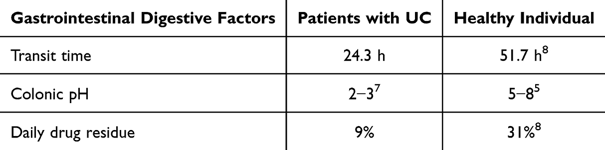 |
Table 1 Comparison of Gastrointestinal Digestion Factors Between UC Patients and Healthy Individuals |
Effect of Intestinal Mucosal Barrier on Oral Medications in UC Patients
The intestinal mucosal barrier is an important intestinal defense system, which is currently classified into four categories: biological, chemical, mechanical and immune barriers, the first three of which act as natural barriers to protect the intestinal epithelial layer from a variety of external stimuli, thereby regulating the balance between mucosal immunity and external stimuli, and hindering the absorption and utilization of orally administered medications in the process.7 The gut is a complex microbial ecosystem where gut microbiota co-evolves with the host and form mutual relationships. Through a series of bacterial enzymes such as polysaccharide enzymes, glycosidase, protease, and peptidase, they break down large molecular polymers.9 This process inevitably affects the metabolism and absorption of oral drugs in the human body. The intestinal mucus layer serves as a robust intestinal barrier, with mucin 2 (MUC2) being the most critical component. MUC2 molecules are interconnected via disulfide bonds and linked to the glycocalyx of the intestinal epithelium, forming a protein network within the mucus layer.10 The average pore size of this network is approximately 200 nm, functioning as a size-exclusion filter, meaning larger particles cannot penetrate the mucus layer or intestinal epithelium;11 simultaneously, the mucus layer interacts with macromolecules through non-covalent forces such as hydrogen bonds, van der Waals forces, hydrophobic forces, and electrostatic forces, limiting the diffusion of most foreign substances, including drugs;12 Furthermore, the strong and sustained secretory capacity of goblet cells confers a high turnover rate on the intestinal mucus layer,13 making it difficult for drugs to reach the surface of intestinal epithelial cells before being expelled, thereby limiting local drug penetration and absorption, resulting in poor therapeutic efficacy. In UC patients, literature reports have described a thinning of the intestinal mucus layer, particularly a reduction in MUC2, a core component of the mucus layer.14 Some viewpoints suggest that this pathological change may facilitate the absorption of orally administered drugs. The intestinal epithelial layer is composed of tight junctions, adherens junctions, desmosomes, and the intestinal epithelial cells they connect. These structures confer selective permeability to the intestinal epithelial layer, functioning as a mechanical barrier between intestinal contents and the internal environment, maintaining balance between nutrition and immunity, and strictly limiting the transport of hydrophilic molecules such as proteins, lipids, and microbial-derived peptides, as well as obstructing the transport of orally administered drugs.15 Similar to the intestinal mucus layer, UC patients often exhibit disruption of the mechanical barrier’s integrity, with extensive local death of intestinal epithelial cells, increased M cell phagocytosis, and disruption of intercellular junctions.16 The open transport channels allow more drugs to pass through, making this an attractive pharmacological target. (Figure 1 Influence of gastrointestinal factors on oral drug administration in patients with UC).
 |
Figure 1 Influence of gastrointestinal factors on oral drug administration in patients with UC. (A) UC shortens the transit time of the colon and makes the pH of the colon more acidic; (B) The colonic mucus layer blocks drugs from passing through the mucus layer and coming into contact with the intestinal epithelium in a size-dependent manner; (C) Negatively charged mucoproteins adsorb positively charged drugs to prevent them from passing through the mucus layer; (D) Tight junctions. The arrow indicates the passage of the drug through the barrier. |
Definition and Nomenclature of PELNs
Extracellular Vehicles (EVs) are defined by the International Society for Extracellular Vesicles (ISEV) as “particles naturally released by cells, defined by a lipid bilayer, that do not replicate, ie, they do not contain a functional nucleus”.17 EVs can be released by all types of cells, including plants, animals and microorganisms, and have a particle size of approximately 30–150 nm.18 Plant Extracellular Vesicles (PEVs) were first documented by transmission electron microscopy (TEM) in the 1960s,19 but subsequent initial studies of PEVs did not show significant value, and the discovery of PEVs was overshadowed by studies on Mammalian cell-derived Extracellular Vesicles (MEVs), and in the decades that followed, research on PEVs focused on the plants themselves, such as the regulation of plant gene expression and plant resistance to pathogens.19 However, further studies on MEVs revealed shortcomings underneath their use as excellent therapeutic agents and delivery vehicles, such as limited production, potential genetic threats, and immunogenicity; at the same time, with the deepening of the research, in the last decade or so, researchers have recognized the potential of cross-boundary regulation by PEVs, and the researchers’ attention has once again returned to plant-derived vesicles (PDVs), which have been used in the development of the plant. PDVs, which, as desired, have the unique advantages of low immunogenicity, high yield and good biocompatibility as a new form of natural products.20 However, due to the differences in the aims of botanical and biomedical research, studies on PDVs have been divided into different paths, with biomedical research focusing on the therapeutic function of PEVs, abandoning the consideration of “extracellular” sources, and obtaining PEVs by destroying the cells of plant tissues, which has produced nano-vesicles with morphologies and compositions similar to those of extracellular vesicles in plants. This resulted in the production of nanovesicles similar in morphology and composition to plant extracellular vesicles,21 which were named PELNs due to the lack of specific markers, clear biogenesis pathways and characterisation.22 Thus, PELNs are nanoscale vesicles derived from plant cells with a particle size of about 30–500 nm, which are usually produced by fresh plants, but it has also been shown that they can be obtained from dried plants.23
Currently, research on PEVs and PELNs is developing in parallel, with PEVs research focusing on revealing their nature, including biogenesis, identification and characterization, and therefore rigorously separating vesicles from their extracellular environments (leaf plasmalemma ectodomains, root secretions, etc). PELNs research, on the other hand, has explored their application in disease therapy, while optimizing the possibilities for their production and engineering modifications. Despite their significant differences in research pathways and aims, the two will also complement each other and together advance a deeper understanding of PDVs.
Delivery Advantages and Engineering Strategies of PELNs in UC Therapy
Compositional Features and Multidimensional Therapeutic Potential of PELNs
The main components of PELNs include proteins, RNA, lipids and plant bioactive molecules. The protein components of PELNs can be mainly classified into peripheral membrane proteins, transmembrane proteins and intracellular proteins, of which peripheral membrane proteins and transmembrane proteins may be involved in vesicle formation and specific targeting.24 Whereas intracellular proteins account for the majority of the protein composition of PELNs, where cell wall remodeling-related enzymes may provide antipathogenic effects25 and metabolism-related enzymes confer antioxidant capacity.26 Indeed PELNs from diverse sources are extremely rich in plant-specific proteins with great therapeutic potential. However, it is still very difficult to establish a complete protein database of PELNs. On the one hand, there is a significant difference in the number of protein species of PELNs from different plant sources, which can range from dozens to thousands;21,27 on the other hand, different cultivation methods, extraction sites, and extraction methods of the same plant can affect their proteomics results, eg, compared with fruits from conventional agriculture, fruits from organic agriculture can be more effective than fruits from conventional agriculture.28 Fruits from organic agriculture contain higher antioxidant proteins compared to those from conventional agriculture;28 a total of 598 proteins were detected in Arabidopsis thaliana plastid exosome sap PELNs and whole-leaf PELNs, of which only 170 proteins appeared in both sources of PELNs after comparison;29 and another similar study showed that the proteomics of the Arabidopsis thaliana plastid exosome sap PELNs and whole-leaf comparative proteomic study of PELNs, the total number of proteins identified was 787 and 1438, respectively.30
RNAs in PELNs mainly include miRNAs, mRNAs and sRNAs, of which miRNAs are believed to have the role of mediating intercellular communication and even cross-species regulation of gene expression, especially in humans,31 and it has been suggested that one miRNA can target hundreds of mRNAs, so that when the concentration of PELNs reaches a certain baseline level, their contained miRNAs will produce significant regulatory effects.32 In another study, a total of 418 miRNAs were identified in PELNs samples from 11 plant species, with each species containing between 32 and 127 miRNAs, including 26 “frequent” miRNAs, 39 “moderately present” miRNAs, and 353 “rare” miRNAs.33 Conventional wisdom suggests that naked miRNAs are unstable and susceptible to RNase degradation, and the highly variable digestive environment of the gastrointestinal tract makes them difficult to be administered orally. In contrast, the lipid bilayer of PELNs effectively protects miRNAs from RNase and the GI environment;34 further studies have shown that many miRNAs from PELNs maintain specific acid resistance and stability through methylation of the 2′-OH group,35 thereby maintaining long-term activity in the human GI environment. More specifically, studies have demonstrated the ability of orally administered PELNs to effectively regulate the intestinal flora36 and host macrophages37 for the treatment of UC by delivering miRNAs.
In terms of lipid composition, PELNs differ significantly from MEVs in that PELNs are rich in phospholipids and do not contain cholesterol, which is present in MEVs,38 and they mainly include phosphatidylcholine (PC), phosphatidic acid (PA), phosphatidylethanolamine (PE), phosphatidylinositol (PI) and phosphatidylglycerol (PG).39 PELNs from various sources differ in the types and proportions of lipids on the basis of which their function, distribution and uptake propensity are determined, eg, PELNs of orange origin contain about 40% PE, 25% PC, 12% PI and 5% PA,40 whereas PELNs of turmeric origin contain about 42% DGDG, 12% MGDG, 15% PA and 5% PC.41 In terms of effects on the distribution of PELNs PA favored the accumulation of PELNs in the gut, while PC enhanced the transfer of PELNs from the gut to the liver.39 With regard to the propensity of PELNs to be taken up, PA enhances the uptake of ginger-derived PELNs by Porphyromonas42 and Lactobacillus rhamnosus,39 which in turn affects their therapeutic efficacy, whereas PC-enriched PELNs are preferentially taken up by intestinal Ruminalogastroenterococcaceae.39
In addition to the aforementioned biomolecules, PELNs also contain bioactive molecules from homologous plants and are able to deliver them to the body to produce the corresponding biological effects, eg, lemon-derived PELNs contain vitamin C and citrate;43 lemon-grapefruit-derived PELNs contain naringenin;44 and broccoli-derived PELNs contain lycopene sulfone.45 (Figure 2 Compositional features and multidimensional therapeutic potential of PELNs).
 |
Figure 2 Compositional features and multidimensional therapeutic potential of PELNs. The main components of PELNs include proteins, RNA, lipids, and plant bioactive molecules, which give PELNs multidimensional therapeutic effects. |
Natural Advantages and Targeting Mechanisms of Oral Delivery of PELNs
In the design and selection of orally delivered nanomedicines, in addition to therapeutic functionality, factors that need to be considered a priori include safety, stability, biodistribution, biocompatibility and natural targeting.46 Whereas, since plants do not contain zoonotic or human pathogens, PELNs have a safety advantage over MEVs due to their non-immunogenic and innocuous properties,47 as well as the absence of tissue and organ abnormalities in relevant in vivo safety tests in animals.48 More importantly, the excellent physicochemical properties of PELNs, including small particle size, negative charge, lipid membrane and hydrophilic surface, provide them with good mucus penetration, mucosal adhesion, gastrointestinal stability, biocompatibility and natural targeting, which allow PELNs to circumvent most of the physiological obstacles in the gastrointestinal tract and safely target UC lesions. Small particle size facilitates the prolongation of the retention time of PELNs in the intestinal mucus layer, while avoiding their expulsion during mucus layer turnover,4 while both the small particle size and hydrophilic surface facilitate the penetration of PELNs into the intestinal mucus layer,49 which in turn crosses the intestinal mucosal tight junction protein ZO-1.50 The lipid bilayer of PLENs confers a robust stability in the gastrointestinal digestive environment, allowing them to tolerate extreme environments from pH 2.0–8.0 and are resistant to degradation by digestive enzymes, and it has been shown that PELNs are able to maintain stability in simulated digestive fluids by adaptively adjusting potential and particle size,51 thereby maintaining vesicle populations as well as the corresponding targeting and communication functions.50 The lipid membrane and its surface proteins also confer good barrier penetration and biocompatibility to PELNs, giving them the ability to penetrate the blood-brain barrier while being blocked by the placental barrier, thus permitting them to be investigated for neurological-related diseases and avoiding pregnancy-related complications.52,53 The natural targeting of PELNs can in fact be classified into two categories depending on the mechanism, non-specific targeting and specific targeting, the former is a non-specific process based on interaction forces such as electrostatic forces, the negatively charged mucus layer on the surface of the intestinal epithelial cell layer in the UC state is weakened, and at the same time, overexpression of certain positively charged proteins in the intestinal epithelial cells, such as transferrin,54 will be conducive to the negatively charged PELNs its adhesion to the intestinal epithelial cells through electrostatic interaction, PELNs passively approaching the lesion and then being taken up by the relevant cells in the form of giant cytosolic drinking, lattice protein-dependent endocytosis and niche-dependent endocytosis,55 thus exhibiting targeting to tissues and organs; the latter is a receptor-dependent specific active targeting process, whereby certain surface proteins carried by the lipid membranes of the PELNs may act as ligands for receptors with the target site, exhibiting targeting to specific cells, eg, the garlic-derived PELNs carry mannose-specific binding protein II lectin, which specifically binds to the CD98 receptor to achieve active targeting, whereas after removal of all surface proteins on the lipid membrane surface of garlic-derived PELNs, the specific uptake of garlic-derived PELNs by cells carrying the CD98 receptor was significantly reduced.56
Strategies for Engineering PELNs and Therapeutic Potentiation Pathways
As natural products, PELNs are not perfect, and they certainly possess many innate advantages, but usually these advantages are not sufficient to fully meet the needs of scientific research and clinics, and the strengths of the original PELNs in anti-inflammatory,57,58 anti-oxidative stress,59 and digestive stability48,60 often need to be optimized by modern biotechnology, and the engineered modification potential is precisely the biggest advantage of PELNs. The engineering modification of PELNs mainly includes surface modification and drug loading. Since only a few PELNs naturally have suitable surface ligands to achieve specific targeting and meet research and clinical needs, the nonspecific targeting of most PELNs does not afford such a responsibility, but the lipid membranes of PELNs happen to allow for modification according to practical needs.53 Examples include surface modification using folic acid to target folate receptors on intestinal epithelial cells,61 surface modification using lectins to target integrin receptors on the surface of M cells62 and surface modification using mannose to target mannose receptors on the surface of macrophages.63 Drug loading takes advantage of the good safety, stability, penetration, and biocompatibility of PELNs. Chemotherapeutic drugs, anti-inflammatory drugs, miRNAs, siRNAs, DNA expression vectors, and proteins are loaded into the PELNs by co-incubation, ultrasonication, and co-extrusion after incubation, which enhances the stability of the drugs, organ targeting, tissue penetration, uptake by the target cells, release optimization, toxicity reduction and potency enhancement.64 For example, researchers loaded microporous silica nanoparticles loaded with infliximab onto ginger-derived PELNs thereby avoiding their destruction in the gastrointestinal environment, and also achieved colon-targeted delivery and helped infliximab to be released in the lamina propria after penetration of the intestinal epithelium in order to achieve a high accumulation of the drug within the inflammatory lesions of the colon.57 In another study, free siRNA-CD98 and garlic-derived PELNs loaded with siRNA-CD98 were comparatively investigated, and it was found that the former only stayed in the stomach, whereas the latter could be delivered to the stomach, ileum, and colon, and was highly enriched in the ileum versus the colon, significantly decreasing colon CD98 expression and thus controlling the progression of colitis as compared to free siRNA-CD98. The effective dose of garlic-derived PELNs loaded with siRNA-CD98 was reduced by approximately 10,000-fold.48 Another study utilized the mechanism by which PELNs target F4/80+ macrophages in the intestinal lamina propria through microcellular drinking and lattice protein-dependent endocytosis pathways, and loaded methotrexate onto grapefruit-derived PELNs for targeted therapy, which significantly reduced methotrexate’s side effects compared to free methotrexate administration.44
Advances in the Study of PELNs for the Treatment of UC
Original PELNs for the Treatment of UC
Natural products have the unique advantages of multiple components acting on multiple targets and links, good and stable effects, and low toxicity and side effects, and both basic and clinical studies have affirmed their good effects in the prevention and treatment of UC.65 However, due to the special properties of UC as a digestive disease and the limitations of natural product drug dosage forms, the gastrointestinal environment in the state of UC poses a great challenge to orally administered natural products, resulting in drug degradation, malabsorption, nonspecific distribution, and side effect generation, etc. PELNs, as a new technology and new dosage form, provide a new direction of research and a pathway of clinical implementation for the future development of natural products.
The most preliminary studies started with PELNs in their pristine state, and in vivo and in vitro experiments confirmed their therapeutic efficacy and natural delivery advantages for UC models. Although the mechanisms of PELNs for UC treatment vary due to the different plants from which they are derived, mainly through the regulation of intestinal flora, remodeling of macrophage polarization, and anti-oxidative stress,21,36 all of them take advantage of the natural advantages of PELNs, such as digestive tract stability, good biocompatibility, natural active targeting and preferential cellular uptake.41,51 Since most of the early research teams chose easily accessible species in food for natural product sources, such as garlic,36,66 turmeric,41,59,67 ginger,48,51,57,68,69 tea,70,71 etc., the subsequent research teams have continued this choice, and fewer studies have chosen therapeutic UC of typical natural products as a source of extracted PELNs. According to the results of previous basic and clinical studies, plant-extracted PELNs such as Rhizome coptidis72 may have more significant therapeutic effects and natural advantages, and PELNs from different plant sources may exert their natural advantages through different mechanisms, and through in-depth study of PELNs from different plant sources, human beings may have a new understanding of the natural delivery of drugs. (Table 2 Studies related to the treatment of UC with original PELNs).
 |
Table 2 Studies Related to the Treatment of UC with Original PELNs |
Engineering Modified PELNs for UC Treatment
Current research has affirmed the natural therapeutic effects and drug delivery advantages possessed by PELNs and has found that such advantages are somewhat universal across plant species, but the strength of such advantages is often not sufficient to directly address research and clinical needs. With the development of bioengineering techniques, available technologies allow researchers to engineer PELNs at the nanoscale to further enhance various aspects of their strengths to meet research and clinical needs, such as by surface modification of PELNs to enhance their targeting ability,57 by encapsulating drugs into PELNs to enhance their therapeutic effects,69 and by using other materials to encapsulate PELNs to enhance their stability, etc.83 (Table 3 Studies related to engineering modified PELNs for the treatment of UC).
 |
Table 3 Studies Related to Engineering Modified PELNs for the Treatment of UC |
Summary, Prospects and Challenges
In this study, we systematically discussed the special gastrointestinal environment of UC patients and its impact on oral drugs, which significantly reduces the stability, targeting, and efficacy of oral drugs through multiple mechanisms such as shortened drug residence time, extreme pH, enzymatic action, intestinal mucosal barrier, and charge interactions. The UC-associated pathological alterations, such as changes in the charge of the intestinal mucus layer and disruption of the intestinal mucosal barrier, also provide potential opportunities for oral drug delivery, which require precise design to overcome the barriers and optimize the delivery strategies using pathological features. In this study, we also traced the research pathway of PELNs and summarized their unique natural advantages and potential for engineering modification. PELNs achieve therapeutic effects through their core components of proteins, RNAs, lipids, and plant-active molecules, and good safety, stability, barrier penetration, biocompatibility, and natural targeting for oral drug delivery through their unique structural properties, such as small particle sizes, negative charge, and hydrophilic surfaces. The engineering potential of PELNs can enhance the targeting of specific cells through surface modification technology, while drug loading technology can efficiently encapsulate chemotherapeutic drugs, anti-inflammatory drugs, nucleic acids and proteins to reduce toxicity and increase efficacy.
Compared to traditional nanocarriers, PELNs exhibit highly prominent multidimensional therapeutic effects, whereas traditional nanocarriers generally lack direct therapeutic effects or possess only a single therapeutic mechanism. Based on structural differences, traditional nanocarriers can be categorized into inorganic and organic types. The former includes non-metals and metals, while the latter includes liposomes, polymers, and polysaccharides.84 Liposomes85 and polymers86 merely serve protective and drug delivery functions, with the carriers themselves lacking direct therapeutic effects. Non-metallic materials may exhibit single therapeutic effects, such as mesoporous silica with antioxidant stress effects;87 metal-based nanocarriers are believed to possess diverse therapeutic mechanisms, such as gold nanoclusters exhibiting antioxidant stress effects and regulating macrophage polarization.88 Polysaccharides, which are also naturally sourced like PELNs, exhibit even richer therapeutic effects. Direct therapeutic mechanisms include anti-inflammatory,89 antioxidant stress,90 and gut microbiota remodeling,91 while indirect mechanisms include the conversion of polysaccharides into beneficial short-chain fatty acids via enzymatic degradation.92 The therapeutic mechanisms of PELNs from different plant sources encompass multiple pathways, including regulating gut microbiota balance, repairing the mucosal barrier, inhibiting oxidative stress, and reshaping the immune microenvironment. Additionally, PELNs benefit from their unique physicochemical properties and structural characteristics, such as small particle size, negative charge, and hydrophilic surface, which confer advantages for oral drug delivery, including good safety, stability, barrier penetration, biocompatibility, and natural targeting. PELNs may provide an efficient delivery platform for UC treatment and open new avenues for the development of natural medicines.
Current research has confirmed that PELNs demonstrate significant therapeutic efficacy in UC animal models by leveraging their unique natural advantages and potential for engineering modifications. However, the research and clinical translation of PELNs still face numerous challenges and limitations, such as the lack of unified standards for the production and characterization of PELNs, the instability of PELNs in vitro, which makes it difficult to store and transport them efficiently, the lack of long-term clinical trials to prove the safety of PELNs, and the need for further engineering modifications to achieve multi-modal combination therapy with PELNs and other drugs to enhance efficacy. Future research strategies can be developed from the following aspects: ① Standardized production: Different cultivation methods, extraction sites, and extraction methods of the same plant can all affect its material composition. Establishing large-scale production and quality control standards for PELNs to ensure batch-to-batch stability and consistency is extremely important for the widespread research and application of PELNs. ② Enhanced stability: Current views suggest that PELNs are highly fragile in vitro and require storage under stringent conditions, such as −20°C, and should avoid repeated freeze-thaw cycles. Their fragile in vitro stability hinders the large-scale application of PELNs. Exploring storage and transportation conditions for PELNs, identifying suitable PELNs protectants, and achieving efficient storage and transportation of PELNs are critical for their clinical translation; ③ Systematic characterization: Characterization standards for PELNs have not yet been established, and many issues remain unresolved. For example, whether Dynamic Light Scattering (DLS) or Nanoparticle Tracking Analysis (NTA) is more suitable for measuring PELN particle size, whether PELNs from different plant parts require separate characterization, and whether PELNs from the same plant grown under different cultivation methods require separate characterization all require standardized discussion; ④ Advantages mechanism research: The therapeutic mechanisms of PELNs vary depending on the source plant and are not the focus of this study. However, their natural advantages, such as safety, stability, barrier penetration, biocompatibility, and natural targeting, are common to all. By deeply understanding the mechanisms underlying these natural advantages, humanity may gain new insights into drug delivery research; ⑤Engineering modifications: PELNs are an excellent carrier. Exploring the potential for engineering modifications of PELNs, including surface-modified active targeting capabilities and the possibility of combining them with other drugs through loading, may enhance targeting and combination therapy, overcoming the limitations of single therapies and achieving reduced toxicity and enhanced efficacy; ⑥ Safety assessment: Although PELNs are considered free of zoonotic or human pathogens due to their natural plant origin and have demonstrated good biocompatibility and safety in animal and cell experiments, given that PELNs are complex substances containing proteins, RNA, lipids, and plant bioactive molecules, their long-term safety after metabolism in the human body still requires further verification; ⑦ Clinical trials: Given the excellent performance of PELNs as novel therapeutic agents and carriers, as well as the promising efficacy demonstrated in various studies, researchers are highly anticipating the clinical application of PELNs. However, since research on PELNs is still in its early stages, quality control and characterization standards for PELNs have not yet been standardized, and safety assessments remain incomplete. Therefore, clinical trials of PELNs are still in the early stages and progressing slowly, with only four types of PELNs derived from lemon, ginger, aloe vera, and grape registered for clinical trials. The effect of lemon-derived PELNs on cardiovascular metabolic risk factors in patients with metabolic syndrome (NCT04698447); The ability of ginger and aloe vera-derived PELNs to alleviate insulin resistance and chronic inflammation in patients with polycystic ovary syndrome (PCOS) (NCT03493984); The ability of PELNs to deliver curcumin to normal and malignant colon tissue (NCT01294072) A preliminary clinical trial investigating whether ginger-derived PELNs, with or without curcumin, can alleviate symptoms of inflammatory bowel disease (IBD) (NCT04879810) Exploring the efficacy of grape-derived PELNs in alleviating oral mucositis in head and neck cancer patients following combined chemotherapy and radiotherapy (NCT01668849). Before advancing PELNs into large-scale clinical trials, we should first establish foundational work in this field, such as standardizing quality control and characterization protocols, and conducting comprehensive long-term safety assessments of PELNs.
The authors do not wish to compare PELNs with existing drug delivery methods; rather, this study tends to describe the potential that PELNs hold: i) PELNs represent a drug delivery method with high bioavailability; ii) PELNs are free of possible side effects at the organ and tissue level throughout the systemic range; iii) PELNs may contain highly effective therapeutic substances unknown to humans; iv) The natural advantages of PELNs may serve as a model for human beings to learn from nature, and by understanding their mechanisms in depth, and then replicating and enhancing them using existing technologies, human beings may construct even better drugs.
Funding
The author(s) declare financial support was received for the research, authorship, and/or publication of this article. This study was funded by the National Natural Science Foundation of China (No. 82374426), the National College Students’ Innovation and Entrepreneurship Training Program Key Support Area Project (S202410541019), the Hunan Provincial Natural Science Foundation Project (2023JJ60044), the Hunan Provincial Department of Education Scientific Research Fund Project (24A0274) and the Hunan Provincial Administration of Traditional Chinese Medicine Project (B2023079); and the Domestic First-class Construction Discipline of Chinese Medicine in Hunan University of Chinese Medicine.
Disclosure
The authors report no conflicts of interest in this work.
References
1. Le Berre C, Honap S, Peyrin-Biroulet L. Ulcerative colitis. Lancet. 2023;402(10401):571–584. doi:10.1016/S0140-6736(23)00966-2
2. Moroz E, Matoori S, Leroux JC. Oral delivery of macromolecular drugs: where we are after almost 100years of attempts. Adv Drug Deliv Rev. 2016;101:108–121. doi:10.1016/j.addr.2016.01.010
3. Bassotti G, Antonelli E, Villanacci V, et al. Gastrointestinal motility disorders in inflammatory bowel diseases. World J Gastroenterol. 2014;20(1):37–44. doi:10.3748/wjg.v20.i1.37
4. Stubbs JB, Valenzuela GA, Stubbs CC, et al. A noninvasive scintigraphic assessment of the colonic transit of nondigestible solids in man. J Nucl Med. 1991;32(7):1375–1381.
5. Koziolek M, Grimm M, Becker D, et al. Investigation of pH and Temperature Profiles in the GI Tract of Fasted Human Subjects Using the Intellicap (®) System. J Pharm Sci. 2015;104(9):2855–2863. doi:10.1002/jps.24274
6. Kiela PR, Ghishan FK. Physiology of Intestinal Absorption and Secretion. Best Pract Res Clin Gastroenterol. 2016;30(2):145–159. doi:10.1016/j.bpg.2016.02.007
7. Fallingborg J, Christensen LA, Jacobsen BA, et al. Very low intraluminal colonic pH in patients with active ulcerative colitis. Dig Dis Sci. 1993;38(11):1989–1993. doi:10.1007/BF01297074
8. Hebden JM, Blackshaw PE, Perkins AC, et al. Limited exposure of the healthy distal colon to orally-dosed formulation is further exaggerated in active left-sided ulcerative colitis. Aliment Pharmacol Ther. 2000;14(2):155–161. doi:10.1046/j.1365-2036.2000.00697.x
9. Williams BA, Grant LJ, Gidley MJ, et al. Gut Fermentation of Dietary Fibres: physico-Chemistry of Plant Cell Walls and Implications for Health. Int J Mol Sci. 2017;18(10):2203. doi:10.3390/ijms18102203
10. Wu L, Shan W, Zhang Z, et al. Engineering nanomaterials to overcome the mucosal barrier by modulating surface properties. Adv Drug Deliv Rev. 2018;124:150–163. doi:10.1016/j.addr.2017.10.001
11. Bhattacharjee S, Mahon E, Harrison SM, et al. Nanoparticle passage through porcine jejunal mucus: microfluidics and rheology. Nanomedicine. 2017;13(3):863–873. doi:10.1016/j.nano.2016.11.017
12. Zhu Q, Chen Z, Paul PK, et al. Oral delivery of proteins and peptides: challenges, status quo and future perspectives. Acta Pharm Sin B. 2021;11(8):2416–2448. doi:10.1016/j.apsb.2021.04.001
13. Lai SK, Wang YY, Hanes J. Mucus-penetrating nanoparticles for drug and gene delivery to mucosal tissues. Adv Drug Deliv Rev. 2009;61(2):158–171. doi:10.1016/j.addr.2008.11.002
14. Ning H, Liu J, Tan J, et al. The role of the Notch signalling pathway in the pathogenesis of ulcerative colitis: from the perspective of intestinal mucosal barrier. Front Med. 2024;10:1333531. doi:10.3389/fmed.2023.1333531
15. Suzuki T. Regulation of intestinal epithelial permeability by tight junctions. Cell Mol Life Sci. 2013;70:63.
16. Mulvaney P. Nanoscience vs Nanotechnology—Defining the Field. ACS Nano. 2015;9(3):2215–2217. doi:10.1021/acsnano.5b01418
17. Théry C, Witwer KW, Aikawa E, et al. Minimal information for studies of extracellular vesicles 2018 (MISEV2018): a position statement of the International Society for Extracellular Vesicles and update of the MISEV2014 guidelines. J Extracell Vesicles. 2018;7(1):1535750. doi:10.1080/20013078.2018.1535750
18. van Niel G, D’Angelo G, Raposo G. Shedding light on the cell biology of extracellular vesicles. Nat Rev Mol Cell Biol. 2018;19(4):213–228. doi:10.1038/nrm.2017.125
19. Halperin W, Jensen WA. Ultrastructural changes during growth and embryogenesis in carrot cell cultures. J Ultrastruct Res. 1967;18(3–4):428–443. doi:10.1016/S0022-5320(67)80128-X
20. Wang X, Xin C, Zhou Y, et al. Plant-Derived Vesicle-like Nanoparticles: the Next-Generation Drug Delivery Nanoplatforms. Pharmaceutics. 2024;16(5):588. doi:10.3390/pharmaceutics16050588
21. Ju S, Mu J, Dokland T, et al. Grape exosome-like nanoparticles induce intestinal stem cells and protect mice from DSS-induced colitis. Mol Ther. 2013;21(7):1345–1357. doi:10.1038/mt.2013.64
22. Ly NP, Han HS, Kim M, et al. Plant-derived nanovesicles: current understanding and applications for cancer therapy. Bioact Mater. 2022;22:365–383. doi:10.1016/j.bioactmat.2022.10.005
23. Woith E, Melzig MF. Extracellular Vesicles from Fresh and Dried Plants-Simultaneous Purification and Visualization Using Gel Electrophoresis. Int J Mol Sci. 2019;20(2):357. doi:10.3390/ijms20020357
24. Song H, Canup BSB, Ngo VL, et al. Internalization of Garlic-Derived Nanovesicles on Liver Cells is Triggered by Interaction With CD98. ACS Omega. 2020;5(36):23118–23128. doi:10.1021/acsomega.0c02893
25. Liu G, Kang G, Wang S, et al. Extracellular Vesicles: emerging Players in Plant Defense Against Pathogens. Front Plant Sci. 2021;12:757925. doi:10.3389/fpls.2021.757925
26. Garaeva L, Kamyshinsky R, Kil Y, et al. Delivery of functional exogenous proteins by plant-derived vesicles to human cells in vitro. Sci Rep. 2021;11(1):6489. doi:10.1038/s41598-021-85833-y
27. Cao M, Yan H, Han X, et al. Ginseng-derived nanoparticles alter macrophage polarization to inhibit melanoma growth. J Immunother Cancer. 2019;7(1):326. doi:10.1186/s40425-019-0817-4
28. Logozzi M, Di Raimo R, Mizzoni D, et al. Nanovesicles from Organic Agriculture-Derived Fruits and Vegetables: characterization and Functional Antioxidant Content. Int J Mol Sci. 2021;22(15):8170. doi:10.3390/ijms22158170
29. Rutter BD, Innes RW. Extracellular Vesicles Isolated from the Leaf Apoplast Carry Stress-Response Proteins. Plant Physiol. 2017;173(1):728–741. doi:10.1104/pp.16.01253
30. Liu Y, Wu S, Koo Y, et al. Characterization of and isolation methods for plant leaf nanovesicles and small extracellular vesicles. Nanomedicine. 2020;29:102271. doi:10.1016/j.nano.2020.102271
31. Redis RS, Calin S, Yang Y, et al. Cell-to-cell miRNA transfer: from body homeostasis to therapy. Pharmacol Ther. 2012;136(2):169–174. doi:10.1016/j.pharmthera.2012.08.003
32. Xu T, Zhu Y, Lin Z, et al. Evidence of Cross-Kingdom Gene Regulation by Plant MicroRNAs and Possible Reasons for Inconsistencies. J Agric Food Chem. 2024;72(9):4564–4573. doi:10.1021/acs.jafc.3c09097
33. Xiao J, Feng S, Wang X, et al. Identification of exosome-like nanoparticle-derived microRNAs from 11 edible fruits and vegetables. PeerJ. 2018;6:e5186. doi:10.7717/peerj.5186
34. Dad HA, Gu TW, Zhu AQ, et al. Plant Exosome-like Nanovesicles: emerging Therapeutics and Drug Delivery Nanoplatforms. Mol Ther. 2021;29(1):13–31. doi:10.1016/j.ymthe.2020.11.030
35. Yu B, Yang Z, Li J, et al. Methylation as a crucial step in plant microRNA biogenesis. Science. 2005;307(5711):932–935. doi:10.1126/science.1107130
36. Wang X, Liu Y, Dong X, et al. peu-MIR2916-p3-enriched garlic exosomes ameliorate murine colitis by reshaping gut microbiota, especially by boosting the anti-colitic Bacteroides thetaiotaomicron. Pharmacol Res. 2024;200:107071. doi:10.1016/j.phrs.2024.107071
37. Yan L, Cao Y, Hou L, et al. Ginger exosome-like nanoparticle-derived miRNA therapeutics: a strategic inhibitor of intestinal inflammation. J Adv Res. 2025;69:1–15. doi:10.1016/j.jare.2024.04.001
38. Yi Q, Xu Z, Thakur A, et al. Current understanding of plant-derived exosome-like nanoparticles in regulating the inflammatory response and immune system microenvironment. Pharmacol Res. 2023;190:106733. doi:10.1016/j.phrs.2023.106733
39. Teng Y, Ren Y, Sayed M, et al. Plant-Derived Exosomal MicroRNAs Shape the Gut Microbiota. Cell Host Microbe. 2018;24(5):637–652. doi:10.1016/j.chom.2018.10.001
40. Berger E, Colosetti P, Jalabert A, et al. Use of Nanovesicles from Orange Juice to Reverse Diet-Induced Gut Modifications in Diet-Induced Obese Mice. Mol Ther Methods Clin Dev. 2020;18:880–892. doi:10.1016/j.omtm.2020.08.009
41. Liu C, Yan X, Zhang Y, et al. Oral administration of turmeric-derived exosome-like nanovesicles with anti-inflammatory and pro-resolving bioactions for murine colitis therapy. J Nanobiotechnology. 2022;20(1):206. doi:10.1186/s12951-022-01421-w
42. Sundaram K, Miller DP, Kumar A, et al. Plant-Derived Exosomal Nanoparticles Inhibit Pathogenicity of Porphyromonas gingivalis. iScience. 2019;21:308–327. doi:10.1016/j.isci.2019.10.032
43. Baldini N, Torreggiani E, Roncuzzi L, et al. Exosome-like Nanovesicles Isolated from Citrus limon L. Exert Antioxidative Effect. Curr Pharm Biotechnol. 2018;19(11):877–885. doi:10.2174/1389201019666181017115755
44. Wang B, Zhuang X, Deng ZB, et al. Targeted drug delivery to intestinal macrophages by bioactive nanovesicles released from grapefruit. Mol Ther. 2014;22(3):522–534. doi:10.1038/mt.2013.190
45. Deng Z, Rong Y, Teng Y, et al. Broccoli-Derived Nanoparticle Inhibits Mouse Colitis by Activating Dendritic Cell AMP-Activated Protein Kinase. Mol Ther. 2017;25(7):1641–1654. doi:10.1016/j.ymthe.2017.01.025
46. De Jong WH, Borm PJ. Drug delivery and nanoparticles: applications and hazards. Int J Nanomed. 2008;3:133–149. doi:10.2147/IJN.S596
47. Chen Q, Lai H. Plant-derived virus-like particles as vaccines. Hum Vaccin Immunother. 2013;9(1):26–49. doi:10.4161/hv.22218
48. Zhang M, Wang X, Han MK, et al. Oral administration of ginger-derived nanolipids loaded with siRNA as a novel approach for efficient siRNA drug delivery to treat ulcerative colitis. Nanomedicine (Lond). 2017;12(16):1927–1943. doi:10.2217/nnm-2017-0196
49. Kim J, Li S, Zhang S, et al. Plant-derived exosome-like nanoparticles and their therapeutic activities. Asian J Pharm Sci. 2022;17(1):53–69. doi:10.1016/j.ajps.2021.05.006
50. Hwang JH, Park YS, Kim HS, et al. Yam-derived exosome-like nanovesicles stimulate osteoblast formation and prevent osteoporosis in mice. J Control Release. 2023;355:184–198. doi:10.1016/j.jconrel.2023.01.071
51. Zhang M, Viennois E, Prasad M, et al. Edible ginger-derived nanoparticles: a novel therapeutic approach for the prevention and treatment of inflammatory bowel disease and colitis-associated cancer. Biomaterials. 2016;101:321–340. doi:10.1016/j.biomaterials.2016.06.018
52. Zhuang X, Teng Y, Samykutty A, et al. Grapefruit-derived Nanovectors Delivering Therapeutic miR17 Through an Intranasal Route Inhibit Brain Tumor Progression. Mol Ther. 2016;24(1):96–105. doi:10.1038/mt.2015.188
53. Wang Q, Zhuang X, Mu J, et al. Delivery of therapeutic agents by nanoparticles made of grapefruit-derived lipids. Nat Commun. 2013;4(1):1867. doi:10.1038/ncomms2886
54. Tirosh B, Khatib N, Barenholz Y, et al. Transferrin as a luminal target for negatively charged liposomes in the inflamed colonic mucosa. Mol Pharm. 2009;6(4):1083–1091. doi:10.1021/mp9000926
55. Xu S, Yang Q, Wang R, et al. Genetically engineered pH-responsive silk sericin nanospheres with efficient therapeutic effect on ulcerative colitis. Acta Biomater. 2022;144:81–95. doi:10.1016/j.actbio.2022.03.012
56. Canup BS, Song H, Le Ngo V, et al. CD98 siRNA-loaded nanoparticles decrease hepatic steatosis in mice. Dig Liver Dis. 2017;49(2):188–196. doi:10.1016/j.dld.2016.11.008
57. Mao Y, Han M, Chen C, et al. A biomimetic nanocomposite made of a ginger-derived exosome and an inorganic framework for high-performance delivery of oral antibodies. Nanoscale. 2021;13(47):20157–20169. doi:10.1039/D1NR06015E
58. Zhang M, Xu X, Su L, et al. Oral administration of Sophora Flavescens-derived exosomes-like nanovesicles carrying CX5461 ameliorates DSS-induced colitis in mice. J Nanobiotechnology. 2024;22(1):607. doi:10.1186/s12951-024-02856-z
59. Lu G, Lu S, Dai H, et al. Engineered Turmeric-Derived Nanovesicles for Ulcerative Colitis Therapy by Attenuating Oxidative Stress and Alleviating Inflammation. Mol Pharm. 2025;22(4):2159–2167. doi:10.1021/acs.molpharmaceut.4c01328
60. Tang X, Wang K, Liu Z, et al. Functional chitosan/HP-β-CD hydrogel for targeted co-delivery of Rhubarb-derived nanovesicles and kaempferol for alleviating ulcerative colitis. Carbohydr Polym. 2025;352:123206. doi:10.1016/j.carbpol.2024.123206
61. Jain S, Rathi VV, Jain AK, et al. Folate-decorated PLGA nanoparticles as a rationally designed vehicle for the oral delivery of insulin. Nanomedicine (Lond). 2012;7(9):1311–1337. doi:10.2217/nnm.12.31
62. Zhang N, Ping Q, Huang G, et al. Lectin-modified solid lipid nanoparticles as carriers for oral administration of insulin. Int J Pharm. 2006;327(1–2):153–159. doi:10.1016/j.ijpharm.2006.07.026
63. Wang C, Guo Z, Liang J, et al. An oral delivery vehicle based on konjac glucomannan acetate targeting the colon for inflammatory bowel disease therapy. Front Bioeng Biotechnol. 2022;10:1025155. doi:10.3389/fbioe.2022.1025155
64. Chen X, Ji S, Yan Y, et al. Engineered Plant-Derived Nanovesicles Facilitate Tumor Therapy: natural Bioactivity Plus Drug Controlled Release Platform. Int J Nanomed. 2023;18:4779–4804. doi:10.2147/IJN.S413831
65. Long D, Mao C, Zhang W, et al. Natural products for the treatment of ulcerative colitis: focus on the JAK/STAT pathway. Front Immunol. 2025;16:1538302. doi:10.3389/fimmu.2025.1538302
66. Zhu Z, Liao L, Gao M, et al. Garlic-derived exosome-like nanovesicles alleviate dextran sulphate sodium-induced mouse colitis via the TLR4/MyD88/NF-κB pathway and gut microbiota modulation. Food Funct. 2023;14(16):7520–7534. doi:10.1039/D3FO01094E
67. Gao C, Zhou Y, Chen Z, et al. Turmeric-derived nanovesicles as novel nanobiologics for targeted therapy of ulcerative colitis. Theranostics. 2022;12(12):5596–5614. doi:10.7150/thno.73650
68. Yan L, Cao Y, Hou L, et al. Ginger exosome-like nanoparticle-derived miRNA therapeutics: a strategic inhibitor of intestinal inflammation. J Adv Res. 2024;S2090-1232:00130–00139.
69. Huang S, Zhang M, Li X, et al. Formulation, characterization, and evaluation of curcumin-loaded ginger-derived nanovesicles for anti-colitis activity. J Pharm Anal. 2024;14(12):101014. doi:10.1016/j.jpha.2024.101014
70. Zu M, Xie D, Canup BSB, et al. ‘Green’ nanotherapeutics from tea leaves for orally targeted prevention and alleviation of colon diseases. Biomaterials. 2021;279:121178. doi:10.1016/j.biomaterials.2021.121178
71. Luo T, Hou L, Cao Y, et al. Tea Extracellular Vesicle-Derived MicroRNAs Contribute to Alleviate Intestinal Inflammation by Reprogramming Macrophages. J Agric Food Chem. 2025;73(11):6745–6757. doi:10.1021/acs.jafc.5c01990
72. Zhu C, Li K, Peng XX, et al. Berberine a traditional Chinese drug repurposing: its actions in inflammation-associated ulcerative colitis and cancer therapy. Front Immunol. 2022;13:1083788. doi:10.3389/fimmu.2022.1083788
73. Kim J, Zhang S, Zhu Y, et al. Amelioration of colitis progression by ginseng-derived exosome-like nanoparticles through suppression of inflammatory cytokines. J Ginseng Res. 2023;47(5):627–637. doi:10.1016/j.jgr.2023.01.004
74. Wang F, Yuan M, Shao C, et al. Momordica charantia-Derived Extracellular Vesicles Provide Antioxidant Protection in Ulcerative Colitis. Molecules. 2023;28(17):6182. doi:10.3390/molecules28176182
75. Zhu MZ, Xu HM, Liang YJ, et al. Edible exosome-like nanoparticles from portulaca oleracea L mitigate DSS-induced colitis via facilitating double-positive CD4CD8T cells expansion. J Nanobiotechnology. 2023;21(1):309. doi:10.1186/s12951-023-02065-0
76. Tinnirello V, Zizzo MG, Conigliaro A, et al. Industrial-produced lemon nanovesicles ameliorate experimental colitis-associated damages in rats via the activation of anti-inflammatory and antioxidant responses and microbiota modification. Biomed Pharmacother. 2024;174:116514. doi:10.1016/j.biopha.2024.116514
77. Li JH, Xu J, Huang C, et al. Houttuynia cordata-Derived Exosome-Like Nanoparticles Mitigate Colitis in Mice via Inhibition of the NLRP3 Signaling Pathway and Modulation of the Gut Microbiota. Int J Nanomed. 2024;19:13991–14018. doi:10.2147/IJN.S493434
78. Lu Y, Xu J, Tang R, et al. Edible pueraria lobata-derived exosome-like nanovesicles ameliorate dextran sulfate sodium-induced colitis associated lung inflammation through modulating macrophage polarization. Biomed Pharmacother. 2024;170:116098. doi:10.1016/j.biopha.2023.116098
79. Sriwastva MK, Deng ZB, Wang B, et al. Exosome-like nanoparticles from Mulberry bark prevent DSS-induced colitis via the AhR/COPS8 pathway. EMBO Rep. 2022;23(3):e53365. doi:10.15252/embr.202153365
80. Choi SH, Eom JY, Kim HJ, et al. Aloe-derived nanovesicles attenuate inflammation and enhance tight junction proteins for acute colitis treatment. Biomater Sci. 2023;11:5490–5501. doi:10.1039/D3BM00591G
81. Kang M, Kang M, Lee J, et al. Allium tuberosum -derived nanovesicles with anti-inflammatory properties prevent DSS-induced colitis and modify the gut microbiome. Food Funct. 2024;15(14):7641–7657. doi:10.1039/D4FO01366B
82. Li Y, Shao S, Zhou Y, et al. Oral administration of Folium Artemisiae Argyi-derived exosome-like nanovesicles can improve ulcerative colitis by regulating intestinal microorganisms. Phytomedicine. 2025;137:156376. doi:10.1016/j.phymed.2025.156376
83. Weng L, Zhao M, Zhang Y, et al. American ginseng vesicles loaded hyaluronic acid hydrogel for ulcerative colitis. Int J Biol Macromol. 2025;288:138565. doi:10.1016/j.ijbiomac.2024.138565
84. Wang H, Zhou F, Shen M, et al. Classification of Nanomaterial Drug Delivery Systems for Inflammatory Bowel Disease. Int J Nanomed. 2025;20:1383–1399. doi:10.2147/IJN.S502546
85. Xian S, Zhu J, Wang Y, et al. Oral liposomal delivery of an activatable budesonide prodrug reduces colitis in experimental mice. Drug Deliv. 2023;30(1):2183821. doi:10.1080/10717544.2023.2183821
86. Wang CJ, Ko GR, Lee YY, et al. Polymeric DNase-I nanozymes targeting neutrophil extracellular traps for the treatment of bowel inflammation. Nano Converg. 2024;11(1):6. doi:10.1186/s40580-024-00414-9
87. Li W, Li Y, Liu Z, et al. Hierarchical structured and programmed vehicles deliver drugs locally to inflamed sites of intestine. Biomaterials. 2018;185:322–332. doi:10.1016/j.biomaterials.2018.09.024
88. Lu C, Xue L, Luo K, et al. Colon-Accumulated Gold Nanoclusters Alleviate Intestinal Inflammation and Prevent Secondary Colorectal Carcinogenesis via Nrf2-Dependent Macrophage Reprogramming. ACS Nano. 2023;17(18):18421–18432. doi:10.1021/acsnano.3c06025
89. Yang T, Jia M, Meng J, et al. Immunomodulatory activity of polysaccharide isolated from Angelica sinensis. Int J Biol Macromol. 2006;39(4–5):179–184. doi:10.1016/j.ijbiomac.2006.02.013
90. Aldini R, Micucci M, Cevenini M, et al. Antiinflammatory effect of phytosterols in experimental murine colitis model: prevention, induction, remission study. PLoS One. 2014;9(9):e108112. doi:10.1371/journal.pone.0108112
91. Wong C, Harris PJ, Ferguson LR. Potential Benefits of Dietary Fibre Intervention in Inflammatory Bowel Disease. Int J Mol Sci. 2016;17(6):919. doi:10.3390/ijms17060919
92. Nie Y, Lin Q, Luo F. Effects of Non-Starch Polysaccharides on Inflammatory Bowel Disease. Int J Mol Sci. 2017;18(7):1372. doi:10.3390/ijms18071372
 © 2025 The Author(s). This work is published and licensed by Dove Medical Press Limited. The
full terms of this license are available at https://www.dovepress.com/terms
and incorporate the Creative Commons Attribution
- Non Commercial (unported, 4.0) License.
By accessing the work you hereby accept the Terms. Non-commercial uses of the work are permitted
without any further permission from Dove Medical Press Limited, provided the work is properly
attributed. For permission for commercial use of this work, please see paragraphs 4.2 and 5 of our Terms.
© 2025 The Author(s). This work is published and licensed by Dove Medical Press Limited. The
full terms of this license are available at https://www.dovepress.com/terms
and incorporate the Creative Commons Attribution
- Non Commercial (unported, 4.0) License.
By accessing the work you hereby accept the Terms. Non-commercial uses of the work are permitted
without any further permission from Dove Medical Press Limited, provided the work is properly
attributed. For permission for commercial use of this work, please see paragraphs 4.2 and 5 of our Terms.
Recommended articles
The Construction Strategy of Curcumin Nanomedicine Delivery System and Its Application in the Treatment of Ulcerative Colitis
Ning H, Huang X, Lin X, Sun Q, Zheng Y, Deng N, Xu Y
International Journal of Nanomedicine 2025, 20:15135-15166
Published Date: 16 December 2025