
Back to Journals » Journal of Inflammation Research » Volume 19

The Association of C-Reactive Protein-Triglyceride Glucose Index with Major Adverse Cardiovascular Events in Patients with Acute Myocardial Infarction
Authors Wang T, Zhang G, Li Q, Liu Y, Zhao T, Che W, Xia J
Received 9 March 2026
Accepted for publication 18 May 2026
Published 30 May 2026 Volume 2026:19 606153
DOI https://doi.org/10.2147/JIR.S606153
Checked for plagiarism Yes
Review by Single anonymous peer review
Peer reviewer comments 2
Editor who approved publication: Dr Qing Lin
Tao Wang, Guoheng Zhang, Qinxue Li, Yayun Liu, Tao Zhao, Wenjing Che, Jinggang Xia
Department of Cardiology, Xuanwu Hospital, Capital Medical University, Beijing, 100053, People’s Republic of China
Correspondence: Jinggang Xia, Email [email protected]
Background: The C-reactive protein-triglyceride glucose index (CTI), a novel marker that integrates metabolic dysfunction and inflammation, has an uncertain prognostic value for predicting major adverse cardiovascular events (MACEs) after acute myocardial infarction (AMI).
Methods: This retrospective study enrolled 578 patients with AMI admitted to Xuanwu Hospital from 2019 to 2022. All patients were categorized according to tertiles of CTI values. Receiver operating characteristic (ROC) curves analysis and restricted cubic spline (RCS) were applied to explore the relationships of CTI, triglyceride-glucose (TyG) index, and high-sensitivity C-reactive protein (hs-CRP) with MACEs. Furthermore, the association between CTI and MACEs was evaluated using Kaplan-Meier curves and Cox proportional hazards regression models, with subgroup analyses assessing this relationship across various clinical conditions.
Results: During a follow-up period with a median time of 34 months, 69 patients experienced MACEs, and higher CTI tertiles were associated with a greater cumulative risk of MACEs (Log rank test, p = 0.003). RCS analysis showed a positive linear association between CTI and MACEs risk (p-overall = 0.017). Compared with the TyG index (AUC = 0.562, 95% CI: 0.488– 0.636) and hs-CRP (AUC = 0.596, 95% CI: 0.530– 0.663), CTI demonstrated the highest AUC (0.612, 95% CI: 0.541– 0.682). Importantly, the addition of CTI to the baseline model provided significant incremental prognostic value, with significant improvements in both net reclassification improvement (NRI) and integrated discrimination improvement (IDI), whereas neither the TyG index nor hs-CRP yielded significant improvements. Multivariate Cox regression analysis confirmed that the highest CTI tertile had a 3.09-fold increased risk of MACEs compared to the lowest tertile (p < 0.001) in the fully adjusted model, and the association remained stable across subgroups.
Conclusion: The CTI is a more promising indicator for predicting MACEs in AMI patients than the TyG index or hs-CRP alone. By integrating the core risk factors of inflammation and insulin resistance, CTI may provide additional value for post-discharge risk stratification, helping to identify high-risk patients who may benefit from precise management.
Keywords: acute myocardial infarction, C-reactive protein-triglyceride glucose index, high-sensitivity C-reactive protein, triglyceride glucose index
Introduction
Acute myocardial infarction (AMI), widely recognized as the most severe form of ischemic heart disease, remains one of the leading causes of global mortality and is a major contributor to disability.1 Although advances in reperfusion therapies, together with improvements in regional coordination of care, have substantially decreased acute-phase mortality in patients with AMI, the burden of extended adverse clinical events remains considerable over time.2,3 Therefore, identifying and managing modifiable risk factors holds substantial importance in the long-term prognosis of AMI patients.
Among the complex pathophysiological mechanisms of AMI, insulin resistance (IR) is recognized as one of the core metabolic disorders driving atherosclerotic progression.4,5 Multiple investigations have demonstrated a significant association between IR and increased incidence of several cardiovascular events.6–9 The triglyceride-glucose (TyG) index, composed of triglyceride (TG) and fasting blood glucose (FBG), is a well-validated indicator of IR and shows a strong association with adverse cardiovascular outcomes.10–13 It is an easily obtainable and inexpensive tool that is anticipated to offer useful information for the clinical management of cardiovascular disease (CVD).
In addition to IR, recent findings have increasingly suggested that inflammation is a crucial factor driving recurrent cardiovascular events and poor outcomes after AMI.14–16 High-sensitivity C-reactive protein (hs-CRP) concentration is among the most recognized and widely utilized biomarkers available for detecting low-grade systemic inflammation.17 Elevated hs-CRP levels have been identified across multiple studies as a strong predictor of recurrent cardiovascular events and mortality following AMI.18,19 Based on the concept of integrated systemic risk and the recognition that inflammation and IR jointly promote atherosclerotic progression, researchers have increasingly explored the value of assessing inflammatory markers together with the TyG index. The C-reactive protein–triglyceride glucose index (CTI) was first described in 2022 as a potential biomarker capable of predicting survival in patients with malignancies.20 The index was created to provide a combined assessment of systemic inflammatory activity and IR severity. Previous studies have suggested that CTI may have predictive value for the incidence of CVD.21,22 However, CTI is a relatively novel index, and its external validation across different cardiovascular populations remains limited. Moreover, AMI is a multifactorial disease, and residual cardiovascular risk cannot be fully explained by any single pathway alone. Recent evidence suggests that plaque characteristics and coronary physiology may also provide complementary prognostic information.23 These findings further support the concept of integrated risk assessment after AMI and underscore the need to explore composite indices that can more comprehensively capture risk.
Our study first compared CTI and its two constituent indices in their ability to predict adverse cardiovascular events in patients with AMI. In addition, we conducted further analyses to verify the robustness and consistency of the observed relationship between CTI and the occurrence of major adverse cardiovascular events (MACEs).
Materials and Methods
Study Design and Participants
For this study, we enrolled 578 patients with AMI who presented at Xuanwu Hospital of Capital Medical University from 2019 to 2022. We analyzed the previous day’s indicators for patients who were in stable condition and prepared for discharge. The diagnosis of AMI was determined according to the consensus document outlining the contemporary definitions of myocardial infarction.24 The exclusion criteria were as follows: (1) missing data related to indicators, including hs-CRP, TG, and FBG; (2) severe conditions, including severe hepatic (alanine aminotransferase (ALT) or aspartate aminotransferase (AST) > 5.0 × the upper limit of normal) or renal (Creatinine > 3.0 × the upper limit of normal) dysfunction, critical structural heart disease requiring intervention, infectious diseases, bleeding predisposition, and cancer; and (3) previous coronary artery bypass grafting (CABG). Participants were categorized into one of three equal groups depending on their CTI levels: T1: CTI ≤ 9.06; T2: 9.06 < CTI ≤ 9.79; and T3: CTI > 9.79 (Figure 1). The studies involving humans were approved by Xuanwu Hospital Ethics Committee under document No. 2022-129. This investigation was conducted in full compliance with the ethical standards set forth in the Declaration of Helsinki. This study was reported in accordance with the RECORD (REporting of studies Conducted using Observational Routinely-collected Data) guidelines to promote transparency and completeness in the reporting of observational research.
 |
Figure 1 Flowchart of Patient Enrollment. Abbreviations: CTI, C-reactive protein-triglyceride glucose index; CABG, coronary artery bypass grafting. |
Data Collection and Definitions
Information on patient demographics, past medical conditions, laboratory indices, echocardiographic parameters, and coronary angiographic findings was obtained from the digital patient record system by qualified physicians. All participants provided fasting blood samples in the morning after an overnight fast, and the samples were analyzed on the same day in a central laboratory. The TyG index was quantified via the following formula: TyG index = ln [TG (mg/dL) × FBG (mg/dL)/2]. Previous cohort studies have established hs-CRP level as a key prognostic indicator in patients with atherosclerotic cardiovascular disease. Therefore, our study employed hs-CRP values to compute the CTI. The CTI was calculated as follows: CTI = TyG index + 0.412 × ln [hs-CRP (mg/L)].
Follow-Up and Endpoint Events
Follow-up was conducted by telephone calls, text messages, and clinical visits. To standardize follow-up procedures and ensure data accuracy, all coordinators received professional training. The primary endpoint assessed in this investigation was the occurrence of composite MACEs, defined as all-cause death, nonfatal myocardial infarction, nonfatal stroke, or unplanned revascularization. Unplanned revascularization refers to a procedure performed due to worsening myocardial ischemia caused by in-stent restenosis, native coronary artery lesion progression, or new territory disease. If a patient experienced more than one endpoint event, only the first occurring event was used for the analysis.
Statistical Analysis
Continuous data based on distribution characteristics were reported as means ± standard deviations or medians (interquartile ranges) and compared using ANOVA or the Kruskal–Wallis test. We utilized restricted cubic splines (RCS) to analyze associations between CTI, TyG index, and hs-CRP with MACEs. Receiver operating characteristic (ROC) curves were used to assess discriminatory ability, and model improvement was evaluated using the net reclassification improvement (NRI) and integrated discrimination improvement (IDI). We plotted Kaplan–Meier survival curves to illustrate the cumulative incidence of MACEs in groups based on CTI tertiles and applied the Log rank test to compare the survival differences between these groups. Given the limited number of MACEs, we adopted a parsimonious modeling strategy for the primary multivariable Cox regression, with covariates pre-specified to include demographic variables and traditional cardiovascular risk factors. To quantify the association between the CTI and study outcomes, three sequential Cox proportional hazards models were constructed: an unadjusted model; a model adjusted for age, gender, and body mass index (BMI); and a fully adjusted model additionally including hypertension, low-density lipoprotein cholesterol (LDL-C) and high-density lipoprotein cholesterol (HDL-C). To address potential confounding from AMI- specific prognostic variables, a sensitivity analysis was performed using an extended Cox model that further adjusted for diabetes, hyperlipidemia, smoking status, GRACE score, left ventricular ejection fraction (LVEF), and percutaneous coronary intervention (PCI). Multicollinearity was assessed using variance inflation factors (VIF), with no substantial multicollinearity observed in the multivariable Cox models (Supplementary Table S1). Subgroup analyses stratified by demographic and clinical factors were conducted to test for interactions using likelihood ratio tests. Statistical analyses were performed using R Programming Language (version 4.5.1). All statistical tests were two-sided, and the significance level was set at 0.05.
Results
Characteristics of the Study Population
All patients were categorized according to CTI tertiles (T1: CTI ≤ 9.06; T2: 9.06 < CTI ≤ 9.79; and T3: CTI > 9.79), and the baseline characteristics of the three groups are detailed in Table 1. A total of 578 patients were ultimately assessed in the study cohort, with the median age being 60 years. Among these individuals, 470 (81.31%) were male and 108 (18.69%) were female. 69 patients experienced MACEs during a follow-up period with a median time of 34 months. Patients in the higher CTI tertiles were more likely to have diabetes, hyperlipidemia and previous stroke (p < 0.05). No significant differences among the groups were observed in age, gender, hypertension, previous MI or revascularization, or GRACE score (p > 0.05). Patients with higher CTI levels had higher laboratory indices, including blood glucose, total cholesterol (TC), TG, serum uric acid, hs-CRP, glycated hemoglobin A1c (HbA1c), interleukin-6 (IL-6), and N-terminal pro-B-type natriuretic peptide (NT-proBNP) levels. The incidence of in-hospital ventricular tachyarrhythmias, atrial fibrillation, and cardiac arrest was low across all groups, with no statistically significant differences observed (p > 0.05). Additionally, the T3 tertile group showed the highest incidence of three-vessel disease (42.71% vs 31.25% vs. 27.32%; p < 0.001) and MACEs (17.71% vs. 10.94% vs. 7.22%; p = 0.006) when compared with the other two groups.
 |
Table 1 Baseline Characteristics of AMI Participants Divided by Tertiles of the CTI Value |
RCS Analysis
We employed RCS to visualize the associations of CTI, TyG index, and hs-CRP level with MACEs. The analysis demonstrated that both CTI (p-overall = 0.017) and hs-CRP level (p-overall = 0.020) were significantly associated with the risk of MACEs. The tests for nonlinearity were not significant for either variable (CTI: p-nonlinear = 0.733; hs-CRP level: p-nonlinear = 0.167), suggesting linear positive associations with risk. The RCS curves demonstrated that the hazard ratio (HR) continuously increased with increasing CTI (Figure 2A) and hs-CRP level (Figure 2C). In contrast, the analyses for the TyG index (p-overall = 0.394, p-nonlinear = 0.883) indicated a lack of statistical evidence for an association with MACEs (Figure 2B).
 |
Figure 2 Restricted cubic spline curves for the associations between the CTI (A), TyG index (B) and hs-CRP (C) with the incidence of MACEs among patients with AMI. Abbreviations: AMI, acute myocardial infarction; CI, confidence interval; CTI, C-reactive protein-triglyceride glucose index; HR, hazard ratio; hs-CRP, high-sensitivity C-reactive protein; MACEs, major adverse cardiovascular events; TyG, triglyceride-glucose. |
Comparative Predictive Ability of CTI, TyG Index and Hs-CRP Level for MACEs
We first assessed the individual discriminative ability of these indices for MACEs using ROC analysis (Figure 3). The CTI demonstrated the highest predictive value, with an AUC of 0.612 (95% CI: 0.541–0.682), compared with hs-CRP (AUC = 0.596; 95% CI: 0.530–0.663) and TyG (AUC = 0.562; 95% CI: 0.488–0.636). Based on the Youden index, the optimal CTI cut-off value was 9.46, with a sensitivity of 71.0% and a specificity of 52.7% (Supplementary Table S2). This value was close to the boundary between the second and third tertiles of CTI in our analysis (9.79). We then evaluated the incremental predictive value of each index when added to a baseline model, which was adjusted for conventional risk factors (age, gender, BMI, hypertension status, HDL-C and LDL-C). The performance of the models is detailed in Table 2. While adding CTI did not significantly change the AUC (p = 0.370), it resulted in significant improvements in reclassification and discrimination metrics, including continuous NRI (0.421; 95% CI: 0.177–0.665; p < 0.001), categorical NRI (0.131; 95% CI: 0.012–0.249; p = 0.031; Supplementary Table S4), and IDI (0.021; 95% CI: 0.005–0.038; p = 0.011). In contrast, the addition of the TyG index or hs-CRP did not significantly improve any of the evaluated predictive metrics.
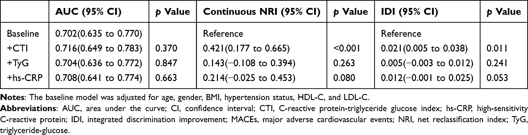 |
Table 2 The Discriminative Value of the CTI, TyG, Hs-CRP for the Risk of MACEs |
 |
Figure 3 Receiver operating characteristic curve analysis of the CTI, TyG index and hs-CRP for MACEs. Abbreviations: CTI, C-reactive protein-triglyceride glucose index; hs-CRP, high-sensitivity C-reactive protein; MACEs, major adverse cardiovascular events; TyG, triglyceride-glucose. |
Prognostic Value of the CTI for MACEs
On the basis of the preceding analysis, we found that the CTI demonstrated relatively greater predictive value for MACEs. Therefore, we further analyzed the relationship between CTI and MACEs. Kaplan–Meier analysis demonstrated a significant relationship between CTI tertiles and MACE incidence (log-rank, p = 0.003). As CTI tertile increased, the cumulative risk of MACEs gradually increased. The group with the highest CTI had the highest risk during the 36-month follow-up (Figure 4). Multivariate Cox regression analysis (Table 3) revealed that the association between CTI and MACEs remained significant after progressive adjustment for confounders, including age, gender, BMI, hypertension status, HDL-C level, and LDL-C level. In Model 3, patients in the highest CTI tertile had a 3.09-fold increased risk of MACEs (p < 0.001), compared with patients in the lowest tertile. Furthermore, when analyzed as a continuous variable, the CTI remained a robust independent predictor (HR: 1.64; 95% CI: 1.23–2.18; p < 0.001), indicating a 64% increase in MACEs risk for each 1-unit increase in the CTI. To further address potential confounding from AMI-specific prognostic variables, we constructed an extended Cox model incorporating diabetes, hyperlipidemia, smoking status, GRACE score, LVEF, and PCI. The association between CTI and MACEs remained robust across this extended adjustment (Supplementary Table S3), supporting the independence of CTI as a prognostic marker beyond established AMI risk variables.
 |
Table 3 Cox Regression Models for the Relationship Between the CTI and MACEs |
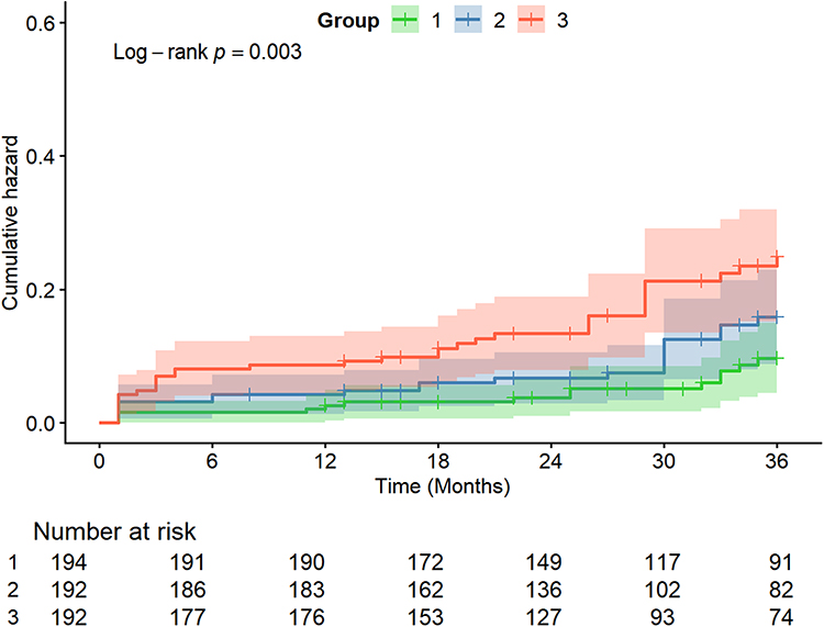 |
Figure 4 Kaplan–Meier survival curves to illustrate the cumulative risk of MACEs in groups based on CTI tertiles. Abbreviations: CTI, C-reactive protein-triglyceride glucose index; MACEs, major adverse cardiovascular events. |
Subgroup Analysis
No significant interactions were observed across the subgroups stratified by age, gender, BMI, hypertension status, diabetes status, or smoking status. These results demonstrate that the prognostic value of the CTI for MACEs across these diverse clinical subpopulations is robust and consistent (Figure 5).
 |
Figure 5 Subgroup and interaction analysis for the association between the CTI and MACEs. Adjusted model included age, gender, BMI, hypertension, HDL-C and LDL-C. Abbreviations: BMI, body mass index; CI, confidence interval. |
Discussion
This research first compared the CTI, hs-CRP level, and TyG index in predicting MACEs in patients with AMI. Our results demonstrate that the CTI is a more promising indicator for identifying high-risk individuals. Further analysis revealed that the CTI remained significantly associated with MACEs after controlling for confounding factors, whether analyzed as a continuous or categorical variable. These results indicate that the CTI may be a practical biomarker for identifying high-risk patients with AMI.
Although secondary prevention has made substantial advances, MACEs still occur after AMI.25 Identifying and managing residual risks effectively is essential for enhancing patient outcomes. The CTI reflects two core components: systemic inflammation (hs-CRP level) and IR (TyG index). Each contributes substantially to residual cardiovascular risk and is linked to an increased incidence of CVD.26–28
IR refers to an inability of the liver, muscles, and adipose tissue to effectively use insulin, resulting in chronic hyperglycemia and significantly increasing the long-term risk of developing type 2 diabetes.29,30 The gold standard for evaluating IR is currently considered to be the hyperinsulinemic–euglycemic clamp. The clamp technique is laborious and costly, limiting its use in large epidemiological studies and routine clinical practice.31 Alternatively, the TyG index is often used as a method for assessing IR. Nevertheless, the ability of the TyG index to predict adverse cardiovascular events across different populations remains controversial. According to a study by Luo et al, elevated TyG index levels serve as a robust and independent prognostic marker for MACEs in STEMI patients within one year following percutaneous coronary intervention (PCI).32 In contrast, Rokicka et al reported that the TyG index had a low predictive capacity for MACEs within 12 months in a population of 1706 AMI patients.33 In this study, the ability of the TyG index to predict MACEs in patients with AMI was insufficient. Several studies have indicated that the integration of the TyG index and measurement of inflammatory biomarkers provides greater predictive significance for MACEs after AMI.34,35
Post-AMI inflammation is characterized by distinct temporal phases and is subject to multifactorial regulation. It commences with a proinflammatory phase, characterized by neutrophil and macrophage infiltration within 72 h to clear necrotic tissue. This is followed by a reparative phase. However, disruption of this regulation can result in chronic low-grade inflammation and poor prognosis.36,37 This state of persistent low-grade inflammation represents a critical target for assessing residual cardiovascular risk after AMI. Hs-CRP level is an important biomarker for assessing this persistent, low-grade inflammatory state.25,27 In our study, we found that the CTI, a combination of hs-CRP level and TyG index, exhibited better predictive performance. In particular, although CTI did not significantly improve the AUC of the baseline model, it was associated with significant improvements in both continuous and categorical NRI, as well as IDI. The categorical NRI of 0.131 indicated significant improvement in category-based risk reclassification when CTI was added to the baseline model. Given the relatively limited sample size and number of MACEs in our cohort, the magnitude of these improvements should be interpreted with caution. Nevertheless, the overall findings still support the potential value of integrating information on inflammation and IR for risk stratification after AMI. Gao et al also found that the most reliable indicator among IR and inflammatory indicators for predicting the recurrence of cardiovascular risk in patients treated with PCI was CTI.38 A nationwide cohort study investigated the combined association of the TyG index and hs-CRP levels with CVD. The results demonstrated that these markers exerted a synergistic effect on cardiovascular risk. The risk of CVD was highest among patients with high TyG and hs-CRP levels when compared to individuals with low levels of these indicators.39 In addition, a multicenter cohort study involving 9421 patients with chronic coronary syndrome investigated the combined effect of TyG and hs-CRP. The study found that among patients with a high TyG index, the risk of MACE was significantly increased only when hs-CRP levels were also elevated.40 These findings indicate that combining assessment of inflammation and IR appears to be a more powerful predictive strategy than using either marker alone.
In the present study, as CTI tertile increased, the cumulative risk of MACEs gradually increased. The group with the highest CTI showed the highest risk during the 36-month follow-up. Previous research in middle-aged and elderly adults revealed that both cumulative CTI and dynamic CTI are independently linked to an elevated risk of CVD.41 Elevated CTI has also been significantly linked to the incidence of CVD.22 Additionally, higher CTI is significantly linked to increased all-cause mortality at 30-day and 1-year follow-ups, along with longer hospital stays in critically ill patients.42 We also found that patients in the highest CTI group had a higher prevalence of three-vessel disease. Previous studies have demonstrated that both the TyG index and hs-CRP are associated with the severity of coronary artery disease, and our findings indicate that combining these indicators similarly reveals this association.43,44 In our Cox proportional hazards regression analyses, the association between CTI and MACEs remained significant across the main models, and this finding was further supported by the extended model. No significant interactions were detected across the subgroups stratified by age, gender, BMI, hypertension status, diabetes status, or smoking status. These results demonstrate that the prognostic value of CTI for MACEs across these diverse clinical subpopulations is robust and consistent.
There are possible underlying mechanisms by which the CTI is linked to poor clinical outcomes in patients with AMI. First, the CTI reflects two core components, systemic inflammation (hs-CRP level) and IR (TyG index), which together make the evaluation of metabolic-inflammatory status more comprehensive.42,45,46 Second, these two pathways are closely linked. IR itself acts as a proinflammatory state that promotes cytokine release. This systemic inflammation in turn exacerbates IR by affecting its signaling pathways.47–49 Finally, considering the substantial inter-individual variability in biomarker levels, the TyG index and hs-CRP levels respectively reflect insulin sensitivity and inflammatory status, thus providing complementary biological information.39,50
CTI is calculated from three simple laboratory parameters, including TG, FBG, and hs-CRP, all of which are routinely recommended for assessment in AMI patients,2,25 making CTI calculation feasible without additional cost or specialized equipment. Clinicians can use this composite index, which integrates both inflammatory and IR status, to assess prognostic risk in AMI patients. However, current research on CTI lacks a validated risk threshold for clinical application. In our cohort, the optimal CTI cut-off value was 9.46, yielding a sensitivity of 71.0% and a specificity of 52.7% for predicting 3-year MACEs. This statistically derived threshold closely approximates the boundary between the second and third tertiles of CTI in our analysis, supporting the internal consistency of CTI tertile-based stratification. These findings provide a preliminary reference for CTI-based risk stratification in AMI patients. Nevertheless, in our study, CTI components were measured before discharge, a timepoint influenced by multiple factors that may systematically affect biomarker values. Specifically, hs-CRP levels may remain elevated due to incomplete resolution of the acute-phase response following AMI, whereas TG levels may be reduced by in-hospital dietary restriction, fasting protocols, and medications. These opposing influences could result in systematic bias in CTI values. Therefore, both the optimal timing of CTI measurement and a validated risk threshold in AMI patients warrant further investigation in future prospective and multicenter studies.
Residual cardiovascular risk after AMI arises from multiple factors. Although CTI reflects an important metabolic-inflammatory dimension of residual risk, biomarker-based assessment alone cannot fully capture the complexity of long-term adverse outcomes after AMI. Recent evidence has shown that high-risk plaque morphology and impaired coronary physiology are independently associated with adverse prognosis in patients with coronary artery disease.23 In this context, CTI should be regarded as a simple and readily available complementary biomarker rather than a standalone prognostic tool. Nevertheless, our findings suggest that CTI may provide additional value for post-discharge risk stratification, helping to identify high-risk patients who may benefit from precise management.
Limitations
This study has several limitations. First, this was a retrospective single-center study, and therefore selection bias and residual confounding cannot be completely excluded. Second, the relatively limited sample size and number of MACEs constrained the extent of confounder adjustment in the multivariable analyses. In particular, because NRI is sensitive to sample size and event rate, the precision of the observed reclassification improvement may be limited. Third, the biomarkers used to calculate CTI were measured at the pre-discharge stage rather than at admission. Although this timing was intended to reflect a more stable condition after the acute phase of AMI, these values may still be influenced by acute-phase physiology, dietary restriction, and in-hospital treatment. Although standard pharmacological therapies, including statins, renin-angiotensin-aldosterone system (RAAS) inhibitors, beta-blockers, and antiplatelet agents, were applied as part of routine clinical care in all enrolled patients, these in-hospital treatments could have systematically affected the pre-discharge biomarker values. Therefore, the absolute CTI values and proposed threshold levels identified in this cohort may have limited generalizability to other clinical settings or measurement time points. Finally, larger prospective multicenter studies with longer follow-up and serial biomarker measurements across different disease stages are needed to further validate and refine the prognostic value of CTI.
Conclusion
The CTI is a more promising indicator for predicting MACEs in AMI patients than the TyG index or hs-CRP alone. By integrating the core risk factors of inflammation and insulin resistance, CTI may provide additional value for post-discharge risk stratification, helping to identify high-risk patients who may benefit from precise management. Future studies integrating additional residual risk indicators may further improve prognostic prediction in AMI patients.
Data Sharing Statement
The data that support the findings of this study are available on request from the corresponding author.
Ethics Statement and Informed Consent
The study involving human participants was approved by the Ethics Committee of Xuanwu Hospital (Document No. 2022-129). The study was conducted in accordance with local legislation and institutional requirements. The requirement for informed consent was waived by the Ethics Committee given the retrospective nature of the study.
Acknowledgments
We thank all study participants, their relatives, and the members of the survey teams.
Author Contributions
Tao Wang: Conceptualization, Formal analysis, Data curation, Methodology, Software, Supervision, Validation, Visualization, Writing - original draft. Guoheng Zhang: Data curation, Methodology, Software, Validation, Writing - original draft. Qinxue Li: Conceptualization, Data curation, Methodology, Validation, Writing - original draft. Yayun Liu: Data curation, Methodology, Software, Validation, Writing - original draft. Tao Zhao: Data curation, Methodology, Validation, Writing - original draft. Wenjing Che: Data curation, Methodology, Validation, Writing - original draft. Jinggang Xia: Conceptualization, Funding acquisition, Methodology, Project administration, Resources, Supervision, Visualization, Writing - review & editing. All authors reviewed and approved the final version of the manuscript and have agreed to its submission. All authors accept responsibility for the integrity and accuracy of the entire work.
Funding
This work was supported by the Beijing Natural Science Foundation (L246059); the Research Project on Chronic Disease Prevention and Control Management Mode in Medical Institutions of the Hospital Management Research Institute of the National Health Commission (No. YM2024ZD004) and Clinical Collaboration Project of Traditional Chinese and Western Medicine for Major Difficult Diseases (ZDYN-2024-A-006).
Disclosure
The authors declare no conflicts of interest.
References
1. Roth GA, Mensah GA, Johnson CO, et al. Global Burden of Cardiovascular Diseases and Risk Factors, 1990–2019: update From the GBD 2019 Study. J Am Coll Cardiol. 2020;76(25):2982–13. doi:10.1016/j.jacc.2020.11.010
2. Rao SV, O’Donoghue ML, Ruel M, et al. 2025 ACC/AHA/ACEP/NAEMSP/SCAI Guideline for the Management of Patients With Acute Coronary Syndromes: a Report of the American College of Cardiology/American Heart Association Joint Committee on Clinical Practice Guidelines. Circulation. 2025;151(13). doi:10.1161/CIR.0000000000001309
3. Byrne RA, Rossello X, Coughlan JJ, et al. 2023 ESC Guidelines for the management of acute coronary syndromes. Eur Heart J. 2023;44(38):3720–3826. doi:10.1093/eurheartj/ehad191
4. Khan AW, Jandeleit-Dahm KAM. Atherosclerosis in diabetes mellitus: novel mechanisms and mechanism-based therapeutic approaches. Nat Rev Cardiol. 2025;22(7):482–496. doi:10.1038/s41569-024-01115-w
5. Hill MA, Yang Y, Zhang L, et al. Insulin resistance, cardiovascular stiffening and cardiovascular disease. Metabolism. 2021;119:154766. doi:10.1016/j.metabol.2021.154766
6. Lee JH, Jeon S, Joung B, Lee HS, Kwon YJ. Associations of Homeostatic Model Assessment for Insulin Resistance Trajectories With Cardiovascular Disease Incidence and Mortality. Arterioscler Thromb Vasc Biol. 2023;43(9):1719–1728. doi:10.1161/ATVBAHA.123.319200
7. Laakso M, Kuusisto J. Insulin resistance and hyperglycaemia in cardiovascular disease development. Nat Rev Endocrinol. 2014;10(5):293–302. doi:10.1038/nrendo.2014.29
8. Li M, Chi X, Wang Y, Setrerrahmane S, Xie W, Xu H. Trends in insulin resistance: insights into mechanisms and therapeutic strategy. Signal Transduct Target Ther. 2022;7:216. doi:10.1038/s41392-022-01073-0
9. Hu F, Chen H, Lin W, Zeng X, Luo Y, Fan L. Different Insulin Resistance Surrogates and Risk of All-Cause and Cardiovascular Mortality among Adults with Metabolic Syndrome-Like Characteristics: evidence from NHANES 1999–2018 – a Cohort Study. Heart Mind. 2026;10(1):22–34. doi:10.4103/hm.HM-D-25-00132
10. Simental-Mendía LE, Rodríguez-Morán M, Guerrero-Romero F. The product of fasting glucose and triglycerides as surrogate for identifying insulin resistance in apparently healthy subjects. Metab Syndr Relat Disord. 2008;6(4):299–304. doi:10.1089/met.2008.0034
11. Tao LC, Xu JN, Wang TT, Hua F, Li JJ. Triglyceride-glucose index as a marker in cardiovascular diseases: landscape and limitations. Cardiovasc Diabetol. 2022;21(1):68. doi:10.1186/s12933-022-01511-x
12. Huang R, Wang Z, Chen J, et al. Prognostic value of triglyceride glucose (TyG) index in patients with acute decompensated heart failure. Cardiovasc Diabetol. 2022;21:88. doi:10.1186/s12933-022-01507-7
13. Sun C, Hu L, Li X, et al. Triglyceride-glucose index’s link to cardiovascular outcomes post-percutaneous coronary intervention in China: a meta-analysis. ESC Heart Failure. 2024;11(3):1317–1328. doi:10.1002/ehf2.14679
14. Kraler S, Mueller C, Libby P, Bhatt DL. Acute coronary syndromes: mechanisms, challenges, and new opportunities. Eur Heart J. 2025;46(29):2866–2889. doi:10.1093/eurheartj/ehaf289
15. Ridker PM, Everett BM, Thuren T, et al. Antiinflammatory Therapy with Canakinumab for Atherosclerotic Disease. N Engl J Med. 2017;377(12):1119–1131. doi:10.1056/NEJMoa1707914
16. Bay B, Tanner R, Gao M, et al. Residual cholesterol and inflammatory risk in statin-treated patients undergoing percutaneous coronary intervention†. Eur Heart J. 2025;46(32):3167–3177. doi:10.1093/eurheartj/ehaf196
17. Lawler PR, Bhatt DL, Godoy LC, et al. Targeting cardiovascular inflammation: next steps in clinical translation. Eur Heart J. 2021;42(1):113–131. doi:10.1093/eurheartj/ehaa099
18. Ridker PM, Bhatt DL, Pradhan AD, Glynn RJ, MacFadyen JG, Nissen SE. Inflammation and cholesterol as predictors of cardiovascular events among patients receiving statin therapy: a collaborative analysis of three randomised trials. Lancet. 2023;401(10384):1293–1301. doi:10.1016/S0140-6736(23)00215-5
19. Ridker PM, Lei L, Louie MJ, et al. Inflammation and Cholesterol as Predictors of Cardiovascular Events Among 13 970 Contemporary High-Risk Patients With Statin Intolerance. Circulation. 2024;149(1):28–35. doi:10.1161/CIRCULATIONAHA.123.066213
20. Ruan GT, Xie HL, Zhang HY, et al. A Novel Inflammation and Insulin Resistance Related Indicator to Predict the Survival of Patients With Cancer. Front Endocrinol. 2022;13:905266. doi:10.3389/fendo.2022.905266
21. Huo G, Tang Y, Liu Z, Cao J, Yao Z, Zhou D. Association between C-reactive protein-triglyceride glucose index and stroke risk in different glycemic status: insights from the China Health and Retirement Longitudinal Study (CHARLS). Cardiovasc Diabetol. 2025;24:142. doi:10.1186/s12933-025-02686-9
22. Sun Y, Guo Y, Ma S, et al. Association of C-reactive protein-triglyceride glucose index with the incidence and mortality of cardiovascular disease: a retrospective cohort study. Cardiovasc Diabetol. 2025;24:313. doi:10.1186/s12933-025-02835-0
23. Acerbo V, Cesaro A, Biccirè FG, et al. Prognostic impact of high-risk plaque morphology and impaired physiology in untreated non-culprit coronary lesions: a systematic review and meta-analysis. Int J Cardiol Heart Vasc. 2026;63:101895. doi:10.1016/j.ijcha.2026.101895
24. Thygesen K, Alpert JS, Jaffe AS, et al. Fourth Universal Definition of Myocardial Infarction (2018). J Am Coll Cardiol. 2018;72(18):2231–2264. doi:10.1016/j.jacc.2018.08.1038
25. Mensah GA, Arnold N, Prabhu SD, Ridker PM, Welty FK. Inflammation and Cardiovascular Disease: 2025 ACC Scientific Statement: a Report of the American College of Cardiology. J Am Coll Cardiol. 2025;2025:S0735. doi:10.1016/j.jacc.2025.08.047
26. Imbesi A, Greco A, Spagnolo M, et al. Targeting Inflammation After Acute Myocardial Infarction. JACC. 2025;86(15):1146–1169. doi:10.1016/j.jacc.2025.07.064
27. Matter MA, Paneni F, Libby P, et al. Inflammation in acute myocardial infarction: the good, the bad and the ugly. Eur Heart J. 2023;45(2):89–103. doi:10.1093/eurheartj/ehad486
28. Li C, Zhao S, Li Y, et al. Triglyceride-glucose index predicts adverse cardiovascular events in patients with H-type hypertension combined with coronary heart disease: a retrospective cohort study. Cardiovasc Diabetol. 2025;24:45. doi:10.1186/s12933-024-02555-x
29. Brie AD, Christodorescu RM, Popescu R, Adam O, Tîrziu A, Brie DM. Atherosclerosis and Insulin Resistance: is There a Link Between Them? Biomedicines. 2025;13(6):1291. doi:10.3390/biomedicines13061291
30. Yang DR, Wang MY, Zhang CL, Wang Y. Endothelial dysfunction in vascular complications of diabetes: a comprehensive review of mechanisms and implications. Front Endocrinol. 2024;15:1359255. doi:10.3389/fendo.2024.1359255
31. Muniyappa R, Lee S, Chen H, Quon MJ. Current approaches for assessing insulin sensitivity and resistance in vivo: advantages, limitations, and appropriate usage. Am J Physiol Endocrinol Metab. 2008;294(1):E15–E26. doi:10.1152/ajpendo.00645.2007
32. Luo E, Wang D, Yan G, et al. High triglyceride–glucose index is associated with poor prognosis in patients with acute ST-elevation myocardial infarction after percutaneous coronary intervention. Cardiovasc Diabetol. 2019;18:150. doi:10.1186/s12933-019-0957-3
33. Rokicka D, Hudzik B, Wróbel M, et al. The prognostic impact of insulin resistance surrogates in patients with acute myocardial infarction with and without type 2 diabetes. Cardiovasc Diabetol. 2024;23:147. doi:10.1186/s12933-024-02240-z
34. Ma X, Chu H, Sun Y, et al. The effect of hsCRP on TyG index-associated cardiovascular risk in patients with acute coronary syndrome undergoing PCI. Sci Rep. 2024;14(1):18083. doi:10.1038/s41598-024-69158-0
35. Wang L, Wang Y, Wang W, Wang Z. Predictive value of triglyceride glucose index combined with neutrophil-to-lymphocyte ratio for major adverse cardiac events after PCI for acute ST-segment elevation myocardial infarction. Sci Rep. 2024;14:12634. doi:10.1038/s41598-024-63604-9
36. Sun K, Yuan LY, Jin J. A double-edged sword of immuno-microenvironment in cardiac homeostasis and injury repair. Signal Transduct Target Ther. 2021;6:79. doi:10.1038/s41392-020-00455-6
37. Ong SB, Hernández-Reséndiz S, Crespo-Avilan GE, et al. Inflammation following acute myocardial infarction: multiple players, dynamic roles, and novel therapeutic opportunities. Pharmacol Ther. 2018;186:73–87. doi:10.1016/j.pharmthera.2018.01.001
38. Gao A, Peng B, Gao Y, et al. Evaluation and comparison of inflammatory and insulin resistance indicators on recurrent cardiovascular events in patients undergoing percutaneous coronary intervention: a single center retrospective observational study. Diabetol Metab Syndr. 2025;17:157. doi:10.1186/s13098-025-01687-8
39. Cui C, Liu L, Qi Y, et al. Joint association of TyG index and high sensitivity C-reactive protein with cardiovascular disease: a national cohort study. Cardiovas Diabetol. 2024;23(1):156. doi:10.1186/s12933-024-02244-9
40. Li Q, Song Y, Zhang Z, et al. The combined effect of triglyceride–glucose index and high-sensitivity C-reactive protein on cardiovascular outcomes in patients with chronic coronary syndrome: a multicenter cohort study. J Diabetes. 2024;16(8):e13589. doi:10.1111/1753-0407.13589
41. Ma X, Ma X, Wang Y, Qiu G, Zhang C. Associations of cumulative exposure and dynamic trajectories of the C-reactive protein-triglyceride-glucose index with incident cardiovascular disease in middle-aged and older Chinese adults: a nationwide cohort study. Cardiovasc Diabetol. 2025;24:303. doi:10.1186/s12933-025-02869-4
42. Wang H, Hu L, Chen Y, et al. The C-reactive protein-triglyceride-glucose index (CTI) as a predictor of mortality and hospital stay in critically ill patients: a retrospective cohort study. Lipids Health Dis. 2025;24:299. doi:10.1186/s12944-025-02739-8
43. Wang X, Xu W, Song Q, et al. Association between the triglyceride-glucose index and severity of coronary artery disease. Cardiovasc Diabetol. 2022;21(1):168. doi:10.1186/s12933-022-01606-5
44. Tajfard M, Tavakoly Sany SB, Avan A, et al. Relationship between serum high sensitivity C-reactive protein with angiographic severity of coronary artery disease and traditional cardiovascular risk factors. J Cell Physiol. 2019;234(7):10289–10299. doi:10.1002/jcp.27945
45. Zhang L, Li S, Liu D, et al. The relationship between C-reactive protein-triglyceride-glucose index and cardiovascular disease: insights from the China health and retirement longitudinal study (CHARLS). Cardiovasc Diabetol. 2025;24:410. doi:10.1186/s12933-025-02960-w
46. Fernández-Real JM, Ricart W. Insulin resistance and chronic cardiovascular inflammatory syndrome. Endocr Rev. 2003;24(3):278–301. doi:10.1210/er.2002-0010
47. Szukiewicz D. Molecular Mechanisms for the Vicious Cycle between Insulin Resistance and the Inflammatory Response in Obesity. Int J Mol Sci. 2023;24(12):9818. doi:10.3390/ijms24129818
48. Yan S, Santoro A, Niphakis MJ, et al. Inflammation causes insulin resistance in mice via interferon regulatory factor 3 (IRF3)-mediated reduction in FAHFA levels. Nat Commun. 2024;15(1):4605. doi:10.1038/s41467-024-48220-5
49. Lu X, Xie Q, Pan X, et al. Type 2 diabetes mellitus in adults: pathogenesis, prevention and therapy. Signal Transduct Target Ther. 2024;9(1):262. doi:10.1038/s41392-024-01951-9
50. Wan B, Wang S, Hu S, et al. The comprehensive effects of high-sensitivity C-reactive protein and triglyceride glucose index on cardiometabolic multimorbidity. Front Endocrinol. 2025;16:1511319. doi:10.3389/fendo.2025.1511319
 © 2026 The Author(s). This work is published and licensed by Dove Medical Press Limited. The
full terms of this license are available at https://www.dovepress.com/terms
and incorporate the Creative Commons Attribution
- Non Commercial (unported, 4.0) License.
By accessing the work you hereby accept the Terms. Non-commercial uses of the work are permitted
without any further permission from Dove Medical Press Limited, provided the work is properly
attributed. For permission for commercial use of this work, please see paragraphs 4.2 and 5 of our Terms.
© 2026 The Author(s). This work is published and licensed by Dove Medical Press Limited. The
full terms of this license are available at https://www.dovepress.com/terms
and incorporate the Creative Commons Attribution
- Non Commercial (unported, 4.0) License.
By accessing the work you hereby accept the Terms. Non-commercial uses of the work are permitted
without any further permission from Dove Medical Press Limited, provided the work is properly
attributed. For permission for commercial use of this work, please see paragraphs 4.2 and 5 of our Terms.