
Back to Journals » Medical Devices: Evidence and Research » Volume 19
Tensile and Tear Strength Evaluation of Clean-Grade Silicone with Cellulose Nanocrystals Reinforcement as an Alternative Material for Maxillofacial Prostheses: An Experimental Study
Authors Stefany S, Kusumadewi AN, Damayanti L  , Takarini V
Received 4 May 2026
Accepted for publication 23 June 2026
Published 9 July 2026 Volume 2026:19 621954
DOI https://doi.org/10.2147/MDER.S621954
Checked for plagiarism Yes
Review by Single anonymous peer review
Peer reviewer comments 2
Editor who approved publication: Professor Mohamad Bashir
Sara Stefany,1 An-Nissa Kusumadewi,2 Lisda Damayanti,2 Veni Takarini3
1Faculty of Dentistry, Universitas Padjadjaran, Bandung, Indonesia; 2Prosthodontics Department, Faculty of Dentistry, Universitas Padjadjaran, Bandung, Indonesia; 3Dental Materials and Technology Department, Faculty of Dentistry, Universitas Padjadjaran, Bandung, Indonesia
Correspondence: Veni Takarini, Faculty of Dentistry, Universitas Padjadjaran, Jalan Sekeloa Selatan No. 1, Bandung, 40132, Indonesia, Email [email protected]
Introduction: Silicone elastomers are widely used in maxillofacial prostheses owing to their elasticity, esthetic properties, and biocompatibility. Clean-grade silicone serves as an affordable alternative to medical-grade silicone. The addition of Cellulose Nanocrystals (CNC) and intrinsic pigments is expected to enhance the performance of clean-grade silicone.
Purpose: This study aims to evaluate the effects of CNC and intrinsic pigments on the tensile strength, elongation percentage, and tear strength of clean-grade silicone as an alternative material for maxillofacial prostheses.
Materials and Methods: Six groups of clean-grade silicone were evaluated: control, A (with pigment), B (with 0.5% CNC), C (with 0.5% CNC and pigment), D (with 1% CNC), and E (with 1% CNC and pigment). Scanning Electron Microscopy (SEM) and Fourier Transform Infrared Spectroscopy (FTIR) were conducted to evaluate the morphological and functional group characteristics; tensile strength, elongation percentage, and tear strength were measured to assess changes in mechanical properties and were analyzed statistically.
Results: SEM analysis showed microstructure formation characterized by dispersed particles with the addition of 1% CNC and intrinsic pigments. As the filler and pigment were added, the FTIR revealed increased absorption intensities of Si-O-Si functional groups, detected at 1022– 1026 cm− 1. The tensile trength (3.87– 4.84 MPa) was successfully increased compared to the control group (3.63 MPa), a statistically significant difference (p< 0.05). Group D had the highest tensile strength, aligning closely with medical-grade silicone (approximately 2.53– 8.36 MPa). Meanwhile, the lowest elongation percentage and the highest tear strength (3.84 kN/m) resulted in Group E, both of which are close to the range of maxillofacial prosthesis requirements.
Conclusion: The addition of 1% CNC and intrinsic pigments successfully increased tensile and tear strength while reducing the elongation percentage of clean-grade silicone that potential as an alternative material; however, future optimization on the tear strength still required before maxillofacial prosthesis application.
Keywords: cellulose nanocrystals, intrinsic pigment, tensile and tear strength, elongation percentage, clean-grade silicone, alternative maxillofacial prosthesis
Introduction
Maxillofacial defects present significant challenges for reconstructive surgeons, prosthodontists, and dental technicians. These defects may be congenital or acquired because of trauma, disease, or surgical resection. Although curative treatment often involves a multidisciplinary approach with surgical intervention, secondary deformities may persist after resection. In such cases, a maxillofacial prosthesis serves as a viable alternative when surgical reconstruction is not feasible or contraindicated. Despite advances in reconstructive and plastic surgery, extraoral prostheses fabricated using synthetic materials remain essential for restoring complex facial structures.1–3 The intention of prosthetic rehabilitation for maxillofacial patients is to enhance psychological well-being, in addition to restoring function and appearance.4 Maxillofacial prosthesis fabrication is required to restore the patient’s appearance, thereby enhancing self-esteem and facilitating social reintegration. Therefore, ideal maxillofacial prosthetic materials should possess optimal physical, esthetic, and biological properties to ensure patient acceptance.5,6
Silicone elastomers are widely used as the material of choice to replace missing facial structures due to trauma or disease. The selection of the appropriate material is critical for the fabrication of extraoral prostheses. The desired properties include high tear resistance at thin margins, adequate tensile strength and elongation to ensure flexibility, ease of manipulation, dimensional stability, color stability under environmental exposure, low water absorption, and biocompatibility with surrounding tissues.6,7 Medical-grade silicone is the gold standard for maxillofacial prostheses because of its superior biocompatibility and durability, since it could maintain its mechanical properties when repeatedly subjected to body fluids, sterilization or disinfection treatments.8,9 Clean-grade silicone has also been considered a safe, although it is not specifically composed to resist prolonged exposure. A more economical silicone material with the common process of room-temperature vulcanization (RTV) is also considered due to their ease of processing, high tear resistance, flexibility, tissue-like consistency, and compatibility with adhesives.9,10 Despite its potential, the mechanical properties of clean-grade silicone still require further investigation to validate its suitability for maxillofacial applications.11,12
One of the challenges in maxillofacial prosthesis applications is the degradation of elastomeric properties over time, which includes durability and color stability of the materials. The use of intrinsic pigments can affect the physical characteristic of mimicking natural skin color. Pigments were integrated through physical contact rather than chemical bonding. If a chemical interaction takes place between polymers and pigments, the crosslink shall produce reinforcement,1 which could be further explored in the use of liquid cosmetic as the intrinsic pigment. Other than the physical properties, various approaches have been explored to enhance the mechanical performance of silicone elastomers, including the incorporation of nanofillers or nanofibers to improve the tensile and tear strength, as well as the overall mechanical characteristics. Silica (SiO2) is one of the most commonly used fillers; however,13,14 the increase in environmental concerns has prompted the search for more sustainable and eco-friendly alternatives. In recent years, natural polymers have gained considerable attention as fillers in biomedical applications owing to their sustainability and biocompatibility.15 These polymers are derived from renewable resources such as chitosan, starch, and cellulose. Among these, cellulose is the most abundant natural polymer found in biomass.16,17
Nanocellulose primarily exists in two forms: cellulose nanocrystals (CNC) and cellulose nanofibrils (CNF). Compared to conventional nanofillers, CNC exhibit excellent mechanical properties, biodegradability, biocompatibility, low toxicity, high specific surface area, and ease of chemical modification owing to the abundance of hydroxyl groups.17 In 2023, Ali et al demonstrated that the incorporation of CNF into RTV medical-grade silicone significantly enhanced its mechanical properties, including tensile strength, tear strength, and elongation. Optimal improvements were observed at a concentration of 0.5%, while higher concentrations (1%) resulted in reduced tensile strength owing to filler agglomeration, leading to premature failure under tensile stress.14 However, no studies have investigated the incorporation of CNC into clean-grade silicone with the addition of intrinsic pigments for maxillofacial prosthetic applications. Therefore, this study aimed to evaluate the effect of CNC and intrinsic pigment addition on the tensile strength, elongation percentage, and tear strength of clean-grade silicone as an alternative material for maxillofacial prostheses. The hypothesis of this study is the use of CNC could reinforce the clean-grade silicone and shall affect the tensile strength, elongation percentage, and tear strength.
Materials and Methods
Materials
The clean-grade silicone used in this study was Silicone Rubber RTV P-25 (Sumber Kimia Jaya, Indonesia). CNC were obtained as nanocellulose (AMI Scientific, Bandung, Indonesia) with particle size of 10–20 nm and length of 300–900 nm, which then used as received. The intrinsic pigment was obtained from the commercially available cosmetic liquid foundation shade 52N (Wardah Colorfit, Jakarta, Indonesia). All materials were used as received.
Specimen Preparation
The specimen in this study consisted of room-temperature vulcanized (RTV) clean-grade silicone as control group. Meanwhile, the treatment groups were incorporated with cellulose nanocrystals (CNC) at concentrations of 0.5% and 1% to the clean grade silicone, which were then compared with the group that incorporated intrinsic pigment at a concentration of 0.2wt%.
Sixty specimens were divided into six groups: Control Group of clean-grade silicone without any additives, Group A intrinsic pigment only, Group B with 0.5% CNC; Group C with 0.5% CNC and intrinsic pigment, Group D with 1% CNC; and Group E with 1% CNC and intrinsic pigment. Each group consisted of five specimens for the tensile strength and elongation percentage tests, as well as for tear strength.
The control group specimens were prepared by mixing RTV clean-grade silicone according to the manufacturer’s instructions, using a 1:1 ratio of base to catalyst, measured with a digital scale. Cellulose nanocrystals (CNC) were incorporated into the silicone matrix at concentrations of 0.5% for Groups B and C and 1% for Groups D and E, based on the total weight of the silicone. For Groups A, C, and E, an intrinsic pigment was added at a concentration of 0.2% of the total silicone weight. All mixtures were homogenized at room temperature using an overhead stirrer with tubular blade geometry for 5 min, followed by degassing in a vacuum chamber of 400 mmHg pressure at 360 rpm for 10 min to eliminate air bubbles and ensure the uniform dispersion of CNC within the silicone matrix. The mixtures were then poured into molds corresponding to each test group using a stainless-steel spatula and allowed to cure at room temperature for approximately 24 h. After vulcanization, the specimens were removed from the molds and stored in a light-proof container prior to testing. All specimens were maintained under controlled conditions at a temperature of 20–25°C and relative humidity below 60%. The specimens from each test group are shown in Figure 1.
 |
Figure 1 Specimens from each group for (a) Tensile strength and percentage elongation test; and (b) Tear strength test. |
Scanning Electron Microscopy (SEM) and Fourier Transform Infrared Spectroscopy (FTIR) Characterization
Scanning Electron Microscopy (SEM) was used to analyze the microstructural morphology and filler distribution of the silicone composite matrix. SEM analysis was performed using a JSM IT-200 JEOL instrument at magnifications of 100x and 1000x, which were selected to evaluate macro- and micro-scale filler dispersion patterns, micro-network formation, and agglomeration sites within the silicone matrix — the morphological features most directly relevant to the mechanical properties investigated in this study. The nanoscale dimensions of the CNC (10–20 nm diameter, 300–900 nm length) were confirmed by the supplier (AMI Scientific) and are consistent with values reported in the literature; therefore, the purpose of SEM in this study was not to re-characterize the CNC particles themselves, but to assess how they distribute and interact within the silicone matrix at the composite level. At 1000x magnification, key features including micro-network formation, filler dispersion, and agglomeration were clearly discernible and sufficient to support the interpretations presented. While higher magnification imaging would provide additional resolution for isolated nanoparticle characterization, the magnifications employed are appropriate for composite matrix evaluation and are consistent with the approach used in comparable studies in this field.
Fourier Transform Infrared Spectroscopy (FTIR) was used as a non-destructive analytical technique to evaluate the chemical characteristics and determine the potential interactions between RTV clean-grade silicone and CNC. The FTIR analysis was conducted using a Prestige 21 spectrometer (Shimadzu, Japan), with spectra acquired over the range of 4000–400 cm−1, 40 scans performed at a resolution of 4 cm−1.
Tensile Strength and Elongation Percentage Testing
The specimens for tensile strength and elongation percentage testing were prepared in a dumbbell shape with a length of 75 mm and thickness of 2 mm, in accordance with ISO 37:2024 standards. The specimens were mounted on a Universal Testing Machine (LRX Plus, LLOYD Instruments) using an extensometer, and testing was conducted at a crosshead speed of 500 mm/min until specimen failure. The width (w) and thickness (t) of each specimen were measured in millimeters using a digital caliper. The maximum force required to cause a fracture (F) was recorded in Newtons (N). The tensile strength was expressed in megapascals (MPa) and was calculated using the following equation:

The percentage of elongation was determined concurrently during the tensile strength tests. It was calculated based on the final length of the specimen at break (Lb) and the original length (Lo), both measured in millimeters, using the following equation:

Tear Strength Testing
The specimens for tear strength testing were prepared in a double-pant configuration in accordance with ASTM D624 standards. The dimension of unnicked 90° angle with 102 mm length for the tear strength specimens were mounted on a Universal Testing Machine (LRX Plus, LLOYD Instruments) and subjected to tensile loading at a crosshead speed of 500 mm/min until failure. The maximum force (F) required to cause rupture was recorded in Newtons, and the median thickness (d) in the right-angle region of each specimen was measured using a digital caliper in millimeters. Tear strength was calculated using the following equation:

Statistical Analysis
Data were collected and presented in tables with standard deviations. Normality and homogeneity were assessed using the Shapiro–Wilk and Levene tests, respectively. If the data were normally distributed and homogeneous, statistical analysis was performed using one-way ANOVA, followed by Tukey’s post hoc test. Otherwise, the Kruskal–Wallis test was used. All analyses were conducted using SPSS (version 26). Statistical significance was defined as p>0.05 (non-significant), p<0.05 (significant), and p<0.01 (highly significant).
Results
SEM Analysis
Surface microstructural analysis was performed using SEM at low (100x) and high (1000x) magnifications to evaluate morphological changes in clean-grade silicone after the addition of CNC and intrinsic pigments. SEM images of each sample group are shown in Figure 2. In the control group, the SEM image at 100x magnification showed a homogenous and continuous surface without filler particles or agglomeration. At 1000x magnification, the surface exhibited the characteristic wavy morphology of silicone elastomers.18 The SEM images of Sample A (silicone with intrinsic pigment) show a relatively homogenous surface with dispersed pigment particles within the silicone matrix. At 1000x magnification, the surface exhibited a characteristic wrinkled elastomeric morphology;18 however, no significant pigment agglomeration was observed. This indicated that the intrinsic pigment was well distributed without causing major structural defects in the silicone matrix. In Sample B (silicone with 0.5% CNC), the SEM images show the distribution of CNC without significant agglomeration. At 1000x magnification, the surface exhibited a wrinkled morphology with the formation of a micro-network, as seen in the bright spot area (indicated by blue arrows). The micro-network formed by the aligned CNC appears as white streaks or lines that follow the wrinkled texture of silicone (blue circles).
 |
Figure 2 SEM image of each group sample (a) 100x magnification, (b) 1000x magnification. As shown by the color coding, blue arrows indicate bright spots area in Sample B; blue circles indicate aligned CNC seen as white streaks in Sample B; green arrows indicate white irregularly shaped with darker area inside in Sample C; purple arrows indicate more greyish appearance wrinkled surface in Sample D; and yellow arrows indicate bright spots area without darker area inside in Sample E. |
The SEM images of Sample C (silicone with 0.5% CNC and intrinsic pigment) revealed a more wrinkled surface with an increased micro network area with larger bright spot sites compared to Sample B. At 1000x magnification, the white irregularly shaped particle with a darker area inside (indicated by green arrows) is described as the filler from the intrinsic pigment added. In Sample D (silicone with 1% CNC), the SEM images showed a more evenly dispersed surface. At 1000x magnification, the wrinkled surface appeared quite dense and homogenous with white streaks that seemed to be more scattered, exhibiting a more grayish appearance (indicated by purple arrows).
The SEM images of Sample E (silicone with 1% CNC and intrinsic pigment) revealed a more homogeneous and well-dispersed structure with larger bright spots. At 1000x magnification, the visibility of white streaks seems to be reduced, and bright spots appear without the darker area inside (indicated by yellow arrows). This may be described as the interaction between CNC and the intrinsic pigment, suggesting the presence of agglomeration. Complex micro-networks were observed, suggesting a high surface area that enhances the filler-matrix interaction and contributes to improved tear strength.19 However, agglomeration may also act as stress concentration points, potentially reducing the tensile strength and elongation percentage.14
FTIR Analysis
FTIR analysis was performed to evaluate the chemical characteristics of each specimen from each experimental group. The FTIR spectra of all test samples, as shown in Figure 3, exhibited characteristic silicone features confirmed by the presence of siloxane (Si-O-Si) absorption bands at 1022–1026 cm−1 and 1091–1093 cm−1, indicating that the fundamental chemical structure remained dominant across all groups. No new peaks or wavenumber shifts were observed in any group, confirming that the incorporation of CNC and intrinsic pigment resulted in physical interactions within the silicone matrix rather than the formation of new covalent bonds. To provide a semi-quantitative assessment of filler–matrix interactions, the relative absorption intensities of three key bands — the O-H stretching band (~3446–3448 cm−1), the C-H stretching band (~2850–2960 cm−1), and the Si-O-Si band (~1022–1026 cm−1) were systematically compared across all groups.
 |
Figure 3 FTIR Spectra of each group samples. Grey dashed box indicates the areas of O-H bending band; black arrows indicate Si-O-Si functional group in pigment-containing groups; black dashed box indicates C-H band as hydrocarbon contribution. |
The O-H stretching band at 3446–3448 cm−1 was present in all samples, with Samples A–E showing progressively lower transmittance than the control, suggesting stronger O-H absorption and increased hydrogen bonding attributable to the hydroxyl groups on the CNC surface and in the components of the intrinsic pigment. This trend was most pronounced in pigment only-containing groups (Sample A), which showed markedly lower transmittance than CNC with pigment and CNC-only groups (B, C, D and E), suggesting that pigment addition exerts a significant and additive influence on hydrogen bonding density within the matrix. Next, the O-H bending band at ~1630 cm−1 exhibited the greatest reduction in transmittance in groups C and E, consistent with higher bound water content in CNC and pigment-incorporated formulations.
The C-H stretching bands at 2850–2960 cm−1 demonstrated a systematic increase in absorption intensity from the control through to Group E, reflecting enhanced hydrocarbon contributions from the combined incorporation of CNC and intrinsic pigment. This increase appears to be more prominent in groups containing pigment (A) and both fillers (C and E), indicating an additive effect on the hydrocarbon vibration environment of the silicone matrix. For the Si-O-Si absorption bands at 1022–1026 cm−1, all modified groups (A–E) demonstrated lower transmittance relative to the control, indicating stronger siloxane network absorption following filler incorporation. Among these, although pigment-containing groups (A) demonstrated significant absorption from liquid cosmetic composition to silicone matrix; however (C, and E) showed the greatest absorption, consistent with pigment-induced physical interactions also with CNC that alter the bonding environment of the siloxane network without disrupting its chemical integrity.
Taken together, the systematic comparison of relative band intensities across groups reveals a concentration-dependent pattern in which increasing CNC content and the addition of intrinsic pigment progressively enhance intermolecular interactions, particularly hydrogen bonding and physical crosslinking within the silicone matrix. These findings support the observed improvements in mechanical properties across the modified groups and are consistent with previous studies reporting that nanocellulose reinforcement in silicone primarily strengthens physical filler–matrix interactions rather than inducing covalent bond formation.
Tensile Strength
The mean tensile strength values are shown in Figure 4. The highest tensile strength was observed in Group D (silicone with 1% CNC), with a mean value of 4.84 MPa, while the lowest value was found in the control group, with a mean of 3.63 MPa (Table 1). Although Group E was the lowest among all treatment groups, its mean value (3.87 MPa) was still higher than that of the control group. Normality was assessed using the Shapiro–Wilk test, which showed that all groups were normally distributed. Homogeneity of variance was confirmed using Levene’s test (p>0.05). One-way ANOVA, as shown in Table 2, revealed a statistically significant difference in tensile strength among the six experimental groups (p<0.05). Post-hoc multiple comparisons using Tukey’s test (Table 3) showed highly significant differences (p<0.01) between the control group and Groups A, B, and D, between Groups E and B, Groups E and D, and Groups C and D. Significant differences (p<0.05) were observed between the control group and Group C, Groups E and A, Groups C and B, and Groups A and D.
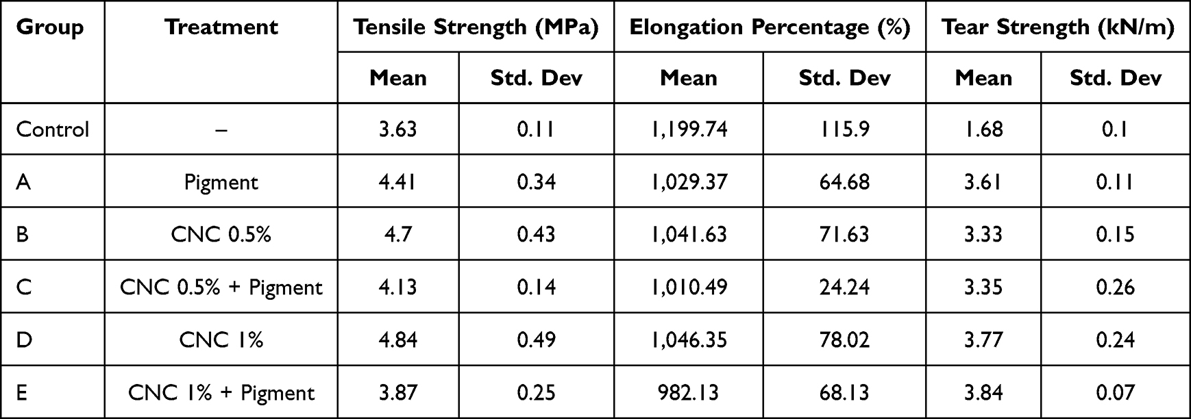 |
Table 1 Mean and Standard Deviation Tensile Strength, Elongation Percentage, and Tear Strength Values of Each Group Specimens |
 |
Table 2 ANOVA Analysis of Tensile Strength, Elongation Percentage, and Tear Strength Test |
 |
Table 3 Post-Hoc Analysis Table Using t-Test for Tensile Strength Test |
 |
Figure 4 Mean tensile strength values of each group specimens. |
Elongation Percentage
The percentage of elongation was calculated as the ratio of the change in specimen length at break to its original length during tensile testing. The mean elongation values are presented in Figure 5, with the lowest value observed in Group E (982.13%), indicating the smallest changes in specimen length. The highest value was observed in the Control Group (1,199.74%).
 |
Figure 5 Mean elongation percentage values of each group specimens. |
The Shapiro–Wilk test confirmed a normal data distribution, and Levene’s test indicated homogeneity of variance (p>0.05). One-way ANOVA revealed a statistically significant difference among the groups (p<0.05), as shown in Table 2. Post-hoc analysis using Tukey’s test showed highly significant differences (p<0.01) between the Control Group and all other groups (A-E), whereas comparisons among the remaining groups were not statistically significant (Table 4).
 |
Table 4 Post-Hoc Analysis Table Using t-Test for Elongation Percentage Test |
Tear Strength
The mean tear strength values for each group are shown in Figure 6. The highest tear strength was observed in Group E, with a mean value of 3.84 kN/m, whereas the lowest was found in the Control Group, with a mean of 1.68 kN/m (Table 1). The Shapiro–Wilk test confirmed a normal data distribution, and Levene’s test indicated homogeneity of variance (p>0.05). One-way ANOVA, as shown in Table 2, revealed a statistically significant difference among the six groups (p<0.05). Post-hoc analysis using Tukey’s test (Table 5) showed highly significant differences (p<0.01) between the control group and all other groups (A-E), as well as between Groups B and D, B and E, C and D, and C and E. Significant differences (p<0.05) were observed between Groups B and A, C and A, and A and E. No statistically significant differences (p>0.05) were found between Groups A and D, and between Groups D and E.
 |
Table 5 Post-Hoc Analysis Table Using t-Test for Tear Strength Test |
 |
Figure 6 Mean tear strength values of each group specimens. |
Discussion
Silicone elastomers are the most commonly used materials for fabricating maxillofacial prostheses. Silicone is composed of silicone, oxygen, carbon, and hydrogen, and its physical properties are primarily determined by the average chain length and the degree of crosslinking within the polymer network. The highly polar Si-O bonds provide strong bond energy, contributing to the excellent thermal and chemical stability of the material.20 An ideal maxillofacial prosthetic material should exhibit favorable physical and mechanical properties and maintain its performance over time. Previous studies have reported a wide range of mechanical properties of medical-grade silicone used in maxillofacial applications.
In this study, the addition of CNC and intrinsic pigments significantly altered the mechanical properties of clean-grade silicone, resulting in increased tensile and tear strength, along with reduced elongation. The addition of 0.5% CNC only (Group B) resulted in a higher mean value of tensile strength than the addition of pigment only (Group A), whereas the addition of 1% CNC only (Group D) resulted in the highest mean value (4.84 MPa). The combination of CNC with pigment in Group C (0.5% CNC and pigment) and Group E (1% CNC and pigment) resulted in lower values compared to the CNC- or pigment-only groups, with the lowest mean values found in Group E (3.87 MPa). These results revealed significant differences in the tensile strength among the six experimental groups (p<0.05). However, the tensile strength results of all treatment groups showed an overall increase compared to the control group (3.63 MPa), which is also recommended based on the tensile strength of silicone maxillofacial prosthesis values that ranges from 2.53−8.36 MPa.20,21
The tensile strength of silicone elastomers reflects the overall durability of the material and is closely associated with the elongation, which represents the flexibility of the prosthesis. It is defined as the maximum stress that a material can withstand before undergoing rapid localized deformation, such as that during prosthesis removal.22 The reinforcing mechanism of fillers involves their function as multifunctional crosslinking agents, forming strong hydrogen bonds between the hydroxyl groups on the filler surface and polysiloxane chains of silicone. This interaction increased the overall crosslink density of the polymer network, resulting in a stiffer and stronger material.23 Consequently, the addition of fillers is generally associated with an increased tensile strength and reduced elongation.15,24
In this study, an increase in the tensile strength was accompanied by a decrease in the elongation percentage. A reduction in elongation percentage was observed in all modified groups compared to that in the control group, with a statistically significant difference among all groups (p<0.05). This indicates that the incorporation of CNC and intrinsic pigments restricts the polymer chain mobility, thereby reducing the flexibility of the silicone matrix.17 Notably, Group D (with 1% CNC) demonstrated high tensile strength while maintaining moderate elongation (1,046.35%), suggesting adequate dispersion of CNC within the matrix at this concentration, as observed in the SEM images. In contrast, the lowest elongation percentage (982.13%) was observed in Group E (with 1% CNC and pigment), which may be attributed to the presence of filler agglomeration, as supported by the SEM images. This result may lead to lower interfacial bonding,25,26 which could also affect the lower tensile strength compared to all treatment groups. However, the elongation percentage values are still close to the range of the silicone maxillofacial prosthesis recommended values of 400–980%.21,27,28
The mechanical performance of silicone is strongly influenced by the type and concentration of fillers as well as the crosslinking system employed. The incorporation of fillers has been shown to enhance the tensile and tear strengths.24,29 Tensile and tear strengths are critical indicators of overall material performance and durability, particularly in addressing common clinical issues such as tearing at the thin margins of maxillofacial prostheses.30 The addition of 1% CNC and intrinsic pigment from Group E have the highest tear strength values in this study (3.84 kN/m) while the lowest was found in control group (1.68 kN/m). Statistically, there was a highly significant difference between the addition of 0.5% CNC and 1% CNC, both with and without intrinsic pigments. This may be attributed to the high degree of crystallinity of CNC, typically exceeding 60%, which is largely stabilized by hydrogen bonding between cellulose chains, contributing to the formation of well-defined crystalline domains and influencing their physicochemical properties.31 Furthermore, the abundance of hydroxyl groups in CNC, as evidenced by FTIR analysis, promoted surface interactions and contributed to the improved functional properties of the composite material.32
The reinforcing efficiency of the filler depends largely on the particle size and surface area. Smaller filler particles provide greater reinforcement owing to their increased surface area and more effective interfacial interactions with the silicone matrix.33 A systematic review reported that the addition of nanoparticles to silicone elastomers at various concentrations can enhance mechanical and physical properties, improve color stability, and maintain the long-term performance of maxillofacial prostheses.24 Cellulose nanocrystals (CNC) are rigid, rod-shaped nanocellulose structures with predominantly crystalline domains that have been widely utilized in dental applications to enhance their mechanical properties, biocompatibility, and antimicrobial performance.16,34,35
The highest tear strength observed in Group E was not able to reach the reported reference board range of silicone maxillofacial, which ranges from 4.53 to 17.51 N/mm.21,27,28 This may be attributed to the differences in particle size between the intrinsic pigment and CNC, as seen in the SEM image with more dispersed and larger bright spots. In this study, the intrinsic pigment used was a liquid foundation containing a divinyl-dimethicone cross-polymer and silica as a reinforcing filler, which is a silicone-based polymer that is commonly used in cosmetics as a film-forming and viscosity-modifying agent.36 Although these components may contribute to the improvement of mechanical properties,28 their reinforcing effect may not be as effective as CNC, thereby limiting the overall enhancement and preventing the material from reaching the reference values. However, the use of liquid cosmetics as intrinsic pigments may serve as an alternative approach to the coloration of silicone materials.
Tear strength is a clinically critical property of maxillofacial prostheses, particularly at thin margins that are prone to failure during insertion, removal, and functional movements.22,24 The highest tear strength observed in Group E (with 1% CNC and pigment) suggests a synergistic reinforcing effect, where the CNC and pigment act as barriers to crack propagation. The rigid crystalline structure of CNC promotes crack deflection, increasing the energy required for tear propagation, while the pigment contributes additional reinforcement within the matrix.14,37,38 The discrepancy between the tensile and tear strengths indicated that these properties were governed by different failure mechanisms. While tensile strength is influenced by matrix homogeneity and interfacial integrity, tear strength is more closely related to a material’s resistance to crack growth.14,37,38 Although filler agglomeration may reduce tensile performance, it can still enhance tear resistance by increasing energy-dissipation mechanisms.
Overall, the incorporation of CNC, intrinsic pigment, and their combination provided a reinforcing effect on clean-grade silicone, as evidenced by increased tensile and tear strength and reduced elongation. From a clinical perspective, based on the findings of this study, the addition of pigment is essential for esthetic requirements; therefore, the combination of 1% CNC and intrinsic pigment (Group E) may be considered the most favorable formulation, as it exhibited the highest tear strength, although it has not yet reached the reported reference values. Nevertheless, tear strength remains a critical property for maxillofacial prostheses, which are highly susceptible to tearing during repeat insertion and removal, such as auricular prostheses, where adequate tear strength is essential to prevent edge failure.
However, further investigation is necessary to evaluate long-term clinical performance. This study was limited to mechanical and physicochemical characterization and did not include assessments of biological properties, color stability, or aging under environmental conditions such as ultraviolet exposure, temperature, and humidity. Prior to clinical application, in vivo studies and long-term simulations are required to confirm the safety and durability of clean-grade silicone as an alternative maxillofacial prosthetic material.
Conclusion
In conclusion, the addition of cellulose nanocrystals (CNC), intrinsic pigments, and their combinations significantly influences the mechanical properties of clean-grade silicone. Specifically, the incorporation of 1% CNC and intrinsic pigment successfully increased the tensile strength and tear strength, while reducing the elongation percentage. Therefore, reinforcement in clean-grade silicone has the preliminary potential to be used as an alternative material for maxillofacial prostheses. However, prospective studies must be taken for the aging simulation, color stability and biological evaluation for maxillofacial silicones, suggesting that further optimization is required before clinical application.
Acknowledgments
The authors would like to thank the Faculty of Dentistry Integrated Laboratory Universitas Padjadjaran for providing laboratory facilities during material testing.
Disclosure
The author(s) report no conflicts of interest in this work.
References
1. Abdalqadir M, Saeed Z, Azhdar B. Surface roughness of pigmented and non-pigmented maxillofacial silicone elastomer before and after artificial aging. Mater Res Express. 2024;11(1):015401. doi:10.1088/2053-1591/ad1a60
2. Zayed SM. Mechanical properties of maxillofacial silicone elastomer and approaches for their improvement: a literature review. EC Dent Sci. 2018;17:1293–14.
3. Steiert AE, Boyce M, Sorg H. Capsular contracture by silicone breast implants: possible causes, biocompatibility, and prophylactic strategies. Med Devices. 2013;6:211–218. doi:10.2147/MDER.S49522
4. Polisiero M, Bifulco P, Liccardo A, et al. Design and assessment of a low-cost, electromyographically controlled, prosthetic hand. Med Devices. 2013;6(1):97–104. PubMed PMID: 23843711. doi:10.2147/MDER.S39604
5. Yeh HC. Effect of silica filler on the mechanical properties of silicone maxillofacial prosthesis. 2014.
6. Mahajan H, Gupta K. Maxillofacial prosthetic materials: a literature review. J Orofac Res. 2012;2:87–90. doi:10.5005/jp-journals-10026-1020
7. Zaheer Kola M, Begum Z, Joshi P, Agnihotri Y. Analysis of the properties of commercially available silicone elastomers for maxillofacial prostheses. Int J Contemp Dent. 2011;2(4):1.
8. Chugh A, Hattori M, Towithelertkul C, Sumita YI, Wakabayashi N. Evaluation of the color stability of three maxillofacial silicone materials after exposure to beverages: an in vitro study. Heliyon. 2024;10(4):e25529. doi:10.1016/j.heliyon.2024.e25529
9. Arora J, Gulati M, Mishra A. Maxillofacial prosthesis materials. Int J Health Sci. 2021;5:112–124. doi:10.53730/ijhs.v5ns2.5438
10. Jasim Mohammed A, Alirhayim R, Alirhayim RN. The effect of nanoparticles addition on the physical properties of the maxillofacial silicone: a literature review. J Res Med Dent Sci. 2022;2022(2):760–764.
11. Tanveer W. Biomaterials for maxillofacial prosthetic rehabilitation. In: Advanced Dental Biomaterials. Bangkok: Elsevier; 2019:615–641. doi:10.1016/B978-0-08-102476-8.00024-4
12. Kiantoro I, Sumarsongko T, Damayanti L, Takarini V. Tensile strength and shore hardness evaluation in clean-grade silicone with nanosilica filler reinforcement as an alternative for maxillofacial prosthesis materials. Mater Scie Forum. 2022;1069:145–152. doi:10.4028/p-q96742
13. Al-Samaray M, Al-Somaiday H, Rafeeq AK. Effect of adding different concentrations of CaCO3-SiO2 nanoparticles on tear strength and hardness of maxillofacial silicone elastomers. Nano Biomed Eng. 2021;13(3):257–263. doi:10.5101/nbe.v13i3.p257-263
14. Ali AA, Safi IN. Impact of nano-cellulose fiber addition on physico-mechanical properties of room temperature vulcanized maxillofacial silicone material. J Taibah Univ Med Sci. 2023;18(6):1616–1626. doi:10.1016/j.jtumed.2023.07.002
15. Takarini V, Asri LATW, Djustiana N, Hadi BK. Alternative dental impression fillers made of nanorod glutinous rice flour particles through precipitation. Mater Res Express. 2023;10(7):075304. doi:10.1088/2053-1591/ace3a6
16. Du H, Liu W, Zhang M, Si C, Zhang X, Li B. Cellulose nanocrystals and cellulose nanofibrils based hydrogels for biomedical applications. Carbohydr Polym. 2019;209:130–144. PubMed PMID: 30732792. doi:10.1016/j.carbpol.2019.01.020
17. Yang X, Li Z, Jiang Z, et al. Mechanical reinforcement of room-temperature-vulcanized silicone rubber using modified cellulose nanocrystals as cross-linker and nanofiller. Carbohydr Polym. 2020;229:115509. PubMed PMID: 31826417. doi:10.1016/j.carbpol.2019.115509
18. Guzmán-De La Cerda D, Jofré-Ulloa PP, Benavente E, et al. Incorporation of water-soluble quantum dots and formation of wrinkled patterns on acrylic and silicone elastomers. ACS Omega. 2025;10(4):3322–3331. doi:10.1021/acsomega.4c04071
19. Russo P, Venezia V, Tescione F, et al. Improving interaction at polymer–filler interface: the efficacy of wrinkle texture. Nanomaterials. 2020;10(2):208. doi:10.3390/nano10020208
20. Zare M, Ghomi ER, Venkatraman PD, Ramakrishna S. Silicone-based biomaterials for biomedical applications: antimicrobial strategies and 3D printing technologies. J Appl Polym Sci. 2021;138. doi:10.1002/app.50969
21. Aziz T, Waters M, Jagger R. Analysis of the properties of silicone rubber maxillofacial prosthetic materials. J Dent. 2003;31:67–74. doi:10.1016/S0300-5712(02)00084-2
22. Al-Kadi FK, Adbulkareem JF, Azhdar BA. Evaluation of the mechanical and physical properties of maxillofacial silicone Type A-2186 impregnated with a hybrid Chitosan–TiO2 nanocomposite subjected to different accelerated aging conditions. Biomimetics. 2023;8(7):539. doi:10.3390/biomimetics8070539
23. Shakir DA, Abdul-Ameer FM. Effect of nano-titanium oxide addition on some mechanical properties of silicone elastomers for maxillofacial prostheses. J Taibah Univ Med Sci. 2018;13(3):281–290. doi:10.1016/j.jtumed.2018.02.007
24. Sonnahalli N, Chowdhary R. Effect of nanoparticles on color stability and mechanical and biological properties of maxillofacial silicone elastomer: a systematic review. J Indian Prosthodont Soc. 2020;20(3):244–254. doi:10.4103/jips.jips_429_19
25. Bangera BS, Guttal SS. Evaluation of varying concentrations of nano-oxides as ultraviolet protective agents when incorporated in maxillofacial silicones: an in vitro study. J Prosthetic Dentistry. 2014;112(6):1567–1572. PubMed PMID: 25156091. doi:10.1016/j.prosdent.2014.07.001
26. Mohammad SA, Wee AG, Rumsey DJ, Schricker SR. Maxillofacial materials reinforced with various concentrations of polyhedral silsesquioxanes. J Dent Biomech. 2010;1(1):1–6. doi:10.4061/2010/701845
27. Hatamleh MM, Watts DC. Mechanical properties and bonding of maxillofacial silicone elastomers. Dent Mater. 2010;26(2):185–191. PubMed PMID: 19892390. doi:10.1016/j.dental.2009.10.001
28. Barman A, Rashid F, Farook TH, et al. The influence of filler particles on the mechanical properties of maxillofacial prosthetic silicone elastomers: a systematic review and meta-analysis. Polymers. 2020;13:1–16. doi:10.3390/polym12071536
29. Umarwan A, Tarigan K. Literature Review: development of silicone elastomer composite materials for capping machine silicone rollers. JTTM. 2025;6(2):186–197. doi:10.37373/jttm.v6i2.1730
30. Rai SY, Guttal SS. Effect of intrinsic pigmentation on the tear strength and water sorption of two commercially available silicone elastomers. J Indian Prosthodont Soc. 2013;13(1):30–35. doi:10.1007/s13191-012-0174-1
31. Lipscomb. Compositional effects on mechanical properties of nanocomposite hydrogels composed of poly (N,N-dimethylacrylamide) and Clay. ACS Appl Mater Interfaces. 2017.
32. Coccia F, Gryshchuk L, Moimare P, et al. Chemically functionalized cellulose nanocrystals as reactive filler in bio-based polyurethane foams. Polymers. 2021;13(15):2556. doi:10.3390/polym13152556
33. Low DYS, Supramaniam J, Soottitantawat A, et al. Recent developments in nanocellulose-reinforced rubber matrix composites: a review. Polymers. 2021;13(4):1–35. doi:10.3390/polym13040550
34. Chojnacka K, Moustakas K, Mikulewicz M. Multifunctional cellulose-based biomaterials for dental applications: a sustainable approach to oral health and regeneration. Ind Crops Prod. 2023;203:117142. doi:10.1016/j.indcrop.2023.117142
35. Leite A, Viotto H, Nunes T, Pasquini D, Pero A. Cellulose nanocrystals into Poly(ethyl methacrylate) used for dental application. Polimeros. 2022;32(1):1–7. doi:10.1590/0104-1428.20210066
36. Becker LC, Bergreld W, Belsito D, et al. Safety assessment of dimethicone crosspolymers as used in cosmetics. Int J Toxicol. 2014;33:65–115. doi:10.1177/1091581814524963
37. Aulia RK, Beatty MW, Simetich B. Effect of superhydrophobic coating and nanofiller loading on facial elastomer physical properties. Materials. 2022;15(20):7343. doi:10.3390/ma15207343
38. Bokobza L. Elastomer nanocomposites: effect of filler–matrix and filler–filler interactions. Polymers. 2023. doi:10.3390/polym15132900
 © 2026 The Author(s). This work is published and licensed by Dove Medical Press Limited. The
full terms of this license are available at https://www.dovepress.com/terms
and incorporate the Creative Commons Attribution
- Non Commercial (unported, 4.0) License.
By accessing the work you hereby accept the Terms. Non-commercial uses of the work are permitted
without any further permission from Dove Medical Press Limited, provided the work is properly
attributed. For permission for commercial use of this work, please see paragraphs 4.2 and 5 of our Terms.
© 2026 The Author(s). This work is published and licensed by Dove Medical Press Limited. The
full terms of this license are available at https://www.dovepress.com/terms
and incorporate the Creative Commons Attribution
- Non Commercial (unported, 4.0) License.
By accessing the work you hereby accept the Terms. Non-commercial uses of the work are permitted
without any further permission from Dove Medical Press Limited, provided the work is properly
attributed. For permission for commercial use of this work, please see paragraphs 4.2 and 5 of our Terms.