Back to Journals » International Journal of Nanomedicine » Volume 20
Targeting Multiple Pathophysiological Axes in COPD: Nanomaterial Advances
Authors Zhang Q ![]() , Xu S, Yang C, Wang X, Liu T, Zhang X
, Xu S, Yang C, Wang X, Liu T, Zhang X ![]() , Qu C, Wu J, Yang J, Xing X
, Qu C, Wu J, Yang J, Xing X ![]()
Received 25 May 2025
Accepted for publication 8 September 2025
Published 1 October 2025 Volume 2025:20 Pages 11989—12007
DOI https://doi.org/10.2147/IJN.S542725
Checked for plagiarism Yes
Review by Single anonymous peer review
Peer reviewer comments 2
Editor who approved publication: Professor Xing Zhang
Qianyue Zhang,1,* Shuanglan Xu,2,* Chunyan Yang,3,* Xiaolan Wang,1 Ting Liu,1 Xinting Zhang,2 Chongchang Qu,2 Jiawang Wu,1 Jiao Yang,4 Xiqian Xing2
1Kunming Medical University, Kunming, Yunnan, 650500, People’s Republic of China; 2Department of Pulmonary and Critical Care Medicine, The Affiliated Hospital of Yunnan University, Key Laboratory of Respiratory Disease Research of Department of Education of Yunnan Province, Kunming, Yunnan, 650021, People’s Republic of China; 3Pulmonary and Critical Care Medicine, The Third Affiliated Hospital of Yunnan University of Traditional Chinese Medicine (Kunming Hospital of Traditional Chinese Medicine), Kunming, Yunnan, 650500, People’s Republic of China; 4Department of Pulmonary and Critical Care Medicine, First Affiliated Hospital of Kunming Medical University, Kunming, Yunnan, 650032, People’s Republic of China
*These authors contributed equally to this work
Correspondence: Xiqian Xing, Department of Pulmonary and Critical Care Medicine, The Affiliated Hospital of Yunnan University, Key Laboratory of Respiratory Disease Research of Department of Education of Yunnan Province, Kunming, Yunnan, 650021, People’s Republic of China, Email [email protected] Jiao Yang, Department of Pulmonary and Critical Care Medicine, First Affiliated Hospital of Kunming Medical University, Kunming, Yunnan, 650032, People’s Republic of China, Email [email protected]
Abstract: Chronic obstructive pulmonary disease (COPD), a leading global cause of mortality and morbidity, imposes substantial socioeconomic burdens due to its progressive nature and limited therapeutic efficacy. Current strategies face dual challenges: suboptimal pulmonary bioavailability of pharmacologic agents and systemic toxicity from non-targeted drug distribution. To address these limitations, this review establishes a mechanistic framework through the first systematic identification of COPD-specific nano-intervention targets, organized around four core pathophysiological axes: (1) dysregulated inflammatory cascades, (2) redox imbalance mechanisms, (3) protease-antiprotease homeostasis disruption, and (4) progressive airway remodeling. We critically evaluate respiratory-adaptive nanocarrier systems, including polymer nanoparticles (PLGA-PEG) with 6.5-fold enhanced Neutrophil targeting efficiency (*p* < 0.001) and lipid nanoparticles (LNPs) achieving > 90% siRNA-mediated inflammatory gene suppression. Despite advancements, clinical translation remains hindered by technical limitations in nanoparticle engineering, chronic pulmonary biocompatibility risks (eg, silica nanoparticles elevating TGF-β by 1.8-fold, *p* < 0.05), and stringent regulatory requirements. Future research must prioritize intelligent stimulus-responsive platforms for inflammation-triggered drug release, multidisease targeting nanotechnologies, and AI-driven patient-specific formulations. By integrating mechanistic insights with translational strategies, this work provides a roadmap to advance nano-interventions toward precision therapeutics for COPD.
Keywords: COPD, nanomaterial, drug delivery, targeted therapy, nanocarriers
Introduction
COPD ranks among the top three causes of global mortality, responsible for 3.5 million deaths in 2021 (5% of total deaths) and imposing a significant socioeconomic burden.1,2 COPD is a heterogeneous inflammatory disorder characterized by persistent Airway and parenchymal inflammation. The pathophysiological triad of chronic airflow limitation, recurrent acute exacerbations, and multisystemic inflammation underpins COPD progression, with impaired mucociliary clearance mechanisms synergizing with mucous hypersecretion to compromise therapeutic efficacy through reduced pulmonary drug retention and suboptimal bioavailability.3,4 This self-perpetuating cycle of airway obstruction (manifested clinically as dyspnea, productive cough, and bronchial hyperresponsiveness) coupled with the socioeconomic Burden of disease management necessitates innovative therapeutic paradigms.5 Nanoscale delivery systems demonstrate particular promise in respiratory medicine, leveraging engineered physicochemical properties to enhance sputum penetration, prolong mucosal adhesion, and achieve targeted cellular uptake—capabilities that overcome traditional limitations of conventional pharmacotherapy.6,7 In the field of COPD treatment, nanomaterials can be used as drug delivery systems to realize precise targeted drug delivery, and can also act as gene carriers to provide strong support for gene therapy; at the same time, their use in COPD biomarker detection can play a key role in early diagnosis and monitoring of the condition.8–15 In the treatment of COPD, traditional pharmacological and non-pharmacological therapies still occupy an important position. However, in view of its complex pathogenesis and diverse therapeutic targets, nanomaterials provide a new strategy to break through the existing therapeutic bottlenecks.16,17 Its function is twofold: firstly, to improve the concentration of drugs reaching the local area, and secondly, to enhance the solubility and diffusion of the drug. The result of these processes is an increase in the bioavailability of the drug.18 The employment of nanomaterials in the treatment of COPD confers a distinct advantage, substantially enhancing the therapeutic efficacy of the intervention and mitigating the disease burden. The multifaceted pathogenesis of COPD necessitates the employment of nanomaterials as a means to enhance the therapeutic effect of COPD by targeting specific COPD targets.19 Although the potential of nanomaterials in the treatment of COPD has gradually emerged, the clinical application of nanomaterials in the treatment of COPD still faces multiple challenges. Specifically, the safety of nanomaterials is the primary concern, and their potential inflammatory response, cytotoxicity, and potential carcinogenicity under long-term exposure need to be highly emphasized in their design and application.20–22 Stability is also a key bottleneck, and although surface modification technology has improved this problem to some extent, its impact on biocompatibility and drug delivery efficiency still needs to be evaluated in depth.23 In addition, high production costs, complex preparation processes, stringent quality control requirements, and the lack of uniform regulatory guidelines and standardization systems have combined to constrain the clinical translation of nanomaterials, so the establishment of a comprehensive regulatory framework and standardized processes to safeguard their safety and efficacy has become an urgent issue.24–28 No previous review has systematically reported the therapeutic targets of nanomaterials. Through an extensive review of domestic and international literature, this review systematically summarizes and analyzes the research on the targets of nanomaterials in COPD therapy to fill this gap, which is of key significance in promoting the development of disease therapeutic solutions. This review focuses on the possible targets of nanomaterials in COPD and summarizes nanomaterials used in COPD.
Inflammation-Related Targets
COPD is pathologically sustained by dysregulated inflammation, stemming from pathological crosstalk between immune cells and inflammatory mediators. Central to this process, the pulmonary inflammatory landscape integrates innate and adaptive immunity through a self-amplifying network of cellular and molecular interactions that exacerbate inflammatory cascades and maladaptive immune activation.29 Below, we dissect these inflammatory hubs and their targeting by nanomaterials,30 with cell-specific nanointervention strategies systematically cataloged in Table 1.
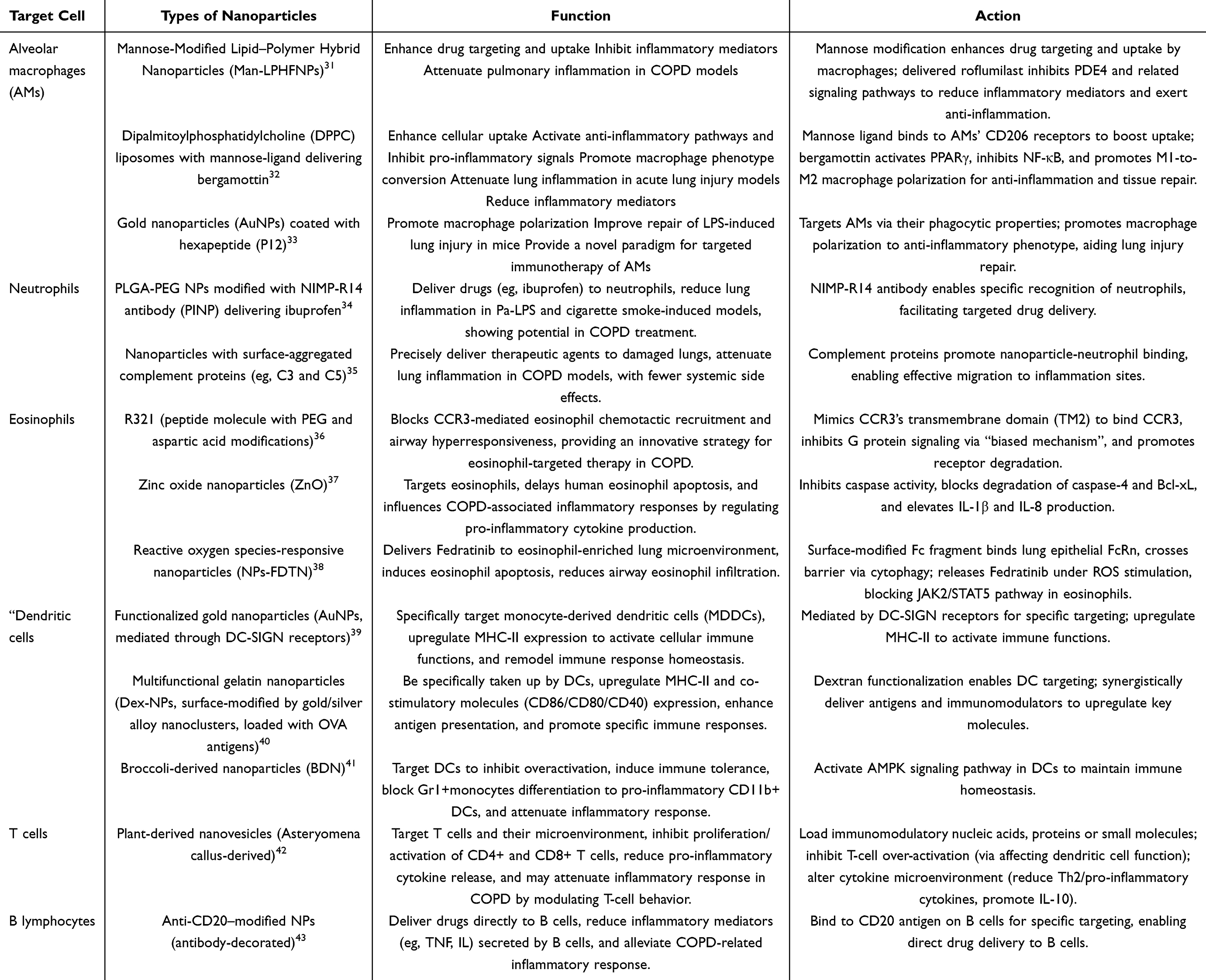 |
Table 1 The Comparison of Different Nanoparticle Therapies |
Inflammatory Cells
Alveolar Macrophages
Alveolar macrophages are an important component of the innate immune system and play a key role in chronic inflammation in COPD through their phagocytic and immunomodulatory functions, and positively correlate with the severity of the disease.44 Studies have demonstrated that the number of alveolar macrophages in COPD patients is markedly elevated, showing a 5 - to 10-fold increase compared to healthy controls.45–47 Furthermore, smokers exhibit significantly increased interstitial macrophage (IM) accumulation within alveolar septa, with total IM numbers and local density elevated by 36% (p < 0.01) and 56% (p < 0.001), respectively, relative to nonsmokers.48 However, prolonged exposure to harmful substances, such as cigarette smoke, can lead these cells to trigger abnormal inflammatory responses and cause dysfunctional phagocytosis.30 Aggregated macrophages secrete a variety of inflammatory mediators, including lipid mediators (eg, leukotrienes, prostaglandins), chemokines (eg, IL-8, IP-10), inflammatory cytokines (eg, TNF-α), growth factors (eg, TGF-β, TGF-α), as well as leukotriene B4 (LTB4) and reactive oxygen species (ROS), which, in varying degrees, influence the COPD inflammatory response, making alveolar macrophages an important target for COPD treatment.49,50 Nanomaterials are used to design targeted drug delivery systems for anti-inflammatory drug delivery by virtue of their unique physicochemical properties - such as tunable size for passive targeting across physiological barriers and surface modifiability for active ligand-mediated recognition. These nanocarriers significantly enhanced drug uptake by alveolar macrophages and inhibited the release of key pro-inflammatory cytokines, thereby alleviating inflammation in COPD.51,52
For example, mannose-modified lipid-polymer hybrid nanoparticles (Man-LPHFNPs) can deliver roflumilast targeted to alveolar macrophages. Such hybrid nanomaterials, on the one hand, improve the targeting and uptake efficiency of the drug in macrophages through mannose modification, and on the other hand, inhibit PDE4 through the delivered drug roflumilast, which in turn inhibits the related signaling pathways and reduces the production of inflammatory mediators, thus exerting anti-inflammatory effects. This study demonstrated that this targeted delivery system significantly attenuated the pulmonary inflammatory response in a COPD model, providing a new strategy for the treatment of chronic inflammation in COPD patients.31 Similarly, for the key target of alveolar macrophages, another study utilized bergamottin combined with dipalmitoylphosphatidylcholine (DPPC) liposomes to develop a lung-targeting bioactive lipid nanomedicine for precise therapeutic intervention. The surface-modified mannose ligand specifically binds to CD206 receptors on alveolar macrophages, significantly enhancing cellular uptake efficiency. Concurrently, delivery of the natural active molecule bergamottin activates the PPARγ pathway, effectively inhibiting NF-κB inflammatory signaling and promoting macrophage polarization from pro-inflammatory M1-type to anti-inflammatory M2-type. The resulting M2-type macrophages secrete anti-inflammatory factors, suppress inflammatory responses, and promote tissue repair.32 This nanomedicine significantly attenuated lung inflammation and reduced inflammatory mediator levels in an acute lung injury model. In a separate study, unique anti-inflammatory nanodrug, P12 (made of hexapeptides and gold nanoparticles), gold nanoparticles coated with a hexapeptide were designed to target alveolar macrophages via their phagocytic properties, similarly promoting macrophage polarization and improving repair of LPS-induced lung injury in mice.33,53 This approach provides a novel paradigm for targeted immunotherapy of alveolar macrophages. Consequently, macrophage-targeting nanosystems represent a promising therapeutic strategy for COPD and other chronic airway diseases. These studies not only verified the effectiveness of nanomaterials in COPD treatment but also provided a theoretical basis for the development of novel nanomedicines. Future studies should further optimize the design of nanomaterials, improve their targeting and safety, and explore their potential for clinical applications.
Neutrophils
Neutrophils are central to the pathogenesis of COPD, and airway neutrophils are increased in all patients with COPD, regardless of age of onset, clinical phenotype, or disease severity. As a key component of innate immune cells, a large body of data suggests that neutrophils play a key driving role in COPD airway inflammation, with their numbers strongly correlating with reduced FEV1, chronic sputum coughing, and degree of airway obstruction.49,54 In patients with COPD, the release of neutrophil products (eg, proteases such as neutrophil elastase NE, protease 3 Pr3, and histone B CatB) was significantly elevated55 Among them, protein hydrolases stimulate mucus secretion and affect mucus cilia clearance function, while neutrophils can increase protease activity, leading to further tissue damage.54 Hydrolytic enzymes such as elastase cause tissue damage by degrading the extracellular matrix, and after destroying the airway barrier function, more inflammatory mediators enter the lung tissues, which further produce chemokines and attract neutrophil infiltration, forming a vicious cycle.56 Therefore, effective targeted delivery of drugs to neutrophils is essential to reduce the inflammatory response, and nanomaterials are significant in this regard. Experiments have been conducted to develop a neutrophil-targeted nanodrug delivery system (PINP) using PLGA-PEG nanoparticles modified with NIMP-R14 antibody to specifically recognize neutrophils. PINP effectively delivered drugs such as ibuprofen to neutrophils in a mouse model and significantly reduced lung inflammation in Pa-LPS and cigarette smoke-induced inflammation models, demonstrating its potential in the treatment of COPD.34 In other experiments, by aggregating proteins on the surface of the nanoparticles to confer their tropism towards neutrophils, complement proteins (eg, C3 and C5) were utilized to promote nanoparticle-neutrophil binding for effective migration towards the site of inflammation, and to deliver the therapeutic agents to the damaged lungs precisely with fewer systemic side effects. Studies have shown that such nanoparticles significantly attenuate the inflammatory response of the lungs in COPD models, providing a new strategy for COPD treatment.35 These studies validate the effectiveness of nanomaterials in COPD treatment and provide a theoretical basis for novel nanomedicine development. Future studies should further optimize the design of nanomaterials that can target cell surface receptors based on ligand-receptor specific interactions to improve targeting and safety, and explore their potential for clinical applications.
Eosinophils
In COPD, eosinophils, as common inflammatory cells, are key cells in inflammation-driven and airway remodeling through the release of cytotoxic proteins and cytokines, which can both directly cause tissue damage and airway hyperresponsiveness and exacerbate inflammation and airway remodeling.57 Clinically, blood and sputum eosinophil counts are used as biomarkers to guide individualized treatment, and targeted therapy for them has become one of the key entry points for COPD treatment.58 Nanomaterials, as a research hotspot in recent years, have made some progress in targeting eosinophils. The effects of nanomaterials on eosinophils are manifold, involving apoptosis, cell signaling pathways, cell adhesion, and cytokine production, as well as influencing related cell signaling pathways that regulate inflammatory responses, all of which provide new directions for the development of therapeutic strategies targeting eosinophils in COPD disease.59,60 For example, R321 is a novel peptide molecule designed based on the second transmembrane helix of CCR3 receptor, with 27 units of polyethylene glycol (PEG) and three aspartic acid residues modified at the C-terminus. CCR3 is a key chemokine receptor recruited by eosinophils, and R321 binds to the receptor by mimicking the transmembrane structural domain (TM2) of CCR3 and utilizing the “biased mechanism By mimicking the binding of CCR3 transmembrane structural domain (TM2) to the receptor, R321 uses the “bias mechanism” to inhibit only the G protein signaling of CCR3 and promote the degradation of the related receptor, thus effectively blocking CCR3-mediated eosinophil chemotactic recruitment and airway hyperresponsiveness. By circumventing drug tolerance mechanisms, R321 has been demonstrated to provide an innovative strategy for eosinophil-targeted therapy in COPD.36 When specifically targeting the CCR3 receptor pathway, nanomaterials mediate suppression of inflammatory cell infiltration and amelioration of airway hyperresponsiveness. In addition, it was found that zinc oxide nanoparticles (ZnO) could target eosinophils and delay the apoptotic process of human eosinophils. The mechanism of action includes inhibition of cysteine asparaginase (caspase) activity, blocking the degradation pathway of caspase-4 and Bcl-xL, and significantly elevating the production of the pro-inflammatory cytokines IL-1β and IL-8. This regulatory effect may influence the pathologic course of COPD-associated inflammatory responses. Importantly, experimental validation in this study demonstrated that human eosinophils could serve as a novel direct action target for ZnO nanoparticles.37 In another asthma study, researchers constructed reactive oxygen species-responsive nanoparticles (NPs-FDTN) targeting FcRn. Its surface-modified Fc fragment was specifically delivered to the eosinophil-enriched inflammatory microenvironment of the lungs by binding to the FcRn receptor of lung epithelial cells and crossing the epithelial barrier via cytophagy. Under local ROS stimulation, the nanosystem releases encapsulated Fedratinib (FDTN), which directly induces eosinophil apoptosis in a concentration-dependent manner by precisely blocking the JAK2/STAT5 signaling pathway in eosinophils, thus effectively reducing the level of airway eosinophil infiltration and exerting therapeutic effects.38 The interventional efficacy of nanoformulations on COPD pathological progression has been experimentally validated, with molecular-level mechanistic elucidation further providing theoretical foundations for novel drug development. Subsequent investigations should prioritize precise optimization of nanocarrier structure-function compatibility, focus on overcoming enhancement bottlenecks in biological targeting precision and in vivo safety thresholds, while concurrently advancing systematic validation of clinical translation potential.61–63
Dendritic Cells
In COPD research, dendritic cells (DCs) have garnered extensive attention as critical mediators bridging innate and adaptive immunity.64 Studies have demonstrated that DCs in the lung tissues of COPD patients persistently remain activated, with their activation levels shown to positively correlate with clinical disease stages. Within immune response regulatory networks, the activation of inflammatory effector cells such as neutrophils and Th17 cells has been identified to be mediated by these professional antigen-presenting cells enriched in airway-parenchymal regions, thereby exerting pivotal regulatory functions.65 Based on these mechanisms, DC-targeted intervention strategies have been incorporated into the core technical framework of COPD prevention and treatment systems. To enhance disease management efficacy, novel therapeutic modalities focusing on DC functional regulation have been established as research priorities.66 Functionalized gold nanoparticles (AuNPs) mediated through DC-SIGN receptors have been confirmed to specifically target monocyte-derived dendritic cells (MDDCs), where upregulation of MHC-II expression is employed to activate cellular immune functions, thereby achieving remodeling of immune response homeostasis. The development of nanocarriers with lung DC-targeting capability and immunoregulatory properties has been supported by multidimensional evidence from this finding.39 A study was conducted by developing multifunctional gelatin nanoparticles (Dex-NPs) whose surface was modified by gold/silver alloy nanoclusters and loaded with ovalbumen (OVA) antigens, while dendritic cell targeting was achieved through dextran functionalization. The nanosystems were specifically taken up by DCs and significantly upregulated the expression of MHC-II molecules and co-stimulatory molecules (CD86/CD80/CD40) on the surface of DCs by synergistically delivering antigens and immunomodulators, thereby enhancing antigen presentation and promoting specific immune responses.40
In another study, broccoli-derived nanoparticles (BDN) of plant origin were used to target DCs to inhibit DCs overactivation and induce immune tolerance by activating the adenosine monophosphate-activated protein kinase (AMPK) signaling pathway. The material blocked the differentiation of Gr1⁺monocytes to pro-inflammatory CD11b⁺ DCs and effectively attenuated the inflammatory response. Both cellular and animal experiments in this study confirmed that BDN maintains immune homeostasis by specifically activating the AMPK pathway in DCs.41 Structural parameters of nanomaterials require prioritized optimization in subsequent investigations to improve targeting precision and biocompatibility, while their clinical translation feasibility necessitates systematic evaluation.
T Cells
The total number of T lymphocytes in the lung parenchyma and peripheral and central airways of patients with COPD is significantly increased, with a predominance of CD8+ cytotoxic T cells and CD4+ helper T cells, with a greater increase in CD8+ cells. During an immune imbalance, the infiltration of CD4+ and CD8+ cells into the airways is exacerbated, releasing large amounts of cytokines, exacerbating airway inflammation and promoting Airway remodelling, leading to a decrease in FEV1 and a correlation between the number of T cells and the severity of alveolar destruction and airflow obstruction.67,68
For example, it has been demonstrated that the use of Asteryomena callus-derived nanovesicles as natural nanocarriers, loaded with immunomodulatory nucleic acids, proteins or small molecules, can target T cells and their microenvironment, inhibit the proliferation and activation of CD4+ and CD8+ cells, and reduce the release of pro-inflammatory cytokines from them, which could reduce inflammatory responses and improve symptoms of the rat asthma model. In addition, the nanovesicles could further inhibit the over-activation of T cells by affecting the function of dendritic cells. Meanwhile, the vesicles indirectly altered the cytokine microenvironment by reducing the production of Th2-type cytokines (eg, IL-4, IL-5, IL-13) and pro-inflammatory cytokines (eg, TNF-α) and promoting the secretion of the anti-inflammatory factor IL-10, which attenuated airway inflammation and airway hyperresponsiveness and improved asthma symptoms.42 Based on the results of the asthma model, we speculate that nanovesicles may attenuate the inflammatory response by modulating T-cell behaviour in COPD. These studies not only validate the efficacy of nanomaterials in immunomodulation but also provide a theoretical basis for the development of novel nanomedicines targeting COPD.
B Lymphocytes
While T cells dominate adaptive immunity in COPD, B lymphocytes contribute to chronic inflammation through autoantibody production. B lymphocytes play a key role in the inflammatory process of COPD through their production of antibodies and their involvement in lymphoid follicle formation.69 Targeting B cells can effectively reduce the production of inflammatory mediators and thereby alleviate the symptoms of COPD. Specific targeting of B cells has been investigated using surface-modified anti-CD20 nanoparticles that bind to the CD20 antigen on B cells. This targeting strategy can deliver drugs directly to B cells, thereby reducing the inflammatory mediators secreted by B cells, such as tumour necrosis factor (TNF) and interleukin (IL), and thus alleviating the associated inflammatory response.43 The pivotal regulatory role of B cells in immune response processes has been definitively elucidated, with their pathological mechanism involvement demonstrated to exert determinant influences during disease progression in autoimmune disorders and malignant neoplasms. As a novel drug delivery platform, the clinical advantages of nanocarriers have been systematically clarified, encompassing characteristics such as reduced drug toxicity, prolonged in vivo circulation half-life, and enhanced therapeutic efficacy.70 Based on these technical attributes, the development of targeted nanomaterials for B-cell subsets in COPD has been prioritized in research agendas, where feasibility validation of this technical pathway provides a theoretical foundation for the establishment of novel therapeutic strategies in COPD management.
Targeting Inflammatory Mediators
As a typical inflammatory disease, the pathologic complexity of COPD is not only reflected in the involvement of diverse inflammatory and structural cells as described previously, but also in the formation of a complex regulatory network by these cells through the release of inflammatory mediators. These mediators play a key role in the development of COPD through synergistic or antagonistic effects.30,71 In COPD, the aberrant expression and action of inflammatory mediators (eg TNF-α, IL-6, IL-8, CRP, IL-1β and MCP-1, etc) form a complex network that together act on the relevant inflammatory cells, from initiating the inflammatory response to maintaining and amplifying the inflammatory response, to drive the chronic inflammatory process in COPD, leading to the gradual decline in lung function and disease progression in patients.72–77 Nanomaterials engineered for the delivery of anti-inflammatory agents have demonstrated significant potential in modulating key inflammatory mediators. For example, one study successfully developed the ability to downregulate interleukin-1 receptor (1L-1R) and toll-like receptor (TLR) signalling pathways, such as IL-1 receptor-associated kinase (IRAK1) and TNF receptor-associated factor (TRAF6), after using nanocomposite microparticles (NCMPs) nanoparticles to deliver miRNA-146a to the lung, thereby reducing the production of inflammatory cytokines (eg, IL-1β, IL-6 and IL-8), thereby inhibiting the inflammatory response78 and providing new ideas for COPD treatment.79 In investigating the potential of chitosan-encapsulated lipid carriers loaded with quercetin as an oral nanoplatform for depression management, quercetin-chitosan nanolipid carriers (QU-CS-NLCs) resulted in a significant reduction in the level of CRP, a marker of inflammation, by 65.4% in an LPS-induced depression model through enhanced anti-inflammatory delivery efficacy.80 In another study, chitosan-coated solid lipid nanocapsules (SLN-chitosan) loaded with beriberi (Ber) were able to reduce inflammatory cell infiltration in bronchoalveolar lavage fluid (BALF), down-regulate pro-inflammatory cytokines, such as IL-1β, IL-6, IL-17, TNF-α, etc., and reduce myeloid oxidant levels. IL-1β, IL-6, IL-17, TNF-α, and other pro-inflammatory cytokines in BALF, as well as decreasing myeloperoxidase (MPO) activity and increasing superoxide dismutase (SOD) activity, which could reduce airway inflammation and oxidative stress, and create a new paradigm in the treatment of COPD.81 Meanwhile, in an in vitro model of COPD induced by cigarette smoke extract (CSE), poly(lactic acid)-hydroxyacetic acid) (PLGA) nanoparticles loaded with 18-β 18-β-glycyrrhetinic acid (18βGA) were able to reduce the generation of reactive oxygen species (ROS), down-regulate senescence-associated β-galactosidase (SA-β-Gal) and p21, and reduce pro-inflammatory genes (CXCLA) and p21, while reducing the expression of CXCLA. It also reduced the levels of pro-inflammatory genes (CXCL1, IL-6, TNF-α) and inflammation-related proteins (IL-8), and restored the expression of immunoregulatory factors such as IL-15, RANTES, and MIF, thus reducing oxidative stress, cellular senescence, and Airway inflammation, and exploring a new direction for the treatment of COPD.82 Inflammatory mediators are pivotal to the pathological process of COPD, and nanomaterials have the potential to attenuate the inflammatory response by modulating the TLR signalling pathway and the expression of inflammatory mediators. This study not only validates the efficacy of nanomaterials in the treatment of COPD but also provides a theoretical basis for the development of novel nanomedicines. Future studies should optimize nanomaterial design to enhance specificity against key inflammatory mediators, improve targeted delivery to activated immune cells within COPD microenvironments, and evaluate clinical potential for suppressing inflammation-driven exacerbations.
Oxidative Stress-Related
Beyond inflammation, oxidative stress exacerbates COPD via ROS-mediated damage to proteins, lipids, and DNA. Mitochondrial dysfunction further amplifies ROS production, thereby creating a self-perpetuating cycle.83 Targets Oxidative stress plays a central role in the pathogenesis of COPD, and its main sources include the overproduction of reactive oxygen species (ROS) and reactive nitrogen species (RNS).84 These substances can promote the expression of pro-inflammatory genes by directly damaging cellular components, such as proteins, lipids, and DNA, and by activating several signalling pathways, including transcription factors like NF-κB and histone acetyltransferases, thereby enhancing the inflammatory response in the lung. For example, ROS can increase the production of inflammatory factors by activating NF-κB and turning on the transcription of several inflammatory genes,85 as well as promoting lipid peroxidation and generating reactive intermediates (eg, 4-hydroxy-2-enolenic aldehyde, 4-HNE) that further exacerbate lung tissue damage.84,86 Cigarette smoke, a significant source of exogenous ROS, contains a large number of oxidants and free radicals that directly contribute to oxidative stress.87 In addition, mitochondria, as the primary site of energy supply in the cell, generate a certain amount of ROS during metabolism, and when mitochondrial function is impaired (especially when the electron transport chain is dysfunctional), this leads to overproduction of ROS, which is a major source of endogenous ROS. These excess ROS (superoxide anion O2-- and hydrogen peroxide H2O2) can accumulate intracellularly and cause oxidative stress, leading to damage to cellular proteins, lipids, and DNA, as well as activating a number of signalling pathways (eg, proteases such as caspase-3) and triggering apoptosis.88
Nanomaterials show promise in mitigating oxidative stress. By targeting and delivering antioxidants, nanomaterials can effectively mitigate oxidative damage in mitochondria.10 Oxidative stress in mitochondria is closely related to the Nrf2 signalling pathway, which is involved in resisting oxidative stress-mediated injury and also regulates processes such as inflammation, calcium homeostasis, apoptosis, iron death, and mitochondrial oxidative stress. For example, as an Nrf2 activator, dimethyl fumarate (DMF) promotes the transcription of antioxidant genes by activating the Nrf2-Keap1 signalling pathway, which in turn reduces ROS production and inhibits mitochondrial oxidative stress-induced injury. In our studies, DMF nanoparticles were delivered to the lungs for their antioxidant effects using an aerosol dispersion technique89,90 (Figure 1). This strategy provides a novel idea for the treatment of oxidative stress in COPD and demonstrates the unique advantages of nanomaterials in modulating complex oxidative stress networks. Oxidative stress plays a central role in the pathogenesis of COPD, and nanomaterials have the potential to attenuate oxidative stress damage through targeted delivery of antioxidants. These studies not only validate the efficacy of nanomaterials in regulating oxidative stress but also provide a theoretical basis for the development of novel nanomedicines for COPD.
 |
Figure 1 Mechanisms of DMF Activation of the Nrf2 Pathway Attenuate ROS Storm and Alleviate Lung Injury. DMF-loaded nanoparticles are delivered to the lungs via aerosol inhalation, enabling targeted release of DMF in pulmonary tissues. DMF binds to Keap1, disrupting the Keap1–nuclear factor erythroid 2-related factor 2 (Nrf2) complex and facilitating Nrf2 nuclear translocation. Nuclear Nrf2 activates the transcription of downstream antioxidant genes, including HO-1 and NQO1, thereby reducing ROS levels and RNS levels and mitigating mitochondrial oxidative damage. Created in BioRender. Qianyue, Z. (2025) https://BioRender.com/rvyw3io. Abbreviations: DMF, dimethyl fumarate; Nrf2, nuclear factor erythroid 2-related factor 2; Keap1, Kelch-like ECH-associated protein 1; ROS, reactive oxygen species; RNS, reactive nitrogen species, HO-1, heme oxygenase-1; NQO1, NAD(P)H quinone dehydrogenase 1. |
Proteases - Targets Related to an Imbalance in the Anti-Protease System
In the context of COPD, the release of neutrophil elastase (NE), proteinase 3, and histone B by neutrophils, along with matrix metalloproteinases (MMPs, eg, MMP-9, MMP-12) and histones (eg, cathepsin L and S) produced by macrophages, has been identified as a key factor in the destruction of lung tissue.91 The excessive release of these proteases, in combination with the relative deficiency of antiproteases (eg, α1-antitrypsin, α1-AT), gives rise to a protease-antiprotease imbalance, which is a significant contributing factor to the progression of COPD.92 Smoking, a major trigger of COPD, has been shown to both increase protease release and decrease the activity of α1-AT through oxidative effects. Concurrently, inflammatory cells release a substantial amount of inflammatory mediators (eg, TNF-α, IL-1β) for production, and these mediators further promote the release of MMPs, which ultimately leads to the worsening of the imbalance.93–95
The targeting of the protease-antiprotease imbalance by nanomaterials has been demonstrated to show promising applications. For instance, lipid nanomaterials interbilayer-crosslinked multilamellar vesicles (ICMVs) loaded with sivelestat, an inhibitor of neutrophil elastase (NE), have been shown to deliver NE inhibitors through targeted delivery mechanisms, thereby prolonging the duration of drug action and significantly increasing its bioavailability. This nanomaterial has demonstrated the significant efficacy of sivelestat in a mouse model of LPS-induced endotoxic shock. Under conditions of significant NETs formation inhibition, the reduction in inflammatory mediator levels and improvement in murine survival rates were simultaneously observed (Figure 2). The inhibitory effect on NE activity has been verified to achieve cascade downregulation of secondary proteases (eg, matrix metalloproteinases) activities through modulation of inflammatory factor secretion networks, ultimately leading to the restoration of dynamic equilibrium in the protease-antiprotease system being accomplished.96 Existing research data have not only allowed the theoretical foundation for COPD treatment paradigm innovation to be established, but also enabled the unique advantages of nanomaterials in inflammatory cascade regulation to be systematically revealed.
 |
Figure 2 Mechanisms of ICMVs Targeting of Protease-Antiprotease Imbalance Attenuates COPD Pathogenesis. ICMVs loaded with sivelestat, a NE inhibitor, deliver NE inhibitors via a targeted delivery mechanism to neutrophils and macrophages. Sivelestat binds to and inhibits NE, suppresses the activation of MMPs, and reduces the release of pro-inflammatory cytokines ([TNF-α]/ [IL-1β]↓). This cascade attenuates lung tissue damage and restores the protease-antiprotease homeostasis. Created in BioRender. Qianyue, Z. (2025) https://BioRender.com/euz90h1. Abbreviations: ICMVs, Interbilayer-crosslinked multilamellar vesicles; NE, Neutrophil elastase; NETs, MMPs, Matrix metalloproteinases; TNF-α, Tumor necrosis factor-alpha; IL-1β, Interleukin-1 beta. |
Nanotherapeutic strategies targeting proteases/inflammatory factors have been identified as potential intervention protocols for COPD. In subsequent studies, the targeting specificity and biosafety profiles of nanocarriers within COPD pathological microenvironments require prioritized elucidation, with the advancement of their clinical translation process thereby being realized.
Targets Related to Airway Remodeling
COPD, as a chronic respiratory disorder with multidimensional pathological features, has had its core pathomechanisms clearly elucidated. The pivotal role of airway remodeling has been established through substantial research evidence.97,98
The interaction mechanisms among pathological elements, including epithelial cell injury, fibrotic progression, and structural alterations in smooth muscle, have been systematically elucidated. These elements have been demonstrated to collectively influence the spatiotemporal progression of airway remodeling through molecular network regulation, thereby exerting determinant effects on the clinical outcomes of COPD.3 The chemotactic cascade reaction triggered by epithelial damage has been proven to significantly exacerbate the degree of airway obstruction.99 Abnormal deposition of extracellular matrix (ECM) and compositional alterations at injury sites have been experimentally verified.100 These modifications are achieved through mechanical property regulatory pathways, manifested as decreased airway elasticity and compliance, ultimately leading to enhanced airway wall stiffness. This pathological phenomenon has been confirmed to exhibit direct correlation with the formation of fixed stenosis and the deterioration of airflow limitation.101 Aberrant proliferation and hypertrophy of airway smooth muscle have been identified as core driving factors in the remodeling process.102 The thickening effect not only induces structural modifications of the airway wall but also results in markedly elevated ventilatory resistance, with consequent impairment of respiratory function being accomplished.103 Epithelial cells, serving as critical regulatory units in airway remodeling, have their stem cell characteristics and plasticity activated through stromal interaction mechanisms, playing central roles in airway repair processes.104 The susceptibility of COPD patient-derived epithelial cells to remodeling processes has been substantiated by multiple investigations, and the efficacy of nanomaterial-based pulmonary epithelial targeting strategies has been successfully validated. For example, nanoparticles such as multilamellar bodies, mesoporous nanoparticles, and liposomes can carry therapeutic agents (eg, drugs or DNA-expressing vectors), which can be delivered to the airway epithelial cells. This delivery system provides a viable clinical therapeutic approach for targeting damage to the respiratory epithelium in airway remodelling, promoting the repair and regeneration of epithelial cells and, by extension, targeted treatment of Airway remodelling.105
Furthermore, nanomaterials have shown potential in inhibiting fibrosis. For example, spermine-functionalized acetylated dextran nanoparticles (SpAcDex NPs) can target the NFκB signaling pathway in fibroblasts by delivering NFκB decoy ODN. This mechanism effectively inhibits the fibrotic process by blocking transforming growth factor-beta (TGF-β)-mediated differentiation of fibroblasts into myofibroblasts while suppressing collagen synthesis and deposition.106 The fibrotic therapeutic strategy in the airway remodeling process of COPD has been confirmed to provide a theoretical foundation for the application of nanomaterials. Through research verification, the development of multi-target synergistic therapeutic regimens based on the complex COPD microenvironment has been established as a key future research direction to achieve profound inhibition of Airway remodeling-associated fibrosis. During the pathological process of airway remodeling, the proliferation and hypertrophy of smooth muscle have been clearly identified as critical pathological features, rendering them potential targets for therapeutic intervention.
Through experimental studies, the biological behaviors of airway smooth muscle cells (ASMCs) have been found to be regulated by silver nanoparticles (AgNPs), specifically manifested as significantly upregulated expression of inducible nitric oxide synthase (iNOS), thereby achieving inhibition of ASMC proliferation and induction of apoptosis. This mechanism further confirms that the biosynthesis of nitric oxide (NO) is effectively promoted, subsequently generating significant effects in reducing airway wall thickness, decreasing airway resistance, and improving ventilation function (Figure 3). Notably, as a molecule with well-characterized bronchodilatory properties, the physiological functions of NO have been demonstrated to include alleviation of smooth muscle contraction and improvement of airway hyperresponsiveness.107
 |
Figure 3 AgNPs Attenuate COPD Airway Remodeling via iNOS/NO-Mediated Inhibition of Smooth Muscle Hyperplasia. AgNPs target ASMCs, significantly upregulating inducible nitric oxide synthase (iNOS) expression, inhibiting ASMC proliferation, and inducing apoptosis. The increased nitric oxide (NO) biosynthesis consequently reduces airway wall thickness, decreases airway resistance, thereby attenuating airway remodeling and improving ventilatory function. Created in BioRender. Qianyue, Z. (2025) https://BioRender.com/em29uc2. Abbreviations: AgNPs, Silver nanoparticles; ASMCs, Airway smooth muscle cells; iNOS, Inducible nitric oxide synthase; NO, Nitric oxide. |
These findings enable the construction of novel therapeutic strategies for COPD-related airway smooth muscle hyperplasia. During COPD progression, the pathologically significant phenomenon of airway remodeling has been revealed to be amenable to multidimensional interventions through nanomaterials, encompassing precise targeting of epithelial cells, effective suppression of fibrotic processes, and dynamic regulation of smooth muscle proliferation. Through relevant studies, significant enhancement of drug utilization efficiency has been achieved, while innovative development of new therapeutic pathways for COPD has been accomplished. Subsequent research should prioritize the optimization of nanomaterial design, improvement of targeting specificity and safety profiles, and comprehensive exploration of clinical translation potential.108
Application of Nanoparticles in COPD
The following five main categories of nanomaterials are currently employed in the treatment of COPD: polymer nanoparticles, dendrimers, inorganic nanoparticles, lipid nanoparticles (LNP), and bioactive nanoparticles of plant origin. The unique physicochemical properties and functionalized design of these materials enable them to target and regulate the core pathological mechanisms of COPD, such as the inflammatory cascade, oxidative stress, and protease imbalance. This has the potential to significantly enhance therapeutic precision and efficacy.109,110 The subsequent section will systematically analyse their mechanism of action and research progress.
Polymer Nanoparticles
Among polymer nanoparticles, polylactic acid-hydroxyacetic acid copolymer (PLGA) nanoparticles have been the focus of research and application due to their biodegradable structure consisting of lactic acid and hydroxyacetic acid linked by ester bonds.111 Through precise structural design and functional optimisation, PLGA nanoparticles can achieve targeted drug delivery. Experimental results demonstrated that PLGA-targeted PINP nanoparticles exhibited significantly higher neutrophil-targeted delivery efficiency compared to non-targeted PNPs, with a 6.5-fold increase (17.58% vs 2.72%, p < 0.001), as well as controlled release and improved stability. This significantly enhances their unique advantages in the treatment of COPD.34
The nanoscale properties of PLGA nanoparticles have been demonstrated to enhance drug efficacy and reduce side effects, thereby significantly improving patients’ therapeutic experience.112–114 Specifically, in a study by Mohamed et al, the inflammatory process in COPD was targeted through the loading of miR-146a into PLGA nanoparticles. The application of miR-146a nanoparticles was confirmed by experimental data to induce a significant reduction in interleukin-1 receptor-associated kinase 1 (IRAK-1) gene expression levels, with precise regulation of inflammatory responses being achieved through this mechanism.79 The construction of poly(lactic-co-glycolic acid) (PLGA) nanoparticles was accomplished in related studies, and the encapsulation of the anti-inflammatory drug bunofen was realized via this nanocarrier, while the therapeutic targeting of neutrophil-mediated inflammatory responses in COPD was established through this strategy.115 Lipopolysaccharide (LPS)- and cigarette smoke-induced lung injury was demonstrated to be markedly alleviated by this nanodrug delivery system, with its therapeutic potential for COPD being further validated through animal experimental data.34 The clinical translational value of the PLGA-based nanotherapeutic system was substantiated by existing research findings, and the implementation of additional clinical trials was recommended to systematically evaluate the efficacy parameters and safety profile of this nanosystem in COPD treatment.116
Dendrimers
Dendritic macromolecules, as a class of synthetic branched topological polymers, exhibit a three-dimensional architecture systematically constructed with a central core layer, internal repeating unit layers, and peripheral functional unit layers. Alcohol chain-modified polyamidoamine (PAMAM) and polyphenylene ether (PPE) are classified as canonical therapeutic modification forms within this category.117 The endowment of multifunctional properties is achieved in these macromolecules through surface functionalization design strategies, leading to a significant expansion of their application potential in drug delivery systems and biomedical domains.118
Au-PAMAM dendrimer nanocomposites demonstrate antioxidant properties conclusively superior to gold-core polypropylene imine (PPI) dendrimers and ascorbic acid controls. Quantitatively, the oxidative degradation rate constant of Au-PAMAM dendrimers was determined to be 85-fold higher than that of ascorbic acid, a magnitude difference that fully elucidates their unique advantage as drug delivery systems for selective targeting of oxidative stress. The efficacy of this nanosystem in precision regulation of oxidative stress responses has been experimentally validated, with pathophysiological mechanisms underlying its therapeutic application in chronic obstructive pulmonary disease (COPD) being established.119 Consequently, the feasibility of pioneering novel COPD treatment paradigms has been rigorously proposed based on these findings.117,120
Inorganic Nanoparticles
Significant advantages in disease diagnosis and therapeutic delivery applications have been demonstrated by inorganic nanoparticles comprising precious metal species (eg, aurum, argentum), ferric oxide derivatives, and silica-based architectures, which are fundamentally attributed to their distinctive physicochemical property profiles.121,122 Mesoporous silica nanoparticles (MSNs) are important carriers for targeted delivery in the lungs due to their high specific surface area (~1000 m²/g), tunable pore size (2–30 nm), and easy functionalization of surface silanol groups. Studies have shown that MSNs can escape alveolar macrophage phagocytosis via polyethylene glycol (PEG) modification and enhance mucus layer penetration via mucin-penetrating peptides, significantly increasing lung drug residence time (from <2 h to >6 h) and bioavailability (from <15% to >50%).123,124 In related studies, researchers have also used rare earth metals such as cerium oxide (CeO2), whose unique cubic fluorite crystal structure and Ce³⁺ /Ce4⁺ redox cycling properties give it excellent antioxidant and catalytic properties. Cerium nanoparticles (CeNPs) mimic the activities of endogenous antioxidant enzymes (eg, SOD and catalase), reduce oxidative damage by scavenging reactive oxygen species (ROS), and activate the Nrf2/ARE pathway to enhance cellular antioxidant defence mechanisms.125
In addition, iron oxide nanoparticles (IONPs) have potential in COPD airway inflammation modulation due to their superparamagnetic properties, which allow precise aggregation at the focal site via an external magnetic field, as well as the modification of anti-inflammatory peptides (eg, chlorotoxins) on their surface to improve targeting.126 The suppression of nuclear factor κB (NF-κB) activation and NLRP3 inflammasome pathways has been demonstrated to mediate the downregulation of pro-inflammatory cytokine expression (eg, TNF-α, IL-1β) through cerium oxide nanoparticle (CeNP) intervention.127 Ideal therapeutic candidacy for chronic obstructive pulmonary disease (COPD) management is conferred upon these nanostructures by their synergistic antioxidant/anti-inflammatory capacities coupled with precisely programmable surface functionalization modalities.
Lipid Nanoparticles (LNPs)
Significant therapeutic advantages in chronic obstructive pulmonary disease (COPD) management have been demonstrated through strategic optimization of auxiliary lipid constituents (DOPC/DOPS) in lipid nanoparticle (LNP) formulations.128 Targeted delivery to bronchial epithelial cells is specifically enabled by the engineered RNA encapsulation efficiency of these nanostructured systems, with concomitant minimization of off-target systemic effects. These nanoparticles are capable of delivering anti-inflammatory mRNAs (eg, IL-10) or silencing inflammation-related siRNAs (eg, NF-κB).129,130 For example, Tam et al showed that DOPS-LNP delivery of siRNA resulted in a more than 90% reduction in the expression of target proteins and significantly suppressed the chronic inflammatory response. After nasal delivery, luciferase expression in the nose and lung increased tenfold, suggesting that LNPs are suitable for localised therapy in COPD.131
In addition, Ongun et al further optimised the PEG lipid content (1.5 mol%) to avoid inhibition of cellular uptake while maintaining the stability of LNP nebulisation, providing a novel strategy for precise delivery to deep airways such as terminal fine bronchi and alveoli.132 Future studies should focus on further validating the long-term efficacy and safety of LNPs in animal models of COPD and exploring the synergistic effects of adjuvant lipids with PEG to accelerate the clinical translation of gene therapy with LNPs.133,134
Plant-Based Nanoparticles
Clinical management of COPD is challenged by the limited efficacy and significant side effects of traditional medications, prompting plant-derived biocomponents to become the focus of emerging therapeutic strategies. Nano-formulations based on plant active ingredients provide a new paradigm for efficient and low-toxicity therapy by enhancing bioavailability, enabling precise lung targeting, providing controlled release protection and synergistic effects, significantly improving COPD lung function indices (eg, FEV₁), inhibiting inflammatory factors (eg, IL-6/TNF-α), and reducing acute exacerbation frequency. The effectiveness of this strategy in plant-derived drug delivery has been well-studied.135–138 Taking liquorice (Glycyrrhiza glabra) as an example, the main active component of this common medicinal plant, 18-β-glycyrrhizinic acid (18βGA), exhibits multiple pharmacological effects such as anti-inflammatory, antioxidant, and antiviral effects,139 providing a scientific basis for its use in the treatment of COPD. It was shown that 18βGA-loaded poly(lactic acid)-hydroxyacetic acid copolymer (PLGA) nanoparticles significantly suppressed the inflammatory response by inhibiting NF-κB nuclear translocation and reducing cytokine expression in an in vitro model of cigarette smoke-induced COPD. In addition, this study found that 18βGA-PLGA nanoparticles significantly reduced CSE-induced reactive oxygen species (ROS) generation. Specifically, 5% cigarette smoke extract significantly increased intracellular ROS levels in BCiNS1.1 cells, while ROS generation was reduced by 39.5% after pretreatment with 18βGA-PLGA nanoparticles, further emphasising the significant benefits of plant-based nanoparticles in antioxidant therapy.82
The cigarette smoke extract (CSE)-induced elevation of reactive oxygen species (ROS) levels in 16HBE bronchial epithelial cells and RAW264.7 macrophages was demonstrated to be effectively suppressed by berberine (BBR)-incorporated liquid crystalline nanoparticles (BBR-LCNs).140 A dose-dependent reduction in ROS generation was observed through pretreatment experiments, with concomitant significant neutralization of CSE-triggered oxidative stress. In CSE-stimulated 16HBE cells, upregulated gene expression of inflammatory cytokines, including IL-1β, IL-6, and TNF-α, was identified, whereas transcriptional levels of these mediators were markedly suppressed following BBR-LCNs pretreatment. Concurrent detection of decreased TNF-α gene expression and reduced nitric oxide (NO) production in RAW264.7 cell models further corroborated the anti-inflammatory efficacy, establishing the therapeutic potential of botanically derived nanomaterials in COPD management through dual antioxidative/anti-inflammatory mechanisms.141 The molecular pathways underlying these formulations’ actions in COPD pathogenesis require elucidation, while their safety profiles and therapeutic outcomes necessitate systematic validation via clinical trials to facilitate expansion of clinical translation pathways for plant-based nanotherapeutics.142
Conclusion and Outlook
In the field of COPD, nanomedicines have gradually moved from proof-of-concept to clinical translation, in which inhalable nanoparticle formulations have been realized for precise delivery through devices such as dry powder inhalers, which are advancing clinical trials in COPD by virtue of the advantages of controllable particle size and strong targeting.143–147 A recent Phase II study showed that dexamethasone liposome nanoparticles (Lipo-Dexa) specifically targeted alveolar macrophages, reducing inflammatory factor levels by 60%.148 The nebulized inhalation route, on the other hand, creates high local concentrations in the lungs and significantly reduces systemic toxicity.149,150 However, clinical translation still faces multiple challenges and bottleneck, not only the optimization of drug encapsulation rate and stability, lung physiological barrier breakthrough, the guarantee of inter-batch consistency of physicochemical properties in large-scale production, biosafety also warrants attention, as studies have demonstrated that seven-day exposure of murine lungs to aluminum oxide nanoparticles (Al2O₃ NPs) results in emphysema and small airway remodeling, accompanied by exacerbated inflammation and apoptosis,151 and the harmonization of regulatory standards, which constrains its clinical translation, highlighting the potential of nanomedicines for clinical development in this field and the problems to be solved, as well as technical and cost challenges of adapting to the heterogeneity of patients in personalized medicine, all of which need to be solved through cross-subsidization and cross-subsidization.20–22,25,152 At the same time, the technical and cost challenges of adapting to patient heterogeneity in personalized medicine need to be gradually solved through interdisciplinary collaboration and technological innovation.12
Nanocarriers operate in COPD therapy via three core mechanisms: targeted delivery (enhancing lesion-specific recognition through surface chemistry modifications, improving drug-loading efficiency153), slow-release modulation (sustaining therapeutic effects via controlled biodegradation, validated by in vitro success154), slow-release modulation (sustaining therapeutic effects via controlled biodegradation, validated by in vitro success155).156,157 Despite these advances, clinical translation faces critical barriers: (1) low target retention rates limiting drug efficacy;158 (2) nanomaterial toxicity risks, exemplified by silica nanoparticles (SiNPs) inducing lung fibrosis (150% collagen increase, 1.8-fold TGF-β upregulation, *p*<0.05) and systemic Th2 inflammation (2-fold mast cell accumulation, *p*<0.05) in murine models;159–162 and (3) stringent FDA regulations delaying development.163
Future efforts must prioritize intelligent stimulus-responsive systems for inflammation-triggered drug release,164,165 expand applications to asthma, lung cancer, and infections,166 and integrate AI-driven genomic/proteomic analysis with computational nanomedicine optimization to enable etiology-specific precision therapies, as demonstrated in oncology.167 These interdisciplinary strategies promise to accelerate translational pipelines, advancing COPD-specific nanotherapeutics toward clinical reality.168 Focusing also on the key role of interactions between axes of pathology in driving COPD progression, could such interactions be a key consideration in the design of holistic nanotherapies? The hypothesis becomes reality that therapies can target multiple pathways simultaneously, breaking the self-perpetuating cycle of the disease.169,170 Such interdisciplinary innovations are poised to accelerate the translational trajectory of nanomedicine from bench to bedside, ultimately enabling precision therapeutics for respiratory disorders.
Acknowledgments
This work was supported by grants from the National Natural Science Foundation of China (82160016, 82560078), Yunnan Province’s Xingdian Talent Support Program Famous Doctors Special Project/Medical and Health Talents Special Project (YNWR-MY-2020-013 and XDYC-YLWS-2023-0043), and Yunnan Provincial Innovation Team for Respiratory and Pulmonary Circulation Diseases (No. 202405AS350018), the Yunnan Fundamental Research Projects (202501CF070056), and the Yunnan University Medical Research Foundation (YDYXJJ2024-0026).
Disclosure
The authors report no conflicts of interest in this work.
References
1. World Health Organization. Chronic obstructive pulmonary disease (COPD). 2024. Available from: https://www.who.int/news-room/fact-sheets/detail/chronic-obstructive-pulmonary-disease-(copd).
2. Meng K, Chen X, Chen Z, et al. Burden of chronic obstructive pulmonary disease in adults aged 70 years and older, 1990-2021: findings from the Global Burden of Disease Study 2021. PLoS One. 2025;20(1):e0316135. doi:10.1371/journal.pone.0316135
3. (GOLD), G.I.f.C.O.L.D. Global strategy for prevention, diagnosis and management of COPD: 2025 Report. 2025. Available from: https://goldcopd.org/2025-gold-report/.
4. Olsson B, Bondesson E, Borgström L, et al. Pulmonary drug metabolism, clearance, and absorption. In: Control Pulmonary Drug Delivery. 2011:21–50.
5. Iheanacho I, Zhang S, King D, et al. Economic burden of chronic obstructive pulmonary disease (COPD): a systematic literature review. Int J Chronic Obstr. 2020;15:439–460. doi:10.2147/COPD.S234942
6. Ma X, Zhao Y, Liang X-J. Theranostic nanoparticles engineered for clinic and pharmaceutics. Acc Chem Res. 2011;44(10):1114–1122. doi:10.1021/ar2000056
7. Stater EP, Sonay AY, Hart C, et al. The ancillary effects of nanoparticles and their implications for nanomedicine. Nat Nanotechnol. 2021;16(11):1180–1194. doi:10.1038/s41565-021-01017-9
8. Fan Y, Yang Z. Inhaled siRNA formulations for respiratory diseases: from basic research to clinical application. Pharmaceutics. 2022;14(6):1193. doi:10.3390/pharmaceutics14061193
9. Wang W, Huang Z, Huang Y, et al. Pulmonary delivery nanomedicines towards circumventing physiological barriers: strategies and characterization approaches. Adv Drug Deliv Rev. 2022;185:114309. doi:10.1016/j.addr.2022.114309
10. Xu Y, Liu H, Song L. Novel drug delivery systems targeting oxidative stress in chronic obstructive pulmonary disease: a review. J Nanobiotechnol. 2020;18(1):145. doi:10.1186/s12951-020-00703-5
11. Kunda NK, Somavarapu S, Gordon SB, et al. Nanocarriers targeting dendritic cells for pulmonary vaccine delivery. Pharm Res. 2013;30(2):325–341. doi:10.1007/s11095-012-0891-5
12. Wang S, Zhou H, Mao J, et al. Precision nanomedicine for pneumonocyte-targeting: emerging strategies and clinical prospects in refractory pulmonary disease therapy. Biomaterials. 2025;323:123420. doi:10.1016/j.biomaterials.2025.123420
13. Wang Y, Xuan W, Mao C, Liu Y. Inhalable nucleic acid therapeutics for chronic pulmonary disease: progress, challenges, and prospects. Acta Biomater. 2025.
14. Al Faraj A, Sultana Shaik A, Afzal S, et al. MR imaging and targeting of a specific alveolar macrophage subpopulation in LPS-induced COPD animal model using antibody-conjugated magnetic nanoparticles. Int J Nanomed. 2014;9:1491–1503. doi:10.2147/IJN.S59394
15. Zhang J, Chen F, Wang Y, et al. Early detection and prediction of acute exacerbation of chronic obstructive pulmonary disease. Chin Med J Pulm Crit Care Med. 2023;1(2):102–107. doi:10.1016/j.pccm.2023.04.004
16. Bakeer M, Funk GC, Valipour A. Chronic obstructive pulmonary disease phenotypes: imprint on pharmacological and non-pharmacological therapy. Ann Transl Med. 2020;8(21):1472. doi:10.21037/atm-20-2219
17. Passi M, Shahid S, Chockalingam S, et al. Conventional and nanotechnology based approaches to combat chronic obstructive pulmonary disease: implications for chronic airway diseases. Int J Nanomed. 2020;15:3803–3826. doi:10.2147/IJN.S242516
18. Hami Z. A brief review on advantages of nano-based drug delivery systems. Annals Military Health Sci Res. 2021;19(1). doi:10.5812/amh.112274
19. Vij N. Nano-based theranostics for chronic obstructive lung diseases: challenges and therapeutic potential. Expert Opin Drug Deliv. 2011;8(9):1105–1109. doi:10.1517/17425247.2011.597381
20. Yu HH, Chen Y-C, Su H-P, et al. Comparative pulmonary toxicity assessment of tungsten trioxide and tungsten trioxide hydrate nanoparticles. Sci Total Environ. 2023;855:158885. doi:10.1016/j.scitotenv.2022.158885
21. Seke M, Jovanović I, Mačak N, et al. Physico-chemically different carbon nanomaterials may elicit similar inflammatory molecular pathways within the lungs of mice. Toxicol Res. 2025;14(4):
22. Fujita K, Obara S, Maru J, et al. Effects of advanced nanomaterials on the respiratory system of a murine COPD model. Toxicol Rep. 2023;11:481–492. doi:10.1016/j.toxrep.2023.11.009
23. Yao L, Gao Z, Wei X, et al. Application of nanotechnology in TACE treatment of liver cancer. Int J Nanomed. 2025;20:9621–9639. doi:10.2147/IJN.S527518
24. Khalid-Salako F, Salimi Khaligh S, Fathi F, et al. The nanocarrier landscape─evaluating key drug delivery vehicles and their capabilities: a translational perspective. ACS Appl Mater Interfaces. 2025;17(26):37383–37403. doi:10.1021/acsami.5c07366
25. Ponkshe P, Feng S, Tan C. Inhalable liposomes for treating lung diseases: clinical development and challenges. Biomed Mater. 2021;16(5):054101. doi:10.1088/1748-605X/ac0c0c
26. Feng X, Shi Y, Zhang Y, et al. Opportunities and challenges for inhalable nanomedicine formulations in respiratory diseases: a review. Int J Nanomed. 2024;19:1509–1538. doi:10.2147/IJN.S446919
27. Chaudhary KR, Singh K, Singh C. Recent updates in inhalable drug delivery system against various pulmonary diseases: challenges and future perspectives. Curr Drug Deliv. 2024;21(10):1320–1345. doi:10.2174/0115672018265571231011093546
28. Iyer R, Hsia CC, Nguyen KT. Nano-therapeutics for the lung: state-of-the-art and future perspectives. Curr Pharm Des. 2015;21(36):5233–5244. doi:10.2174/1381612821666150923095742
29. Sen Chaudhuri A, Sun J. Lung-resident lymphocytes and their roles in respiratory infections and chronic respiratory diseases. Chin Med J Pulm Crit Care Med. 2024;2(4):214–223. doi:10.1016/j.pccm.2024.11.006
30. Barnes PJ. Inflammatory mechanisms in patients with chronic obstructive pulmonary disease. J Allergy Clin Immunol. 2016;138(1):16–27. doi:10.1016/j.jaci.2016.05.011
31. Craparo EF, Cabibbo M, Scialabba C, et al. Inhalable formulation based on lipid–polymer hybrid nanoparticles for the macrophage targeted delivery of roflumilast. Biomacromolecules. 2022;23(8):3439–3451. doi:10.1021/acs.biomac.2c00576
32. Liao R, Sun Z-C, Wang L, et al. Inhalable and bioactive lipid-nanomedicine based on bergapten for targeted acute lung injury therapy via orchestrating macrophage polarization. Bioact Mater. 2025;43:406–422. doi:10.1016/j.bioactmat.2024.09.020
33. Wang L, Zhang H, Sun L, et al. Manipulation of macrophage polarization by peptide-coated gold nanoparticles and its protective effects on acute lung injury. J Nanobiotechnol. 2020;18(1):38. doi:10.1186/s12951-020-00593-7
34. Vij N, Min T, Bodas M, et al. Neutrophil targeted nano-drug delivery system for chronic obstructive lung diseases. Nanomedicine. 2016;12(8):2415–2427. doi:10.1016/j.nano.2016.06.008
35. Kim J, Sahay G. Nanomedicine hitchhikes on neutrophils to the inflamed lung. Nature Nanotechnol. 2021;17(1):1–2. doi:10.1038/s41565-021-00981-6
36. Grozdanovic M, Laffey KG, Abdelkarim H, et al. Novel peptide nanoparticle–biased antagonist of CCR3 blocks eosinophil recruitment and airway hyperresponsiveness. J Allergy Clin Immunol. 2019;143(2):669–680.e12. doi:10.1016/j.jaci.2018.05.003
37. Silva LR, Girard D. Human eosinophils are direct targets to nanoparticles: zinc oxide nanoparticles (ZnO) delay apoptosis and increase the production of the pro-inflammatory cytokines IL-1β and IL-8. Toxicol Lett. 2016;259:11–20. doi:10.1016/j.toxlet.2016.07.020
38. Sun W, Song S, Li G, et al. FcRn-targeting and ROS-responsive Fedratinib-incorporated nanoparticles alleviate asthma by inducing eosinophil apoptosis. Allergy. 2023;78(6):1659–1663. doi:10.1111/all.15575
39. Fytianos K, Chortarea S, Rodriguez-Lorenzo L, et al. Aerosol delivery of functionalized gold nanoparticles target and activate dendritic cells in a 3D lung cellular model. ACS Nano. 2016;11(1):375–383. doi:10.1021/acsnano.6b06061
40. El-Sayed N, Korotchenko E, Scheiblhofer S, et al. Functionalized multifunctional nanovaccine for targeting dendritic cells and modulation of immune response. Int J Pharm. 2021;593:120123. doi:10.1016/j.ijpharm.2020.120123
41. Deng Z, Rong Y, Teng Y, et al. Broccoli-derived nanoparticle inhibits mouse colitis by activating dendritic cell AMP-activated protein kinase. Mol Ther. 2017;25(7):1641–1654. doi:10.1016/j.ymthe.2017.01.025
42. Kim WS, Ha J-H, Jeong S-H, et al. Immunological effects of aster yomena callus-derived extracellular vesicles as potential therapeutic agents against allergic asthma. Cells. 2022;11(18):2805. doi:10.3390/cells11182805
43. Biffi S, Capolla S, Garrovo C, et al. Targeted tumor imaging of anti-CD20-polymeric nanoparticles developed for the diagnosis of B-cell malignancies. Int J Nanomed;2015. 4099–4109. doi:10.2147/IJN.S78995
44. Akata K, van Eeden SF. Lung macrophage functional properties in chronic obstructive pulmonary disease. Int J Mol Sci. 2020;21(3):853. doi:10.3390/ijms21030853
45. Pesci A, Balbi B, Majori M, et al. Inflammatory cells and mediators in bronchial lavage of patients with chronic obstructive pulmonary disease. Eur Respir J. 1998;12(2):380–386. doi:10.1183/09031936.98.12020380
46. Keatings VM, Collins PD, Scott DM, et al. Differences in interleukin-8 and tumor necrosis factor-alpha in induced sputum from patients with chronic obstructive pulmonary disease or asthma. Am J Respir Crit Care Med. 1996;153(2):530–534. doi:10.1164/ajrccm.153.2.8564092
47. Tetley TD. Macrophages and the pathogenesis of COPD. Chest. 2002;121(5 Suppl):156s–159s. doi:10.1378/chest.121.5_suppl.156S
48. Hume PS, Gibbings SL, Jakubzick CV, et al. Localization of macrophages in the human lung via design-based stereology. Am J Respir Crit Care Med. 2020;201(10):1209–1217. doi:10.1164/rccm.201911-2105OC
49. Qi Y, Yan Y, Tang D, et al. Inflammatory and immune mechanisms in COPD: current status and therapeutic prospects. J Inflamm Res. 2024;17:6603–6618. doi:10.2147/JIR.S478568
50. Razia DEM, Shah SSTH, Faheem T. Unveiling the dynamic role of innate and adaptive immune cells in COPD pathogenesis induced by cigarette smoke. Life Res. 2024;7(4):29–41. doi:10.53388/LR20240019
51. Nie D, Liu C, Yu M, et al. Elasticity regulates nanomaterial transport as delivery vehicles: design, characterization, mechanisms and state of the art. Biomaterials. 2022;291:121879. doi:10.1016/j.biomaterials.2022.121879
52. Zhu M, Nie G, Meng H, et al. Physicochemical properties determine nanomaterial cellular uptake, transport, and fate. Acc Chem Res. 2013;46(3):622–631. doi:10.1021/ar300031y
53. Wang L, Rao Y, Liu X, et al. Administration route governs the therapeutic efficacy, biodistribution and macrophage targeting of anti-inflammatory nanoparticles in the lung. J Nanobiotechnol. 2021;19(1):56. doi:10.1186/s12951-021-00803-w
54. Butler A, Walton GM, Sapey E. Neutrophilic inflammation in the pathogenesis of chronic obstructive pulmonary disease. COPD. 2018;15(4):392–404. doi:10.1080/15412555.2018.1476475
55. Wan A, Chen D. The multifaceted roles of neutrophil death in COPD and lung cancer. J Respir Biol Transl Med. 2025;2(1):10022. doi:10.70322/jrbtm.2024.10022
56. Voynow JA, Shinbashi M. Neutrophil elastase and chronic lung disease. Biomolecules. 2021;11(8):1065. doi:10.3390/biom11081065
57. Lee YL, Heriyanto DS, Yuliani FS, et al. Eosinophilic inflammation: a key player in COPD pathogenesis and progression. Ann Med. 2024;56(1):2408466. doi:10.1080/07853890.2024.2408466
58. Regard L, Roche N, Burgel P-R. The ongoing quest for predictive biomarkers in chronic obstructive pulmonary disease. Am J Respir Crit Care Med. 2023;208(5):511–513. doi:10.1164/rccm.202306-0957ED
59. Vanharen M, Girard D. Activation of human eosinophils with nanoparticles: a new area of research. Inflammation. 2020;43(1):8–16. doi:10.1007/s10753-019-01064-4
60. Sharma P, Dhanjal DS, Chopra C, et al. Targeting eosinophils in chronic respiratory diseases using nanotechnology-based drug delivery. Chem Biol Interact. 2022;365:110050. doi:10.1016/j.cbi.2022.110050
61. Vij N. Precision nanoparticles for prognosis-based early intervention of COPD-emphysema. Expert Opin Drug Deliv. 2024;21(5):679–681. doi:10.1080/17425247.2024.2355997
62. Mukherjee D, Bhatt S. Biocomposite-based nanostructured delivery systems for the treatment and control of inflammatory lung diseases. Nanomedicine. 2022;17(12):845–863. doi:10.2217/nnm-2021-0425
63. van Rijt SH, Bein T, Meiners S. Medical nanoparticles for next generation drug delivery to the lungs. Eur Respir J. 2014;44(3):765–774. doi:10.1183/09031936.00212813
64. Christine M, Freeman JLC. Lung dendritic cells: shaping immune responses throughout COPD progression. Am J Respir Cell Mol Biol. 2016;21.
65. Freeman CM, Martinez FJ, Han MK, et al. Lung dendritic cell expression of maturation molecules increases with worsening chronic obstructive pulmonary disease. Am J Respir Crit Care Med. 2009;180(12):1179–1188. doi:10.1164/rccm.200904-0552OC
66. Givi ME, A. Redegeld F, Folkerts G, et al. Dendritic cells in pathogenesis of COPD. Curr Pharm Des. 2012;18(16):2329–2335. doi:10.2174/138161212800166068
67. Hodge G, Jersmann H, Tran HB, et al. COPD is associated with increased pro-inflammatory CD28null CD8 T and NKT-like cells in the small airways. Clin Exp Immunol. 2022;207(3):351–359. doi:10.1093/cei/uxab037
68. Xue W, Ma J, Li Y, et al. Role of CD4+ T and CD8+ T lymphocytes-mediated cellular immunity in pathogenesis of chronic obstructive pulmonary disease. J Immunol Res. 2022;2022:1–9. doi:10.1155/2022/1429213
69. Polverino F, Seys LJM, Bracke KR, et al. B cells in chronic obstructive pulmonary disease: moving to center stage. Am J Physiol Lung Cell Mol Physiol. 2016;311(4):L687–L695. doi:10.1152/ajplung.00304.2016
70. Wang J, Yang J, Kopeček J. Nanomedicines in B cell-targeting therapies. Acta Biomater. 2022;137:1–19. doi:10.1016/j.actbio.2021.10.024
71. Brightling C, Greening N. Airway inflammation in COPD: progress to precision medicine. Eur Respir J. 2019;54(2):1900651. doi:10.1183/13993003.00651-2019
72. Traves SL, Culpitt SV, Russell RE, Barnes PJ, Donnelly LE. Increased levels of the chemokines GROalpha and MCP-1 in sputum samples from patients with COPD. Thorax. 2002;57(7):590–595. doi:10.1136/thorax.57.7.590
73. Barnes PJ. Cellular and molecular mechanisms of chronic obstructive pulmonary disease. Clinics Chest Med. 2014;35(1):71–86. doi:10.1016/j.ccm.2013.10.004
74. Liang P, Wu J. Association between nutritional risk and CRP levels in patients with acute exacerbation of chronic obstructive pulmonary disease. Front Med. 2025;12:1611981. doi:10.3389/fmed.2025.1611981
75. Dahl M, Vestbo J, Lange P, et al. C-reactive protein as a predictor of prognosis in chronic obstructive pulmonary disease. Am J Respir Crit Care Med. 2007;175(3):250–255. doi:10.1164/rccm.200605-713OC
76. Wu D, Gong Z, Hao X, et al. Genetic perturbation of IL-6 receptor signaling pathway and risk of multiple respiratory diseases. J Transl Med. 2024;22(1):581. doi:10.1186/s12967-024-05366-6
77. Lee JE, Nguyen HQ, Fan VS. Inflammatory markers and fatigue in individuals with moderate to severe chronic obstructive pulmonary disease. Nurs Res. 2024;73(1):54–61. doi:10.1097/NNR.0000000000000695
78. Taganov KD, Boldin MP, Chang K-J, Baltimore D. NF-κB-dependent induction of microRNA miR-146, an inhibitor targeted to signaling proteins of innate immune responses. Proc Natl Acad Sci USA. 2006;103(33):12481–12486. doi:10.1073/pnas.0605298103
79. Mohamed A, Pekoz AY, Ross K, et al. Pulmonary delivery of nanocomposite microparticles (NCMPs) incorporating miR-146a for treatment of COPD. Int J Pharm. 2019;569:118524. doi:10.1016/j.ijpharm.2019.118524
80. Zewail MB, Nomier YA, E.Elesawy A, et al. Investigating the potential of quercetin-loaded chitosan-coated lipid carriers as an oral nanoplatform for depression management. Int J Biol Macromol. 2025;319(Pt 3):145569. doi:10.1016/j.ijbiomac.2025.145569
81. Liu H, Li Y, Zhang X, et al. Chitosan-coated solid lipid nano-encapsulation improves the therapeutic antiairway inflammation effect of berberine against COPD in cigarette smoke-exposed rats. Can Respir J. 2022;2022:8509396. doi:10.1155/2022/8509396
82. El Sherkawi T, Bani Saeid A, Yeung S, et al. Therapeutic potential of 18-β-glycyrrhetinic acid-loaded poly (lactic-co-glycolic acid) nanoparticles on cigarette smoke-induced in-vitro model of COPD. Pathol Res Pract. 2024;263:155629. doi:10.1016/j.prp.2024.155629
83. Barnes PJ. Oxidative stress-based therapeutics in COPD. Redox Biol. 2020;33:101544. doi:10.1016/j.redox.2020.101544
84. Kirkham PA, Barnes PJ. Oxidative Stress in COPD. Chest. 2013;144(1):266–273. doi:10.1378/chest.12-2664
85. Tomita K, Barnes PJ, Adcock IM. The effect of oxidative stress on histone acetylation and IL-8 release. Biochem Biophys Res Commun. 2003;301(2):572–577. doi:10.1016/S0006-291X(02)03029-2
86. Rahman I. Oxidative stress in pathogenesis of chronic obstructive pulmonary disease: cellular and molecular mechanisms. Cell Biochem Biophys. 2005;43(1):167–188. doi:10.1385/CBB:43:1:167
87. Barreiro E, Peinado VI, Galdiz JB, et al. Cigarette smoke–induced oxidative stress A role in chronic obstructive pulmonary disease skeletal muscle dysfunction. Am J Respir Crit Care Med. 2010;182(4):477–488. doi:10.1164/rccm.200908-1220OC
88. Kowalczyk P, Sulejczak D, Kleczkowska P, et al. Mitochondrial oxidative stress-A causative factor and therapeutic target in many diseases. Int J Mol Sci. 2021;22(24):13384. doi:10.3390/ijms222413384
89. Thiruvengadam R, Venkidasamy B, Samynathan R, Govindasamy R, Thiruvengadam M, Kim JH. Association of nanoparticles and Nrf2 with various oxidative stress-mediated diseases. Chem Biol Interact. 2023;380:110535.
90. Muralidharan P, Hayes D, Black SM, et al. Microparticulate/nanoparticulate powders of a novel Nrf2 activator and an aerosol performance enhancer for pulmonary delivery targeting the lung Nrf2/Keap-1 pathway. Mol Syst Des Eng. 2016;1(1):48–65. doi:10.1039/C5ME00004A
91. Kotlyarov S, Oskin D. The role of inflammation in the pathogenesis of comorbidity of chronic obstructive pulmonary disease and pulmonary tuberculosis. Int J Mol Sci. 2025;26(6):2378. doi:10.3390/ijms26062378
92. Pandey KC, De S, Mishra PK. Role of proteases in chronic obstructive pulmonary disease. Front Pharmacol. 2017;8:512. doi:10.3389/fphar.2017.00512
93. Fischer BM, Pavlisko E, Voynow JA. Pathogenic triad in COPD: oxidative stress, protease-antiprotease imbalance, and inflammation. Int J Chron Obstruct Pulmon Dis. 2011;6:413–421. doi:10.2147/COPD.S10770
94. Serban KA, Petrache I. Alpha-1 antitrypsin and lung cell apoptosis. Ann Am Thorac Soc. 2016;13(Suppl 2):S146–9. doi:10.1513/AnnalsATS.201505-312KV
95. Genschmer KR, Russell DW, Lal C, et al. Activated PMN exosomes: pathogenic entities causing matrix destruction and disease in the lung. Cell. 2019;176(1–2):113–126.e15. doi:10.1016/j.cell.2018.12.002
96. Okeke EB, Louttit C, Fry C, et al. Inhibition of neutrophil elastase prevents neutrophil extracellular trap formation and rescues mice from endotoxic shock. Biomaterials. 2020;238:119836. doi:10.1016/j.biomaterials.2020.119836
97. Nayak AP, Deshpande DA, Penn RB. New targets for resolution of airway remodeling in obstructive lung diseases. F1000Research. 2018;7:7. doi:10.12688/f1000research.13350.2
98. Yao L, Wang S, Wei P, et al. Huangqi-Fangfeng protects against allergic airway remodeling through inhibiting epithelial-mesenchymal transition process in mice via regulating epithelial derived TGF-β1. Phytomedicine. 2019;64:153076. doi:10.1016/j.phymed.2019.153076
99. Comer DM, Kidney JC, Ennis M, et al. Airway epithelial cell apoptosis and inflammation in COPD, smokers and nonsmokers. Eur Respir J. 2013;41(5):1058–1067. doi:10.1183/09031936.00063112
100. Liu G, Philp AM, Corte T, et al. Therapeutic targets in lung tissue remodelling and fibrosis. Pharmacol Ther. 2021;225:107839. doi:10.1016/j.pharmthera.2021.107839
101. Hogg JC. Pathophysiology of airflow limitation in chronic obstructive pulmonary disease. Lancet. 2004;364(9435):709–721. doi:10.1016/S0140-6736(04)16900-6
102. Jones RL, Noble PB, Elliot JG, et al. Airway remodelling in COPD: it’s not asthma! Respirology. 2016;21(8):1347–1356. doi:10.1111/resp.12841
103. James AL, Wenzel S. Clinical relevance of airway remodelling in airway diseases. Eur Respir J. 2007;30(1):134–155. doi:10.1183/09031936.00146905
104. Hogan Brigid LM, Barkauskas C, Chapman H, et al. Repair and regeneration of the respiratory system: complexity, plasticity, and mechanisms of lung stem cell function. Cell Stem Cell. 2014;15(2):123–138. doi:10.1016/j.stem.2014.07.012
105. Deng Z, Kalin GT, Shi D, et al. Nanoparticle delivery systems with cell-specific targeting for pulmonary diseases. Am J Respir Cell Mol Biol. 2021;64(3):292–307. doi:10.1165/rcmb.2020-0306TR
106. De Rubis G, Chakraborty A, Paudel KR, et al. Exploring the anti-inflammatory and anti-fibrotic activity of NFκB decoy oligodeoxynucleotide-loaded spermine-functionalized acetalated nanoparticles. Chem Biol Interact. 2024;396:111059. doi:10.1016/j.cbi.2024.111059
107. Ramírez-Lee MA, Rosas-Hernández H, Salazar-García S, et al. Silver nanoparticles induce anti-proliferative effects on airway smooth muscle cells. Role of nitric oxide and muscarinic receptor signaling pathway. Toxicol Lett. 2014;224(2):246–256. doi:10.1016/j.toxlet.2013.10.027
108. Wadhwa R, Aggarwal T, Thapliyal N, et al. Nanoparticle-based drug delivery for chronic obstructive pulmonary disorder and asthma. Nanotechnol Mod Animal Biotechnol. 2019;59–73.
109. Jessamine V, Mehndiratta S, De Rubis G, et al. The application of nanoparticles as advanced drug delivery systems in Attenuating COPD. Heliyon. 2024;10(3):e25393. doi:10.1016/j.heliyon.2024.e25393
110. Lokesh N, Gulati N, Saini A, et al. Recent trends and applications of nanostructure-based drug delivery in alleviating chronic obstructive pulmonary disease (COPD). Curr Drug Deliv. 2024;21:1–13.
111. Emami F, Mostafavi Yazdi SJ, Na DH. Poly(lactic acid)/poly(lactic-co-glycolic acid) particulate carriers for pulmonary drug delivery. J Pharm Invest. 2019;49(4):427–442. doi:10.1007/s40005-019-00443-1
112. Guo X, Zuo X, Zhou Z, et al. PLGA-based Micro/Nanoparticles: an overview of their applications in respiratory diseases. Int J Mol Sci. 2023;24(5):4333. doi:10.3390/ijms24054333
113. Danhier F, Ansorena E, Silva JM, et al. PLGA-based nanoparticles: an overview of biomedical applications. J Control Release. 2012;161(2):505–522. doi:10.1016/j.jconrel.2012.01.043
114. Saxena J, Bisen M, Misra A, et al. Targeting COPD with PLGA-based nanoparticles: current status and prospects. Biomed Res Int. 2022;2022(1):5058121. doi:10.1155/2022/5058121
115. Mehta M, Tewari D, Gupta G, et al. Oligonucleotide therapy: an emerging focus area for drug delivery in chronic inflammatory respiratory diseases. Chem Biol Interact. 2019;308:206–215. doi:10.1016/j.cbi.2019.05.028
116. Vij N. Synthesis and evaluation of airway targeted PLGA nanoparticles for drug delivery in obstructive lung diseases. In: Nanoparticles in Biology and Medicine: Methods and Protocols. 2012: 303–310.
117. Khan OF, Zaia EW, Jhunjhunwala S, et al. Dendrimer-inspired nanomaterials for the in vivo delivery of siRNA to lung vasculature. Nano Lett. 2015;15(5):3008–3016. doi:10.1021/nl5048972
118. Ashish Sriram mishra AS, Damodharan N. Nanoparticulate delivery systems for the effective treatment of COPD. Ann Roman Soc Cell Biol. 2021;25:13892–13904.
119. Vasile E, Serafim A, Petre D, et al. Direct synthesis and morphological characterization of gold-dendrimer nanocomposites prepared using PAMAM succinamic acid dendrimers: preliminary study of the calcification potential. Sci World J. 2014;2014:1–15. doi:10.1155/2014/103462
120. Wang J, Li B, Qiu L, et al. Dendrimer-based drug delivery systems: history, challenges, and latest developments. J Biol Eng. 2022;16(1):18. doi:10.1186/s13036-022-00298-5
121. Muthukumaran T, Philip J. A review on synthesis, capping and applications of superparamagnetic magnetic nanoparticles. Adv Colloid Interface Sci. 2024;334:103314. doi:10.1016/j.cis.2024.103314
122. Araújo EV, Carneiro SV, Neto DMA, et al. Advances in surface design and biomedical applications of magnetic nanoparticles. Adv Colloid Interface Sci. 2024;328:103166. doi:10.1016/j.cis.2024.103166
123. Manzano M, Vallet-Regí M. Mesoporous Silica Nanoparticles for Drug Delivery. Adv Funct Mat. 2020;30(2):1902634. doi:10.1002/adfm.201902634
124. García-Fernández A, Sancenón F, Martínez-Máñez R. Mesoporous silica nanoparticles for pulmonary drug delivery. Adv Drug Delivery Rev. 2021;177:113953. doi:10.1016/j.addr.2021.113953
125. Singh KRB, Nayak V, Sarkar T, et al. Cerium oxide nanoparticles: properties, biosynthesis and biomedical application. RSC Adv. 2020;10(45):27194–27214. doi:10.1039/D0RA04736H
126. Fu Y, An N, Li K, et al. Chlorotoxin-conjugated nanoparticles as potential glioma-targeted drugs. J Neuro-Oncol. 2012;107(3):457–462. doi:10.1007/s11060-011-0763-6
127. Hirst SM, Karakoti AS, Tyler RD, et al. Anti‐inflammatory properties of cerium oxide nanoparticles. Small. 2009;5(24):2848–2856. doi:10.1002/smll.200901048
128. Leong EWX, Ge R. Lipid nanoparticles as delivery vehicles for inhaled therapeutics. Biomedicines. 2022;10(9):2179. doi:10.3390/biomedicines10092179
129. Mohamed A, Kunda NK, Ross K, et al. Polymeric nanoparticles for the delivery of miRNA to treat chronic obstructive pulmonary disease (COPD). Eur J Pharm Biopharm. 2019;136:1–8. doi:10.1016/j.ejpb.2019.01.002
130. Li Y, Liu W, Wang Y, et al. Nanotechnology‐mediated immunomodulation strategy for inflammation resolution. Adv Healthcare Mater. 2024;13(27):e2401384. doi:10.1002/adhm.202401384
131. Tam A, Kulkarni J, An K, et al. Lipid nanoparticle formulations for optimal RNA-based topical delivery to murine airways. Eur J Pharm Sci. 2022;176:106234. doi:10.1016/j.ejps.2022.106234
132. Ongun M, Lokras AG, Baghel S, et al. Lipid nanoparticles for local delivery of mRNA to the respiratory tract: effect of PEG-lipid content and administration route. Eur J Pharm Biopharm. 2024;198:114266. doi:10.1016/j.ejpb.2024.114266
133. Ngan CL, Asmawi AA. Lipid-based pulmonary delivery system: a review and future considerations of formulation strategies and limitations. Drug Delivery Transl Res. 2018;8(5):1527–1544. doi:10.1007/s13346-018-0550-4
134. Kim J, Jozic A, Lin Y, et al. Engineering lipid nanoparticles for enhanced intracellular delivery of mRNA through inhalation. ACS Nano. 2022;16(9):14792–14806. doi:10.1021/acsnano.2c05647
135. Mehta M, Sharma P, Kaur S, et al. Plant-based drug delivery systems in respiratory diseases, in Targeting Chronic Inflammatory Lung Diseases Using Advanced Drug Delivery Systems. In: Targeting Chronic Inflammatory Lung Diseases Using Advanced Drug Delivery Systems. 2020;517–539.
136. Kokkinis S, Singh M, Paudel KR, et al. Plant-based therapeutics for chronic obstructive pulmonary diseases: nanoformulation strategies to overcome delivery challenges. Food Biosci. 2024;58:103761. doi:10.1016/j.fbio.2024.103761
137. Karnwal A, Jassim AY, Mohammed AA, et al. Nanotechnology for healthcare: plant-derived nanoparticles in disease treatment and regenerative medicine. Pharmaceuticals. 2024;17(12):1711. doi:10.3390/ph17121711
138. Hano C, Abbasi BH. Plant-based green synthesis of nanoparticles: production. Charact Appl Biomol. 2021;12(1):31.
139. Öztürk M. Pharmacological Activities and Phytochemical Constituents. In: Öztürk M, et al. editor. Liquorice: From Botany to Phytochemistry. Cham: Springer International Publishing; 2017:45–72.
140. Wang W, Zha G, Zou -J-J, et al. Berberine attenuates cigarette smoke extract-induced airway inflammation in mice: involvement of TGF-β1/Smads signaling pathway. Curr Med Sci. 2019;39(5):748–753. doi:10.1007/s11596-019-2101-8
141. Paudel KR, Panth N, Manandhar B, et al. Attenuation of cigarette-smoke-induced oxidative stress, senescence, and inflammation by berberine-loaded liquid crystalline nanoparticles: in vitro study in 16HBE and RAW264.7 cells. Antioxidants. 2022;11(5):873. doi:10.3390/antiox11050873
142. Chen Y, Tang Y, Li Y, et al. Enhancing the efficacy of active pharmaceutical ingredients in medicinal plants through nanoformulations: a promising field. Nanomaterials. 2024;14(19):1598. doi:10.3390/nano14191598
143. Chan HW, Chow S, Zhang X, et al. Inhalable nanoparticle-based dry powder formulations for respiratory diseases: challenges and strategies for translational research. AAPS Pharm Sci Tech. 2023;24(4):98. doi:10.1208/s12249-023-02559-y
144. Ahmed R, Tewes F, Aucamp M, et al. Formulation and clinical translation of inhalable nanomedicines for the treatment and prevention of pulmonary infectious diseases. Drug Deliv Transl Res. 2025;15(9):2967–2993. doi:10.1007/s13346-025-01861-5
145. Ibrahim M, Verma R, Garcia-Contreras L. Inhalation drug delivery devices: technology update. Med Devices. 2015;8:131–139.
146. Wang B, Wang L, Yang Q, et al. Pulmonary inhalation for disease treatment: basic research and clinical translations. Mater Today Bio. 2024;25:100966.
147. Mahmoud DE, Hosseini SH, Rathore HA, et al. Inhalable nanotechnology-based drug delivery systems for the treatment of inflammatory lung diseases. Pharmaceutics. 2025;17(7):893. doi:10.3390/pharmaceutics17070893
148. Patra JK, Das G, Fraceto LF, et al. Nano based drug delivery systems: recent developments and future prospects. J Nanobiotechnol. 2018;16(1):71. doi:10.1186/s12951-018-0392-8
149. El-Sherbiny IM, El-Baz NM, Yacoub MH. Inhaled nano- and microparticles for drug delivery. Glob Cardiol Sci Pract. 2015;2015:2. doi:10.5339/gcsp.2015.2
150. Shen AM, Minko T. Pharmacokinetics of inhaled nanotherapeutics for pulmonary delivery. J Control Release. 2020;326:222–244. doi:10.1016/j.jconrel.2020.07.011
151. Li X, Yang H, Wu S, et al. Suppression of PTPN6 exacerbates aluminum oxide nanoparticle-induced COPD-like lesions in mice through activation of STAT pathway. Part Fibre Toxicol. 2017;14(1):53. doi:10.1186/s12989-017-0234-0
152. Sun Z, Yang L, Chen K-F, et al. Nano zerovalent iron particles induce pulmonary and cardiovascular toxicity in an in vitro human co-culture model. Nanotoxicology. 2016;10(7):881–890. doi:10.3109/17435390.2015.1133861
153. Ahmed A, Sarwar S, Hu Y, et al. Surface-modified polymeric nanoparticles for drug delivery to cancer cells. Expert Opin Drug Delivery. 2020;18(1):1–24. doi:10.1080/17425247.2020.1822321
154. Zhu C, Yang H, Shen L, et al. Microfluidic preparation of PLGA microspheres as cell carriers with sustainable Rapa release. J Biomater Sci Poly Ed. 2019;30(9):737–755. doi:10.1080/09205063.2019.1602930
155. Jiang G, Tang S, Chen X, et al. Enhancing the receptor-mediated cell uptake of PLGA nanoparticle for targeted drug delivery by incorporation chitosan onto the particle surface. J Nanopart Res. 2014;16(6):2453. doi:10.1007/s11051-014-2453-8
156. Cojocaru E, Petriș OR, Cojocaru C. Nanoparticle-based drug delivery systems in inhaled therapy: improving respiratory medicine. Pharmaceuticals. 2024;17(8):1059. doi:10.3390/ph17081059
157. Singh AP, Biswas A, Shukla A, et al. Targeted therapy in chronic diseases using nanomaterial-based drug delivery vehicles. Signal Transduct Target Ther. 2019;4(1):33. doi:10.1038/s41392-019-0068-3
158. Blanco E, Shen H, Ferrari M. Principles of nanoparticle design for overcoming biological barriers to drug delivery. Nat Biotechnol. 2015;33(9):941–951. doi:10.1038/nbt.3330
159. Biswas S, Bellare J. 1 - Bioactivity, biocompatibility, and toxicity of metal oxides. In: Mondal K, editor. Metal Oxides for Biomedical and Biosensor Applications. 2022:3–33.
160. Singh N, Joshi A, Toor AP, Verma G. Drug delivery: advancements and challenges. In: Nanostructures for drug delivery. 2017: 865–886.
161. Li X, Wang L, Fan Y, et al. Biocompatibility and toxicity of nanoparticles and nanotubes. J Nanomater. 2012;2012(1):548389. doi:10.1155/2012/548389
162. Li X, Li Y, Lv S, et al. Long-term respiratory exposure to amorphous silica nanoparticles promoted systemic inflammation and progression of fibrosis in a susceptible mouse model. Chemosphere. 2022;300:134633. doi:10.1016/j.chemosphere.2022.134633
163. Administration, U.S.F.a.D. Drug Products, Including Biological Products, that Contain Nanomaterials - Guidance for Industry. 2022. Available from: https://www.fda.gov/regulatory-information/search-fda-guidance-documents/drug-products-including-biological-products-contain-nanomaterials-guidance-industry.
164. Liu M, Du H, Zhang W, Zhai G. Internal stimuli-responsive nanocarriers for drug delivery: design strategies and applications. Mater Sci Eng C. 2017;71:1267–1280.
165. Mura S, Nicolas J, Couvreur P. Stimuli-responsive nanocarriers for drug delivery. Nat Mat. 2013;12(11):991–1003. doi:10.1038/nmat3776
166. Sharma M, Shah R, Saraf A, et al. Current advances in the therapeutic potential of nanomedicines for pulmonary disorders. Emergent Mat. 2025;8(2):935–958. doi:10.1007/s42247-024-00708-y
167. Adir O, Poley M, Chen G, et al. Integrating artificial intelligence and nanotechnology for precision cancer medicine. Adv Mater. 2020;32(13):e1901989. doi:10.1002/adma.201901989
168. Alghamdi MA, Fallica AN, Virzì N, et al. The promise of nanotechnology in personalized medicine. J Pers Med. 2022;12(5):673. doi:10.3390/jpm12050673
169. Hogg JC, Timens W. The pathology of chronic obstructive pulmonary disease. Annu Rev Pathol. 2009;4:435–459. doi:10.1146/annurev.pathol.4.110807.092145
170. Chung KF, Adcock IM. Multifaceted mechanisms in COPD: inflammation, immunity, and tissue repair and destruction. Eur Respir J. 2008;31(6):1334–1356. doi:10.1183/09031936.00018908
 © 2025 The Author(s). This work is published and licensed by Dove Medical Press Limited. The
full terms of this license are available at https://www.dovepress.com/terms
and incorporate the Creative Commons Attribution
- Non Commercial (unported, 4.0) License.
By accessing the work you hereby accept the Terms. Non-commercial uses of the work are permitted
without any further permission from Dove Medical Press Limited, provided the work is properly
attributed. For permission for commercial use of this work, please see paragraphs 4.2 and 5 of our Terms.
© 2025 The Author(s). This work is published and licensed by Dove Medical Press Limited. The
full terms of this license are available at https://www.dovepress.com/terms
and incorporate the Creative Commons Attribution
- Non Commercial (unported, 4.0) License.
By accessing the work you hereby accept the Terms. Non-commercial uses of the work are permitted
without any further permission from Dove Medical Press Limited, provided the work is properly
attributed. For permission for commercial use of this work, please see paragraphs 4.2 and 5 of our Terms.
Recommended articles
An Update on Novel Ocular Nanosystems with Possible Benefits in the Treatment of Corneal Neovascularization

Zhang C, Yin Y, Zhao J, Li Y, Wang Y, Zhang Z, Niu L, Zheng Y
International Journal of Nanomedicine 2022, 17:4911-4931
Published Date: 19 October 2022
From Pioneering Discoveries to Innovative Therapies: A Journey Through the History and Advancements of Nanoparticles in Breast Cancer Treatment
Basingab FS, Alshahrani OA, Alansari IH, Almarghalani NA, Alshelali NH, Alsaiary AH, Alharbi N, Zaher KA
Breast Cancer: Targets and Therapy 2025, 17:27-51
Published Date: 21 January 2025
Progress in the Application of Novel Nanomaterials in Targeted Therapy for Liver Cancer

Wei X, Cao W, Wang S, Zhang Y, Gao Z, Wang S, Yao L, Zhang Z, Li X, Deng W, Xie Y, Li M
International Journal of Nanomedicine 2025, 20:2623-2643
Published Date: 3 March 2025
Exosomes as Emerging Therapeutic Strategies in Primary Osteoporosis: A Narrative Review
Chen G, Li H, Chen R, Chen G
Drug Design, Development and Therapy 2025, 19:9099-9115
Published Date: 7 October 2025
Bridging the Gap: The Role of Advanced Formulation Strategies in the Clinical Translation of Nanoparticle-Based Drug Delivery Systems
Herdiana Y
International Journal of Nanomedicine 2025, 20:13039-13053
Published Date: 29 October 2025