
Back to Journals » Clinical Ophthalmology » Volume 19
Surgical Outcomes of Lens Removal with or Without Intraocular Lens Implantation in Marfan Syndrome: A Retrospective Cohort Study
Authors Bin Helayel H  , Magliyah MS , Alnutaifi R, Badawi AH
Received 4 August 2025
Accepted for publication 11 November 2025
Published 17 November 2025 Volume 2025:19 Pages 4245—4255
DOI https://doi.org/10.2147/OPTH.S557116
Checked for plagiarism Yes
Review by Single anonymous peer review
Peer reviewer comments 2
Editor who approved publication: Dr John Miller
Halah Bin Helayel,1 Moustafa S Magliyah,2 Raed Alnutaifi,3 Abdulrahman H Badawi2
1Anterior Segment Division, King Khaled Eye Specialist Hospital, Riyadh, Saudi Arabia; 2Vitreoretinal Division, King Khaled Eye Specialist Hospital, Riyadh, Saudi Arabia; 3College of Medicine, King Saud University, Riyadh, Saudi Arabia
Correspondence: Moustafa S Magliyah, Vitreoretinal Division, King Khaled Eye Specialist Hospital, Al-Oruba Street, PO Box 7191, Riyadh, 11462, Saudi Arabia, Tel +966114821234 ext.2655, Email [email protected]; [email protected]
Purpose: To evaluate the outcomes of lensectomy with and without intraocular lens (IOL) implantation in patients with Marfan syndrome and ectopia lentis.
Methods: This retrospective review included 55 patients (85 eyes) diagnosed with Marfan syndrome who underwent lens surgery at King Khaled Eye Specialist Hospital between 2014 and 2023. Preoperative lens status; surgical technique; use of capsular support devices and IOLs; and postoperative clinical, biometric, and refractive parameters were analyzed. The primary outcome was final best-corrected visual acuity. Secondary outcomes included refractive correction (spherical equivalent), axial length, intraocular pressure, IOL position and stability, and incidence of postoperative complications.
Results: All eyes had subluxated crystalline lenses [predominantly inferotemporal lenses (52.9%)]. Surgical procedures included lensectomy with anterior vitrectomy (49.4%), lensectomy with IOL implantation (38.8%), pars plana vitrectomy with lensectomy (7.1%), and phacoemulsification with IOL (4.7%). Capsular support devices were used in 12 eyes. Secondary IOL implantation was performed in 12 eyes, with 91.7% scleral-fixated IOLs. At the final follow-up, 41.2% of the eyes were aphakic, 32.9% had scleral-fixated IOLs, 15.3% had posterior chamber IOLs, and 8.2% had anterior chamber IOLs. There was a significant association between phakic status and refractive correction (P< 0.001). The final mean visual acuity improved significantly from 1.1± 0.7 logMAR (20/250) at presentation to 0.3± 0.3 logMAR (20/40) at the last visit (P< 0.001). The IOL position was stable in 47.1% of the eyes and was significantly associated with IOL type (P< 0.001).
Conclusion: Lensectomy, with or without IOL implantation, is an effective and versatile approach for managing ectopia lentis in Marfan syndrome, with favorable long-term visual and anatomical outcomes. These findings emphasize the need for individualized planning based on subluxation severity and capsular support. Improved IOL stability and vision support the use of tailored techniques. Future studies should assess long-term safety and quality of life to guide standardized care.
Keywords: Marfan syndrome, ectopia lentis, lensectomy, visual outcomes, scleral fixation
A Letter to the Editor has been published for this article.
A Response to Letter by Dr Xu has been published for this article.
Introduction
Marfan syndrome is a congenital connective tissue disorder that is inherited in an autosomal dominant manner. It was first described in 1896 by Antoine-Bernard Marfan.1 This condition is caused by mutations in the fibrillin-1 (FBN1) gene, which disrupt fibrillin synthesis.1 Over 3,000 potential mutations in FBN1 have been identified.2 Because fibrillin is a major component of the extracellular matrix, its abnormal production affects multiple organ systems, with the skeletal, ocular, and cardiovascular systems being the most commonly involved.3 The revised Ghent criteria of 2010 serves as the current diagnostic standard for Marfan syndrome.4 In the ocular system, ectopia lentis (dislocation of the crystalline lens) is the only major diagnostic criterion.4
Ectopia lentis in Marfan syndrome is typically bilateral, symmetrical, and non-progressive. It can range from a subtle, asymptomatic displacement visible only on pupillary dilation to a pronounced subluxation with the lens equator positioned within the pupillary axis.5 Histological studies have demonstrated that individuals with Marfan syndrome exhibit a reduction in zonular fibers and decreased fibrillin levels at the lens equator.6
Lensectomy is indicated when ectopia lentis results in progressive or unstable refractive error, glaucoma, or decreased best-corrected visual acuity (BCVA).7 Aphakia can be managed with contact lenses, glasses, or intraocular lens (IOL) implantation. Options for IOL implantation include anterior or retro-pupillary iris-claw lenses, anterior chamber IOLs (ACIOLs), posterior chamber IOLs (PCIOLs), scleral-fixated IOLs (SFIOLs), and scleral-fixated capsular tension rings (CTRs).5
Multiple complications following lensectomy in patients with Marfan syndrome have been reported in the literature. Retinal detachment is a major complication that occurs early or late after surgery. The incidence of retinal detachment in Marfan syndrome ranges from 4% to 24% depending on the study design.7 Cross-sectional studies report rates of 4–10%,8,9 while survey-based studies report higher rates of 15–24%.10,11 Several features inherent to Marfan syndrome, such as male sex, high myopia, and younger age, are associated with an increased risk of pseudophakic retinal detachment.1
Despite advancements in surgical techniques, the literature remains limited regarding long-term visual, biometric, and surgical outcomes across various IOL implantation approaches in patients with Marfan syndrome. Moreover, existing studies often lack sufficient follow-up durations or fail to compare different surgical strategies within a single population. Therefore, this study aimed to evaluate the long-term visual, biometric, and surgical outcomes, including refractive stability, axial length, intraocular pressure (IOP), and postoperative complications of lensectomy with or without IOL implantation in patients with Marfan syndrome presenting with ectopia lentis. Given that this study was conducted at King Khaled Eye Specialist Hospital (KKESH), a major tertiary referral center in Saudi Arabia, it provides valuable insights from a Middle Eastern population, where hereditary disorders such as Marfan syndrome may present with distinct clinical profiles due to regional genetic and demographic factors. The 9-year study period (2014–2023) enhances the generalizability of the findings and underscores the long-term outcomes of various surgical interventions in this unique setting.
Methodology
Study Design
This retrospective observational study included patients with Marfan syndrome who underwent lens surgery at KKESH between 2014 and 2023. The study adhered to the tenets of the Declaration of Helsinki and was approved by the Institutional Review Board of KKESH (IRB no. RP 24072-R). Owing to the retrospective design and use of de-identified patient data, the requirement for informed consent was waived.
Eligibility Criteria
Electronic medical records were reviewed to identify patients diagnosed with Marfan syndrome. Search terms included “Marfan syndrome”, “Marfanoid habitus”, “Marfanoid features”, “high myopia with lens subluxation”, and “high myopia with dropped crystalline lens.” Patients were excluded if they had a diagnosis of homocystinuria, a history of retinal detachment, a history of lens surgery outside KKESH, or < 12 months of postoperative follow-up.
Diagnosis of Marfan’s Syndrome
The 2010 revised Ghent criteria for Marfan syndrome served as the basis for the diagnosis at our center.4 In patients with no family history of Marfan syndrome, we relied on the clinical findings of ectopia lentis as well as aortic root dilatation or dissection on cardiovascular examination. In patients with documented family history, the diagnosis of Marfan syndrome was based upon the clinical finding of ectopia lentis. Although Marfan syndrome typically presents with superotemporal lens subluxation, inferotemporal displacement observed in some patients likely reflects variable zonular weakness rather than an atypical etiology.
Data Collection
The data collected included demographics, systemic features, and family history of Marfan syndrome, along with ocular examination findings, such as anterior and posterior segment status, IOP, and baseline lens position, including the direction and extent of subluxation. Surgical details were documented, including the type of lens removal technique performed, the type of IOL implanted [ACIOL, PCIOL, or SFIOL], and the use of CTRs or capsular tension segments (CTS).
Surgical Techniques of Lens Removal
All surgeries were performed under general or peribulbar anesthesia by anterior segment surgeons or vitreoretinal surgeon based on the accessibility of the lens at the time of surgery. The surgical approach was individualized according to the degree of zonular weakness and capsular stability.
Use of Capsular Support
Depending on the degree of zonular weakness and when the lens capsule preservation is possible, a capsular tension ring (CTR) was inserted to restore centration in eyes with up to three clock hours of zonular weakness. In cases with greater zonular weakness, a combined CTR and capsular tension segment (CTS), was used to stabilize the bag and allow safe in-the-bag IOL implantation.
Lens removal was achieved by
Phacoemulsification
In Eyes with minimal-to-mild subluxation, phacoemulsification machine was used to remove the lens through a clear corneal incision. Dispersive viscoelastic was used to coat the corneal endothelium and the area above the stretched zonules to tamponade the vitreous and limit the escape of trypan blue. After capsular staining and with the aid of capsular hooks when needed, a continuous curvilinear capsulorrhexis was created. Lens removal was achieved with minimal power when phacoemulsification probe is used or using aspiration and irrigation probe. After cortical cleanup, a foldable posterior chamber IOL, either single piece in the presence of CTD as described earlier or three-piece (Sensar AR40e or MA60AC) was implanted in the bag or sulcus depending on the degree of zonular support.
Lensectomy with IOL Implantation
In select eyes with partial zonular support, a lensectomy was combined with primary IOL implantation using either capsular support devices or scleral fixation, depending on intraoperative stability.
Lensectomy with Anterior Vitrectomy
For moderate-to-severe subluxation or poor capsular support, lens removal was performed through a limbal approach using a vitrector. Both the lens material and anterior vitreous were completely removed to prevent vitreous prolapse and traction. Eyes without adequate support were left aphakic, and secondary IOL implantation was scheduled after ocular stabilization.
Pars Plana Vitrectomy with Pars Plana Lensectomy (PPV + PPL)
Eyes with markedly unstable or posteriorly dislocated lenses underwent a standard three-port 23- or 25-gauge pars plana vitrectomy with lensectomy using a vitrector or fragmatome. Sclerotomies were closed with 7–0 Vicryl sutures at the end of the procedure.
Scleral-Fixated IOL (SFIOL) Techniques
Secondary IOL implantation was performed once the eye was stable Four main approaches were used according to surgeon preference and ocular anatomy: Gore-Tex-sutured fixation, Prolene-sutured fixation, sutured-and-flap fixation, and flap-and-glue fixation.
Gore-Tex-Sutured SFIOL
Limited peritomies were performed temporally and nasally, and the 3- and 9-o’clock meridians were marked 2–3 mm from the limbus. Two sclerotomies were created at each meridian with a 23-gauge needle, and a 3-mm corneal incision was made. A hydrophobic single-piece foldable IOL (PhysIOL MicroPure) was fixed using CV-8 Gore-Tex sutures passed through the IOL eyelets and externalized with microforceps. The sutures were tied securely, knots buried, and a peripheral iridectomy performed when indicated. The conjunctiva and sclerotomies were closed with 8–0 Vicryl.
Prolene-Sutured SFIOL
A 3-mm corneal incision and two partial-thickness scleral flaps were created 1.5 mm posterior to the limbus. A 10–0 Prolene suture was passed through the IOL haptic eyelets and fixed beneath the flaps. The IOL was centered, knots buried, and the flaps repositioned for stability.
Flap-and-Glue SFIOL
A limited limbal peritomy was performed, and the 3- and 9-o’clock meridians were marked. Two partial-thickness scleral flaps and tangential scleral tunnels were created using a 23-gauge microvitreoretinal blade. Through a 3-mm superior corneal incision, a three-piece IOL (Alcon MA60A or Sensar AR40e) was inserted. The haptics were externalized using 23-gauge microforceps (MST or DORC), centered, and tucked into the tunnels beneath the flaps. The flaps were sealed with fibrin glue (Tisseel), and conjunctiva and sclerotomies were closed with 8–0 Vicryl.
Sutured-and-Flap SFIOL
This technique is similar to the previous one but instead of fibrin glue to secure the flaps, 10–0 nylon was used.
Iris-Fixated Intraocular Lens
In eyes with adequate iris tissue, a three-piece IOL was sutured to the mid-peripheral iris at two haptic points using 10–0 Prolene in a modified McCannel configuration. The optic was centered, viscoelastic was exchanged for balanced salt solution, and wound integrity was confirmed.
Anterior Chamber IOL
In select aphakic eyes with insufficient posterior support but healthy corneal endothelium, open-loop anterior chamber IOLs were implanted using standard technique.
Postoperative Care
All patients received topical antibiotics for two weeks and corticosteroids tapered over six weeks. Follow-up visits were scheduled at day 1, 2–3 weeks, 1 month, 3 months, 6 months, and 1 year.
Outcome Measures
The primary outcome was the final BCVA. Secondary outcomes included refractive correction (spherical equivalent), axial length, IOP, IOL position and stability, and the incidence of postoperative complications.
Statistical Analysis
Data analysis was performed using SPSS version 26.0 (IBM Corp., Armonk, NY, USA). Visual acuity values were converted to the logarithm of the minimum angle of resolution (logMAR) scale for statistical analysis. Categorical variables are presented as frequencies and percentages [n (%)], while numerical variables are presented as mean ± standard deviation (SD).
For the numerical variables, tests for normality were performed using the Shapiro–Wilk test and Q–Q plots. The data were found to be normally distributed. Inferential analysis was conducted using the Chi-square test to evaluate associations between categorical variables. One-way analysis of variance (ANOVA) was used to compare differences in visual outcomes among multiple categorical groups, and a paired-samples t-test was applied to compare visual acuity at presentation and at the final follow-up visit. Statistical significance was defined as p < 0.05.
A post-hoc power analysis using G*Power (version 3.1.9.7) indicated that a minimum sample size of 82 eyes was required to achieve 80% power at a 5% significance level. As this was a retrospective study, an a priori sample-size calculation was not feasible, and this has been acknowledged as a study limitation.
Results
Demographic and Baseline Characteristics
A total of 163 eyes of 82 patients with Marfan syndrome were examined, of which 85 eyes from 55 patients met the inclusion criteria. Twenty-eight patients (50.1%) were male, and the mean age was 28.9 ± 16.1 years. Forty-four patients (80%) had musculoskeletal manifestations, and 11 (20%) had cardiac features.
The demographic features are summarized in Table 1.
 |
Table 1 Demographic Features of 55 Patients (85 Eyes) Included in the Study |
The average axial length was 25.9 ± 2.2 mm, and the mean visual acuity at presentation was 1.1 ± 0.7 (LogMAR) (equivalent to Snellen 20/250). The mean intraocular pressure (IOP) at presentation was 13.8 ± 2.4 mmHg.All operated eyes (100%) had subluxated crystalline lenses. The direction of lens subluxation was inferotemporal in 45 eyes (52.9%), superotemporal in 33 eyes (38.8%), and inferonasal in 7 eyes (8.2%).
Surgical Characteristics
Of the operated eyes, 42 eyes (49.4%) underwent lensectomy with anterior vitrectomy, 33 eyes (38.8%) underwent lensectomy with intraocular lens (IOL) implantation, six eyes (7.1%) underwent pars plana vitrectomy and lensectomy, and four eyes (4.7%) underwent phacoemulsification with IOL implantation. Capsular support devices were used in 12 eyes—five received capsular tension rings (CTR) alone, and seven received CTR combined with capsular tension segments (CTS). Twelve eyes underwent secondary IOL implantation, including 11 (91.7%) with scleral-fixated IOLs (SFIOLs) and one (8.3%) with an iris-fixated IOL. Among the 42 eyes that underwent lensectomy with anterior vitrectomy, eight eyes (19%) later received secondary IOL implantation—seven SFIOLs and one iris-fixated IOL.
Postoperative Outcomes
The average IOP at the last visit was 15.2 ± 2.8 mmHg, and 11 patients (13.3%) developed postoperative glaucoma.The mean number of surgeries per eye was 1.2 ± 0.4.
After an average follow-up of 7.3 ± 6.2 years, four eyes (11.8%) that had previously undergone lensectomy with IOL implantation later required IOL exchange with implantation of a new SFIOL, approximately 8.3 ± 2.9 years after the initial surgery.
A significant correlation was found between the type of IOL at the last visit and IOL position (P < 0.001). The relationships between intraoperative and postoperative factors and IOL position are detailed in Table 2. A significant correlation was also observed between refractive correction and phakic status at the last visit (P < 0.001).
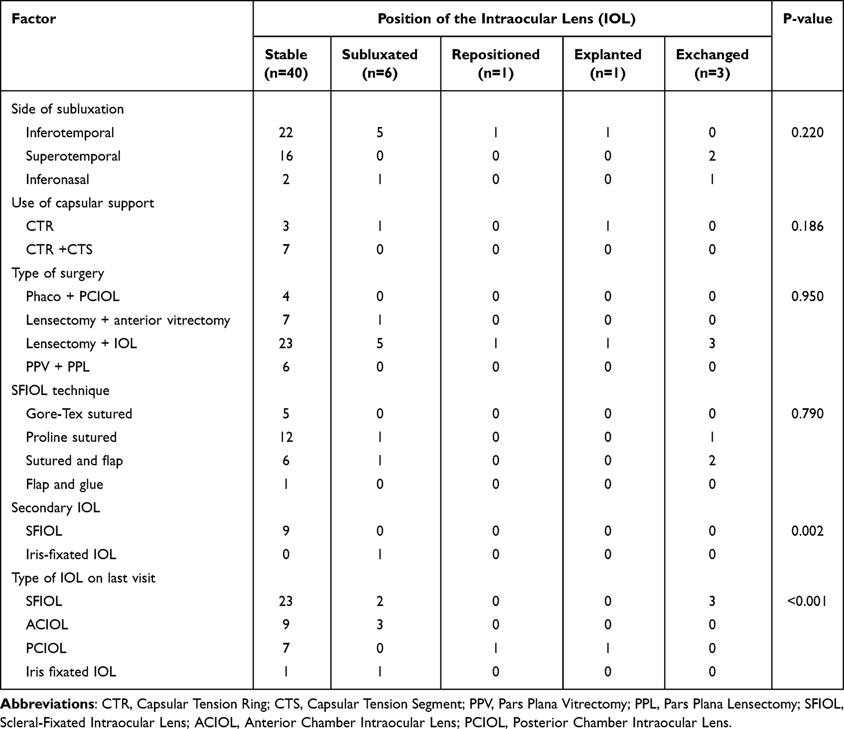 |
Table 2 Relationship Between Different Factors and Position of the Intraocular Lens (IOL) in the Last Visit |
Table 3 shows the mean refractive outcome according to final phakic status.
 |
Table 3 Phakic Status at Last Visit and the Refractive Correction |
Aphakic correction was achieved using spectacles in 29 eyes (82.9%) and contact lenses in six eyes (17.1%). There was a significant association between the preoperative side of lens subluxation and both the type of surgery (P = 0.019) and IOL position (P = 0.009).These relationships are summarized in Table 4.
 |
Table 4 Relationship Between Side of Subluxation and Operative and Postoperative Factors |
Visual Outcomes
The mean final visual acuity was 0.3 ± 0.3 (LogMAR) (Snellen = 20/40).
Table 5 presents a comparison of mean visual acuity values at the last follow-up among different types of surgery, lens positions, final lens status, and SFIOL techniques.
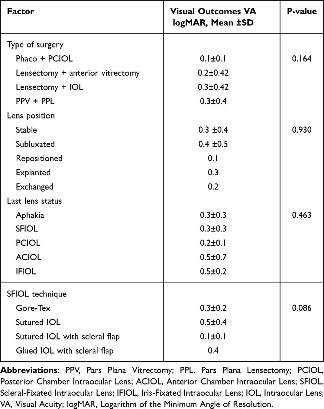 |
Table 5 Possible Predictors of Better Visual Outcomes |
Discussion
This study examined the surgical techniques and visual outcomes associated with lensectomy with and without IOL implantation in patients diagnosed with Marfan syndrome. Ectopia lentis is the most common ocular manifestation of Marfan syndrome. In our series, all patients presented with subluxated crystalline lenses, with nearly half exhibiting inferotemporal subluxation.
Surgical management of ectopia lentis presents a unique challenge owing to the inherent weakness and abnormalities of zonular fibers in patients with Marfan syndrome. These structural anomalies require various tailored surgical approaches to ensure optimal results.12 Over the years, techniques such as lensectomy, phacoemulsification (with or without IOL and CTR), and different methods of transscleral fixation have been developed to manage this complex condition.13–15 Surgical planning often depends on factors such as the degree of lens displacement, the patient’s age, and progression of the condition.
Pars plana lensectomy or intracapsular cataract extraction is often preferred for lenses that are significantly dislocated. However, intracapsular cataract extraction requires a large incision, which increases the risk of surgically induced astigmatism, intraoperative hypotony, and retinal detachment.16 Pars plana lensectomy, although less invasive, has its risks, particularly the potential for iatrogenic retinal breaks near the vitrectomy probe insertion site.17 Retinal detachment is a known risk factor in patients with Marfan syndrome, especially in those with severe dislocation or high axial myopia, which warrants careful surgical planning.1,18
In our study, different surgical techniques were utilized to manage ectopia lentis. Half of the patients underwent lensectomy with anterior vitrectomy. Lensectomy with IOL implantation was performed in 34 eyes, and nine eyes underwent phacoemulsification with IOL implantation. Capsular support devices were used in 12 eyes when the capsular bag was preserved. The extent of lens subluxation influenced the choice of the surgical method. At our institution, we typically use phacoemulsification with CTR implantation in cases of minimal-to-mild lens subluxation. For moderate to severe cases, we prefer a combined lensectomy-vitrectomy technique performed through the limbus or pars plana, as it offers a safer and more controlled approach. This technique avoids complications during capsulorrhexis, reduces stress on the unstable capsular bag, and bypasses the difficulties associated with CTR insertion and IOL fixation.14 It also helps prevent common phacoemulsification-related complications such as vitreous traction or intraoperative bag rupture.14 Our preference for phacoemulsification with CTR in mild subluxation and lensectomy vitrectomy in moderate to severe cases is supported by Dogăroiu et al and Chen et al, who highlighted that the extent of zonular weakness and patient age are key determinants in surgical planning.3,19 Additionally, Erdogan et al compared three approaches—intrascleral fixation, scleral IOL fixation with Z-suture, and IOL with Cionni CTR—and found no significant differences in postoperative outcomes or complications, supporting flexibility in technique selection.20
Owing to the structural instability of the capsular bag in Marfan syndrome, additional support is often required. Scleral-fixated CTR and CTS have proven useful in stabilizing the capsular bag and enabling safe IOL implantation in cases of zonular weakness. Once the crystalline lens is removed, the decision to proceed with IOL implantation for visual rehabilitation depends on multiple factors, including the extent of zonular deficiency and the stability achieved with these supportive devices.
When capsular support is inadequate, alternatives include SFIOLs, ACIOLs, and iris-fixated IOLs.21–23 Primary IOL implantation is generally reserved for patients over the age of three.1 While ACIOLs are a potential solution, they are associated with complications, such as corneal decompensation and secondary glaucoma.24 SFIOLs, by contrast, have demonstrated more stable outcomes with fewer complications.25,26 Our findings align with those of Al-Dwairi et al and van Zeeburg et al, who reported that both anterior and retropupillary iris-claw IOLs are effective and relatively safe, although disenclavation risk tends to be higher in younger patients.5,27
In our cohort, secondary IOL implantation was performed in 12 eyes. Among them, 11 eyes (91.7%) received SFIOLs, and one eye (8.3%) received an iris-fixated IOL. Most IOLs were placed in the bag (12 eyes) or sulcus (one eye). Among the out-of-the-bag implantations, 28 eyes received SFIOLs, seven eyes received anterior chamber IOLs, and two eyes received iris-fixated IOLs. The choice of technique was based on both the surgeon’s preference and the individual patient’s ocular condition.
Visual acuity improved significantly after surgical intervention. On average, vision improved from 1.1 ± 0.7 logMAR (Snellen equivalent 20/250) at presentation to 0.3 ± 0.3 logMAR (Snellen equivalent 20/40) at the last follow-up (P < 0.001). Similar findings have been reported. In one study, children were left aphakic but achieved a postoperative BCVA better than 20/30 with aphakic correction.28 Another case series of nine Marfan patients who underwent lensectomy with secondary ACIOL implantation also reported improvement from 0.5 ± 0.3 to 0.2 ± 0.2 logMAR.29 In a third study involving 39 patients who underwent 23G-vitrectomy lensectomy with or without ACIOL implantation, the visual acuity improved from 0.5 to 0.3 logMAR.1
Interestingly, in our cohort, visual outcomes were not significantly influenced by the phakic status at the final follow-up (P = 0.463). However, eyes with posterior chamber IOLs had the best mean visual acuity (0.2 ± 0.1 logMAR), followed by both aphakic IOL and SFIOL groups (0.3 ± 0.3 logMAR), suggesting a slight advantage for in-the-bag IOL placement when feasible.
In our series, capsular support devices were used in 12 eyes; five received CTRs alone, while seven received both CTRs and CTS. Secondary IOL implantation was performed in 12 eyes following lensectomy and anterior vitrectomy: 11 with scleral fixation and one with iris fixation. After an average follow-up period of 7.3 ± 6.2 years, four eyes (11.8%) that had undergone lensectomy with IOL implantation subsequently underwent IOL exchange and received a new SFIOL, approximately 8.3 ± 2.9 years after the initial procedure.
At the final follow-up, approximately 40% of the eyes were aphakic, with a mean spherical equivalent of +9.5 ± 4.6 diopters, requiring correction via, necessitating correction with glasses or contact lenses. In contrast, eyes with posterior chamber IOLs, either SFIOLs or in-the-bag IOLs, tended to show mild hyperopia.30 This differs from the findings of Morrison et al, in which postoperative outcomes leaned toward myopia.24 Regardless of the surgical technique, all eyes in our series achieved satisfactory IOL centration and long-term stability. Only a few patients required repositioning, IOL exchange, or explantation. Refractive results were consistent across all techniques, consistent with the findings in the existing literature on Marfan-associated ectopia lentis.30–33
Our study had some limitations. First, the retrospective nature and variability in patient follow-up duration may have affected consistency. Second, the surgical techniques were not standardized across cases, and multiple surgeons with differing levels of experience performed the procedures. Detailed operative notes were not available for all patients, which may have affected the outcome analysis. Also, not all IOL techniques were utilized such as iris claw IOLs as they were not available in our center during the study period which may preclude reaching a generalizable conclusion. Nonetheless, given the rarity of Marfan syndrome and the number of cases included, we believe that our findings contribute important insights into the literature on this topic.
Conclusion
This large case series demonstrates that a range of surgical techniques, including lensectomy with or without IOL implantation, can yield favorable long-term visual outcomes in patients with Marfan syndrome and ectopia lentis. Visual outcomes were optimized through individualized surgical planning, taking into account the extent of lens subluxation and the integrity of capsular support. Although not all techniques were studied such as iris claws IOLs, however our results showed the studied posterior chamber IOLs were associated with the best visual acuity results when feasible. This study emphasizes the importance of tailoring surgical approaches to each patient’s anatomical and clinical characteristics and supports the safety and effectiveness of secondary IOL implantation in complex cases. These findings provide valuable guidance for surgical decision-making and highlight the need for future prospective studies to standardize management and assess long-term functional outcomes in this population.
Abbreviations
ACIOL, anterior chamber intraocular lens; BCVA, best-corrected visual acuity; CTR, capsular tension ring; CTS, capsular tension segments; IOL, intraocular lens; IOP, intraocular pressure; KKESH, King Khaled Eye Specialist Hospital; PCIOL, posterior chamber intraocular lens; SFIOL, scleral-fixated intraocular lens.
Data Sharing Statement
Data is available from the corresponding author upon request.
Ethics Approval and Consent to Participate
This study was conducted in accordance with the tenets of the Declaration of Helsinki and received approval from the institutional review board of King Khaled Eye Specialist Hospital (RP 24072-R). Due to the retrospective design and the use of de-identified patient data, the requirement for informed consent was waived.
Consent for Publication
All listed authors have consented to the publication of this manuscript.
Author Contributions
All authors made a significant contribution to the work reported, whether that is in the conception, study design, execution, acquisition of data, analysis and interpretation, or in all these areas; took part in drafting, revising, or critically reviewing the article; gave final approval of the version to be published; have agreed on the journal to which the article has been submitted; and agree to be accountable for all aspects of the work.
Funding
There is no funding to report.
Disclosure
The authors declare that they have no competing interests for this work.
References
1. Fan F, Luo Y, Liu X, Lu Y, Zheng T. Risk factors for postoperative complications in lensectomy-vitrectomy with or without intraocular lens placement in ectopia lentis associated with Marfan syndrome. Br J Ophthalmol. 2014;98(10):1338–1342. doi:10.1136/bjophthalmol-2013-304144
2. Xiao Y, Liu X, Guo X, et al. A novel FBN1 mutation causes autosomal dominant Marfan syndrome. Mol Med Rep. 2017;16(5):7321–7328. doi:10.3892/mmr.2017.7544
3. Dogaroiu AC, Dudau M, Dogaroiu C, Tataru CP. Visual outcomes in ectopia lentis in Marfan syndrome: a study of four surgical techniques in children and adults. Medicina. 2024;60(7):1098. doi:10.3390/medicina60071098
4. Loeys BL, Dietz HC, Braverman AC, et al. The revised Ghent nosology for the Marfan syndrome. J Med Genet. 2010;47(7):476–485. doi:10.1136/jmg.2009.072785
5. Al-Dwairi RA, Shihadeh WA, Alqudah AA, et al. Outcomes of iris-claw IOL implantation in patients with Marfan’s syndrome in Jordan. Clin Ophthalmol. 2022;16:3811–3819. doi:10.2147/OPTH.S385352
6. Judge DP, Dietz HC. Marfan’s syndrome. Lancet. 2005;366(9501):1965–1976. doi:10.1016/S0140-6736(05)67789-6
7. Manning S, Lanigan B, O’Keefe M. Outcomes after lensectomy for children with Marfan syndrome. J AAPOS. 2016;20(3):247–251. doi:10.1016/j.jaapos.2016.02.006
8. Drolsum L, Rand-Hendriksen S, Paus B, Geiran OR, Semb SO. Ocular findings in 87 adults with Ghent-1 verified Marfan syndrome. Acta Ophthalmol. 2015;93(1):46–53. doi:10.1111/aos.12448
9. Konradsen TR, Zetterström C. A descriptive study of ocular characteristics in Marfan syndrome. Acta Ophthalmol. 2013;91(8):751–755. doi:10.1111/aos.12068
10. Hasan A, Poloniecki J, Child A. Ageing in Marfan syndrome. Int J Clin Pract. 2007;61(8):1308–1320. doi:10.1111/j.1742-1241.2007.01407.x
11. Chandra A, Ekwalla V, Child A, Charteris D. Prevalence of ectopia lentis and retinal detachment in Marfan syndrome. Acta Ophthalmol. 2014;92(1):e82–3. doi:10.1111/aos.12175
12. Rezar‐Dreindl S, Stifter E, Neumayer T, Papp A, Gschliesser A, Schmidt‐Erfurth U. Visual outcome and surgical results in children with Marfan syndrome. Clin Exp Ophthalmol. 2019;47(9):1138–1145. doi:10.1111/ceo.13596
13. Stephenson KA. Surgical management of non-syndromic ectopia lentis. Int J Ophthalmol. 2020;13(7):1156–1160. doi:10.18240/ijo.2020.07.21
14. Hoffman RS, Snyder ME, Devgan U, Allen QB, Yeoh R, Braga-Mele R. Management of the subluxated crystalline lens. J Cataract Refract Surg. 2013;39(12):1904–1915. doi:10.1016/j.jcrs.2013.09.005
15. Hsu HY, Edelstein SL, Lind JT. Surgical management of non-traumatic pediatric ectopia lentis: a case series and review of the literature. Saudi J Ophthalmol. 2012;26(3):315–321. doi:10.1016/j.sjopt.2012.05.001
16. Luebke J, Reinhard T, Agostini H, Boehringer D, Eberwein P. Long-term follow-up after scleral lens fixation in patients with Marfan syndrome. BMC Ophthalmol. 2017;17(1):235. doi:10.1186/s12886-017-0625-x
17. Carter JB, Michels RG, Glaser BM, De Bustros S. Iatrogenic retinal breaks complicating pars plana vitrectomy. Ophthalmology. 1990;97(7):848–853. doi:10.1016/S0161-6420(90)32492-2
18. Cross HE, Jensen AD. Ocular manifestations in the Marfan syndrome and homocystinuria. Am J Ophthalmol. 1973;75(3):405–420. doi:10.1016/0002-9394(73)91149-5
19. Chen T, Deng M, Zhang M, et al. Visual outcomes of lens subluxation surgery with Cionni modified capsular tension rings in Marfan syndrome. Sci Rep. 2021;11(1):2994. doi:10.1038/s41598-021-82586-6
20. Erdogan G, Kandemir Besek N, Onal Gunay B, Agca A. Outcomes of three surgical approaches for managing ectopia lentis in Marfan syndrome. Euro J Ophthalmol. 2021;32(1):242–248. doi:10.1177/1120672121992950
21. Yaguchi S, Yaguchi S, Asano Y, et al. Repositioning and scleral fixation of subluxated lenses using a T-shaped capsule stabilization hook. J Cataract Refract Surg. 2011;37(8):1386–1393. doi:10.1016/j.jcrs.2011.06.015
22. Asadi R, Kheirkhah A. Long-term results of scleral fixation of posterior chamber intraocular lenses in children. Ophthalmology. 2008;115(1):67–72. doi:10.1016/j.ophtha.2007.02.018
23. Sminia ML, Odenthal MTP, Ljjm P, Cobben JM, Mourits MP, Völker-Dieben HJ. Long-term follow-up after bilateral Artisan aphakia intraocular lens implantation in two children with Marfan syndrome. J AAPOS. 2012;16(1):92–94. doi:10.1016/j.jaapos.2011.10.014
24. Morrison D, Sternberg P, Donahue S. Anterior chamber intraocular lens (ACIOL) placement after pars plana lensectomy in pediatric Marfan syndrome. J AAPOS. 2005;9(3):240–242. doi:10.1016/j.jaapos.2005.02.004
25. Epley KD, Shainberg MJ, Lueder GT, Tychsen L. Pediatric secondary lens implantation in the absence of capsular support. J AAPOS. 2001;5(5):301–306. doi:10.1067/mpa.2001.117567
26. Omulecki W, Nawrocki J, Palenga-Pydyn D, Sempinska-Szewczyk J. Pars plana vitrectomy, lensectomy, or extraction in transscleral intraocular lens fixation for the management of dislocated lenses in a family with Marfan’s syndrome. Ophthalmic Surg Lasers. 1998;29(5):375–379. doi:10.3928/1542-8877-19980501-04
27. Van Zeeburg EJT, Sminia ML, Schalij-Delfos NE. Long-term results of anterior chamber iris claw intraocular lens implantation in children with ectopia lentis in Marfan syndrome. J AAPOS. 2024;28(3):103922. doi:10.1016/j.jaapos.2024.103922
28. Miraldi Utz V, Coussa RG, Traboulsi EI. Surgical management of lens subluxation in Marfan syndrome. J AAPOS. 2014;18(2):140–146. doi:10.1016/j.jaapos.2013.12.007
29. Rabie HM, Malekifar P, Javadi MA, Roshandel D, Esfandiari H. Visual outcomes after lensectomy and iris claw artisan intraocular lens implantation in patients with Marfan syndrome. Int Ophthalmol. 2017;37(4):1025–1030. doi:10.1007/s10792-016-0366-5
30. Anteby I, Isaac M, BenEzra D. Hereditary subluxated lenses: visual performances and long-term follow-up after surgery. Ophthalmology. 2003;110(7):1344–1348. doi:10.1016/S0161-6420(03)00449-4
31. Van Os L, Hildebrand GD, Tassignon MJ. Adapted bag-in-the-lens implantation technique in children with congenital ectopia lentis. Klin Monbl Augenheilkd. 2021;238(10):1058–1064. doi:10.1055/a-1633-4316
32. Yulia DE, Barliana JD, Soeharto DA. Visual outcome of intraocular iris-claw lens implantation in Indonesian children with ectopia lentis. Taiwan J Ophthalmol. 2023;13(3):335–340. doi:10.4103/tjo.tjo_58_21
33. Soraya Alamsjah SHZ, Yulia DE, Tan S. Scleral-fixated vs Iris-fixated intraocular lens in pediatric ectopia lentis: a systematic review. Eur J Ophthalmol. 2024;34(6):1642–1654. doi:10.1177/11206721241242158
 © 2025 The Author(s). This work is published and licensed by Dove Medical Press Limited. The
full terms of this license are available at https://www.dovepress.com/terms
and incorporate the Creative Commons Attribution
- Non Commercial (unported, 4.0) License.
By accessing the work you hereby accept the Terms. Non-commercial uses of the work are permitted
without any further permission from Dove Medical Press Limited, provided the work is properly
attributed. For permission for commercial use of this work, please see paragraphs 4.2 and 5 of our Terms.
© 2025 The Author(s). This work is published and licensed by Dove Medical Press Limited. The
full terms of this license are available at https://www.dovepress.com/terms
and incorporate the Creative Commons Attribution
- Non Commercial (unported, 4.0) License.
By accessing the work you hereby accept the Terms. Non-commercial uses of the work are permitted
without any further permission from Dove Medical Press Limited, provided the work is properly
attributed. For permission for commercial use of this work, please see paragraphs 4.2 and 5 of our Terms.
Recommended articles

Macular and Functional Outcomes After Secondary IOL Implantation Using the Yamane Technique in Aphakia, IOL Subluxation, and IOL Dislocation
Visioli G, Albanese GM, Magherini S, Cappiello D, Baratta C, Armentano M, Lucchino L, Iannetti L, Gharbiya M
Clinical Ophthalmology 2026, 20:587908
Published Date: 24 February 2026