
Back to Journals » Breast Cancer: Targets and Therapy » Volume 18
Suppressing M2 Macrophage Polarization by Glycine Combined with β-Elemene via the IL-6/JAK2/STAT3 Signaling Pathway to Inhibit Triple-Negative Breast Cancer Progression
Authors Li H, Cheng Y, Meng Y, Nan L, Huang S, Ling X, Lin X, Xie S 
Received 22 May 2026
Accepted for publication 14 July 2026
Published 22 July 2026 Volume 2026:18 626034
DOI https://doi.org/10.2147/BCTT.S626034
Checked for plagiarism Yes
Review by Single anonymous peer review
Peer reviewer comments 2
Editor who approved publication: Professor Pranela Rameshwar
Huan Li,1,* Yu Cheng,1,* Yanyun Meng,2 Liang Nan,1 Sicheng Huang,1 Xiangli Ling,1 Xiangping Lin,3,4 Su Xie1,5
1Department of Traditional Chinese Medicine, Affiliated Hospital of Guizhou Medical University, Guizhou Medical University, Guiyang, Guizhou, People’s Republic of China; 2Emergency Department, Second Department of Internal Medicine, Anlong County Hospital of Traditional Chinese Medicine, Qianxinan Buyi and Miao Autonomous Prefecture, Guizhou, People’s Republic of China; 3Department of Pharmacy, Clinical Research Center of Integrated Traditional Chinese and Western Medicine, Affiliated Hospital of Guizhou Medical University, Guiyang, Guizhou, People’s Republic of China; 4Guizhou Key Laboratory of Integrated Traditional Chinese and Western Medicine, Affiliated Hospital of Guizhou Medical University, Guizhou Medical University, Guiyang, Guizhou, People’s Republic of China; 5Xie Su National Famous Traditional Chinese Medicine Expert Inheritance Studio in the Field of Maternal and Child Health, Affiliated Hospital of Guizhou Medical University, Guizhou Medical University, Guiyang, Guizhou, People’s Republic of China
*These authors contributed equally to this work
Correspondence: Su Xie, Department of Traditional Chinese Medicine, Affiliated Hospital of Guizhou Medical University, Guizhou Medical University, Guiyang, Guizhou, People’s Republic of China, Tel +86 0851 86773643, Email [email protected] Xiangping Lin, Department of Pharmacy, Clinical Research Center of Integrated Traditional Chinese and Western Medicine, Affiliated Hospital of Guizhou Medical University, Guiyang, Guizhou, People’s Republic of China, Tel +86 0851 86774148, Email [email protected]
Background: Triple-negative breast cancer (TNBC) is an aggressive subtype with poor prognosis, closely associated with an imbalanced tumor immune microenvironment. Tumor-associated macrophages (TAMs) play a crucial role in tumor progression.
Purpose: This study aimed to investigate whether glycine combined with β-elemene inhibits TNBC progression by suppressing M2 macrophage polarization through the IL-6/JAK2/STAT3 pathway.
Methods: UPLC–Q–Exactive HRMS was used to identify glycine and β-elemene. In vitro, 4T1 cell viability was determined by CCK-8 assay. RAW264.7 cells were polarized to M2 macrophages and co-cultured with 4T1 cells. Colony formation, wound healing, and Transwell assays were performed. Western blot and immunohistochemistry were used to detect protein expression. In vitro experiments were performed with at least three independent biological replicates. In vivo, 4T1 xenograft mouse models were established (n=10 per group) to evaluate anti-tumor efficacy.
Results: Glycine combined with β-elemene significantly suppressed proliferation, colony formation, and migration of 4T1 cells. Mechanistically, the combination inhibited M2 macrophage polarization by downregulating IL-6, p-JAK2, and p-STAT3. In vivo, the combination with paclitaxel showed the strongest anti-tumor effect, with reduced tumor volume and weight, decreased Ki-67 expression, and suppressed M2 polarization.
Conclusion: Glycine combined with β-elemene inhibits M2 macrophage polarization by suppressing the IL-6/JAK2/STAT3 pathway, thereby exerting anti-TNBC effects. Its combination with paclitaxel demonstrates synergistic anti-tumor efficacy.
Keywords: triple-negative breast cancer, glycine, β-elemene, M2 macrophage polarization, IL-6/JAK2/STAT3 pathway
Introduction
Breast cancer (BC) is one of the most prevalent malignant tumors among women globally and remains a major health threat to women’s lives.1–3 TNBC, a subtype of breast cancer with the worst prognosis, is characterized by the lack of expression of estrogen receptors (ER), progesterone receptors (PR), and human epidermal growth factor receptor 2 (HER-2).4,5 The absence of these receptor markers makes TNBC unresponsive to endocrine therapies or HER2-targeted drugs, contributing to its propensity for metastasis, recurrence, and high mortality rates.6,7 Currently, treatment options for TNBC are limited, with surgery, radiotherapy, and chemotherapy being the primary therapeutic approaches.7,8 Despite these treatments, 50% of patients experience tumor recurrence and metastasis, severely impacting their quality of life and survival rate, highlighting the urgent need to identify more effective therapeutic approaches.8,9 This study aims to explore new therapeutic targets and effective drugs for TNBC, with the potential to break through the current treatment landscape and offer significant clinical value.10
In recent years, the tumor microenvironment (TME) has emerged as a critical focus in cancer research.11,12 Numerous studies have confirmed that tumor progression requires coordination between tumor cells and the TME, wherein immune cells in the TME secrete various cytokines that promote tumor cell proliferation and induce chronic inflammation, creating an immune-suppressive environment conducive to cancer cell survival and immune evasion.13,14 TNBC displays characteristic features of the TME, with early infiltrating immune cells, such as macrophages, lymphocytes, and natural killer cells, possessing anti-tumor activity.15,16 Among these, TAMs, the most abundant immune cells in the TNBC microenvironment, play a crucial role in mediating immune suppression in TNBC.17,18 As tumors progress, TAMs exhibit a high level of M2 macrophage polarization, characterized by decreased secretion of inflammatory cytokines, involvement in tumor progression, and support for tumor cell immune evasion while inhibiting anti-tumor immune responses.19,20 Immune checkpoint inhibitors (ICIs) have shown limited efficacy in TNBC due to the “cold” tumor immune microenvironment characterized by low T-cell infiltration and high immunosuppressive cell populations, including M2-TAMs. This provides stronger justification for targeting macrophage polarization as a strategy to remodel the immunosuppressive TME and enhance anti-tumor immunity.
Inhibiting M2 macrophage polarization to suppress TNBC progression presents a promising therapeutic strategy. Mechanistically, IL-6 binds to its receptor and activates JAK2, which in turn phosphorylates STAT3. Phosphorylated STAT3 dimerizes and translocates to the nucleus, where it promotes the transcription of M2 macrophage marker genes including Arg-1, CD206, and IL-10, thereby driving M2 polarization and creating an immunosuppressive tumor microenvironment.21,22 Beyond the IL-6/JAK2/STAT3 axis governing M2 macrophage polarization, this signaling cascade closely interacts with STING-mediated innate immune response, TGF-β fibrotic signaling and PD-L1 immune checkpoint pathway to jointly construct the immunosuppressive TME.23 Modulating IL-6/JAK2/STAT3 to influence macrophages could effectively inhibit M2 polarization, suppress tumor growth, and restore anti-tumor immunity, thus holding potential clinical significance for TNBC therapy.24,25
Herbal pairings, as commonly used in traditional Chinese medicine (TCM), are designed to enhance therapeutic efficacy and reduce side effects.26 The Bie Jia (Carapax Trionycis) and E Zhu (Rhizoma Curcumae) pair is a well-known combination used by renowned Chinese medicine expert Liu Shangyi in the clinical treatment of breast cancer.27 This herbal pair has shown definite efficacy in inhibiting breast cancer and is commonly used to prevent recurrence and metastasis after surgery or chemotherapy, leading to improved survival quality and extended survival time in breast cancer patients.28 Several studies have confirmed that the Bie Jia-E Zhu combination inhibits breast cancer cell proliferation, migration, and invasion;26,28,29 however, the specific active components, target sites, and mechanisms of action remain unclear. Bie Jia and E Zhu contain complex compounds, and their combined effects involve multiple targets, limiting the development and research of the drug.30
Academician Zhang Boli proposed a new model for the development of modern Chinese medicine, starting from effective formulations and focusing on the active ingredients and their mechanisms. This approach, known as the “precise herbal pair” concept, facilitates a more direct and effective understanding of the active components and their mechanisms, which can provide new insights for precision medicine in Chinese herbal therapy.31 Therefore, this study aims to focus on the effective component combinations of the Bie Jia-E Zhu pair against TNBC, with substantial research value.
The research team previously identified the active components and target molecules with anti-TNBC effects in Bie Jia and E Zhu through studies such as network pharmacology and molecular docking.28 It was found that glycine and β-elemene constitute the primary active components responsible for the antitumor efficacy of this herbal combination. However, whether this combination exerts its anti-TNBC effects by modulating macrophage polarization, particularly by suppressing the immunosuppressive M2 macrophages, remains unclear. Therefore, this study aimed to investigate whether glycine combined with β-elemene inhibits TNBC progression by suppressing M2 macrophage polarization through the IL-6/JAK2/STAT3 signaling pathway. We hypothesized that: (1) glycine combined with β-elemene can inhibit the proliferation, migration, and invasion of 4T1 TNBC cells; (2) the combination can suppress M2 macrophage polarization via inhibiting the IL-6/JAK2/STAT3 pathway; and (3) glycine and β-elemene can enhance the anti-tumor efficacy of paclitaxel in vivo. To test these hypotheses, we performed comprehensive in vitro cell experiments and in vivo xenograft mouse model studies.
Materials and Methods
Reagents
Glycine (purity > 99.0%, CAS: 56-40-6, Cat. No.: G8200) was obtained from Solarbio Life Sciences Co., Ltd. (Beijing, China). β-elemene (purity > 98%, CAS: 515-13-9, Cat. No.: S31794) was purchased from Yuanye Bio-Technology Co., Ltd. (Shanghai, China). Paclitaxel (purity > 99%, CAS: 33069-62-4, Cat. No.: P875571) was acquired from Macklin Biochemical Co., Ltd. (Shanghai, China). Biejia–Ezhu granules were purchased from Guangdong Yifang Pharmaceutical Co., Ltd.
Chemical Profiling and Identification of Glycine and β-Elemene
To clarify the material basis of the anti-tumor effects of Biejia–Ezhu granules, UPLC–Q–Exactive HRMS was employed to establish the chemical profile, with a focus on identifying glycine and β-elemene. Briefly, 0.5 g of granules were extracted with 50 mL of 50% methanol via ultrasonication for 30 min. The extract was filtered through a 0.22 µm membrane for analysis. Chromatographic separation was performed on a Thermo Fisher Hypersil GOLD aQ column (100 × 2.1 mm, 1.9 µm) at 40°C, with a mobile phase of 0.1% formic acid in acetonitrile (A) and 0.1% formic acid in water (B) under gradient elution. Mass detection used a heated electrospray ionization source in positive/negative modes. Glycine and β-elemene were identified by comparing retention times, accurate masses, and fragmentation patterns with reference standards.
Cell Culture
The murine mononuclear macrophage leukemia cell line RAW264.7 (Cat NO.:CL-0190) was obtained from Wuhan Procell Life Science and Technology Co., Ltd. The murine 4T1 cell line (Batch No. ZQ0201) was obtained from Shanghai Zhongqiao Xinzhou Biotechnology Co., Ltd. 4T1 murine TNBC cells and RAW264.7 macrophages were cultured in DMEM high-glucose medium supplemented with 10% FBS and 1% penicillin-streptomycin at 37°C with 5% CO2.
CCK-8 Assay for IC50 Determination
4T1 cells were seeded into 96-well plates at a density of 5×10³ cells per well (100 µL per well) with 3 replicate wells for each group after routine digestion, centrifugation, resuspension and counting, before incubation at 37 °C with 5% CO2. On the next day, different concentrations of glycine (1, 2, 4, 6, 8, 16, 32 mg/mL) and β-elemene (10, 20, 30, 40, 50, 60, 80, 100 μg/mL) (pre-prepared with complete medium) were added sequentially from low to high concentration after confirming good cell adhesion, with the control group treated with 100 µL complete medium, followed by continued incubation under the same conditions. After 24 hours of drug intervention, the cell growth status was observed, and the supernatant was discarded gently; 100 µL of the mixture of CCK-8 reagent and fresh medium (1:10, v/v) was added to each well in the dark, and the plate was incubated for another 2 hours after wrapping with aluminum foil. The OD value of each group was detected at 450 nm by a microplate reader, and the cell proliferation inhibition rate was calculated using a formula and the IC50 value was further calculated:
Cell proliferation inhibition rate = [(OD value of control group − OD value of experimental group)/(OD value of control group − OD value of blank group)] × 100%.
M2 Macrophage Induction and Co-Culture System
RAW264.7 macrophages in the logarithmic growth phase were adjusted to a density of 2×10⁵ cells/mL, seeded into 6-well plates, and stimulated with IL-4 (20 ng/mL) to induce polarization toward the M2 phenotype. After polarization, M2 macrophages were collected and seeded into the upper chambers of 8.0 μm pore-size Transwell inserts matched with 6-well plates, while 4T1 cells were seeded into the corresponding lower chambers. Drugs were added to the upper chambers containing RAW264.7 cells according to the grouping setting, followed by co-culture with 4T1 cells for 24 h. The experimental groups were set as follows: Blank group (untreated RAW264.7 macrophages), M2 group, glycine group, β-elemene group, glycine + β-elemene combination group, and paclitaxel group.
Colony Formation
Cells in the logarithmic growth phase were digested with 0.25% trypsin, centrifuged at 4 °C and 1000 rpm for 5 min, and collected as cell suspension. The cells were resuspended in complete medium, mixed well, and counted, then seeded into 6-well plates at a density of 500 cells per well with 2 mL of complete medium added to each well. After gentle shaking, the plates were incubated at 37 °C in a 5% CO2 incubator. After cell adherence, corresponding medium or drugs were applied for intervention, and fresh medium was replaced after 24 h for continuous culture. The medium was renewed every 3 days, and cell status was routinely monitored to prevent contamination. On day 10 of culture, the supernatant was discarded, and the wells were gently washed twice with PBS along the wall. Each well was fixed with 1 mL of 4% paraformaldehyde at room temperature for 30 min with mild operation to avoid colony detachment. After fixation, paraformaldehyde was removed, and the wells were rinsed twice with PBS. Each well was stained with 1 mL of crystal violet solution for 15 min at room temperature, then repeatedly washed with PBS until the background was colorless and air-dried. The 6-well plate was placed on clean white paper for photographing, and ImageJ software was used to analyze colony number and size in each well.
Wound Healing Assay
Cells in the logarithmic growth phase were digested with 0.25% trypsin, centrifuged at 4 °C and 1000 rpm for 5 min, and the cell suspension was collected. The cells were resuspended and mixed thoroughly in DMEM complete medium containing 10% fetal bovine serum (FBS), then seeded into 6-well plates at an appropriate density with 2 mL medium per well. The plates were shaken gently in a cross pattern and incubated at 37 °C with 5% CO2 until the cell confluence reached 80%–90%. Three straight lines were scratched vertically to the edge of each well on the cell monolayer using a 10 µL pipette tip to ensure a consistent scratch width. The wells were gently rinsed 2–3 times with PBS to remove detached cell debris, followed by the addition of corresponding medium or drugs for intervention. Cell migration in the scratched area was observed and photographed under an inverted microscope at 0 h and 24 h after scratching. ImageJ software was used to measure the scratch width and calculate the cell migration distance.
Transwell Assay
Cells in the logarithmic growth phase were digested with 0.25% trypsin, centrifuged at 4 °C and 1000 rpm for 5 min, and prepared into cell suspension. The cell density was adjusted to \(1\times104\) cells/mL, and 200 μL of cell suspension was seeded into the upper chamber of Transwell inserts, while 800 μL of corresponding medium containing drugs was added to the lower chamber for intervention. All samples were incubated at 37 °C in a 5% CO2 incubator for 24 h. The Transwell inserts were then removed, rinsed with PBS buffer and air-dried, followed by fixation with 4% paraformaldehyde for 30 min and staining with crystal violet for another 30 min. After repeated rinsing with PBS buffer and air-drying, the non-migrated cells and residual crystal violet on the upper surface of the inserts were wiped off with a cotton swab. The migrated cells were observed under an inverted microscope, five random visual fields were selected for each well to count cell numbers, and the average value was calculated.
Western Blot Analysis
Protein lysates were obtained by extracting proteins from cells and tumor tissues and lysing them for 40 min. Following a 20-min centrifugation at 4 °C, the supernatant was collected and measured using the BCA test. After adding the protein into 8–12% SDS-PAGE gels, electrophoresis was performed using a voltage of 80 V. Following the completion of electrophoresis, the gel was placed in a transfer holder and covered with a 0.45 μm PVDF membrane to transfer proteins at a current of 0.4 A for 15 min. Then the PVDF membrane was soaked in 5% BSA and placed on a shaker for 2 h at 60 rpm. β-actin Rabbit Polyclonal Antibody (10494-1-AP, Proteintech Group, Inc., Wuhan), IL-6 Polyclonal antibody (21865-1-AP, Proteintech Group, Inc., Wuhan), Phospho-STAT3 (Tyr705) Antibody (AF3293/4, Affinity), JAK2 Polyclonal antibody (17670-1-AP, Proteintech Group, Inc., Wuhan), STAT3 Polyclonal antibody (10253-2-AP, Proteintech Group, Inc., Wuhan), Arginase-1 (P05089, Zen-bioscience, Chengdu), Mannose Receptor Rabbit pAb (P22897, Zen-bioscience, Chengdu), iNOS (WL0992a, Wanlei Biotechnology) and TGF-β1 Polyclonal antibody (21898-1-AP, Proteintech Group, Inc., Wuhan) were incubated with the membranes at 4 °C overnight. The membranes were washed and incubated for 1 h with the addition of the secondary antibody, then washed 3 times to develop chemiluminescent images.
Animal Model and Treatment in vivo
In this study, 4T1‑derived triple‑negative breast cancer (TNBC) xenograft models were established in BALB/c mice. As a murine mammary tumor cell line, 4T1 cells recapitulate typical TNBC phenotypes with reliable tumor‑forming capacity and an immunosuppressive tumor microenvironment, which has been widely adopted for in‑vivo investigations focusing on immune pathways and anti‑tumor effects of traditional Chinese medicines against TNBC. Forty 6-week-old specific pathogen-free (SPF) female Balb/c mice (20 ± 5 g) were purchased from Changsha Tianqin Biotechnology Co., Ltd. (Hunan, China) under license number CSXK (Yue) 2022–0063. All animals were housed at (23 ± 2) °C with free access to standard food and water, and all experimental procedures were approved by the Animal Ethics Committee of Guizhou Medical University (Approval No. 2101280). 4T1 cells in the logarithmic growth phase (80%–90% confluence) were harvested and resuspended in PBS to a concentration of approximately 1×10⁷ cells/mL, and 0.2 mL of the suspension was subcutaneously inoculated into the second mammary fat pad on the right side of each mouse. Mice were randomly assigned to different treatment groups using a random number table method after tumor establishment. The sample size of each group was calculated via statistical power analysis with α = 0.05 and power = 0.8, referring to previously published xenograft tumor studies. To prevent data loss caused by unexpected death or failed tumorigenesis during animal feeding and administration, 10 mice were randomly assigned to each group. Specific groups: control group, paclitaxel group, glycine + β-elemene group, and combination group (glycine + β-elemene + paclitaxel). The paclitaxel group received 0.4 mL of distilled water by oral gavage once daily plus intraperitoneal injection of paclitaxel (0.2 mL, 10 mg/kg) once every 3 days; the glycine + β-elemene group received glycine (0.2 mL, 2 g/kg) and β-elemene (0.2 mL, 2 mg/kg) by oral gavage once daily; the combination group received a total of 0.4 mL of the combined glycine and β-elemene solution by oral gavage once daily plus intraperitoneal injection of paclitaxel (0.2 mL) once every 3 days; the control group received 0.4 mL of distilled water by oral gavage once daily plus intraperitoneal injection of 0.2 mL normal saline once every 3 days. During the 21-day treatment period, tumor volume and weight were recorded at regular intervals, and all interventions were administered according to the predetermined doses and schedules. All mice were fasted for 12 h with free access to water prior to the final administration. After the last treatment, mice were anesthetized with 2% sodium pentobarbital, and tumors were carefully dissected and immediately stored at −80 °C for subsequent analysis, followed by euthanasia. All outcome assessments, including tumor volume measurement, weight measurement, and immunohistochemical scoring, were performed by investigators who were blinded to the treatment group assignments.
Immunohistochemistry
Tumor tissues were fixed in 4% paraformaldehyde at room temperature for 24 h, rinsed with PBS three times (5 min each), dehydrated in a graded series of ethanol (70%, 80%, 95%, 100%) for at least 1 h per step, embedded in paraffin, and sectioned at 4 μm. After deparaffinization with xylene and rehydration with graded ethanol, antigen retrieval was performed, followed by blocking with 3% H2O2 for 15 min in the dark to quench endogenous peroxidase activity. Sections were then blocked with normal goat or rabbit serum for 30 min at room temperature, incubated with primary antibodies at 4 °C overnight, and subsequently incubated with corresponding secondary antibodies for 1 h at room temperature. Immunoreactivity was visualized using DAB chromogenic reagent within 2 min, counterstained with hematoxylin, dehydrated, cleared, and mounted with neutral resin. Sections were incubated with antibodies against Ki-67, Arg-1, CD206, TGF-β1, and iNOS, and the stained images were analyzed using ImageJ software.
Statistical Analysis
Data is presented as mean ± SD. Statistical analysis was performed using GraphPad Prism 9 and SPSS 24.0. One-way ANOVA followed by Tukey’s test was used for multiple comparisons. P < 0.05 was considered statistically significant.
Results
Identification of Glycine and β-Elemene by UPLC–Q–Exactive HRMS
We utilized UPLC-Q-Exactive HRMS to identify glycine and β-elemene in Biejia-Ezhu granules. The total ion chromatogram acquired over 0–28 min showed multiple chromatographic peaks, indicating the complex chemical composition of Biejia-Ezhu granules (Figure 1a). Glycine exhibited a characteristic peak at a retention time of 1.42 min, with its corresponding MS/MS spectrum showing a major ion at m/z 75.0073 (Figure 1b and c). β-Elemene was detected at a retention time of 13.01 min, and its MS/MS spectrum showed a major ion at m/z 205.1953 (Figure 1d and e). These chromatographic behaviors and MS/MS fragmentation patterns were consistent with those of reference standards, confirming the presence of glycine and β-elemene in Biejia-Ezhu granules.
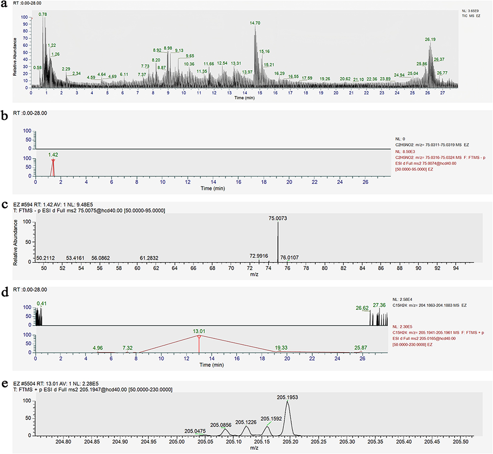 |
Figure 1 Chemical characterization of glycine and β-elemene by UPLC-Q-Exactive HRMS. (a) Total ion chromatogram (TIC) acquired in positive electrospray ionization mode over 0–28 min. (b) Extracted ion chromatogram of glycine showing a characteristic peak at retention time 1.42 min, detected at m/z 75.0316–75.0324. (c) MS/MS spectrum of glycine at RT 1.42 min, with the major ion observed at m/z 75.0073. (d) Extracted ion chromatogram of β-elemene showing a characteristic peak at retention time 13.01 min, detected at m/z 205.1941–205.1961. (e) MS/MS spectrum of β-elemene at RT 13.01 min, with the major ion observed at m/z 205.1953. |
Anti-Proliferative and Anti-Invasive Properties of Glycine and β-Elemene on 4T1 Cells
We evaluated the anti-tumor effects of glycine and β-elemene in 4T1 cells. The CCK-8 assay showed that the IC50 values were 6 mg/mL for glycine (Figure 2a) and 49µg/mL for β-elemene (Figure 2b), so 6 mg/mL glycine and 49 µg/mL β-elemene were used in subsequent experiments. The clone formation assay (Figure 2c and f) showed that M2 macrophages significantly promoted 4T1 cell proliferation (##P < 0.01 vs Blank). Glycine or β-elemene alone partially reversed this effect, while their combination exerted stronger migration (comparable to paclitaxel (&&P < 0.01 vs M2). The scratch wound healing assay (Figure 2d and h) revealed that M2 macrophages markedly enhanced cell migration (##P < 0.01 vs Blank). Monotherapy with glycine or β-elemene reduced migration, whereas combination treatment achieved a more invasiveness (effect, similar to paclitaxel (&&P < 0.01 vs M2). The transwell invasion assay (Figure 2e and g) confirmed that M2 macrophages significantly increased cell invasiveness (##P < 0.01 vs Blank). Glycine or β-elemene alone suppressed invasion, and their combination demonstrated superior activity, comparable to paclitaxel (&&P < 0.01 vs M2). Collectively, these data indicate that glycine and β-elemene partially reverse M2-induced pro-tumor effects, with their combination exhibiting enhanced anti-proliferative, anti-migratory, and anti-invasive activities in 4T1 cells.
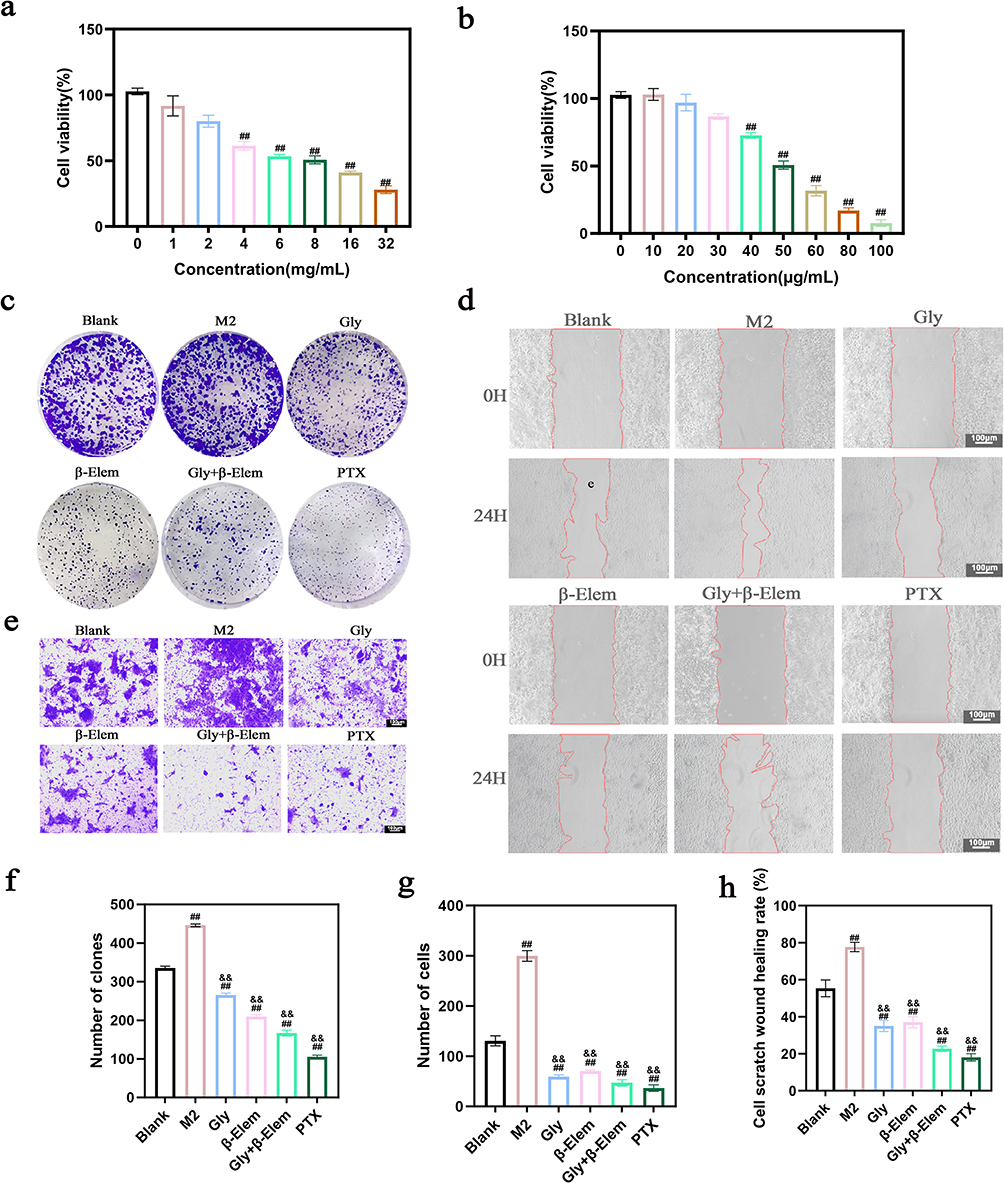 |
Figure 2 Effects of macrophage intervention on viability, proliferation, migration, and invasion of 4T1 cells. (a) Dose–response analysis of glycine on 4T1 cell viability. (b) Dose–response analysis of β-elemene on 4T1 cell viability. (c) Representative images of colony formation assays. (d) Representative images of wound-healing assays at 0 h and 24 h. Red lines denote initial wound edges at 0 h and 24 h. Scale bar, 100 μm. (e) Representative images of transwell invasion assays. (f) Quantification of colony formation assays. (g) Quantification of transwell invasion assays. (h) Quantification of wound-healing assays. Data are presented as mean ± SD. Compared with the Blank group, ##P < 0.01; compared with the control group, &&P < 0.01. |
Glycine Combined with β-Elemene Regulated M2 Macrophage Polarization and the JAK2/STAT3 Pathway in vitro
To investigate the molecular mechanisms underlying the observed functional effects, Western blot analysis was performed on protein lysates from the co - culture system of RAW264.7 - derived M2 macrophages and 4T1 cells across different treatment groups: Blank, M2, Gly, β-Elem, Gly + β-Elem, and PTX. The expression of M2 macrophage polarization markers was significantly upregulated in the M2 group compared to the Blank group (Figure 3a and b). Treatment with glycine or β-elemene alone reduced the expression of Arg - 1, CD206 and TGF - β1, while their combination (Gly + β-Elem) resulted in a more pronounced downregulation. Notably, the inhibitory effect of the Gly + β-Elem combination was significantly stronger than that of either single agent.
 |
Figure 3 Western blot analysis of M2 macrophage polarization-related proteins and JAK2/STAT3 signaling pathway proteins in RAW264.7 and 4T1 co-culture systems. (a) Representative Western blot bands and quantitative analysis of protein expression (b) of M2 macrophage-associated proteins. (c) Representative Western blot bands and quantitative analysis of protein expression (d) of JAK2/STAT3 signaling pathway-related proteins. Compared with the Blank group, #P < 0.05, ##P < 0.01. Compared with the M2 group, &P < 0.05, &&P < 0.01. Compared with the Gly + β-Elem group, *P < 0.05, **P < 0.01. |
Concurrently, the activation status of the JAK2/STAT3 signaling pathway was assessed. Levels of phosphorylated JAK2 (p - JAK2) and STAT3 (p - STAT3), as well as the upstream cytokine IL - 6, were markedly elevated in the M2 group (Figure 3c and d). All treatment groups showed reduced expression of these proteins compared to the M2 group. The Gly + β-Elem combination exhibited the most potent inhibitory effect on p - JAK2, p - STAT3, and IL - 6 expression, which was significantly more effective than glycine or β-elemene alone. These in vitro findings indicate that glycine combined with β-elemene effectively suppresses M2 macrophage polarization and inhibits the activation of the IL - 6/JAK2/STAT3 signaling pathway.
Anti-Tumor Effects of Glycine Combined with β-Elemene and Paclitaxel in vivo
To validate the anti-tumor efficacy of the combination therapy in vivo, we established a 4T1 breast cancer subcutaneous xenograft model in female BALB/c mice (Figure 4a). Compared with the Control group, treatment with paclitaxel (PTX), glycine + β-elemene (Gly+β-Elem), or the triple combination (PTX+Gly+β-Elem) all significantly inhibited tumor growth, as evidenced by reduced tumor weight and volume (Figure 4b–e).
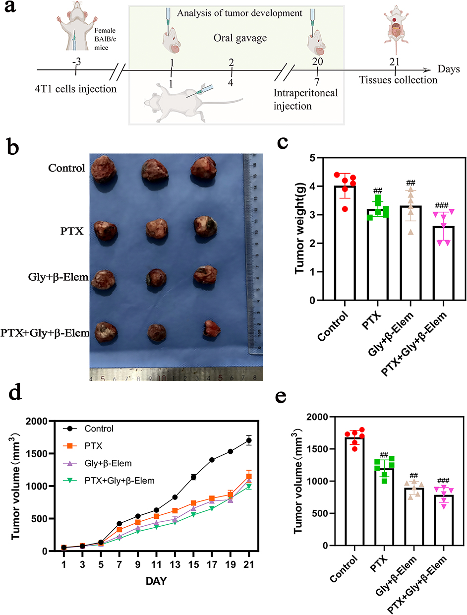 |
Figure 4 Antitumor effects of glycine and β-elemene combined with paclitaxel in vivo. (a) Animal experiment scheme. (b) Representative images of excised tumors from each group. (c) Statistical analysis of tumor weights. (d) Tumor growth curves showing changes in tumor volume during treatment for 21 days. (e) Statistical analysis of tumor volumes at the experimental endpoint on Day 21. Compared with the Control group, ##P < 0.01, ###P < 0.001. |
Notably, while both PTX monotherapy and Gly+β-Elem dual therapy exerted obvious anti-tumor effects, the triple combination of PTX+Gly+β-Elem showed the most pronounced inhibitory effect on both tumor weight and volume, which was significantly stronger than any single or dual-agent treatment alone. These results demonstrate that glycine combined with β-elemene effectively enhances the anti-tumor activity of paclitaxel in vivo, exhibiting a synergistic tumor-suppressive effect in the 4T1 subcutaneous tumor model.
Suppression of M2 Polarization and JAK2/STAT3 Pathway in vivo
To further verify the immunomodulatory effects of the treatments in the tumor microenvironment, we performed immunohistochemical staining on tumor tissues. The expression of M2 macrophage polarization markers (Arg-1, CD206 and TGF-β1) was significantly downregulated in all treatment groups compared to the Control group. In contrast, the expression of the M1 marker iNOS was significantly upregulated (Figure 5a and b). Treatment with PTX alone reduced the expression of Arg-1, CD206 and TGF-β1, while glycine combined with β-elemene. Gly +β-elem resulted in a more pronounced modulation of these markers (vs the PTX group). Notably, the triple combination of PTX, gly, and β-elem exhibited the strongest regulatory effect, significantly outperforming both PTX monotherapy and gly+β-elem dual therapy.
 |
Figure 5 Western blot analysis of M2 macrophage polarization-related proteins and JAK2/STAT3 signaling pathway proteins in tumor tissues from different treatment groups. (a) Representative Western blot bands of M2 macrophage polarization-related proteins. (b) Quantitative analysis of protein expression shown in (a). (c) Representative Western blot bands of JAK2/STAT3 signaling pathway-related proteins. (d) Quantitative analysis of protein expressions shown in (c). Compared with the control group, #P < 0.05, ##P < 0.01. Compared with the PTX group, *P < 0.05, **P < 0.01. |
Concurrently, the activation status of the IL-6/JAK2/STAT3 signaling pathway was assessed in vivo. Levels of phosphorylated JAK2 (p-JAK2) and STAT3 (p-STAT3), as well as the upstream cytokine IL-6, were markedly elevated in the Control group (Figure 5c and d). All treatment groups showed reduced expression of these proteins compared to the Control group. The Gly+β-Elem combination exhibited a more potent inhibitory effect on p-JAK2, p-STAT3, and IL-6 expression than PTX alone, while the triple combination PTX+ gly + β-elem showed the most significant suppression of these signaling molecules.
These findings indicate that glycine combined with β-elemene effectively suppresses M2 macrophage polarization and inhibits the activation of the IL-6/JAK2/STAT3 signaling pathway in tumor tissues, and this inhibitory effect is further enhanced when combined with paclitaxel in vivo.
Immunohistochemical Analysis of Tumor Proliferation and M2 Polarization
We further investigated the in vivo anti-tumor effects of the treatments by immunohistochemistry. Ki-67 expression, a marker of tumor cell proliferation, was significantly reduced in all treatment groups (Figure 6a and c), with the lowest levels observed in the PTX+Gly+β-Elem triple combination group. Meanwhile, the M2 macrophage markers Arg-1, CD206 and TGF-β1 were downregulated, while the M1 marker iNOS was upregulated (Figure 6b and d), with the most notable regulatory effects observed in the combination treatment groups. The purpose is to verify the in vivo anti-tumor and immunomodulatory effects of glycine, β-elemene, and their combination with paclitaxel. It uses immunohistochemistry (IHC) staining to demonstrate that the combination therapies inhibit tumor cell proliferation (via reduced Ki-67) and skew the tumor-associated macrophage phenotype from the pro-tumor M2 to the anti-tumor M1 subtype, with the triple combination showing the strongest therapeutic effect.
 |
Figure 6 Inhibitory effects of glycine and β-elemene combined with paclitaxel on M2 macrophage polarization and tumor cell proliferation. (a) Representative immunohistochemical staining images, yellow boxes indicate the regions magnified in the lower row. (b) Representative immunohistochemical staining images, yellow boxes indicate the regions magnified in the lower row. (c) Quantification analysis of Ki-67 in different groups. (d) Quantification analysis of M2 macrophage polarization-related proteins in different groups. Compared with the control group, #P < 0.05, ##P < 0.01. Compared with the combination group, &P < 0.05, &&P < 0.01. Compared with the PTX group, **P < 0.01. |
Discussion
In ancient Chinese medical literature, breast cancer is documented as Rushi Yong (breast stone abscess) and Ruyan (breast rock carcinoma), whose pathogenesis shares high correspondence with TNBC in modern medicine. From the perspective of TCM, this disease arises from emotional internal injury, stagnation of liver and spleen qi, thoroughfare and conception vessel disorders, as well as liver and kidney deficiency. Overall, TNBC is defined as a syndrome of deficiency in origin and excess in manifestation.32
Master TCM physician Liu Shangyi proposed that breast cancer pertains to “yang within yin”, characterized by stubborn yin pathogenic factors and intense yang-toxin.33 Moreover, surgical resection, radiotherapy and chemotherapy further damage qi, blood and body fluids, particularly resulting in severe consumption of genuine yin. Accordingly, Professor Liu established the therapeutic strategy of nourishing yin and resolving masses. The herb pair Carapax Trionycis-Rhizoma Curcumae precisely conforms to this treatment principle.34,35 The two herbs coordinate yin and yang with balanced cold and warm properties, collaboratively exerting the effects of nourishing yin and resolving masses while eliminating pathogenic factors without damaging healthy qi.
Previous studies by our research group have verified that this herb pair can suppress the proliferation of TNBC cells, tumor growth and EMT, and combined administration exhibits superior efficacy compared with single herbal treatment.36–38
Through network pharmacology and molecular docking analysis, a total of 71 active ingredients and 146 anti-TNBC core targets of the Carapax Trionycis-Rhizoma Curcumae herb pair were screened out. Among them, glycine (the quality marker of Carapax Trionycis) and β-elemene (the major anti-tumor component of Rhizoma Curcumae) presented strong binding affinity to the STAT3 target.28,39,40 Therefore, glycine combined with β-elemene was selected as the representative active component compatibility for subsequent in vivo and in vitro mechanistic research.
TME serves as a critical regulator in the progression of TNBC. TAMs are predominantly polarized into the immunosuppressive M2 phenotype.41 M2-TAMs inhibit T cell immune function by secreting functional mediators, including Arg-1, CD206 and TGF-β1, thereby facilitating tumor proliferation and metastasis.42–45 In the present study, M2 macrophage polarization was confirmed to upregulate the above M2 phenotypic markers and enhance the clonogenic and migratory capacities of 4T1 breast cancer cells. Monotherapy with glycine or β-elemene, as well as their combined intervention, markedly downregulated the expression of M2 markers (Arg-1, CD206, TGF-β1) and upregulated the M1 macrophage marker iNOS, thereby blunting the pro-tumour effects of M2 macrophages. Notably, the combined treatment demonstrated greater efficacy than the single-agent intervention. In animal experiments, the combination of glycine and β-elemene achieved a tumor inhibition rate of 37.21% and reduced the expression of the Ki-67 proliferation marker. These findings demonstrated that the two components suppress TNBC progression via inhibiting M2 macrophage polarization and promoting M1 phenotypic transformation.
The IL-6/JAK2/STAT3 signaling axis is central to tumor immune regulation. Our results revealed that M2-TAMs trigger persistent activation of the IL-6/JAK2/STAT3 pathway.46–48 Nevertheless, glycine combined with β-elemene effectively inhibited the protein expression of key molecules in this signalling cascade, with a better inhibitory effect than monotherapy. Paclitaxel, a first-line chemotherapeutic agent for TNBC, also exerts essential immunomodulatory activities.49,50 The triple combination regimen (glycine + β-elemene + PTX) exhibited superior effects in restricting tumor growth, blocking the IL-6/JAK2/STAT3 pathway and suppressing M2 macrophage polarization compared with all single-drug groups. These results indicated that the combination of TCM active components and chemotherapy does not interfere with the anti-tumor efficacy of paclitaxel but synergistically enhances its anti-tumor immune response.
In summary, glycine combined with β-elemene inhibits the activation of the IL-6/JAK2/STAT3 signalling pathway, inhibit the M2 macrophages, remodels immunosuppressive TME, and ultimately suppresses TNBC malignant progression. Additionally, this component exhibits synergistic anti-tumor effects when combined with paclitaxel (Figure 7). The current study provides experimental evidence for the immunotherapeutic application of TCM active component combinations in the treatment of TNBC.
 |
Figure 7 Mechanistic diagram of this study. In the TME, M2 tumor-associated macrophages secrete IL-6 to activate the JAK2/STAT3 signaling cascade via the IL-6R/gp130 complex, which promotes M2 polarization. Glycine (Gly) and β-elemene exert inhibitory effects (red downward inhibitory arrows) on JAK2 and STAT3 activation. Blocked JAK2/STAT3 signaling reduces the expression of M2 markers (Arg-1, TGF-β1, CD206, marked with downward arrows↓), reprogramming TAMs into anti-tumor M1-like macrophages to restrain cancer cell progression. |
Nevertheless, several limitations of this study should be acknowledged. First, the mechanistic validation was merely performed, focusing on glycine and β-elemene. Other potential active ingredients derived from the Carapax Trionycis-Rhizoma Curcumae herb pair were not investigated, which fails to fully reflect the holistic characteristics of TCM herbal formulas with multi-component, multi-target and multi-pathway pharmacological features. Second, the current research was limited to the IL-6/JAK2/STAT3 pathway and M2 macrophage polarization. Other pivotal immune regulatory signalling pathways and heterogeneous immune cell subsets in the TME were not explored, resulting in insufficient dimensionality in mechanistic interpretation. Third, only short-term efficacy observation was conducted in animal experiments. Long-term safety evaluation, pharmacokinetic characteristics analysis and large-sample repeated verification are lacking, which restricts the preclinical evidence and further clinical translation of this therapeutic combination.
Conclusion
In our research, glycine combined with β-elemene inhibits TNBC progression by reducing the expression of classic M2 polarization markers and inhibiting the M2 macrophages through downregulation of the IL-6/JAK2/STAT3 signaling pathway. Moreover, it exerts a synergistic effect with paclitaxel. This study provides a mechanistic basis for the application of active components from the Bie Jia and E Zhu herb pair in cancer therapy.
Data Sharing Statement
The datasets generated and analyzed in this study are not publicly available but can be obtained from the corresponding author upon reasonable request.
Ethics Statement
All animal experiments were approved by the Animal Care Welfare Committee of Guizhou Medical University (No.2101280).
Funding
This work was supported by the National Natural Science Foundation of China (Grant No. 82460883), Ling Xiangli National Famous Traditional Chinese Medicine Practitioner Inheritance Workshop Construction Project (State Office of TCM Human Resources and Education Letter [2022] No. 245), Guizhou Provincial Science and Technology Program Project (Qiankehe Foundation-ZK [2024] General 204), Xie Su National Famous and Senior TCM Expert Inheritance Workshop in the Field of Maternal and Child Health (State TCM Comprehensive Human Education Letter [2026] No. 17), 2025 Integrated Chinese and Western Medicine “Flagship” Department Construction Project (Qian TCM Letter [2025] No. 20), Guizhou Provincial Science and Technology Program Project (Qiankehe QN [2025]127), Guizhou Provincial Science and Technology Program Project (Qiankehe Platform ZSYS [2025]040).
Disclosure
All the authors have no known competing financial interests or personal relationships that could influence the work reported in this article.
References
1. Siegel RL, Kratzer TB, Wagle NS, Sung H, Jemal A. Cancer statistics, 2026. CA Cancer J Clin. 2026;76(1):e70043. doi:10.3322/caac.70043
2. Sung H, Ferlay J, Siegel RL, et al. Global Cancer Statistics 2020: GLOBOCAN Estimates of Incidence and Mortality Worldwide for 36 Cancers in 185 Countries. CA Cancer J Clin. 2021;71(3):209–16. doi:10.3322/caac.21660
3. Li T, Zhang H, Lian M, et al. Global status and attributable risk factors of breast, cervical, ovarian, and uterine cancers from 1990 to 2021. J Hematol Oncol. 2025;18(1):5. doi:10.1186/s13045-025-01660-y
4. Foulkes WD, Smith IE, Reis-Filho JS. Triple-Negative Breast Cancer. N Engl J Med. 2010;363(20):1938–1948. doi:10.1056/NEJMra1001389
5. Dent R, Trudeau M, Pritchard KI, et al. Triple-negative breast cancer: clinical features and patterns of recurrence. Clin Cancer Res. 2007;13(15 Pt 1):4429–4434. doi:10.1158/1078-0432.CCR-06-3045
6. Bianchini G, Balko JM, Mayer IA, Sanders ME, Gianni L. Triple-negative breast cancer: challenges and opportunities of a heterogeneous disease. Nat Rev Clin Oncol. 2016;13(11):674–690. doi:10.1038/nrclinonc.2016.66
7. Schmid P, Adams S, Rugo HS, et al. Atezolizumab and Nab-Paclitaxel in Advanced Triple-Negative Breast Cancer. N Engl J Med. 2018;379(22):2108–2121. doi:10.1056/NEJMoa1809615
8. Liedtke C, Mazouni C, Hess KR, et al. Response to neoadjuvant therapy and long-term survival in patients with triple-negative breast cancer. J Clin Oncol. 2008;26(8):1275–1281. doi:10.1200/JCO.2007.14.4147
9. Lin NU, Vanderplas A, Hughes ME, et al. Clinicopathologic features, patterns of recurrence, and survival among women with triple-negative breast cancer in the National Comprehensive Cancer Network. Cancer. 2012;118(22):5463–5472. doi:10.1002/cncr.27581
10. Garrido-Castro AC, Lin NU, Polyak K. Insights into Molecular Classifications of Triple-Negative Breast Cancer: improving Patient Selection for Treatment. Cancer Discov. 2019;9(2):176–198. doi:10.1158/2159-8290.CD-18-1177
11. Hanahan D, Weinberg RA. Hallmarks of cancer: the next generation. Cell. 2011;144(5):646–674. doi:10.1016/j.cell.2011.02.013
12. Quail DF, Joyce JA. Microenvironmental regulation of tumor progression and metastasis. Nat Med. 2013;19(11):1423–1437. doi:10.1038/nm.3394
13. Binnewies M, Roberts EW, Kersten K, et al. Understanding the tumor immune microenvironment (TIME) for effective therapy. Nat Med. 2018;24(5):541–550. doi:10.1038/s41591-018-0014-x
14. Mantovani A, Allavena P, Sica A, Balkwill F. Cancer-related inflammation. Nature. 2008;454(7203):436–444. doi:10.1038/nature07205
15. Deepak KGK, Vempati R, Nagaraju GP, et al. Tumor microenvironment: challenges and opportunities in targeting metastasis of triple negative breast cancer. Pharmacol Res. 2020;153:104683. doi:10.1016/j.phrs.2020.104683
16. Gruosso T, Gigoux M, Manem VSK, et al. Spatially distinct tumor immune microenvironments stratify triple-negative breast cancers. J Clin Invest. 2019;129(4):1785–1800. doi:10.1172/JCI96313
17. Tang W, Wang X, Han B, Jiang SH, Cao H. Tumor-associated macrophages in cancer: from mechanisms to application. Mol Biomed. 2025;6(1):145. doi:10.1186/s43556-025-00396-y
18. Jin H, Meng X, Feng J. Mechanisms of tumor-associated macrophages in breast cancer and treatment strategy. Front Immunol. 2025;16:1560393. doi:10.3389/fimmu.2025.1560393
19. Mantovani A, Marchesi F, Malesci A, Laghi L, Allavena P. Tumour-associated macrophages as treatment targets in oncology. Nat Rev Clin Oncol. 2017;14(7):399–416. doi:10.1038/nrclinonc.2016.217
20. Pathria P, Louis TL, Varner JA. Targeting Tumor-Associated Macrophages in Cancer. Trends Immunol. 2019;40(4):310–327. doi:10.1016/j.it.2019.02.003
21. Johnson DE, O’Keefe RA, Grandis JR. Targeting the IL-6/JAK/STAT3 signalling axis in cancer. Nat Rev Clin Oncol. 2018;15(4):234–248. doi:10.1038/nrclinonc.2018.8
22. Yu H, Pardoll D, Jove R. STATs in cancer inflammation and immunity: a leading role for STAT3. Nat Rev Cancer. 2009;9(11):798–809. doi:10.1038/nrc2734
23. Yi M, Li T, Gu Y, et al. Triple targeting of STING, TGF-β, and PD-L1 boosts CXCL16-CXCR6 signaling for potent antitumor response. Nat Commun. 2026;17(1):1441. doi:10.1038/s41467-026-69456-3
24. Xiang X, Wang J, Lu D, Xu X. Targeting tumor-associated macrophages to synergize tumor immunotherapy. Signal Transduct Target Ther. 2021;6(1):75. doi:10.1038/s41392-021-00484-9
25. Zou S, Tong Q, Liu B, Huang W, Tian Y, Fu X. Targeting STAT3 in Cancer Immunotherapy. Mol Cancer. 2020;19(1):145. doi:10.1186/s12943-020-01258-7
26. Du H, Wang W. Research progress on traditional Chinese medicine in the treatment of triple-negative breast cancer and related signaling pathways. China J Chin Materia Medica. 2024;42(2):204–211. doi:10.13193/j.issn.1673-7717.2024.02.038
27. Chen Z, Wang F, Xie SY, et al. Efficacy of TCM and its components in cancer research. China J Chin Materia Medica. 2026. Available from: https://kns.cnki.net/KCMS/detail/detail.aspx?dbcode=CAPJ&dbname=CAPJLAST&filename=ZYHS2026021300K.
28. Hong D, Zhang X, Meng YY, et al. Study on the mechanism of synergistic anti-triple-negative breast cancer effect and regulation by network pharmacology. Chin J Pharmacol Sci. 2025;41(5):950–959.
29. Wu SH, Zhu LW, Li JX, et al. Research on Chinese medicine for triple-negative breast cancer and its mechanism. Nanjing J Tradit Chin Med. 2021;37(4):602–608. doi:10.14148/j.issn.1672-0482.2021.0602
30. Lei S, Zheng R, Zhang S, et al. Global patterns of breast cancer incidence and mortality: a population-based cancer registry data analysis from 2000 to 2020. Cancer Commun. 2021;41(11):1183–1194. doi:10.1002/cac2.12207
31. Zhang L, Wang Y. Fundamental research on key scientific issues of traditional Chinese medicine prescriptions: development of modern Chinese medicine based on component compatibility. China J Chin Materia Medica. 2005;5:258–261.
32. Zhang Q, Hu FS. Experience of Yu Rencun in treating triple-negative breast cancer. J Tradit Chin Med. 2013;54(9):737–739. doi:10.13288/j.11-2166/r.2013.09.006
33. Han R. A brief analysis of Professor Liu Shangyi’s tumor treatment method of “yin-yang dual elimination and nourishing yin to raise qi”. Chin J Inf Tradit Chin Med. 2013;30(4):75–78. doi:10.19656/j.cnki.1002-2406.2013.04.033
34. Wang Y, Wu Q, Ouyang SL, et al. Research on commonly used herb pairs in clinical treatment of malignant tumors by National Chinese Medicine Master Professor Liu Shangyi. Chin J Tradit Chin Med. 2021;39(12):42–45. doi:10.13193/j.issn.1673-7717.2021.12.009
35. Leng FY, Yang Z, Long FX, et al. Analysis of medication characteristics of National Chinese Medicine Master Liu Shangyi in treating breast cancer. Guiding J Tradit Chin Med. 2020;26(3):114–118. doi:10.13862/j.cnki.cn43-1446/r.2020.03.032
36. Hu YQ, Chen TX, Zhang JJ, et al. Effects of serum medicated with Carapax Trionycis-Rhizoma Curcumae drug pair on MCF-7 human breast cancer cells. Chin Tradit Pat Med. 2020;42(10):2599–2605.
37. Zhu MJ, Xie S, Xie Q, Zhu JL, Huang YZ. Effecacy of Biejia (Carapax Trionycis) and Ezhu (Rhizoma Curcumae Phaeocaulis) couplet medicine on epithelial-mesenchymal transition, invasion and migration of MDA-MB-231 triple negative breast cancer cells via PI3K/Akt/mTOR signaling pathway. J Tradit Chin Med. 2021;41(6):853–861. doi:10.19852/j.cnki.jtcm.2021.06.004
38. Zhu JL, Xie Q, Huang YZ, et al. Effects and mechanism of Carapax Trionycis-Rhizoma Curcumae herb pair on the invasion, migration and epithelial-mesenchymal transition of MDA-MB-231 cells induced by TGF-β1. J Guizhou Med Univ. 2022;47(11):1264–1273. doi:10.19367/j.cnki.2096-8388.2022.11.002
39. Li B, Guo LC. Research overview on chemical constituents and pharmacological effects of Carapax Trionycis. Chin J Inf Tradit Chin Med. 2009;26(1):25–27. doi:10.19656/j.cnki.1002-2406.2009.01.010
40. Gong M, Ren QL. Radiosensitizing effect of β-elemene on mouse breast cancer xenografts and its influence on angiogenesis. Chin J Clin Pharmacol. 2015;31(18):1859–1862. doi:10.13699/j.cnki.1001-6821.2015.18.019
41. Zong S, Dai W, Guo X, Wang K. LncRNA-SNHG1 promotes macrophage M2-like polarization and contributes to breast cancer growth and metastasis. Aging. 2021;13(19):23169–23181. doi:10.18632/aging.203609
42. H J, Y P, X K. Effect and Molecular Mechanisms of Traditional Chinese Medicine on Tumor Targeting Tumor-Associated Macrophages. Drug Des Devel Ther. 2020;14. doi:10.2147/DDDT.S223646.
43. X Y, Z H, J K, et al. Immunosuppressive tumor-associated macrophages expressing interlukin-10 conferred poor prognosis and therapeutic vulnerability in patients with muscle-invasive bladder cancer. J Immuno Ther Cancer. 2022;10(3). doi:10.1136/jitc-2021-003416
44. Wu L, Zhang XHF. Tumor-Associated Neutrophils and Macrophages-Heterogenous but Not Chaotic. Front Immunol. 2020;11:553967. doi:10.3389/fimmu.2020.553967
45. M J, W Y, M L, et al. A blueprint for tumor-infiltrating B cells across human cancers. Science. 2024;384(6695):4857. doi:10.1126/science.adj4857
46. Holmer R, Goumas FA, Waetzig GH, Rose-John S, Kalthoff H. Interleukin-6: a villain in the drama of pancreatic cancer development and progression. Hepatobiliary Pancreat Dis Int. 2014;13(4):371–380. doi:10.1016/s1499-3872(14)60259-9
47. Manore SG, Doheny DL, Wong GL, Lo HW. IL-6/JAK/STAT3 signaling in breast cancer metastasis: biology and treatment. Front Oncol. 2022;12:866014. doi:10.3389/fonc.2022.866014
48. Fu XL, Duan W, Su CY, et al. Interleukin 6 induces M2 macrophage differentiation by STAT3 activation that correlates with gastric cancer progression. Cancer Immunol Immunother. 2017;66(12):1597–1608. doi:10.1007/s00262-017-2052-5
49. Chinese Anti-Cancer Association Breast Cancer Committee, Chinese Medical Association Society of Oncology Breast Cancer Group. Chinese Anti-Cancer Association guidelines and standards for the diagnosis and treatment of breast cancer (2026 edition). Chin J Cancer. 2025;35(12):1157–1255. doi:10.19401/j.cnki.1007-3639.2025.12.009.
50. Larionova I, Cherdyntseva N, Liu T, Patysheva M, Rakina M, Kzhyshkowska J. Interaction of tumor-associated macrophages and cancer chemotherapy. Oncoimmunology. 2019;8(7):1596004. doi:10.1080/2162402X.2019.1596004
 © 2026 The Author(s). This work is published and licensed by Dove Medical Press Limited. The
full terms of this license are available at https://www.dovepress.com/terms
and incorporate the Creative Commons Attribution
- Non Commercial (unported, 4.0) License.
By accessing the work you hereby accept the Terms. Non-commercial uses of the work are permitted
without any further permission from Dove Medical Press Limited, provided the work is properly
attributed. For permission for commercial use of this work, please see paragraphs 4.2 and 5 of our Terms.
© 2026 The Author(s). This work is published and licensed by Dove Medical Press Limited. The
full terms of this license are available at https://www.dovepress.com/terms
and incorporate the Creative Commons Attribution
- Non Commercial (unported, 4.0) License.
By accessing the work you hereby accept the Terms. Non-commercial uses of the work are permitted
without any further permission from Dove Medical Press Limited, provided the work is properly
attributed. For permission for commercial use of this work, please see paragraphs 4.2 and 5 of our Terms.