Back to Journals » Infection and Drug Resistance » Volume 19
Stepwise PEG Precipitation Coupled with Hydrophobic Interaction Chromatography Enables the Production of Highly Specific Anti-Ag85B IgY for Tuberculosis Immunodiagnostics
Authors Kusmiati M, Gaffar S ![]() , Kusuma SAF
, Kusuma SAF ![]()
Received 19 March 2026
Accepted for publication 21 May 2026
Published 25 May 2026 Volume 2026:19 610371
DOI https://doi.org/10.2147/IDR.S610371
Checked for plagiarism Yes
Review by Single anonymous peer review
Peer reviewer comments 2
Editor who approved publication: Dr Hazrat Bilal
Meti Kusmiati,1– 3 Shabarni Gaffar,4 Sri Agung Fitri Kusuma2
1Doctoral Program in Pharmacy, Faculty of Pharmacy, University of Padjadjaran, Sumedang, West Java, Indonesia; 2Department of Pharmaceutical Biology, Faculty of Pharmacy, Universitas Padjadjaran, Sumedang, West Java, Indonesia; 3Medical Laboratory Technology Program, Faculty of Health Sciences, Universitas Bakti Tunas Husada, Tasikmalaya, West Java, Indonesia; 4Department of Chemistry, Faculty of Mathematics and Natural Sciences, Universitas Padjadjaran, Sumedang, Indonesia
Correspondence: Sri Agung Fitri Kusuma, Department of Pharmaceutical Biology, Faculty of Pharmacy, Universitas Padjadjaran, Sumedang, West Java, Indonesia, Email [email protected]
Purpose: Reliable antigen detection in tuberculosis (TB) immunoassays requires antibodies with high specificity and minimal background interference. Chicken egg yolk immunoglobulin Y (IgY) is a scalable and cost-effective antibody source; however, crude yolk extracts often show reduced specificity due to non-target proteins. Although antigen 85B (Ag85B) is a well-established Mycobacterium tuberculosis antigen, the generation and purification of Ag85B-specific IgY from egg yolk have not yet been reported. This study established a purification workflow to obtain high-purity Ag85B-specific IgY from chicken egg yolk.
Methods: Chickens were immunized with recombinant Ag85B, and antibody development was monitored using agar gel precipitation tests (AGPT) with serum collected before and after immunization. Dot blot analysis verified the transfer of anti-Ag85B antibodies from serum to egg yolk prior to IgY extraction. IgY was extracted using either stepwise polyethylene glycol (PEG) precipitation or a NaCl-based water-dilution method, followed by hydrophobic interaction chromatography and centrifugal ultrafiltration.
Results: SDS-PAGE demonstrated progressive IgY enrichment and contaminant removal, with PEG extraction yielding higher purity than NaCl-based extraction. Western blot analysis confirmed a distinct immunoreactive band for Ag85B-His with no cross-reactivity against the non-target M. tuberculosis antigen MPT64. Furthermore, a prototype membrane-based strip assay using purified IgY produced clear signals in both the test and control zones when tested with Ag85B (10 μg/mL), indicating retained antigen-recognition capability in a preliminary membrane-based proof-of-concept format.
Conclusion: These findings demonstrate that systematic purification improves IgY specificity and supports the preliminary development of membrane-based proof-of-concept immunodetection systems using Ag85B-specific IgY. A flowchart of IgY extraction and assay development for Ag85B antigen with purification results.The process begins with immunization and antibody development using recombinant Ag85B antigen in chickens. Monitoring is done via Agar Gel Precipitation Test (AGPT) and Dot Blot, showing serum transfer to yolk. Comparative IgY extraction methods include Method A: PEG Precipitation and Method B: NaCl-based Water Dilution, followed by hydrophobic interaction chromatography and centrifugal ultrafiltration, resulting in enriched anti-Ag85B IgY. The result section shows purity and specificity via SDS-PAGE and Western Blot, indicating PEG yields highly purified IgY with high specificity for Ag85B. The proof of concept assay uses a prototype membrane-based strip assay with control and test zones, confirming retained antigen recognition. The conclusion states that systematic purification improves IgY specificity, supporting scalable TB immunodiagnostic platforms with minimal background interference.
Keywords: Ag85B-His, diagnostics, egg yolk, immunoglobulin Y, Mycobacterium tuberculosis, PEG precipitation
Introduction
Tuberculosis (TB) remains one of the most significant global infectious diseases, continuing to cause substantial morbidity and mortality worldwide.1 The persistent burden of TB highlights the urgent need for reliable and accessible diagnostic tools, particularly immunodiagnostic assays capable of detecting specific mycobacterial antigens with high sensitivity and specificity. In such assays, antibodies serve as the fundamental molecular recognition elements, where binding specificity and signal clarity determine the reliability of antigen detection.
In avian species, this immunological function is primarily performed by immunoglobulin Y (IgY), the functional equivalent of mammalian IgG. IgY is naturally deposited in egg yolk and can be collected in large quantities through non-invasive egg harvesting, making it an attractive antibody source for diagnostic applications. Importantly, IgY possesses several immunochemical advantages over mammalian IgG. Unlike IgG, IgY does not interact with mammalian Fc receptors, activate the complement system, or cross-react with rheumatoid factors and human anti-mouse antibodies (HAMA). These characteristics significantly reduce background interference in mammalian immunoassays and improve the signal-to-noise ratio in antigen detection systems.2–4
The diagnostic and therapeutic potential of IgY has been demonstrated in multiple applications, including the detection of avian pathogens, SARS-CoV-2, and Ebola virus, as well as in passive immunotherapy against bacterial pathogens such as Pseudomonas aeruginosa and Streptococcus mutans.5–7 Despite these advantages, the use of IgY targeting tuberculosis-related antigens remains relatively underexplored. Among the known mycobacterial antigens, antigen 85B (Ag85B) is one of the most prominent secretory proteins of Mycobacterium tuberculosis and has been widely investigated as a biomarker for immunological detection. However, the production and purification of Ag85B-specific IgY have not yet been reported. To our knowledge, no previous study has described the generation and purification of IgY antibodies specifically targeting the Ag85B antigen of M. tuberculosis. Moreover, the combined use of stepwise polyethylene glycol (PEG) precipitation and hydrophobic interaction chromatography (HIC) as a purification strategy to obtain highly specific anti-Ag85B IgY for immunodiagnostic applications has not been explored.
One of the main challenges in IgY production lies in the purification process. Egg yolk contains a complex mixture of lipids, lipoproteins, and non-target proteins that can interfere with downstream immunoassays. Conventional extraction techniques, such as polyethylene glycol (PEG) precipitation, are commonly used to isolate IgY but may still co-precipitate lipid-associated contaminants, reducing antibody purity and analytical performance.8,9 Therefore, improved purification strategies are required to obtain highly specific IgY suitable for diagnostic applications.
Several strategies have been reported for IgY extraction and purification from egg yolk, including water dilution and sodium chloride precipitation, polyethylene glycol (PEG) precipitation, ammonium sulfate precipitation, thiophilic adsorption chromatography, affinity chromatography, and hydrophobic interaction chromatography (HIC).2,8,10 Each method presents distinct advantages and limitations in terms of protein recovery, lipid removal efficiency, operational complexity, cost, and preservation of antibody activity. PEG precipitation is widely recognized for its simplicity, scalability, and effectiveness in reducing lipid-associated contaminants in egg yolk, whereas chromatographic approaches such as thiophilic adsorption chromatography and HIC offer improved purification selectivity under relatively mild conditions.2,8,10 In the present study, PEG precipitation coupled with HIC was selected to establish a practical purification workflow capable of improving IgY purity while maintaining antigen-recognition capability.
In this study, we developed a purification workflow for producing Ag85B-specific IgY from chicken egg yolk by combining stepwise PEG precipitation with HIC. The resulting antibody was subsequently characterized to assess its purity, specificity, and antigen-recognition capability. By establishing an optimized purification strategy for anti-Ag85B IgY, this work provides a foundation for the future development of IgY-based membrane immunoassays and preliminary tuberculosis antigen-detection systems. This study primarily focuses on the production, purification, and characterization of Ag85B-specific IgY and its proof-of-concept applicability in a membrane-based detection format.
Materials and Methods
Apparatus
The following equipment was employed in this study: 1 mL and 3 mL syringes (Terumo, Japan), vacutainer tubes (Becton Dickinson, USA), mini centrifuge (DLAB, China), Eppendorf tubes, a 3-way stopcock (Denex International, India), a desiccator, and AKTA pure chromatography system (Cytiva, Sweden). Analytical measurements were performed using a pH meter (Mettler Toledo, Switzerland), a vortex mixer (Thermo Fisher Scientific, USA), and a UV-Vis spectrophotometer (NanoDrop; Thermo Fisher Scientific, USA). Multiskan Skyhigh microplate spectrophotometer (Thermo Fisher Scientific, USA), Dialysis was conducted using specified dialysis membranes (14 kDa MWCO).
Chicken Immunization and Egg Collection
All animal procedures were conducted in accordance with protocols approved by the Animal Ethics Committee of Padjadjaran University, Bandung, Indonesia (Approval No. 835/UN6KEP/EC/2024) and followed institutional guidelines for the care and and followed the Guide for the Care and Use of Laboratory Animals as well as the ARRIVE guidelines for animal research reporting. Two 17-week-old Gallus gallus domesticus chickens were immunized intramuscularly in the pectoral muscle. The primary immunization consisted of a total volume of 600 µL, comprising 300 µL of recombinant Ag85B-His (100 µg/mL; 30 µg total protein) emulsified with 300 µL of incomplete Freund’s adjuvant (IFA) at a 1:1 (v/v) ratio, as previously described.11 Following the primary immunization, booster injections using the same antigen-adjuvant formulation were administered at two-week intervals throughout the antibody production period to maintain antigen exposure and support sustained IgY generation in egg yolk. The chickens were monitored daily for general health condition, feeding behavior, and signs of local inflammation or distress throughout the study period in accordance with the approved animal ethics protocol. No severe adverse effects were observed during the immunization course. Blood samples were collected from the wing vein before immunization and after booster injection to monitor the development of anti-Ag85B antibodies. Eggs were collected separately during each two-week interval following booster immunization, yielding approximately 10–12 eggs per collection period. The collected eggs were processed individually for IgY extraction, with an average yolk volume of 14 mL per extraction. Egg yolks obtained from each booster period were analyzed independently to monitor the progression of anti-Ag85B IgY accumulation throughout the immunization schedule.
Agar Gel Precipitation Test (AGPT)
To monitor the development of anti-Ag85B antibodies during the immunization stage, an Agar Gel Precipitation Test (AGPT) was performed using serum samples collected before immunization and after booster administration. This assay was used as a qualitative method to confirm the formation of antigen-specific antibodies prior to IgY extraction from egg yolks.
Sterile 1% (w/v) agarose prepared in 1× PBS was poured onto glass plates to a uniform thickness of approximately 3–4 mm. Wells of 3 mm diameter were arranged in a circular pattern. Recombinant Ag85B-His antigen was loaded into the central well, while serum samples collected at different immunization stages were placed in the surrounding wells to allow radial diffusion. The plates were incubated in a humidified chamber at 37 °C for 24–48 hours. The presence and clarity of precipitin lines were visually examined to confirm antigen-antibody interaction between Ag85B and the antibodies generated in immunized chickens.12
IgY Extraction via Stepwise PEG Precipitation
IgY was extracted from egg yolks of immunized Gallus gallus domesticus using a stepwise polyethylene glycol (PEG 6000) precipitation method adapted from Lan et al.9 Briefly, egg yolks were separated from the albumen, filtered to remove residual egg white, and diluted 1:2 (v/v) with phosphate-buffered saline (PBS, pH 7.4). The mixture was homogenized, treated with 3.5% (w/v) PEG 6000, gently mixed for 10 minutes, and then centrifuged at 10,000 rpm for 20 minutes at 4 °C to remove lipids and insoluble components. The resulting supernatant was collected, adjusted to 8.5% (w/v) PEG 6000, and then centrifuged under the same conditions to precipitate contaminating proteins.
The pellet obtained from this step was resuspended in PBS and further treated with PEG 6000 to a final concentration of 12% (w/v) to precipitate IgY selectively. After centrifugation, the final pellet containing the enriched IgY fraction was dissolved in 800 µL of PBS. The dissolved IgY fraction was dialyzed overnight at 4 °C against 0.1% saline using a 14 kDa molecular weight cut-off (MWCO) dialysis membrane, followed by an additional 3-hour dialysis in PBS to remove residual PEG and salts. The enriched IgY preparation was stored at −20 °C until further analysis.13
IgY Extraction via NaCl Method
IgY was extracted using the NaCl method to compare its purification efficiency with that of the stepwise PEG precipitation approach. The procedure was adapted from the water dilution (WD) method with minor modifications. Briefly, egg yolk was separated from the albumen and diluted with distilled water at a 1:9 (v/v) ratio. The mixture was homogenized, and the pH was adjusted to 5.0 using 0.5 N HCl to facilitate lipid precipitation. The suspension was incubated at 4 °C for 6 hours, then centrifuged at 10,000 rpm for 20 minutes at 4 °C.
The resulting supernatant, referred to as the water-soluble fraction (WSF), was collected. To further remove contaminating proteins, the pH of the WSF was adjusted to 4.0, and the mixture was stirred for 30 minutes. The pH was then readjusted to 7.0, followed by the addition of NaCl crystals to a final concentration of 8.8% (w/v) to precipitate IgY. The resulting precipitate was collected by centrifugation and resuspended in PBS (pH 7.4). The resuspended IgY fraction was then dialyzed against PBS at 4 °C using a 14 kDa molecular weight cut-off (MWCO) dialysis membrane to remove residual salts. The dialyzed preparation was subsequently used for protein quantification and SDS-PAGE analysis.
Dot Blot Immunoassay
The dot blot assay was performed to verify whether anti-Ag85B antibodies generated in chicken serum following immunization were successfully transferred into egg yolk–derived IgY before purification. Recombinant Ag85B-His antigen was spotted onto a nitrocellulose membrane and allowed to air-dry at room temperature. The membrane was blocked with Tris-buffered saline (TBS, pH 7.4) containing 5% (w/v) skim milk and 0.5% (v/v) Tween-20 for 30 minutes at room temperature under gentle agitation to minimize non-specific binding. After blocking, the membrane was incubated overnight at 4 °C with primary antibody preparations consisting of post-immunization chicken serum and egg yolk extract diluted 1:1000.
Following incubation, the membrane was washed three times with TBS containing 0.1% Tween-20 (TBST) and incubated with alkaline phosphatase-conjugated rabbit anti-chicken IgY (H+L) secondary antibody (1:1000) for 2 hours at room temperature. After three additional washes with TBST, signal detection was performed using NBT/BCIP substrate until purple spots became visible. The reaction was terminated by rinsing the membrane with distilled water. Pre-immune chicken serum processed under identical conditions was used as a negative control.14
IgY Purification via Hydrophobic Interaction Chromatography (HIC)
IgY purification was further performed using hydrophobic interaction chromatography (HIC). Chromatography was carried out using an XK16 column packed with 2 mL Butyl-Toyopearl 650M resin (Tosoh Bioscience), which was equilibrated with a high-salt binding buffer consisting of 50 mM phosphate buffer containing 1.5 M ammonium sulfate (pH 7.0).
The dialyzed IgY fraction obtained from PEG precipitation was adjusted to 1.5 M ammonium sulfate and loaded onto the column at a flow rate of 0.5 mL/min. Bound proteins were eluted using a linear decreasing ammonium sulfate gradient (1.5 M to 0 M) prepared in phosphate buffer.
Protein elution was monitored continuously by measuring absorbance at 280 nm and conductivity. Fractions were collected throughout the elution process and analyzed by SDS-PAGE. Fractions containing IgY were pooled and subsequently concentrated using Amicon Ultra-15 centrifugal filter units with a 10 kDa molecular weight cut-off (MWCO). Concentration was performed at 5,000 × g for 30 minutes at 4 °C. The resulting retentate was collected and stored at −20 °C for further analysis. Operational parameters of the purification process are summarized in Table 1.
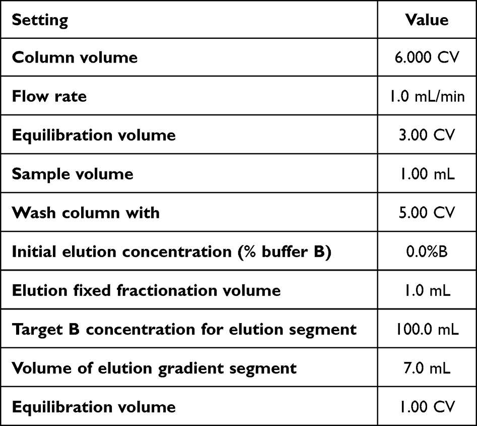 |
Table 1 Operational Parameters for the Anti-Ag85b Purification |
SDS-PAGE Analysis
SDS-PAGE analysis was performed to evaluate the purity and molecular weight of the isolated IgY. A 15% separating gel and a 4% stacking gel were prepared according to standard protocols. Protein samples (30 µL) were mixed with 5 µL of sample buffer containing β-mercaptoethanol and heated at 95 °C for 5 minutes to denature the proteins. The samples were then loaded into the gel wells together with a molecular weight protein marker.
Electrophoresis was carried out at a constant voltage of 100 V for approximately 90 minutes. After electrophoresis, the gels were rinsed with distilled water and stained with Coomassie Brilliant Blue for 1–2 hours at room temperature with gentle agitation. Destaining was performed using a decolorizing solution for 18–24 hours until clear protein bands were observed.
Western Blot Analysis
Western blot analysis was performed to verify the specific binding of purified IgY to the Ag85B antigen. Recombinant Ag85B-His and MPT64 (used as a control antigen) were separated by SDS-PAGE and transferred onto a nitrocellulose membrane using an electroblotter at 100 V for 60 minutes. The membrane was blocked overnight at 4 °C with TBST containing 5% (w/v) skim milk to prevent non-specific binding. The membrane was then incubated with purified chicken anti-Ag85B IgY (1:1000 dilution) for 2 hours at room temperature, followed by three washes with TBST.
Subsequently, the membrane was incubated with alkaline phosphatase (AP)-conjugated rabbit anti-chicken IgY secondary antibody (1:1000 dilution) for 2 hours at room temperature. After three additional washes with TBST, immunoreactive bands were visualized using BCIP/NBT substrate. The reaction was allowed to proceed for approximately 5 minutes until purple bands appeared, then stopped by rinsing the membrane with distilled water.14
Synthesis of Gold Nanoparticles and Conjugation with Purified Anti-Ag85B IgY
Gold nanoparticles (AuNPs) were synthesized using the citrate reduction method adapted from Turkevich and subsequent modifications. Briefly, HAuCl4 solution was heated under constant stirring, followed by rapid addition of sodium citrate until formation of a wine-red AuNP colloid was observed. The pH of the colloid was adjusted to pH 7.4 prior to conjugation.
For conjugation, purified anti-Ag85B IgY was mixed with the AuNP colloid and incubated at room temperature to facilitate adsorption-based conjugation. The mixture was subsequently centrifuged, and the resulting pellet was resuspended in BSA-containing buffer, followed by stabilization using conjugate diluent solution. The resulting AuNP-IgY conjugate was then used as the detection reagent in the prototype membrane-based strip assay.
Proof-of-Concept Strip Assay
A prototype strip assay was constructed to demonstrate the functional applicability of the purified anti-Ag85B IgY in a membrane-based detection format. Anti-Ag85B IgY (1 mg/mL) and goat anti-chicken IgY (1 mg/mL) were immobilized onto a membrane pad to serve as the test and control zones, respectively. Following deposition, the membrane was dried at 37 °C for approximately 2 hours. The dried membrane was then laminated onto a PVC backing card and integrated with an absorbent pad.
The conjugate mixture was prepared with an appropriate conjugate diluent and applied to the conjugate pad, which was then dried overnight at 37 °C. The dried conjugate pad was then assembled onto the PVC backing card together with the sample pad. To demonstrate antigen recognition in the strip format, the assembled strip was tested with a running buffer comprising phosphate-buffered saline (PBS) supplemented with 1% Tween-20, 1% bovine serum albumin (BSA), and 1% casein, containing 7.5 µL of Ag85B protein (0.1 mg/mL). The solution was allowed to migrate via capillary action for approximately 15 minutes, after which the appearance of visible spots at the test and control zones was visually recorded. This experiment was intended as a preliminary proof of concept to verify that the purified IgY retained its functional antigen-recognition capability when integrated into a membrane-based assay format.
Data Analysis
Statistical analyses were performed using SPSS software (version 16.0, IBM Corp., USA). Data are presented as mean ± standard deviation (SD). Differences between groups were analyzed using a Student’s t-test. A p-value < 0.05 was considered statistically significant. All extraction and purification experiments were performed using independently collected egg samples obtained during each booster period. Statistical comparisons were based on replicate measurements obtained from independent experimental preparations.
Results
Validation of Immune Response and IgY Transfer into Egg Yolk
The immune response of Gallus gallus domesticus following immunization with recombinant Ag85B was monitored using AGPT and dot blot assays to evaluate antibody production and its transfer into egg yolk. The AGPT results are presented in Figure 1, while dot blot reactivity and IgY concentrations during successive booster immunizations are summarized in Table 2. No antibody signal was detected during the pre-immunization stage, primary immunization, or the first two booster injections. Antibody reactivity first appeared at Booster 3, indicating successful seroconversion and the onset of IgY accumulation in egg yolk.
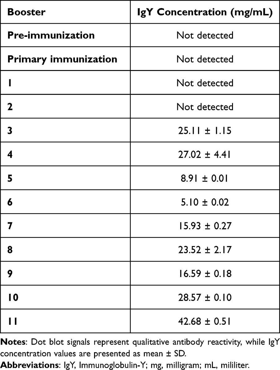 |
Table 2 Development of Anti-Ag85B IgY in Chicken Serum and Egg Yolk During the Immunization Schedule |
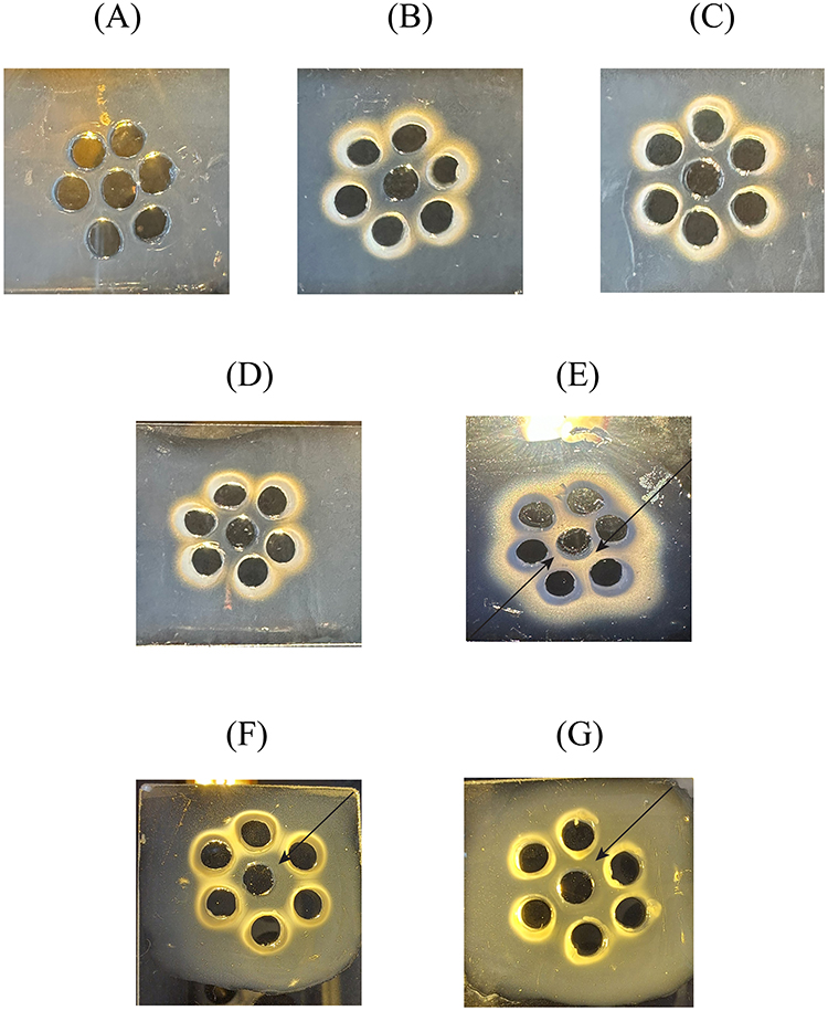 |
Figure 1 Agar gel precipitation test (AGPT) showing antibody response in chicken serum following Ag85B immunization. Notes: The black arrow indicates the formation of a precipitin line, confirming the presence of a specific antibody. (A) Negative control, (B) Pre-immune, (C) Primary immunization, (D) Booster 1, (E) Booster 2, (F) Booster 3, (G) Booster 4. |
The antibody concentration increased further at Booster 4 (27.02 ± 4.41 mg/mL) and showed moderate fluctuations during subsequent booster cycles. The peak antibody concentration of 42.68 ± 0.51 mg/mL was achieved following the 11th booster, marking the highest accumulation of anti-Ag85B IgY observed (Table 2). The presence of antibodies in both serum and egg yolk confirms the efficient transfer of systemic IgY into developing oocytes, enabling non-invasive antibody harvesting through egg collection. The observed increase in IgY concentration during successive booster immunizations demonstrated progressive accumulation of antibody in egg yolk over the course of the study, enabling subsequent purification and functional characterization.
Comparative Evaluation of Crude IgY Extraction Methods
The efficiency of crude IgY extraction from chicken egg yolk was compared using two commonly applied precipitation approaches, namely stepwise polyethylene glycol (PEG 6000) precipitation and the NaCl-based water dilution method. Quantitative protein analysis demonstrated a significantly higher protein recovery in the PEG-derived fraction compared with the NaCl method (p = 0.00000428), as summarized in Table 3. Both procedures started with an identical crude egg yolk protein concentration of 29.28 ± 0.02 mg/mL. After purification, the PEG dialysis fraction retained a high protein concentration of 28.57 ± 0.10 mg/mL, whereas the NaCl extraction yielded a substantially lower final concentration of 14.54 ± 0.70 mg/mL.
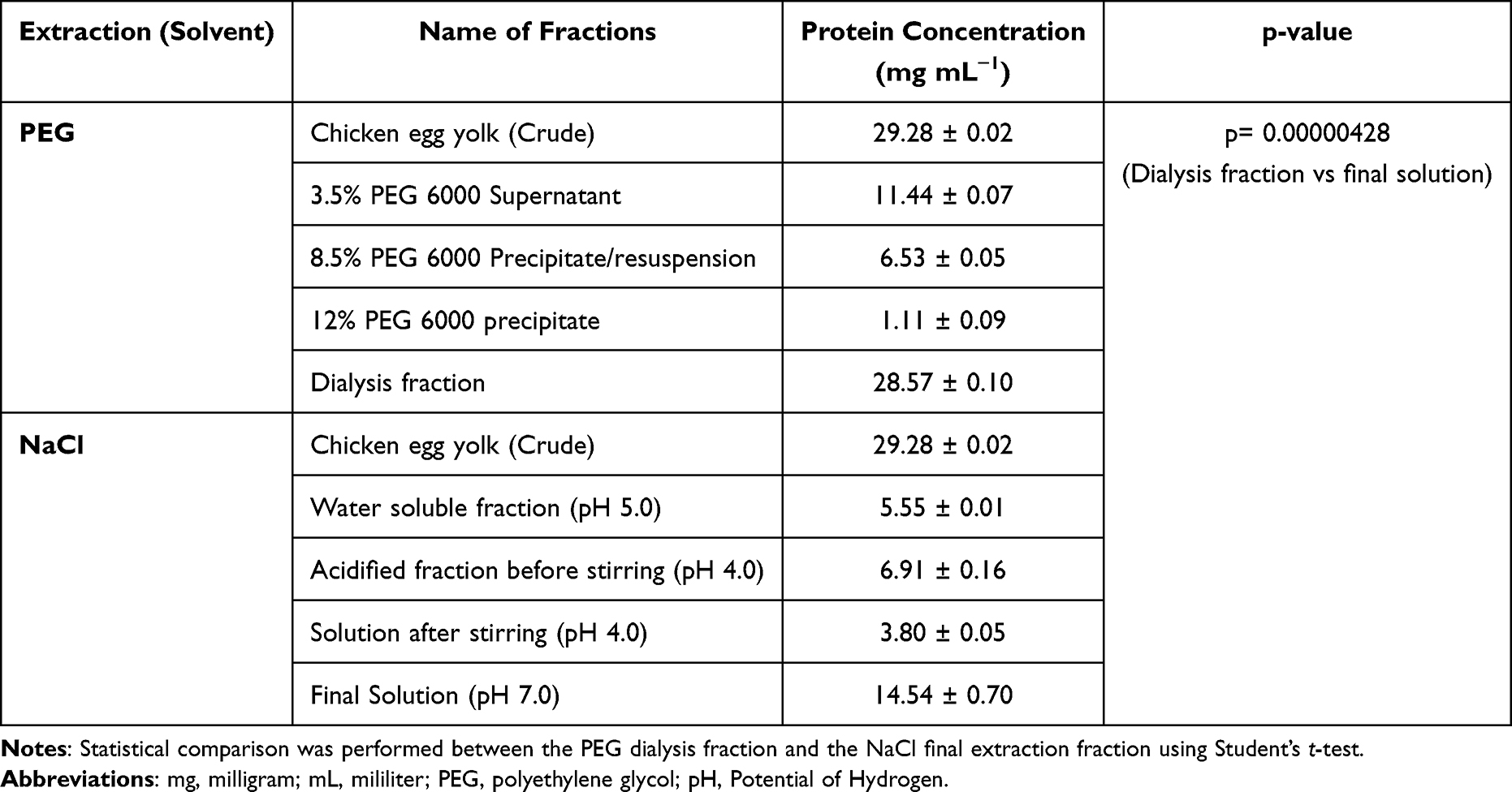 |
Table 3 Comparison of Protein Recovery from Chicken Egg Yolk Using PEG Precipitation and NaCl Extraction Methods |
The SDS-PAGE profile further supported these findings (Figure 2). Distinct protein bands corresponding to the IgY heavy chain (~65 kDa) and light chain (~25 kDa) were observed across several extraction stages. The dialysis fraction obtained from the PEG method (Lane F) and the final NaCl fraction (Lane J) both displayed these characteristic IgY bands, confirming successful IgY isolation. However, the PEG-derived fraction exhibited stronger band intensity, consistent with the higher protein concentration measured in the quantitative analysis.
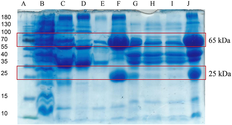 |
Figure 2 SDS-PAGE profile of IgY extraction from chicken egg yolk using PEG and NaCl precipitation methods. Notes: (A) Protein marker (10–180 kDa). (B) Chicken egg yolk (crude). (C) 3.5% PEG 6000 Supernatant. (D) 8.5% PEG 6000 precipitate/resuspension. (E) 12% PEG 6000 precipitate/resuspension. (F) Dialysis fraction. (G) Water-soluble fraction after pH adjustment (pH 5.0). (H) Solution before stirring (pH 4.0). (I) Solution after stirring (pH 4.0). (J) Final solution (pH 7.0). The red boxes indicate the characteristic IgY heavy chain (~65 kDa) and light chain (~25 kDa) bands observed during the extraction process. |
Purification of Anti-Ag85B IgY Using Hydrophobic Interaction Chromatography
Following crude extraction, the egg yolk protein extract was subjected to hydrophobic interaction chromatography (HIC) to further isolate anti-Ag85B IgY from remaining yolk proteins. Prior quantitative analysis indicated that the PEG precipitation method preserved a higher protein concentration (28.57 mg/mL) than the NaCl-based extraction (14.54 mg/mL), while both methods started from the same crude protein concentration (29.28 mg/mL).
The partially purified extract was subsequently applied to the HIC column for further separation based on hydrophobic interactions. As shown in Figure 3, the chromatographic profile exhibited a distinct dominant peak corresponding to the IgY-containing fraction, indicating efficient separation from other egg yolk proteins.
 |
Figure 3 Hydrophobic interaction chromatography (HIC) chromatogram showing purification of IgY from chicken egg yolk extract. Notes: Blue line: UV, Pink line: Gradient concentration, IgY: Target protein. |
The purity of the collected fractions was evaluated by SDS-PAGE (Figure 4). The electrophoretic profile illustrates the purification progression from the crude extract (Lane C) to the concentrated purified fraction (Lane H). Prominent bands migrating at approximately 65 kDa and 25 kDa correspond to the heavy and light chains of IgY, respectively. The presence of these characteristic bands in the final concentrated fraction confirms successful purification and recovery of anti-Ag85B IgY.
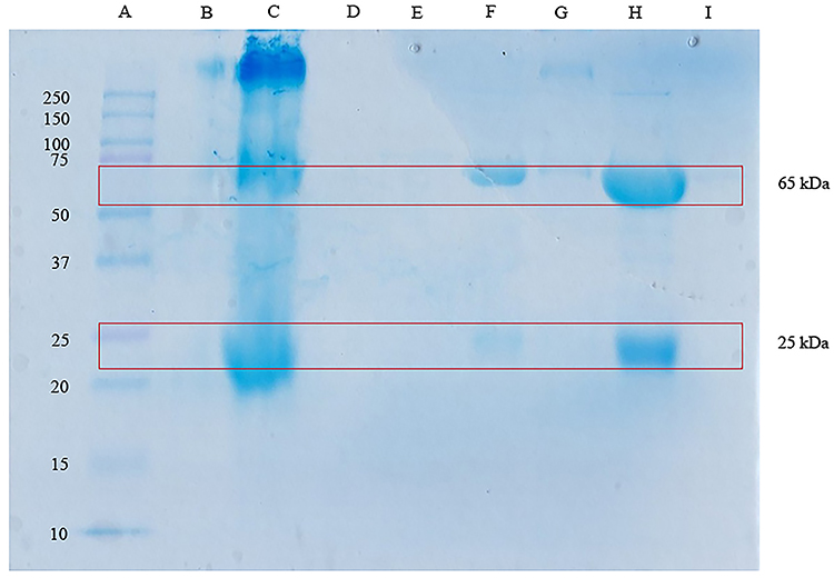 |
Figure 4 SDS-PAGE analysis showing purification of anti-Ag85B IgY following hydrophobic interaction chromatography. Notes: Lane designations are as follows: (A) Protein standard marker; (B–C) Crude chicken egg yolk extract; (D–E) Unbound protein fractions; (F) Purified IgY fraction; (G) Column regeneration fraction; (H) Concentrated purified IgY; and (I) Filtration flow-through. The red boxes indicate the characteristic IgY heavy chain (~65 kDa) and light chain (~25 kDa) bands observed after hydrophobic interaction chromatography purification. |
Specificity and Immunoreactivity of Purified Anti-Ag85B IgY
The antigen-binding specificity of the purified anti-Ag85B IgY was evaluated by Western blot analysis using recombinant Ag85B-His as the target antigen and MPT64 as a non-target control protein. Antibody reactivity was examined across three stages of antibody production: chicken serum, crude egg yolk extract, and purified IgY (Figure 5). Distinct immunoreactive bands migrating at approximately 40 kDa were observed exclusively in lanes containing Ag85B-His (Lanes 2A–C), confirming the specific recognition of the target antigen. In contrast, no detectable signal was observed in lanes containing the non-target antigen MPT64 (Lanes 1A–C), indicating no cross-reactivity. Importantly, the purified IgY fraction retained a comparable immunoreactive signal to that observed in serum and crude egg yolk extracts. This result indicates that the purification procedure successfully enriched anti-Ag85B IgY without compromising its antigen-binding activity.
 |
Figure 5 Western blot analysis demonstrating the specificity of anti-Ag85B IgY against recombinant Ag85B-His and the non-target antigen MPT64. Notes: The black arrow indicates the specific Ag85B-His protein band. (A) Chicken serum, (B) Crude egg yolk extract, and (C) Purified anti-Ag85B IgY. Lane configurations are as follows: Lanes 1 (A and C) contain the negative control protein (MPT64); Lanes 2 (A and C) contain the target antigen (Ag85B-His); and Lanes M (A and C) contain protein molecular weight markers. |
Proof-of-Concept Strip Assay for Ag85B Detection
A prototype membrane-based strip assay was developed to assess the utility of purified anti-Ag85B IgY for antigen detection. In this format, anti-Ag85B IgY was immobilized at the test spot, while anti-chicken IgY was applied at the control spot. Gold nanoparticle-conjugated anti-Ag85B IgY served as the detection reagent (Figure 6). In the negative control samples (Groups A1 and A2), where Ag85B antigen was absent, a red signal appeared only at the control spot. This result confirms proper migration of the conjugate and successful capture by immobilized anti-chicken IgY. In contrast, samples containing Ag85B protein (Groups B1 and B2) produced distinct red signals at both the control and test spots. The appearance of the test signal indicates the successful formation and capture of the Ag85B–IgY complex on the membrane. These results demonstrate that the purified anti-Ag85B IgY retained functional antigen-recognition capability after purification and could be integrated into a preliminary membrane-based strip format as a proof-of-concept immunodetection system.
 |
Figure 6 Proof-of-concept strip assay for Ag85B detection using gold nanoparticle-conjugated anti-Ag85B IgY. Notes: (A) Negative Control: Strips 1 and 2 (without Ag85B protein) exhibit only the Control Spot (1 mg/mL anti-chicken IgY), confirming assay validity and reagent functionality in the absence of the target analyte. (B) Positive Result: Strips 1 and 2, containing the test sample with 10 µg/mL Ag85B protein, display both the Control Spot (1 mg/mL anti-chicken IgY) and Test Spot (1 mg/mL anti-Ag85B IgY), indicating detection of the target analyte. Numerical labels (1, 2) denote independent replicates, demonstrating the consistency and reproducibility of the assay across both conditions. |
Discussion
The development of reliable antibody reagents is a critical component in improving antigen-based tuberculosis (TB) diagnostic assays. Among the secreted proteins of M. tuberculosis, antigen 85B (Ag85B) is one of the most immunodominant antigens and has been widely investigated as a diagnostic target. However, to date, the generation and purification of Ag85B-specific IgY antibodies from chicken egg yolk have not been systematically reported. In this study, we established an integrated workflow to produce and purify anti-Ag85B IgY from Gallus gallus domesticus eggs. By combining controlled immunization, comparative extraction approaches, and hydrophobic interaction chromatography (HIC), we successfully obtained purified IgY with preserved structural integrity and antigen-recognition capability. These findings demonstrate that egg-derived IgY can serve as a scalable antibody source for the future development of membrane-based immunodetection systems targeting tuberculosis-associated antigens.
Comparative Evaluation of PEG and NaCl Methods for IgY Extraction
The initial isolation of IgY from chicken egg yolk is complicated by the high lipid content (approx. 30%) of the yolk, which can interfere with downstream purification and assay performance.2,13 Our comparative analysis of PEG precipitation and the NaCl-based water dilution extraction method adapted from previous IgY purification studies demonstrated improved protein recovery using PEG precipitation for this specific application.9
While both methods initially yielded comparable protein concentrations in the first fraction (29.28 mg/mL), the divergence in subsequent fractions was marked. The PEG method maintained a robust protein concentration throughout the process, yielding a final concentration of 28.57 mg/mL, nearly double the 14.54 mg/mL obtained with the NaCl method.15
This discrepancy can be attributed to the distinct physicochemical mechanisms of each agent. PEG, a non-ionic water-soluble polymer, functions through a steric exclusion mechanism.16 It reorganizes the water structure around protein molecules, effectively forcing proteins out of the solution into a precipitate without disrupting their tertiary structure. This gentle precipitation is particularly effective in lipid-rich environments such as egg yolk, as PEG also aids delipidation by preferentially precipitating high-molecular-weight proteins (immunoglobulins) while leaving lipids and smaller contaminants in the supernatant.17,18
In contrast, the NaCl method relies on the salting-out effect, where high ionic strength competes with proteins for solvation.9 While effective for general bulk extraction, our data suggests that the NaCl method suffered from significant losses during the multi-step fractionation, possibly due to co-precipitation of IgY with lipid aggregates or incomplete recovery during centrifugation steps. Consequently, the stepwise PEG precipitation protocol established in this study provides a more efficient and reproducible approach for IgY recovery from egg yolk and represents a suitable upstream step before chromatographic purification.19,20
Purification of Anti-Ag85B IgY Using Hydrophobic Interaction Chromatography
While PEG precipitation provides a good crude yield, it does not yield a product sufficiently pure for sensitive clinical immunoassays, as residual yolk proteins may generate background signals during antigen detection.13,21 To improve antibody purity, the PEG-precipitated fraction was further subjected to hydrophobic interaction chromatography (HIC).22 Other chromatographic approaches, including thiophilic adsorption chromatography and affinity-based purification, have also been reported for IgY purification with good selectivity. However, HIC was selected in the present study because it operates under relatively mild purification conditions and is compatible with the lipid-rich characteristics of egg yolk-derived protein extracts.
The chromatographic profile obtained from HIC (Figure 3) displayed a distinct and well-resolved peak corresponding to the IgY-containing fraction, indicating effective separation of IgY from other yolk proteins based on differences in surface hydrophobicity. Unlike affinity chromatography, which frequently employs low-pH elution buffers that may partially affect antibody stability, HIC operates under relatively mild conditions using a decreasing salt gradient, thereby helping preserve the native conformation of antibodies during purification.21,23
The presence of purified IgY was subsequently confirmed by SDS-PAGE analysis through the detection of characteristic heavy and light chain band profiles (Figure 4). The final concentrated fraction (Lane H) revealed two prominent bands at approximately 65 kDa and 25 kDa, corresponding to the heavy and light chains of the avian IgY molecule.24 Compared with the crude egg yolk extract (Lane C), which exhibited multiple protein bands and diffuse background patterns, the purified fraction displayed a markedly cleaner electrophoretic profile. This transition from complex crude protein patterns to distinct IgY bands demonstrates that the combined PEG-HIC purification workflow effectively removes non-target yolk proteins and improves the overall purity of the IgY preparation.15,21,22,25
Specificity and Immunoreactivity of Purified Anti-Ag85B IgY
The success of antibody production in this study was evaluated not only by protein purification but also by the functional specificity of the resulting IgY. Antigen 85B (Ag85B) is one of the major secreted proteins of M. tuberculosis and is highly immunogenic.26,27 However, cross-reactivity with other mycobacterial proteins remains a challenge in tuberculosis serological assays, potentially reducing diagnostic specificity.28
Western blot analysis (Figure 5) confirmed the specificity of the anti-Ag85B IgY produced in this study.14 The antibody consistently recognized the recombinant Ag85B-His antigen, producing a distinct immunoreactive band at approximately 40 kDa across all evaluated stages of antibody production, including chicken serum, crude egg yolk extract, and the purified IgY fraction. In contrast, no detectable signal was observed when the membrane was probed against the non-target antigen MPT64, another secreted protein of M. tuberculosis used as a specificity control.25 The absence of detectable binding to MPT64 suggests that the generated IgY exhibits high antigen specificity toward Ag85B. This level of specificity is particularly important for diagnostic applications. In immunoassay platforms such as lateral flow assays (LFA) or enzyme-linked immunosorbent assays (ELISA), antibody cross-reactivity may lead to false-positive results and compromise diagnostic accuracy.5,29 Therefore, the ability of the produced IgY to selectively recognize Ag85B while discriminating against other secreted mycobacterial proteins represents a favorable characteristic for future TB diagnostic development.
Furthermore, the Western blot results also indicate that the purification workflow did not compromise antibody functionality. The comparable signal intensity observed between the purified IgY fraction and earlier antibody sources suggests that the PEG-HIC purification process preserved the antibody’s antigen-binding capability.22,30 This observation is consistent with previous reports indicating that IgY antibodies generally maintain structural stability due to their rigid molecular structure and the absence of a hinge region, which may contribute to improved resistance to environmental and chemical stresses compared with mammalian IgG.2,19
Proof-of-Concept Strip Assay for Ag85B Detection
A prototype membrane strip assay was developed to assess whether the purified anti-Ag85B IgY retained its antigen-recognition capability when incorporated into a membrane-based detection system. The assay successfully generated detectable signals for Ag85B at a concentration of 10 µg/mL, indicating that the purified antibody retained antigen-recognition capability following purification, AuNP conjugation, and incorporation into the membrane-based detection format.
Although this sensitivity does not yet represent a fully optimized diagnostic platform, the result confirms the feasibility of using purified IgY for membrane-based antigen detection. Circulating TB antigens, including Ag85B, have been reported in clinical samples such as serum and urine at concentrations ranging from micrograms to picograms, depending on disease stage and sample type.31 Therefore, further optimization of assay sensitivity will be required before clinical diagnostic implementation.
The appearance of distinct red chromogenic signals at both the test and control zones confirmed successful conjugation of purified IgY with gold nanoparticles (AuNPs) and its functional interaction with the immobilized capture components on the nitrocellulose membrane. The chemical environment of the assay also contributed to the formation of a clear signal. The inclusion of 1% Tween-20 as a non-ionic surfactant, together with blocking agents such as bovine serum albumin (BSA) and casein, helped minimize non-specific hydrophobic interactions and electrostatic interference.32 Similar blocking strategies are widely applied in membrane-based immunoassays to suppress background signals and improve signal clarity.32,33
These observations indicate that the purified anti-Ag85B IgY maintained structural stability and antigen-binding capability despite the chemical and mechanical processes involved in nanoparticle conjugation and membrane immobilization. Compared with mammalian IgG, which may undergo conformational changes at solid–liquid interfaces in lateral flow systems, the more rigid structural architecture of IgY and the absence of a hinge region may contribute to improved molecular stability.16,34 Together with the absence of detectable cross-reactivity with MPT64, these findings support the potential use of purified IgY as a promising antibody component for the future development of membrane-based tuberculosis diagnostic assays.35
Advantages of IgY Antibodies for Tuberculosis Diagnostics
The use of IgY technology offers several advantages over conventional mammalian antibody production systems. One important factor is the large phylogenetic distance between avian species and mammals, which often results in stronger immune responses against conserved mammalian or bacterial proteins.13,36 This evolutionary divergence can enhance the recognition of antigenic epitopes that may be poorly immunogenic in traditional mammalian hosts such as mice or rabbits.21,30 In the context of this study, the strong and specific immune response generated against Ag85B supports the suitability of chickens as an effective host for producing diagnostic antibodies targeting M. tuberculosis antigens.
In addition to improved immunogenicity, IgY antibodies present important analytical advantages in diagnostic applications. Unlike mammalian immunoglobulins, the Fc region of IgY does not interact with Rheumatoid Factor (RF) or Human Anti-Mouse Antibodies (HAMA), which are common sources of assay interference in human immunoassays.3,24 This structural difference may contribute to improved signal specificity and reduced false-positive reactions in immunodiagnostic platforms.
Furthermore, IgY production offers clear ethical and economic benefits. Antibodies can be obtained non-invasively from egg yolk, aligning with the 3Rs principles (Replacement, Reduction, and Refinement) by avoiding repeated animal bleeding.4,8 A single laying hen can produce approximately 100 mg of IgY per egg yolk, allowing continuous antibody collection with minimal animal stress. This production capacity makes IgY an attractive and scalable option for the development of affordable diagnostic reagents, particularly for high-burden tuberculosis regions such as Indonesia.20,37
Although the present study remains at the proof-of-concept stage, the development of IgY-based membrane immunodetection platforms may offer potential advantages for future tuberculosis screening approaches, particularly in resource-limited settings where rapid, low-cost, and user-friendly diagnostic tools are needed. In the broader context of evolving near-point-of-care tuberculosis diagnostics, further optimization of assay sensitivity, specificity, and validation using clinical specimens will be necessary before practical diagnostic implementation can be considered. Comparative evaluation with existing antigen- and molecular-based rapid diagnostic methods may also help define the future applicability and positioning of this platform within tuberculosis diagnostic workflows.
Limitations and Future Directions
While this study successfully established a scalable purification workflow for anti-Ag85B IgY and demonstrated its functional utility in a prototype membrane-based strip assay, several limitations should be acknowledged. First, although the produced IgY showed high specificity against the non-target antigen MPT64, broader cross-reactivity evaluation against additional non-tuberculous mycobacteria (NTM) species and other respiratory pathogens is still required to further confirm diagnostic specificity. Second, the long-term stability and shelf-life of the purified IgY, either in liquid or lyophilized form, remain to be systematically evaluated. Such stability studies are particularly important for diagnostic reagents intended for use in tropical and high-humidity environments where tuberculosis burden remains high. In addition, Ag85B itself has several recognized diagnostic limitations, including its inability to distinguish between vaccinated, exposed, and actively infected individuals, as well as potential limitations in immunocompromised patients with impaired T-cell responses. Therefore, the present study should be interpreted primarily as a proof-of-concept investigation focused on the production, purification, and preliminary membrane-based application of anti-Ag85B IgY rather than as a clinically validated tuberculosis diagnostic platform.
Building upon the detectable signal obtained at 10 µg/mL in the prototype strip assay, future work should focus on improving analytical sensitivity and translating this laboratory prototype into a standardized lateral flow immunoassay (LFIA) platform. Further validation using clinically relevant matrices, including sputum, serum, or urine from patients with confirmed tuberculosis, will be necessary to determine the system’s diagnostic sensitivity and specificity under real-world conditions. In addition, field-based evaluation in resource-limited settings will be important to assess the practical feasibility of implementing IgY-based diagnostic tools as affordable point-of-care solutions for tuberculosis detection.
Conclusion
In summary, this study demonstrates that stepwise polyethylene glycol (PEG) precipitation followed by hydrophobic interaction chromatography (HIC) provides an effective strategy for isolating anti-Ag85B IgY from chicken egg yolk. The purification workflow successfully reduced egg yolk contaminants while preserving the characteristic IgY band profile and antigen-recognition capability, resulting in a highly enriched IgY preparation.
The functionality of the purified antibody was further demonstrated using a prototype membrane-based strip assay, which produced clear visual signals in both the test and control zones upon exposure to Ag85B at 10 µg/mL. These findings indicate that the purified IgY retained specific antigen-binding capability after extraction, purification, and integration into a membrane-based detection format.
Overall, this study provides a practical framework for producing and purifying Ag85B-specific IgY and demonstrates its preliminary applicability in a proof-of-concept membrane-based immunodetection system. Further analytical optimization, broader specificity evaluation, and clinical validation will be necessary before implementation as a tuberculosis diagnostic platform.
Generative AI Statement
The authors used ChatGPT for language editing. The authors reviewed and edited the output as needed and take full responsibility for the content of the publication.
Ethics Approval
All animal procedures were conducted in accordance with the protocols approved by the Animal Ethics Committee of the University of Padjadjaran, Bandung, Indonesia (Approval No. 835/UN6KEP/EC/2024).
Acknowledgments
This publication charge is funded by University of Padjadjaran through the Indonesian Endowment Fund for Education (LPDP) on behalf of the Indonesian Ministry of Higher Education, Science and Technology and managed under the EQUITY Program (Contract No. 4303/B3/DT.03.08/2025 and 3927/UN6.RKT/HK.07.00/2025). The authors also would like to express their sincere gratitude to Beasiswa Pendidikan Indonesia (BPI) from the Ministry of Education, Culture, Research, and Technology for the doctoral scholarship support.
Author Contributions
All authors made a significant contribution to the work reported, whether that is in the conception, study design, execution, acquisition of data, analysis and interpretation, or in all these areas; took part in drafting, revising or critically reviewing the article; gave final approval of the version to be published; have agreed on the journal to which the article has been submitted; and agree to be accountable for all aspects of the work.
Funding
The article processing charge (APC) was supported by the Indonesian Endowment Fund for Education (LPDP) on behalf of the Indonesian Ministry of Higher Education, Science and Technology, under the EQUITY Program, and administered through Universitas Padjadjaran (Contract No. 4303/B3/DT.03.08/2025 and 3927/UN6.RKT/HK.07.00/2025).
Disclosure
The authors declare that the research was conducted in the absence of any commercial or financial relationships that could be construed as a potential conflict of interest.
References
1. World Health Organization. Global tuberculosis report 2025. Geneva, Switzerland: World Health Organization; 2025.
2. Pereira EPV, van Tilburg MF, Florean EOPT, et al. Egg yolk antibodies (IgY) and their applications in human and veterinary health: a review. Int Immunopharmacol. 2019;73:293–17. doi:10.1016/j.intimp.2019.05.015
3. Eriksson M, Larsson A. Avian antibodies as potential therapeutic tools. Antibodies. 2025;14:18. doi:10.3390/antib14010018
4. Capotă R, Ciausu-Sliwa D, Bostanaru-Iliescu AC, et al. Latest findings in immunoglobulin Y technologies and applications. Int J Mol Sci. 2025;26:6380.
5. Syahruni S, Hartati YW, Yusuf M, et al. Development of lateral flow assay based on anti-IBDV IgY for the rapid detection of Gumboro disease in poultry. J Virol Methods. 2021;291:114065. doi:10.1016/j.jviromet.2021.114065
6. Zhao B, Peng H, Zhang Y, et al. Rapid development and mass production of SARS-CoV-2 neutralizing chicken egg yolk antibodies with protective efficacy against Omicron variant in mice. Front Immunol. 2024;15:1356414. doi:10.3389/fimmu.2024.1356414
7. Qi X, Zhao S, Huang Q, et al. Preparation and application of specific chicken yolk antibodies in detecting Brucella. Front Vet Sci. 2025;12:1552097. doi:10.3389/fvets.2025.1552097
8. Bizanov G. IgY extraction and purification from chicken egg yolk. J Hell Vet Med Soc. 2017;68:265–272. doi:10.12681/jhvms.15466
9. Lan TTQ, Kha TT. Investigation of two precipitation methods for extracting immunoglobulin Y (IgY) from egg yolks. Vet Sci Res. 2021;3:23–30.
10. Morgan PM, Chaplin M, de Sousa RL. Extraction and purification of IgY. In: Hatta H, Kovacs-Nolan J, Mine Y, editors. IgY Antibodies: Production and Application. Cham, Switzerland: Springer; 2021:137–159.
11. Kubo N, Nishii M, Osada-Oka M, et al. A Comparative study on egg yolk IgY production with different adjuvants and their inhibitory effects on Staphylococcus aureus. J Poult Sci. 2021;58:192–199. doi:10.2141/jpsa.0200062
12. Natih KKN. Agar gel presipitation test (AGPT). In: BBPMSOH Glossary Series. 2018:1–3.
13. Amro WA, Al-Qaisi W, Al-Razem F. Production and purification of IgY antibodies from chicken egg yolk. J Genet Eng Biotechnol. 2018;16:99–103. doi:10.1016/j.jgeb.2017.10.003
14. Escandón P, Heatley JJ, Berghman LR, et al. Comparison of four anti-avian IgY secondary antibodies used in Western blot and dot-blot ELISA to detect avian bornavirus antibodies in four different bird species. Vet Med Res Rep. 2019;10:141–150.
15. Kusuma SAF, Parwati I, Subroto T, et al. Comparison of simple and rapid extracting methods of free-tags Mycobacterium tuberculosis protein 64 Recombinant Protein from polyacrylamide gel: electroelution and the optimized passive elution. J Adv Pharm Technol Res. 2021;12:180–184. doi:10.4103/japtr.JAPTR_318_20
16. León-Núñez D, Vizcaíno-López MF, Escorcia M, et al. IgY antibodies as biotherapeutics in biomedicine. Antibodies. 2022;11:62. doi:10.3390/antib11040062
17. Madera-Contreras AM, Solano-Texta R, Cisneros-Sarabia A, et al. Optimized method for the extraction of contaminant-free IgY antibodies from egg yolk using PEG 6000. MethodsX. 2022;9:101874. doi:10.1016/j.mex.2022.101874
18. Redwan EM, Aljadawi AA, Uversky VN. Simple and efficient protocol for immunoglobulin Y purification from chicken egg yolk. Poult Sci. 2021;100:100956. doi:10.1016/j.psj.2020.12.053
19. Zhang X, Wu R, Chelliappan B. Proteomic investigation and understanding on IgY purification and product development. Poult Sci. 2023;102:102843. doi:10.1016/j.psj.2023.102843
20. Qiu H, Jin X, Zhang X, et al. Egg yolk immunoglobulins (IgY) purification, activity enhancement, and potential benefits for human health. Nutrients. 2025;17:2890. doi:10.3390/nu17172890
21. Zhang X, Isah MB, Dang M, et al. Editorial: igY technology: theory, technical aspects, applications, and innovations. Front Immunol. 2023;14:1267926. doi:10.3389/fimmu.2023.1267926
22. Prayugo AD, Subroto T, Arnafia W, et al. Purification immunoglobulin yolk anti avian influenza H5N1 in poultry using hydrophobic interaction chromatography. Poult Sci. 2025;104:104476. doi:10.1016/j.psj.2025.104776
23. Almeida MR, Ferreira F, Domingues P, et al. Towards the purification of IgY from egg yolk by centrifugal partition chromatography, separation and purification technology. Sep Purif Technol. 2022;299:121673. doi:10.1016/j.seppur.2022.121697
24. Karachaliou CE, Vassilakopoulou V, Livaniou E. IgY technology: methods for developing and evaluating avian immunoglobulins for the in vitro detection of biomolecules. World J Methodol. 2025;11:243–262. doi:10.5662/wjm.v11.i5.243
25. Kusuma SAF, Fadhlillah M, Maisyarah IT, et al. Purification of anti-Mycobacterium tuberculosis MPT64 immunoglobulin-Y from egg-yolk supernatant using thiophilic adsorption chromatography. Prog Microbes Mol Biol. 2024;7:1. doi:10.36877/pmmb.a0000400
26. Babaki MKZ, Soleimanpour S, Rezaee SA. Antigen 85 complex as a powerful Mycobacterium tuberculosis immunogen: biology, immune-pathogenicity, application in diagnosis, and vaccine design. Microb Pathog. 2017;112:20–29. doi:10.1016/j.micpath.2017.08.040
27. Ernst JD, Cornelius A, Bolz M. Dynamics of Mycobacterium tuberculosis Ag85B revealed by a sensitive enzyme-linked immunosorbent assay. mBio. 2019;10:00611–00619. doi:10.1128/mBio.00611-19
28. Mi J, Liang Y, Liang J, et al. The research progress in immunotherapy of tuberculosis. Front Cell Infect Microbiol. 2021;11:763591. doi:10.3389/fcimb.2021.763591
29. Inbaraj LR, Daniel J, Sathya Narayanan MK, et al. Truenat MTB assays for pulmonary tuberculosis and rifampicin resistance in adults and adolescents. Cochrane Database Syst Rev. 2025;3:15543.
30. Pacheco BLB, Nogueira CP, Venancio EJ. IgY antibodies from birds: a review on affinity and avidity. Animals. 2023;13:3130. doi:10.3390/ani13193130
31. Phan LMT, Kim EB, Cheon SA, et al. Reliable naked-eye detection of Mycobacterium tuberculosis antigen 85B using gold and copper nanoshell-enhanced immunoblotting techniques. Sens Actuators B Chem. 2020;317:128220. doi:10.1016/j.snb.2020.128220
32. Omidfar K, Riahi F, Kashanian S. Lateral flow assay: a summary of recent progress for improving assay performance. Biosensors. 2023;13:837. doi:10.3390/bios13090837
33. Chang CC, Chen CP, Wu TH, et al. Gold nanoparticle-based colorimetric strategies for chemical and biological sensing applications. Nanomaterials. 2019;9:861. doi:10.3390/nano9060861
34. Tran TV, Do BN, Nguyen TPT, et al. Development of an IgY-based lateral flow immunoassay for detection of fumonisin B in maize. F1000Res. 2019;8:1042. doi:10.12688/f1000research.19643.1
35. Muhammad N, Khan MT, Ali S, et al. Novel mutations in MPT64 secretory protein of Mycobacterium tuberculosis complex. Int J Environ Res Public Health. 2023;20:2530. doi:10.3390/ijerph20032530
36. Lee L, Samardzic K, Wallach M, et al. Immunoglobulin Y for potential diagnostic and therapeutic applications in infectious diseases. Front Immunol. 2021;12:696003.
37. Hidayat R, Pasaribu FH. The Benefit of milk formulation to prevent bird flu and diarrhea; production of immunoglobulin Y anti-bird flu and anti-diarrhea in various forms. J Ilmu Pertan Indones. 2010;15:65–70.
 © 2026 The Author(s). This work is published and licensed by Dove Medical Press Limited. The
full terms of this license are available at https://www.dovepress.com/terms
and incorporate the Creative Commons Attribution
- Non Commercial (unported, 4.0) License.
By accessing the work you hereby accept the Terms. Non-commercial uses of the work are permitted
without any further permission from Dove Medical Press Limited, provided the work is properly
attributed. For permission for commercial use of this work, please see paragraphs 4.2 and 5 of our Terms.
© 2026 The Author(s). This work is published and licensed by Dove Medical Press Limited. The
full terms of this license are available at https://www.dovepress.com/terms
and incorporate the Creative Commons Attribution
- Non Commercial (unported, 4.0) License.
By accessing the work you hereby accept the Terms. Non-commercial uses of the work are permitted
without any further permission from Dove Medical Press Limited, provided the work is properly
attributed. For permission for commercial use of this work, please see paragraphs 4.2 and 5 of our Terms.