Back to Journals » Clinical, Cosmetic and Investigational Dentistry » Volume 12
Standardized Arrabidaea chica Extract Shows Cytoprotective Effects in Zoledronic Acid-Treated Fibroblasts and Osteoblasts
Authors Wiziack Zago PM, Oliveira Sousa IM ![]() , Servat-Medina L, Jorge MP, Lima Neto LG, Hass V
, Servat-Medina L, Jorge MP, Lima Neto LG, Hass V ![]() , Li X, Tasca Gois Ruiz AL
, Li X, Tasca Gois Ruiz AL ![]() , Saxena D, Foglio MA
, Saxena D, Foglio MA
Received 29 April 2020
Accepted for publication 26 June 2020
Published 11 August 2020 Volume 2020:12 Pages 327—333
DOI https://doi.org/10.2147/CCIDE.S259158
Checked for plagiarism Yes
Review by Single anonymous peer review
Peer reviewer comments 2
Editor who approved publication: Professor Christopher E. Okunseri
Patricia Maria Wiziack Zago,1 Ilza Maria Oliveira Sousa,2 Leila Servat-Medina,2 Michelle Pedroza Jorge,2 Lidio Gonçalves Lima Neto,3 Viviane Hass,4 Xin Li,5 Ana Lucia Tasca Gois Ruiz,2 Deepak Saxena,5 Mary Ann Foglio2
1Sao Leopoldo Mandic Medical School, Araras, São Paulo, Brazil; 2Faculty of Pharmaceutical Sciences at University of Campinas, Campinas, São Paulo, Brazil; 3Postgraduate Program in Parasitic Biology, CEUMA University, São Luis, Maranhão, Brazil; 4School of Dentistry, University of Missouri-Kansas City, Kansas City, MO, USA; 5College of Dentistry, New York University, New York, NY, USA
Correspondence: Mary Ann Foglio
Faculty of Pharmaceutical Sciences at University of Campinas, Rua Candido Portinari 200, CEP: 13083-871, Campinas, São Paulo, Brazil
Tel +55 19 3521-8132
Email [email protected]
Introduction: Osteonecrosis of the jaw is a condition associated with intraoral ulceration and bone necrosis induced by antiresorptive medications, such as zoledronic acid, a bisphosphonate. Previous data on Arrabidaea chica (H&B.) Verlot wound healing activity prompted the study reported herein on A. chica standardized hydro alcoholic extract in vitro cytoprotective activity data on epithelial and osteoblastic cells exposed to zoledronic acid (ZA).
Methods: Primary human gingival fibroblasts and murine pre-osteoblasts were treated with ZA 10 μM together with 5 or 10 μg.mL− 1 A. chica extract for 24h and 48 h. At both times, cells were submitted to viability assay and caspase 3/7 activation evaluation. Statistical analysis used one-way ANOVA and p=0.05.
Results: In cell viability assay, a drastic damage effect of ZA appeared after 48 h in both epithelial (55.8%) and pre-osteoblastic cells (39.7%). When treated with ZA in combination with A. chica extract, cells showed higher viability values: 74.1%– 82.3% for fibroblasts and 66% for pre-osteoblasts. Furthermore, the combined treatment presented lower caspase 3/7 activation in fibroblasts and pre-osteoblasts.
Conclusion: At low concentrations, A. chica extract showed promising cytoprotective effects against ZA-induced damage actions; however, further in vitro and in vivo studies are required to establish the mechanism of action.
Keywords: Arrabidaea chica, zoledronic acid, epithelial cells, osteoblasts, fibroblasts
Introduction
Bisphosphonates, as the potent third-generation intravenous zoledronic acid (ZA),1 are antiresorptive medications widely used to treat bone disorders and complications of bone metastases in cancer patients.2 Despite bisphosphonates beneficial therapeutic effects, the occurrence of Osteonecrosis of the jaw induced by bisphosphonates (BRONJ) is a devastating side effect, clinically characterized by a persistent exposed bone (more than 8 weeks) in the maxillofacial region of patients previously treated with bisphosphonates, without history of radiation therapy of the jaws.2
Studies revealed that oral mucosa tissues can be directly affected by bisphosphonates through different mechanisms including apoptosis,3 inhibition of migration, proliferation, growth and influence on cells viability,1,3–7 inhibition of angiogenesis5,8 and decrease in collagen and extracellular matrix production.9 Furthermore, oral bisphosphonate presents a potency comparable to intravenous bisphosphates that compromise cell activities, reinforcing the theory that bisphosphonates detrimentally affect not only osteoclasts but also several other somatic cell types.10
Once BRONJ occurs, there is not an established treatment for the oral lesion, and surgical excisions of the necrotic bone, systemic or topical application of antibiotics and the improvement of oral hygiene are palliative applied therapies.11–13
In order to improve the healing process, many studies have focused therapeutic and cytoprotective benefits of secondary metabolites from plant origin.14–16 In that context, Arrabidaea chica (Bignoniaceae), a native South American vine used in folk medicine for epithelial diseases,17 has been reported for important healing properties attributed to the anthocyanin content found in the standardized extract. Among these activities are inducement of in vitro dermal fibroblasts growth and synthesis of collagen18,20 apart from improvement of wound healing in vivo.18
Considering the side effects that patients under BRONJ treatment encounter prompted data reported herein on the benefits that A. chica standardized extract can play on bisphosphonates-modified healing process. Therefore, this study investigated the cytoprotective effect of A. chica extract on cells affected by ZA, using cell viability and apoptosis tests.
Material and Methods
Plant Material and Extraction Procedures
Arrabidaea chica Verlot. (Bignoniaceae) leaves were obtained at Chemical, Biological and Agricultural Pluridisciplinary Research Center of University of Campinas, Brazil, experimental field [Voucher deposit 1348 at CPQBA-Herbarium – Germoplasm bank]. The use of plant genetic material of the present study was approved by the Genetic Patrimony Management Board (CGEN/MMA) by CNPq through Access and Shipment Component of Genetic Heritage of Genetic Heritage for scientific research purposes (no. 010150/2012-9).
A. chica hydroalcoholic standardized extract was obtained according to the method described by Jorge et al,18 with modifications. One kilogram of dried ground leaves was extracted using 5 L of 70% hydroethanol solution acidified with 0.3% citric acid with three one hour and a half period extraction, at room temperature with mechanical stirring. Thereafter, workup involved extract filtration, organic solvent evaporation using vacuum and residual water removed by spray drying using a Mini Spray Drier B-290, loop B-295; Büchi®, Switzerland, with inlet temperature 100°C ± 2°C and outlet temperature 60°C ± 2°C, with aspiration of 250 mL.h−1 of non-filtrated N2 atmosphere, injection pressure of 414 L.h−1 and feed flow of 5 mL.min−1 at room temperature, yielding the final product.
Analytical Analysis of the Extract
Analytical analysis was according to Wen et al,21 with adaptions performed with a Shimadzu series High Pressure Liquid Chromatography (HPLC) system.
Cell Culture
Human primary gingival fibroblasts (FG; the use of these cells was approved by the Ethics Committee in Research of School of Dentistry of Piracicaba - State University of Campinas, register number 008/2012) were maintained in Dulbecco’s modified Eagle’s medium (DMEM; Gibco®) supplemented with 10% fetal bovine serum (FBS; Atlanta biologicals®), 100 U.mL−1 penicillin, 100 µg.mL−1 streptomycin and 250 ng.mL−1 Fungizone® (Amphotericin B; Gibco®). Also, murine osteoblast MC3T3-E1 (ATCC® CLR-2593TM) were maintained in minimum essential medium alpha (Alpha-MEM; Gibco®) supplemented with 10% FBS, 100 U. mL−1 penicillin and 100 µg.mL−1 streptomycin. The cells were cultured in a humidified atmosphere of 5% CO2 in air.
Zoledronic Acid Treatment and Experimental Groups
The cells were exposed to 10 µM zoledronic acid (ZA, Novartis®, Basileia, Switzerland) in combination with A. chica extract at 5 µg.mL−1 (ZAAC5) or 10 µg.mL−1(ZAAC10) concentrations. Control groups were cells without treatment (Control), 10 µM ZA (ZA) and A. chica extract at 5 µg.mL−1 (AC5) or 10 µg.mL−1 (AC10) concentration. All experiments were done in triplicate and repeated at three different occasions.
Cell Viability Tests
Cells (FG at 4x10³ cells/well and MC3T3-E1 at 1x10⁴ cells/well) were cultured in 96-well plates, in triplicate, for all the experimental groups. To avoid serum protective effects, FBS concentration was set to 5%. After 24 h or 48 h of treatment, viable cells were determined using crystal violet staining (CV) according to manufacturer’s recommendation. Briefly, cells were washed with PBS (200 µL/well, pH 7.4), stained with crystal violet solution (0.05%, 50 µL/well), incubated at room temperature for 10 min, washed twice in tap water and drained upside down on paper towels. After 24 h, sodium dodecyl sulfate (SDS) solution (1%, 200 µL/well) was added to solubilize the stain and absorbance was read using a microplate reader (SpectraMax® Plus 384 Absorbance Microplate Reader; SoftMax® ProData Acquisition & Analysis Software, Molecular Devices’ industry) at 562 nm.
Apoptosis Test
Cells (FG at 4x10³ cells/well and MC3T3-E1 at 1x10⁴ cells/well) were cultured in 96-well plates. After 24 h, cells were treated according to the experimental groups and further incubated for 24 h or 48 h. The caspase 3/7 activity in cell extracts was determined using Caspase-Glo 3/7 Assay® (Promega® Corporation, Madison, USA) according to the manufacturer’s instructions. Briefly, caspase 3/7 reagent was added into the wells (100 µL/well), the plates were protected from light, shook during 30 s (300 rpm) and then incubated at room temperature for 1 h. The luminescence was measured using Promega®- GloMax®-Multi detection system.
Statistical Analysis
The results were analyzed by one-way ANOVA for each cell type, with the post hoc test Tukey, and considering 0.05 as the significance level, using the IBM SPSS Statistic software 23.
Results
Analytical Analysis of the Extract
The final A. chica standardized extract presented pH 4.88 ± 0.02. The chromatographic fractionation afforded Carajurin (Table S1; Figure S1) that was used as analytical marker. Analytical HPLC quantitative analysis provided 6.51 ± 0.07 mg g−1 Carajurin yield content (Figure S2).
Cell Viability
For fibroblasts, a significant decrease in cell viability was observed at 48 h with ZA treatment. The combination ZA with A. chica 5 µg mL−1 or 10 µg mL−1 concentration maintained cell viability above 70% (Table 1).
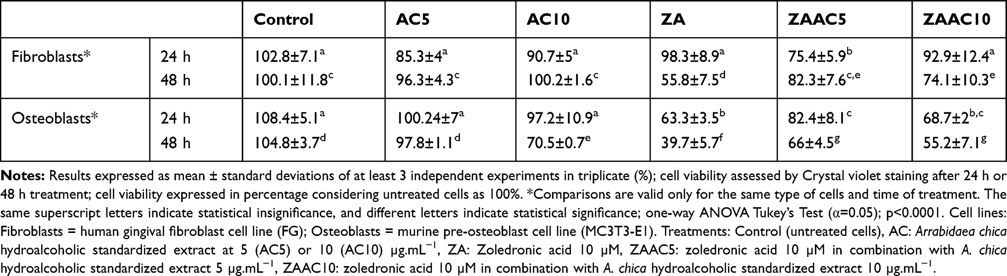 |
Table 1 Cell Viability of Fibroblasts and Osteoblasts After Isolated or Combined Zoledronic Acid and Arrabidaea Chica Extract Treatments |
Osteoblasts viability was significantly decreased by ZA at both times, whilst ZA with A. chica combination achieved higher viability values than ZA alone. At 48 h, osteoblasts treated with ZA showed viability of 39.7% ± 5.7% (p <0.0001) whereas the combination of ZA with A. chica 5 µg mL−1 maintained cells viability above 65% (p <0.0001) (Table 1).
Apoptosis Test
After 24 h, ZA significantly increased caspase 3/7 activation (p<0.0001) in fibroblasts in comparison to Control, whilst the combination of ZA with A. chica 5 µg mL−1 showed caspase 3/7 activation values statistically similar to the Control and to ZA in combination with A. chica 10 µg mL−1 (p>0.05). ZA in combination with A. chica 10 µg mL−1 showed a significant reduction in caspase 3/7 activation when compared to ZA (p<0.0001). After 48 h, ZA in combination with A. chica 5 µg mL−1 showed caspase 3/7 activation similar to Control, whereas ZA increased the luminescence values (p<0.001) (Figure 1A).
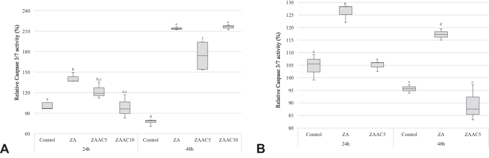 |
Figure 1 Caspase 3/7 activation on human fibroblasts (A) and murine pre-osteoblasts (B). Comparisons are valid only for the same type of cells and time of treatment. The same superscript letters indicate statistical insignificance, and different letters indicate statistical significance; one-way ANOVA Tukey’s Test (α=0.05); p<0.0001. (A) Human gingival fibroblast cell line (FG). (B) Murine pre-osteoblast cell line (MC3T3-E1). Percentage of active caspase 3/7 related to control after 24 h or 48 h treatment. Zoledronic acid 10 µM (ZA); zoledronic acid 10 µM in combination with 5 µg.mL−1 Arrabidaea chica (ZAAC5); zoledronic acid 10 µM in combination with 10 µg.mL−1 Arrabidaea chica (ZAAC10). Caspase 3/7 Glo® Assay Promega®. Averages of at least two independent experiments in triplicate. |
The analyses of osteoblasts’s active caspase 3/7 revealed, at both evaluated times, that ZA presented the highest caspase 3/7 activation (p<0.0001), whereas the combination of ZA with A. chica 5 µg mL−1 extract was statistically similar to the Control group (p>0.05) (Figure 1B).
Discussion
The promising therapeutic actions of Arrabidaea chica Verlot leaves’ extract, mainly in wound healing and antioxidant properties,18 justify the growing interest in studies involving this species. The pharmacological activity demonstrated a straight relationship with Carajurin, the main anthocyanidin identified in A. chica extract. Therefore, the pigment was used in this study for sample extract monitoring. All in vitro BRONJ experiments were conducted using standardized samples with 6.51 ± 0.07 mg Carajurin content per 1 g of A. chica extract.
BRONJ pathogenicity is yet to be explained, however, this work was based on studies that described a hypothetic mechanism of a toxic effect to the oral mucosa tissues (periosteum, connective tissue and epithelium), caused by bisphosphonate deposited on maxillary’s bones; therefore, tissues would be unable to properly heal after trauma, favoring secondary infection to bone.4,22,23 Considering A. chica wound healing properties and the necessity of new approaches for BRONJ treatment, prompted this study that evaluated the capacity of A. chica extract to revert zoledronic acid-induced damage on fibroblast and osteoblast (MC3T3-E1).
Data demonstrated that fibroblasts and osteoblasts viability varied according to treatment and experimental period (either 24 h or 48 h). During isolated treatments, both A. chica extract and ZA, at the selected concentrations, did not affect significantly human fibroblasts viability, independently on time exposure. For ZA, these results corroborated the evidences reported for ZA-treated (10 µM) gingival fibroblasts (HGF) viability evaluated by resazurin (Zafar et al)24 and MTT (3-(4,5-dimethylthiazol-2-yl)-2,5-diphenyltetrazolium bromide) (Açil et al)25 assays. In the present study, interestingly, regardless of preserving fibroblast viability after 24 h ZA exposure, caspase 3/7 was active at the same evaluated time.
The analysis of conjugated effector caspase 3/7 activation supports the identification for cell death mechanism related to apoptosis.26 Despite bisphosphonates effects on osteoclasts cause ultimately cell apoptosis,27 bisphosphonates’ activity in epithelial and osteoblastic cells is still to be elucidated. Studies have described the occurrence of apoptosis and caspase activation, mainly caspase 3,3,4,28 or necrosis, DNA and cellular cycle alterations7,29–31 as mechanisms for bisphosphonates toxicity to epithelial cells. Herein, apoptosis initiated after 24 h ZA treatment could explain the reduction on fibroblast viability (55.8%) observed at 48 h. Moreover, A. chica extract in combination with ZA achieved a significant increase in fibroblast viability at 48 h in comparison to ZA group. Therefore, when ZA was associated with A. chica, a significant decrease in caspase 3/7 activation was observed after 24h and 48 h, in comparison to ZA alone. The extract was capable of partially avoiding fibroblast activating caspase 3/7 at 24 h, that evoked in the increased cell viability at 48 h, regardless of ZA treatment.
Since the promising cytoprotective outcomes on fibroblast cells were evidenced for A. chica at low concentrations, a similar evaluation was conducted using murine osteoblasts cell line, MC3T3-E1. Even though the promotion of osteoclasts’s apoptosis represents the main mechanism of action of bisphosphonates,2 osteoblasts have been shown to play an important role in bone homeostasis as well.32 These cells regulate bone metabolism by directly interacting with osteoclasts through direct cell-cell contact, cytokines and extracellular matrix interaction. Osteoblasts also affect osteoclast production, differentiation or apoptosis through several pathways.33 Therefore, one can assume that any interference on osteoblast activity may directly affect osteoclasts and bone homeostasis.
Previous studies verified ZA actions against osteoblasts with 24 h and 72 h, showing harmful effects of bisphosphonate occurring in a time-dependent manner.25,34,35 In this study, using a murine osteoblast cell line (MC3T3-E1), the damaging effects caused by ZA started at 24 h and increased at 48 h. Furthermore, there was significant caspase 3/7 activation at both 24 h and 48 h after treating cells with ZA, which corroborates the already described effect on caspase 3/7 activation in human osteoblasts after ZA (up to 50 µM) treatment (72 h) (Jung et al).10
When administrated alone, A. chica extract did not affect osteoblast viability except at 10 µg.mL−1 concentration after 48 h exposure. Some studies have reported that polyphenolic and others antioxidant compounds might react with culture medium components generating hydrogen peroxide in situ.36,37 This situation is dependent on diverse experimental conditions such as the selected culture medium and sample concentration range.38,39 Probably, under our experimental conditions, MC3T3-E1 cells were slightly more sensitive to AC-induced H2O2 production than fibroblasts (FG), as described elsewhere.40 This could explain the slight reduction in MC3T3-E1 cell viability after 48 h exposure to A. chica extract at 10 µg.mL−1 concentration. Moreover, just at 5 µg.mL−1, A. chica extract significantly reverted ZA-induced reduction on osteoblast viability after 24 h and 48 h treatment. Considering these results, we evaluated caspase 3/7 activation only for A. chica extract at 5 µg.mL−1 concentration. Therefore, the cytoprotective effects of A. chica extract at 5 µg.mL−1 concentration in ZA-treated osteoblasts could be partially explained by reduction on caspase 3/7 activity.
Despite the lack of consensus regarding BRONJ pathophysiology, several studies were reported in attempt to establish an effective treatment for the condition. The management for BRONJ consists of therapies’ associations and palliative measures such as local hygiene with antimicrobial agents, excision of bone sequestrum, surgical resections and antibiotic regimen.2,41,42 Alternative treatments were studied in vitro, such as geranylgeraniol application,43,44 low-intensity lasers application,45,46 platelet rich plasma (PRP) and platelet-derived growth factor (PDGF).6,45 Draenert et al47 described cytoprotective effects for epithelial cells treated with 50 µM ZA in combination with 600 µM dexrazoxane, a cardio-cytoprotectant agent with antioxidant property. In vivo studies conducted with rats described preventive BRONJ pos tooth extraction strategies’, as using teriparatide, a synthetic analogue of parathyroid hormone,48,49 platelet-rich plasma50 and laser therapy.51 Moreover, A. chica extract cytoprotective effect at low concentration could be a new, low cost and promising herbal product to treat BRONJ wounds; showing many advantages, among them, an easier accessibility of population to therapy.
Conclusion
A. chica at low concentration showed important cytoprotective effects for fibroblast and osteoblastic cells exposed to harmful effects of ZA. However, further in vitro and in vivo studies are required to elucidate the mechanism of action.
Acknowledgments
The authors thank the College of Dentistry New York University (NYU-USA), Faculdade de Odontologia de Piracicaba (FOP/UNICAMP), and the Chemical, Biological and Agricultural Pluridisciplinary Research Center (CPQBA/UNICAMP) for all the infrastructure offered for the development of this research.
Disclosure
Patricia Maria Wiziack Zago reports grants from FAPESP - Sao Paulo Research Foundation, during the conduct of the study. Ana Lucia Tasca Gois Ruiz reports grants from CNPq, and FAEPEX/Unicamp, outside the submitted work. The authors declare that they have no other potential conflicts of interest in this work.
References
1. Saracino S, Canuto RA, Maggiora M, et al. Exposing human epithelial cells to zoledronic acid can mediate osteonecrosis of jaw: an in vitro model. J Oral Pathol Med. 2012;41:788–792. doi:10.1111/j.1600-0714.2012.01173.x
2. Ruggiero SL, Dodson TB, Fantasia J, et al. American Association of Oral and Maxillofacial Surgeons position paper on medication-related osteonecrosis of the jaw—2014 update. J Oral Maxillofac Surg. 2014;72(10):1938–1956. doi:10.1016/j.joms.2014.04.031
3. Scheper MA, Badros A, Chaisuparat R, Cullen KJ, Meiller TF. Effect of zoledronic acid on oral fibroblasts and epithelial cells: A potential mechanism of bisphosphonate-associated osteonecrosis. Br J Haematol. 2009;144:667–676. doi:10.1111/j.1365-2141.2008.07504.x
4. Scheper MA, Chaisuparat R, Cullen KJ, Meiller TF. A novel soft-tissue in vitro model for bisphosphonate-associated osteonecrosis. Fibrogenes Tissue Repair. 2010. doi:10.1186/1755-1536-3-6
5. Kobayashi Y, Hiraga T, Ueda A, et al. Zoledronic acid delays wound healing of the tooth extraction socket, inhibits oral epithelial cell migration, and promotes proliferation and adhesion to hydroxyapatite of oral bacteria, without causing osteonecrosis of the jaw, in mice. J Bone Miner Metab. 2010;28(2):165–175. doi:10.1007/s00774-009-0128-9
6. Cozin M, Pinker BM, Solemani K, et al. Novel therapy to reverse the cellular effects of bisphosphonates on primary human oral fibroblasts. J Oral Maxillofac Surg. 2011;69:2564–2578. doi:10.1016/j.joms.2011.03.005
7. Arai N, Inoue S, Tomihara K, Tsuno H, Noguchi M. In vitro synergistic effects of zoledronic acid and calcium on viability of human epithelial cells. Oral Dis. 2013;19(2):200–205. doi:10.1111/j.1601-0825.2012.01971.x
8. Stresing V, Fournier PG, Bellahcène A, et al. Nitrogen-containing bisphosphonates can inhibit angiogenesis in vivo without the involvement of farnesyl pyrophosphate synthase. Bone. 2011;48(2):259–266. doi:10.1016/j.bone.2010.09.035
9. Simon MJK, Niehoff P, Kimmig B, Wiltfang J, Açil Y. Expression profile and synthesis of different collagen types I, II, III, and V of human gingival fibroblasts, osteoblasts, and SaOS-2 cells after bisphosphonate treatment. Clin Oral Investig. 2010;14(1):51–58. doi:10.1007/s00784-009-0312-2
10. Jung J, Park JS, Righesso L, et al. Effects of an oral bisphosphonate and three intravenous bisphosphonates on several cell types in vitro. Clin Oral Investig. 2018;22(7):2527–2534. doi:10.1007/s00784-018-2349-6
11. Marx RE, Sawatari Y, Fortin M, Broumand V. Bisphosphonate-induced exposed bone (osteonecrosis/osteopetrosis) of the jaws: risk factors, recognition, prevention, and treatment. J Oral Maxillofac Surg. 2005;63:1567–1575. doi:10.1016/j.joms.2005.07.010
12. Marx RE, Cillo JE, Ulloa JJ. Oral bisphosphonate-induced osteonecrosis: risk factors, prediction of risk using serum CTX testing, prevention, and treatment. J Oral Maxillofac Surg. 2007;65:2397–2410. doi:10.1016/j.joms.2007.08.003
13. Ruggiero SL, Mehrotra B. Bisphosphonate-related osteonecrosis of the jaw: diagnosis, prevention, and management. Annu Rev Med. 2009;60(1):85–96. doi:10.1146/annurev.med.60.063007.134350
14. MacKay D, Miller AL. Nutritional support for wound healing. Altern Med Rev. 2003;8(4):359–377.:.
15. Houghton PJ, Hylands PJ, Mensah AY, Hensel A, Deters AM. In vitro tests and ethnopharmacological investigations: wound healing as an example. J Ethnopharmacol. 2005;100(1–2):100–107. doi:10.1016/j.jep.2005.07.001
16. Ozgen U, Ikbal M, Hacimuftuoglu A, et al. Fibroblast growth stimulation by extracts and compounds of Onosma argentatum roots. J Ethnopharmacol. 2006;104(1–2):100–103. doi:10.1016/j.jep.2005.08.052
17. Devia B, Llabres G, Wouters J, et al. New 3-deoxyanthocyanidins from leaves ofArrabidaea chica. Phytochem Anal. 2002;13(2):114–120. doi:10.1002/pca.632
18. Jorge MP, Madjarof C, Ruiz ALTG, et al. Evaluation of wound healing properties of Arrabidaea chica Verlot extract. J Ethnopharmacol. 2008;118:361–366. doi:10.1016/j.jep.2008.04.024
19. Aro AA, Freitas KM, Foglio MA, et al. Effect of the Arrabidaea chica extract on collagen fiber organization during healing of partially transected tendon. Life Sci. 2013;92(13):799–807. doi:10.1016/j.lfs.2013.02.011
20. Aro AA, Simões GF, Esquisatto MAM, et al. Arrabidaea chica extract improves gait recovery and changes collagen content during healing of the Achilles tendon. Injury. 2013;44(7):884–892. doi:10.1016/J.INJURY.2012.08.055
21. Wen D, Li C, Di H, Liao Y, Liu H, Universal A. HPLC method for the determination of phenolic acids in compound herbal medicines. J Agric Food Chem. 2005;53(17):6624–6629. doi:10.1021/jf0511291
22. Reid IR, Bolland MJ, Grey AB. Is bisphosphonate-associated osteonecrosis of the jaw caused by soft tissue toxicity? Bone. 2007;41(3):318–320. doi:10.1016/j.bone.2007.04.196
23. Reid IR. Osteonecrosis of the jaw — who gets it, and why? Bone. 2009;44(1):4–10. doi:10.1016/j.bone.2008.09.012
24. Zafar S, Coates DE, Cullinan MP, Drummond BK, Milne T, Seymour GJ. Zoledronic acid and geranylgeraniol regulate cellular behaviour and angiogenic gene expression in human gingival fibroblasts. J Oral Pathol Med. 2014;43(9):711–721. doi:10.1111/jop.12181
25. Açil Y, Möller B, Niehoff P, et al. The cytotoxic effects of three different bisphosphonates in-vitro on human gingival fibroblasts, osteoblasts and osteogenic sarcoma cells. J Cranio-Maxillofacial Surg. 2012;40(8):e229e235. doi:10.1016/j.jcms.2011.10.024
26. Paroni G, Brancolini C. Measurement of caspase activity: from cell populations to individual cells. In: Stoddart MJ, editor. Mammalian Cell Viability: Methods and Protocols. New York: Springer Science+Business Media; 2011:64–79. doi:10.1007/978-1-61779-108-6
27. Shannon J, Shannon J, Modelevsky S, Grippo AA. Bisphosphonates and Osteonecrosis of the Jaw. J Am Geriatr Soc. 2011;59(12):2350–2355. doi:10.1111/j.1532-5415.2011.03713.x
28. Ravosa MJ, Ning J, Liu Y, Stack MS. Bisphosphonate effects on the behaviour of oral epithelial cells and oral fibroblasts. Arch Oral Biol. 2011;56(5):491–498. doi:10.1016/j.archoralbio.2010.11.003
29. Pan B, To LB, Farrugia AN, et al. The nitrogen-containing bisphosphonate, zoledronic acid, increases mineralisation of human bone-derived cells in vitro. Bone. 2004;34(1):112–123. doi:10.1016/j.bone.2003.08.013
30. Agis H, Blei J, Watzek G, Gruber R. Is zoledronate toxic to human periodontal fibroblasts? J Dent Res. 2010;89(1):40–45. doi:10.1177/0022034509354298
31. Ohnuki H, Izumi K, Terada M, et al. Zoledronic acid induces S-phase arrest via a DNA damage response in normal human oral keratinocytes. Arch Oral Biol. 2012;57(7):906–917. doi:10.1016/j.archoralbio.2011.11.015
32. Wang L, Liu S, Zhao Y, et al. Osteoblast-induced osteoclast apoptosis by fas ligand/FAS pathway is required for maintenance of bone mass. Cell Death Differ. 2015;22(10):1654–1664. doi:10.1038/cdd.2015.14
33. Chen X, Wang Z, Duan N, Zhu G, Schwarz EM, Xie C. Osteoblast–osteoclast interactions. Connect Tissue Res. 2018;59(2):99–107. doi:10.1080/03008207.2017.1290085
34. Schindeler A, Little DG. Osteoclasts but not osteoblasts are affected by a calcified surface treated with zoledronic acid in vitro. Biochem Biophys Res Commun. 2005;338(2):710–716. doi:10.1016/j.bbrc.2005.09.198
35. Patntirapong S, Singhatanadgit W, Chanruangvanit C, Lavanrattanakul K, Satravaha Y. Zoledronic acid suppresses mineralization through direct cytotoxicity and osteoblast differentiation inhibition. J Oral Pathol Med. 2012;41(9):713–720. doi:10.1111/j.1600-0714.2012.01154.x
36. Long LH, Hoi A, Halliwell B. Instability of, and generation of hydrogen peroxide by, phenolic compounds in cell culture media. Arch Biochem Biophys. 2010;501:162–169. doi:10.1016/j.abb.2010.06.012
37. Rodemeister S, Hill K. Pyruvate diminishes the cytotoxic activity of ascorbic acid in several tumor cell lines in vitro. Biochem Biophys Res Commun. 2017;493(3):1184–1189. doi:10.1016/j.bbrc.2017.09.138
38. Kelts JL, Cali JJ, Duellman SJ, Shultz J. Altered cytotoxicity of ROS-inducing compounds by sodium pyruvate in cell culture medium depends on the location of ROS generation. Springerplus. 2015;4(1):1–8. doi:10.1186/s40064-015-1063-y
39. Wee LM, Long LH, Whiteman M, Halliwell B. Factors affecting the ascorbate- and phenolic-dependent generation of hydrogen peroxide in Dulbecco’s modified Eagles Medium. Free Radic Res. 2003;37(10):1123–1130. doi:10.1080/10715760310001607041
40. Fatokun AA, Stone TW, Smith RA. Responses of differentiated MC3T3-E1 osteoblast-like cells to reactive oxygen species. Eur J Pharmacol. 2008;587(13):35–41. doi:10.1016/j.ejphar.2008.03.024
41. Heufelder MJ, Hendricks J, Remmerbach T, Frerich B, Hemprich A, Wilde F. Principles of oral surgery for prevention of bisphosphonate-related osteonecrosis of the jaw. Oral Surg Oral Med Oral Pathol Oral Radiol. 2014;117:e429e435. doi:10.1016/j.oooo.2012.08.442
42. Bermúdez-Bejarano EB, Serrera-Figallo MÁ, Gutiérrez-Corrales A, et al. Prophylaxis and antibiotic therapy in management protocols of patients treated with oral and intravenous bisphosphonates. J Clin Exp Dent. 2017;9(1):e141e149. doi:10.4317/jced.53372
43. Zafar S, Coates DE, Cullinan MP, Drummond BK, Milne T, Seymour GJ. Effects of zoledronic acid and geranylgeraniol on the cellular behaviour and gene expression of primary human alveolar osteoblasts. Clin Oral Investig. 2016;20(8):2023–2035. doi:10.1007/s00784-015-1706-y
44. Ziebart T, Koch F, Klein MO, et al. Geranylgeraniol - A new potential therapeutic approach to bisphosphonate associated osteonecrosis of the jaw. Oral Oncol. 2011;47:195–201. doi:10.1016/j.oraloncology.2010.12.003
45. Martins MAT, Martins MD, Lascala CA, et al. Association of laser phototherapy with PRP improves healing of bisphosphonate-related osteonecrosis of the jaws in cancer patients: A preliminary study. Oral Oncol. 2012;48(1):79–84. doi:10.1016/j.oraloncology.2011.08.010
46. Pansani TN, Basso FG, Turirioni APS, Kurachi C, Hebling J, de Souza Costa CA. Effects of low-level laser therapy on the proliferation and apoptosis of gingival fibroblasts treated with zoledronic acid. Int J Oral Maxillofac Surg. 2014;43(8):1030–1034. doi:10.1016/j.ijom.2014.02.011
47. Draenert GF, Huetzen DO, Kämmerer PW, Palarie V, Nacu V, Wagner W. Dexrazoxane shows cytoprotective effects in zoledronic acid-treated human cells in vitro and in the rabbit tibia model in vivo. J Cranio-Maxillofacial Surg. 2012;40(8):e369e374. doi:10.1016/j.jcms.2012.01.028
48. Ersan N, Van Ruijven LJ, Bronckers ALJJ, Olgaç V, Ilgüy D, Everts V. Teriparatide and the treatment of bisphosphonate-related osteonecrosis of the jaw: A rat model. Dentomaxillofacial Radiol. 2014;43:1. doi:10.1259/dmfr.20130144
49. Dayisoylu EH, FÇ Ş, Üngör C, et al. The effects of adjunctive parathyroid hormone injection on bisphosphonate-related osteonecrosis of the jaws: an animal study. Int J Oral Maxillofac Surg. 2013;42(11):1475–1480. doi:10.1016/j.ijom.2013.05.001
50. Sarkarat F, Motamedi MHK, Jahanbani J, Sepehri D, Kahali R, Nematollahi Z. Platelet-rich plasma in treatment of zoledronic acid-induced bisphosphonate-related osteonecrosis of the jaws. Trauma Mon. 2014;19(2):13–17. doi:10.5812/traumamon.17196
51. Mergoni G, Vescovi P, Sala R, et al. The effect of laser therapy on the expression of osteocalcin and osteopontin after tooth extraction in rats treated with zoledronate and dexamethasone. Support Care Cancer. 2016;24(2):807–813. doi:10.1007/s00520-015-2847-x
 © 2020 The Author(s). This work is published and licensed by Dove Medical Press Limited. The
full terms of this license are available at https://www.dovepress.com/terms
and incorporate the Creative Commons Attribution
- Non Commercial (unported, 3.0) License.
By accessing the work you hereby accept the Terms. Non-commercial uses of the work are permitted
without any further permission from Dove Medical Press Limited, provided the work is properly
attributed. For permission for commercial use of this work, please see paragraphs 4.2 and 5 of our Terms.
© 2020 The Author(s). This work is published and licensed by Dove Medical Press Limited. The
full terms of this license are available at https://www.dovepress.com/terms
and incorporate the Creative Commons Attribution
- Non Commercial (unported, 3.0) License.
By accessing the work you hereby accept the Terms. Non-commercial uses of the work are permitted
without any further permission from Dove Medical Press Limited, provided the work is properly
attributed. For permission for commercial use of this work, please see paragraphs 4.2 and 5 of our Terms.