Back to Journals » International Medical Case Reports Journal » Volume 15
Spontaneous Rupture of Unscarred Uterus in a Term Primagravida with Lethal Skeletal Dysplasia Fetus (Thanatophoric dysplasia). A Case Report and Review of the Literature
Authors Hussein AI, Omar AA, Hassan HA , Kassim MM , Yusuf AA, Osman AA
Received 21 July 2022
Accepted for publication 30 September 2022
Published 6 October 2022 Volume 2022:15 Pages 551—556
DOI https://doi.org/10.2147/IMCRJ.S383195
Checked for plagiarism Yes
Review by Single anonymous peer review
Peer reviewer comments 3
Editor who approved publication: Professor Ronald Prineas
Ahmed Issak Hussein,1 Abdikarim Ali Omar,1 Hodan Abdi Hassan,1 Mohamed Mukhtar Kassim,2 Abdisalam Abdullahi Yusuf,2 Ahmed Adam Osman3
1Obstetrics and Gynecology Department, Mogadishu Somalia Turkish Training and Research Hospital, Mogadishu, Somalia; 2Pediatric Department, Mogadishu Somalia Turkish Training and Research Hospital, Mogadishu, Somalia; 3Radiology Department, Mogadishu Somalia Turkish Training and Research Hospital, Mogadishu, Somalia
Correspondence: Ahmed Issak Hussein, Mogadishu Somalia Turkish Training and Research Hospital, Mogadishu, Somalia, Tel +252615597479, Email [email protected]
Background and Importance: Spontaneous uterine rupture, especially in an unscarred uterus, is a rare pregnancy complication that can cause severe morbidity and mortality in both the mother and the fetus. The vast majority of uterine ruptures occur in the presence of a previous uterine scar, most commonly from a previous cesarean delivery. To our knowledge, here we reported the first case of spontaneous rupture of unscarred uterus in a term primigravida secondary to lethal skeletal dysplasia fetus (Type 1 Thanatophoric dysplasia) faced by a practicing clinician in an underdeveloped country (Somalia) with a successful outcome.
Case Presentation: The patient was 24 yrs. Old Primagravida, at 40 weeks gestation by LMP, presented with abdominal pain and active vaginal bleeding; she did not receive antenatal care during pregnancy; after initial abdominal ultrasonography and vaginal examination, laparotomy was performed due to high suspicion of uterine rupture. After dead fresh fetal extraction, the uterine defect was repaired successfully, and the patient was discharged home in good condition after several days.
Conclusion: Through this case, we would like to highlight the urgent need to focus on and recognize the importance of receiving antenatal care in the community so that the burden of thousands of lives lost each year can be reduced.
Keywords: uterine rupture, Thanatophoric dysplasia, Primagravida, antenatal care, case report
Introduction
“Uterine rupture” refers to a tear in the uterine wall during pregnancy or delivery.1 A rupture in an unscarred uterus is usually a catastrophic event that can result in the death of the mother and baby and extensive damage to the uterus. Sometimes the damage to the uterus is irreparable, and a hysterectomy will be necessary in these cases.2 The incidence of uterine rupture has been reported at 0.03–0.09% of deliveries,3 although data are often unavailable from low- and middle-income countries.4,5
Spontaneous uterine rupture is a rare pregnancy complication.1 Its occurrence may result in significant morbidity and mortality in both the fetus and the mother.6 The diagnosis of spontaneous uterine rupture is often missed or delayed.7 The main risk factor for uterine rupture is a history of a previous cesarean section (the “scarred uterus”).8,9 Most spontaneous ruptures happen during labor and usually involve the lower segment of the uterus.10,11 According to research by the World Health Organization (WHO), the incidence of rupture of an unscarred uterus is 1/8000 to 15,000 deliveries or 0.006%.12 Uterine rupture is more prevalent and remains a major obstetric complication in resource-poor countries.4,13 In resource-poor nations, including Somalia, uterine rupture is more frequently associated with obstructed labor, inappropriate use of uterine stimulants (often performed by untrained birth attendants), lack of antenatal care, grand multiparity, and poor access to emergency obstetric care.2,14,15 In industrialized, high-resource countries, uterine rupture occurs most frequently in women who have had a previous cesarean delivery.16 Uterine rupture in a primigravida patient is rare and, in most cases, completely unexpected.1,12,17,18 Without precipitating factors that have either compromised the myometrium’s integrity or applied abnormal physical stresses to the uterine wall, uterine rupture is unlikely to happen, and the apparent known cause of myometrial integrity loss is prior to cesarean delivery.19 We present a rare case of uterine rupture spontaneously in a primigravida mother who delivered a term fetus with congenital lethal skeletal dysplasia without apparent risk factors.
Case Presentation
A 24-year-old patient, gravida 1, at 40 weeks gestation by LMP, was brought to the emergency department by her family complaining of abdominal pain for 12 hours and active vaginal bleeding. She had no significant past medical history and never had antenatal care during pregnancy. She does not smoke or use drugs except antiemetics and analgesics (Navidoxine and Paracetamol). The patient had no significant personal or family history of congenital anomalies. She also denies a previous history of surgical operations, dilation and curettage, and abortions. Initially, the patient felt mild uterine contractions, minimal vaginal bleeding, and lower abdominal pain at night. The patient’s condition deteriorated in the morning, and she was brought to the hospital. The patient denies a trail of labor with misoprostol or oxytocin at home or another hospital. On examination, she was pale and distressed, with a blood pressure of 70/40 mm Hg and a maternal tachycardia of 120 bpm. The uterus was tender to palpation, with no contractions noted. The patient was hemodynamically unstable.
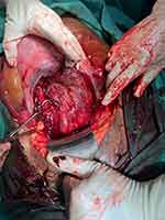
An ultrasound scan showed a fetus with no heartbeat in a transverse position. The location of the placenta could not be identified due to much free fluid in the abdominal cavity. Vaginal examination revealed a 3 cm dilated cervix with 50% effacement and active vaginal bleeding. Due to the emergency, we could not perform a detailed fetal ultrasound. The hemoglobin level at arrival was 5.4 g/dl. The patient was rushed to the operating room for an emergent operation due to suspicion of uterine rupture or placental abruption. The patient had immediate laparotomy under general anesthesia through a Pfannenstiel incision. At surgical exploration, massive hemoperitoneum was evident. A dead fresh female infant weighing 3140 grams with lethal skeletal dysplasia was found extruded in the abdominal cavity and was extracted with no need for a hysterotomy incision (Figure 1).
 |
Figure 1 Rupture of the uterus from fundus down to cervix anteriorly. |
There was marked frontal bossing with large anterior and posterior fontanelles. The nasal bridge was flat, and the ears were low set. The limbs and fingers were remarkably short, proximally and distally, with redundant skin. There was a narrow thorax with a protuberant abdomen. Based on these findings, a Type 1 Thanatophoric dysplasia diagnosis was made (Figure 2). The fetal head and abdominal circumferences were 40 cm and 55 cm, respectively. After the evacuation of the hemoperitoneum, a rupture of the uterus from the fundus anteriorly to the lower segment down to the cervix was observed (Figure 3). There was no uterine anomaly in this case, and the location of the placenta was anteriorly in the uterine fundus. The bladder was intact. The uterine defect was repaired with a continuous double-layer closure with a 1–0 vicryl absorbable suture. After obtaining hemostasis, layers were closed anatomically. The total estimated blood loss was 3000 mL; the patient was transfused with three units of red cell pack and three units of fresh frozen plasma. The patient was discharged on the fourth postoperative day with a hemoglobin level of 9.6 g/dl in good condition. After one week of follow-up, the patient was advised to use a contraceptive method for two years and prompt antenatal care for subsequent pregnancies.
 |
Figure 2 Female fetus at 40 weeks of pregnancy with short limbs and redundant skin folds, macrocephaly, narrow bell-shaped thorax, and protuberant abdomen. |
 |
Figure 3 Radiography of the fetus showing telephone receiver-like curved femurs and humerus with irregular metaphysis. |
Discussion
Spontaneous rupture of an unscarred uterus is extremely rare, especially in Primagravida mothers.18,20,21 A review of case reports of uterine rupture in primigravida women revealed an association between specific risk factors. In addition to prior uterine surgery,9,19 uterine rupture has been reported in women with uterine anomalies17,22 and abnormal placentation.23,24 Uterine rupture was also linked to uterine adenomyosis,25 dilatation and curettage,26,27 and maternal connective tissue disease, specifically Ehlers-Danlos syndrome.28 Labor induction with misoprostol11,29 and labor augmentation with oxytocin30 were linked to uterine rupture in unscarred women. Uterine perforation during operative hysteroscopy has been associated with the possibility of uterine rupture during subsequent pregnancies.18 However, none of the reported cases in the literature mentioned an association between uterine rupture and fetal skeletal anomalies. The current case describes spontaneous rupture of an unscarred uterus in a term Primagravida with lethal skeletal dysplasia fetus (Type 1 Thanatophoric dysplasia). In our case report, we could not pinpoint a specific condition as the cause of the uterine rupture.
Skeletal dysplasias are heritable disorders characterized by abnormalities of cartilage and bone. Thanatophoric dysplasia is the most lethal form of skeletal dysplasias caused by an autosomal dominant mutation in the FGFR 3 gene.31,32
Regarding our case report, it is worth noting that the lack of antenatal care during pregnancy was an important factor that contributed to this patient’s rupture of the uterus. Antenatal care is an effective method of improving pregnancy outcomes.
Conclusion
Uterine rupture is a rare complication, particularly in primigravida women and unscarred uterus. However, routine antenatal care, clinical suspicion, early detection, and prompt management should be the best options for preserving maternal and perinatal outcomes.
Abbreviations
FGFR, Fibroblast growth factor receptor; LMP, last menstrual period.
Ethical Approval
According to the guidelines of the Ethics Committee of Somali Turkey Recep Tayyip Erdogan Hospital institutional approval was not required to publish the case reports.
Informed Consent
The patient provided written informed consent for publication of the details of her case and the images shown in the figure.
Author Contributions
All authors made a significant contribution to the work reported, whether that is in the conception, Case design, execution, acquisition of data, analysis and interpretation, or in all these areas; took part in drafting, revising or critically reviewing the case; gave final approval of the version to be published; have agreed on the journal to which the article has been submitted; and agree to be accountable for all aspects of the work.
Funding
The authors declare that this study has not received any funding resources.
Disclosure
The authors report no conflicts of interest in this study.
References
1. Walsh CA, Reardon W, Foley ME. Unexplained prelabor uterine rupture in a term primigravida. Obstet Gynecol. 2007;109(2PART 1):455. doi:10.1097/01.AOG.0000244699.66548.e3
2. Hofmeyr GJ, Say L, Gülmezoglu AM. WHO systematic review of maternal mortality and morbidity: the prevalence of uterine rupture. BJOG. 2005;112(9):1221–1228. doi:10.1111/j.1471-0528.2005.00725.x
3. Al-Zirqi I, Stray-Pedersen B, Forsén L, Daltveit AK, Vangen S. Uterine rupture: trends over 40 years. BJOG. 2016;123(5):780–787. doi:10.1111/1471-0528.13394
4. Naze C. CME review article. Pediatr Emerg Care. 2017;33(12):792–793. doi:10.1097/01.pec.0000526609.89886.37
5. Tinelli A, Kosmas IP, Carugno J, et al. Uterine rupture during pregnancy: the URIDA (uterine rupture international data acquisition) study. Int J Gynecol Obstet. 2022;157(1):76–84. doi:10.1002/ijgo.13810
6. Guèye M, Mbaye M, Ndiaye-Guèye MD, et al. Spontaneous uterine rupture of an unscarred uterus before labour. Case Rep Obstet Gynecol. 2012;2012:1–3. doi:10.1155/2012/598356
7. Suner S, Jagminas L, Peipert JF, Linakis J. Fatal spontaneous rupture of a gravid uterus: case report and literature review of uterine rupture. J Emerg Med. 1996;14(2):181–185. doi:10.1016/0736-4679(95)02091-8
8. Turgut A, Ozler A, Siddik Evsen M, et al. Uterine rupture revisited: predisposing factors, clinical features, management and outcomes from a tertiary care center in Turkey. Pakistan J Med Sci. 2012;29(3):753–757.
9. Chiossi G, Amico RD, Tramontano AL, Sampogna V, Laghi V, Facchinetti F. Prevalence of uterine rupture among women with one prior low transverse cesarean and women with unscarred uterus undergoing labor induction with pge2: a systematic review and meta-Analysis. PLoS One. 2021;16(7):1–18. doi:10.1371/journal.pone.0253957
10. Singh A, Jain S. Spontaneous rupture of unscarred uterus in early pregnancy:-a rare entity. Acta Obstet Gynecol Scand. 2000;79(5):431–432.
11. Egbe TO, Halle-Ekane GE, Tchente CN, Nyemb JE, Belley-Priso E. Management of uterine rupture: a case report and review of the literature. BMC Res Notes. 2016;9(1):1–5. doi:10.1186/s13104-016-2295-9
12. Posthumus L, Donker ME. Uterine rupture in a primigravid patient, an uncommon but severe obstetrical event: a case report. J Med Case Rep. 2017;11(1):9–12. doi:10.1186/s13256-017-1507-9
13. Desta M, Amha H, Bishaw KA, et al. Prevalence and predictors of uterine rupture among Ethiopian women: a systematic review and meta-analysis. PLoS One. 2020;15:1–19. doi:10.1371/journal.pone.0240675
14. Getahun WT, Solomon AA, Kassie FY, Kasaye HK, Denekew HT. Uterine rupture among mothers admitted for obstetrics care and associated factors in referral hospitals of Amhara regional state, institution-based cross-sectional study, Northern Ethiopia, 2013–2017. PLoS One. 2018;13(12):1–14. doi:10.1371/journal.pone.0208470
15. Omer Handady S, Hassan Sakin H, Ali M, Alawad A. Uterine rupture: a review of 15 cases at bandier maternity hospital in Somalia. Clin Obstet Gynecol Reprod Med. 2015;1(2):55–58. doi:10.15761/COGRM.1000115
16. Dimitrova D, Kästner A, Kästner A, Paping A, Henrich W, Braun T. Risk factors and outcomes associated with type of uterine rupture. Arch Gynecol Obstet. 2022. doi:10.1007/s00404-022-06452-0
17. Nitzsche B, Dwiggins M, Catt S. Uterine rupture in a primigravid patient with an unscarred bicornuate uterus at term. Case Reports Women’s Heal. 2017;15(March):1–2. doi:10.1016/j.crwh.2017.03.004
18. Suner S, Jagminas L, Peipert JF, et al. Spontaneous prelabor uterine rupture in a primigravida: a case report and review of the literature. Am J Obstet Gynecol. 2011;7(6):1–6. doi:10.1016/j.ajog.2011.08.013
19. Feeney JK. Rupture of the uterus. Ir J Med Sci. 1951;27(1):27–32. doi:10.1007/BF02959388
20. Gibbins KJ, Weber T, Holmgren CM, Porter TF, Varner MW, Manuck TA. Maternal and fetal morbidity associated with uterine rupture of the unscarred uterus. Am J Obstet Gynecol. 2015;213(3):382.e1–382.e6. doi:10.1016/j.ajog.2015.05.048
21. Sweeten KM, Graves WK, Athanassiou A. Spontaneous rupture of the unscarred uterus. Am J Obstet Gynecol. 1995;172(6):1851–1856. doi:10.1016/0002-9378(95)91422-6
22. Singh N, Singh U, Verma ML. Ruptured bicornuate uterus mimicking ectopic pregnancy: a case report. J Obstet Gynaecol Res. 2013;39(1):364–366. doi:10.1111/j.1447-0756.2012.01914.x
23. Hou JH, Lee TH, Wang SY, Lai HC, Mao SP. Spontaneous uterine rupture at a non-cesarean section scar site caused by placenta percreta in the early second trimester of gestation: a case report. Taiwan J Obstet Gynecol. 2021;60(4):784–786. doi:10.1016/j.tjog.2021.05.037
24. Zhou Y, Zheng X, Chen L. Spontaneous rupture of unscarred uterus secondary to placental percreta in the third trimester of pregnancy after IVF and embryo transfer: a case report. Clin Exp Obstet Gynecol. 2021;48(6):1454–1457. doi:10.31083/j.ceog4806229
25. Li X, Li C, Sun M, Li H, Cao Y, Wei Z. Spontaneous unscarred uterine rupture in a twin pregnancy complicated by adenomyosis A case report. Medicine. 2021;100(3):6–9.
26. Ghahramani L, Moslemi S, Tahamtan M, Hashemizadeh MH, Keshavarzi A. Antepartum uterine rupture occurring at the site of a previously repaired dilatation and curettage-induced perforation: a case report. Bull Emerg Tauma. 2013;1(2):96–98.
27. Heemskerk SG, Ten Eikelder MLG, Janssen CAH. Uterine rupture in pregnancy after an intervention complicated by uterine perforation: case report and systematic review of literature. Sex Reprod Healthc. 2019;19:9–14. doi:10.1016/j.srhc.2018.11.001
28. Slaoui A, Mahtate M, Lazhar H, Lakhdar A, Baydada A, Kharbach A. Spontaneous uterine rupture revealing vascular Ehlers-Danlos syndrome: an uncommon case report. Int J Surg Case Rep. 2022;92:106840. doi:10.1016/j.ijscr.2022.106840
29. Plaut MM, Schwartz ML, Lubarsky SL, et al. Uterine rupture associated with the use of misoprostol in the gravid patient with a previous cesarean section. Am J Obstet Gynecol. 1999;180(6I):1535–1542. doi:10.1016/S0002-9378(99)70049-9
30. Zhang H, Liu H, Luo S, Gu W. Oxytocin use in trial of labor after cesarean and its relationship with risk of uterine rupture in women with one previous cesarean section: a meta-analysis of observational studies. BMC Pregnancy Childbirth. 2021;21(1):1–10. doi:10.1186/s12884-020-03440-7
31. Stembalska A, Dudarewicz L, Śmigiel R. Lethal and life-limiting skeletal dysplasias: selected prenatal issues. Adv Clin Exp Med. 2021;30(6):6. doi:10.17219/acem/134166
32. Jimah BB, Mensah TA, Ulzen-Appiah K, et al. Prenatal diagnosis of skeletal dysplasia and review of the literature. Case Rep Obstet Gynecol. 2021;2021:1–5. doi:10.1155/2021/9940063
 © 2022 The Author(s). This work is published and licensed by Dove Medical Press Limited. The full terms of this license are available at https://www.dovepress.com/terms.php and incorporate the Creative Commons Attribution - Non Commercial (unported, v3.0) License.
By accessing the work you hereby accept the Terms. Non-commercial uses of the work are permitted without any further permission from Dove Medical Press Limited, provided the work is properly attributed. For permission for commercial use of this work, please see paragraphs 4.2 and 5 of our Terms.
