Back to Journals » International Journal of General Medicine » Volume 16
Splenic Artery Embolization in Conservative Management of Blunt Splenic Injury Graded by 2018 AAST-OIS: Results from a Hospital in Vietnam
Authors Nguyen VT , Pham HD, Phan Nguyen Thanh V, Le TD
Received 19 February 2023
Accepted for publication 28 April 2023
Published 8 May 2023 Volume 2023:16 Pages 1695—1703
DOI https://doi.org/10.2147/IJGM.S409267
Checked for plagiarism Yes
Review by Single anonymous peer review
Peer reviewer comments 3
Editor who approved publication: Prof. Dr. Yuriy Sirenko
Van Thang Nguyen,1,2,* Hong Duc Pham,2,3,* Van Phan Nguyen Thanh,4 Thanh Dung Le5,6
1Radiology Department, Hai Duong Medical Technical University, Hai Duong, Vietnam; 2Radiology Department, Hanoi Medical University, Hanoi, Vietnam; 3Radiology Department, Saint Paul Hospital, Hanoi, Vietnam; 4Department of Biochemistry, Pham Ngoc Thach University of Medicine, Ho Chi Minh city, Vietnam; 5Radiology Department, Viet Duc University Hospital, Hanoi, Vietnam; 6Department of Radiology, VNU University of Medicine and Pharmacy, Hanoi, Vietnam
*These authors contributed equally to this work
Correspondence: Van Phan Nguyen Thanh, Department of biochemistry, Pham Ngoc Thach University of Medicine, 2 Duong Quang Trung Street, Ho Chi Minh city, 700000, Vietnam, Tel +84919691770, Email [email protected]
Purpose: This study was conducted to evaluate the results of conservative management of blunt splenic trauma according to the American Association for the Surgery of Trauma-Organ Injury Scale (AAST-OIS) in 2018 by embolization.
Methods: This observational study included 50 patients (42 men and 8 women) with splenic injury who underwent multidetector computed tomography (MDCT) and embolization.
Results: According to the 2018 AAST-OIS, 27 cases had higher grades than they did according to the 1994 AAST-OIS. The grades of two cases of grade II increased to grade IV; those of 15 cases of grade III increased to grade IV; and four cases of grade IV increased to grade V. As a result, all patients underwent successful splenic embolization and were stable at discharge. No patients required re-embolization or conversion to splenectomy. The mean hospital stay was 11.8± 7 days (range, 6– 44 days), with no difference in length of hospital stay among grades of splenic injury (p > 0.05).
Conclusion: Compared with the AAST-OIS 1994, the AAST-OIS 2018 classification is useful in making embolization decisions, regardless of the degree of blunt splenic injury with vascular lacerations visible on MDCT.
Keywords: blunt splenic trauma, multidetector computed tomography, MDCT, splenic embolization
Introduction
In the past two decades, conservative treatment of blunt splenic injury has increasingly been prioritized to prevent unnecessary laparotomy, maximize the rate of splenic preservation, and reduce the length of hospital stay.1 Splenic artery embolization with materials, such as gelatin sponge, coil, and embozene particles, has proven its worth as an adjunct to nonsurgical management, and it is preferred to splenectomy whenever possible.1 According to recent studies, the success of this method in hemodynamically stable patients is 88.1–100%.2,3 However, most researchers still use the results of the American Association for the Surgery of Trauma-Organ Injury Scale (AAST-OIS)-1994 classification to guide indication for surgical intervention. Assessment of the role of the 2018 AAST-OIS in blunt splenic injury grading has been studied by researchers in other countries.3,4 Especially, no studies applying the 2018 AAST-OIS classification have been conducted in Vietnam. Therefore, the aim of our study was to evaluate the results of embolization in the conservative management of blunt splenic injury using the 2018 AAST-OIS.
Materials and Methods
Study Population
Fifty patients with blunt splenic injuries admitted to Saint Paul Hospital from April 2017 to June 2022 (patients from April 2017 to February 2022 were retrospectively assessed, whereas those admitted from March 2022 to June 2022 were prospectively evaluated) underwent embolization as conservative treatment. All patients received two-pass dual (arterial and portal venous) phase computed tomography (CT), with grades III–V according to the AAST-OIS 2018 classification and vascular injury (active bleeding, pseudoaneurysm, or arteriovenous fistulas) findings. Patients with grades I and II or history of injury >30 days before time of admission were excluded.
Multidetector CT (MDCT)
Evaluations were performed using Siemens Somatom 32 slice CT scanners. Primary image interpretation was performed in the axial plane, and images acquired had 3–5 mm thickness, 0.65–1.2 pitch, 100–120 kilovolts, 350 mm FOV, and sagittal and coronal reformatted in 2 mm. Xenetic 300 iodinated contrast was administered intravenously at 3–5 mL/s, 1.5 mL/kg. The arterial, portal venous, and delayed phases (if needed) were acquired in about 25–35 seconds, 60–70 seconds, and 3–5 minutes, respectively, after the start of intravenous contrast injection.
MDCT Findings According to AAST
The following were the various MDCT findings. (1) Splenic lacerations had a linear/branching shape, low-attenuating defects, and irregular margins that often extended to the capsular surface. (2) Subcapsular hematomas appeared as peripheral, low-attenuating, and crescent-shaped collections between the enhancing parenchyma of the spleen and splenic capsule. (3) Intraparenchymal hematoma was a heterodense area within the normal splenic parenchyma. (4) Splenic infarction likely resulted from vascular injury and typically appeared as a low-attenuating wedge-shaped area. A larger portion of the wedge extended away from the splenic hilum. (5) Active extravasation was defined as contrast that actively escaped from the injured vasculature. It appeared as a focal area that increased in size and changed in morphology on subsequent phases of imaging (Figure 1A). (6) Pseudoaneurysms had a round or oval shape and a regular margin with a density that is similar to that of blood within vessels at different phases (Figure 1B). (7) An arteriovenous fistula was an abnormal enlargement of a vein, which contoured the splenic vein, and was hyperdense on the artery phase (Figure 1C).
Angiography Findings
Angiography was performed at the Monoplane Angiography Unit, Shimadzu, Japan. An active bleeding was defined as a focal or diffuse collection of vascular contrast that increases in size or attenuation at the delayed phase (Figure 1D). A pseudoaneurysm was defined as an oval or round collection of vascular contrast that communicates with a branch of the splenic artery and washes out at the delayed phase (Figure 1E). An arteriovenous fistula was one or more dilated draining veins, with both the artery and splenic vein appearing on the same MDCT image (Figure 1F).
Embolization Techniques
The Seldinger technique was used. It was performed through a unilateral femoral artery access at the point of the inguinal ligament. The catheter was inserted through a femoral introducer sheath into the abdominal aorta at the level of L1. A small volume of the contrast agent was injected into the celiac trunk for testing. After selectively probing the celiac trunk, a digital subtraction angiography was performed to visualize the anatomy of the splenic artery and the level, size, and kind of splenic vascular injury. Thereafter, a micro-catheter was inserted at the vascular injury site to select a target point for embolization with the embolic materials (coils, vascular plugs, Lipiodol+Histoacryl) suitable for the vascular injury form.
Protocol of Blunt Splenic Injury Management
Patients with blunt splenic injury who remained hemodynamically unstable and unresponsive after resuscitation required surgery. The remaining patients were scanned with contrast-enhanced MDCT. All patients with grade I or II splenic injury were observed on the ward. Patients with grade III, IV, or V injury without vascular injury on abdominal MDCT underwent nonoperative management with observation in the intensive care unit (ICU). If signs of continuous bleeding were present, MDCT was performed again, and angiography and embolization were performed. After embolization, these patients continued with nonoperative management with observation in the ICU to detect recurrent bleeding and complications.
Statistical Analysis
Data were analyzed using SPSS software version 20.0 (IBM, Armonk, NY, USA). Variables were reported as means with standard deviations. Differences among the AAST-OIS 2018 grades were determined using χ2 or Fisher’s exact test, whereas correlation between the AAST OIS 1994 and AAST-OIS 2018 was determined using Cramer’s V test. Differences among means were calculated using analysis of variance (ANOVA). For all tests, p-values <0.05 were considered significant.
Results
Patients’ Characteristics
In total, 50 patients with blunt splenic injuries were included in the analysis. They included 42 men and eight women, and they had a mean age of 33.2 ± 17.5 (range: 11–71) years. Road traffic accidents (RTA) were the most frequent cause of injury (76%). Most of the RTAs were related to motorbikes (70%). Most patients were awake on admission (Glasgow Coma Score, GCS: 14.8 ± 0.6 points) and hemodynamically stable (88%). On abdominal MDCT, 41 (82%) patients had combined injuries (chest injury, n = 21; abdominal visceral injury (n = 9), including liver injury (n = 1), left kidney (n = 5), pancreas (n = 2), and left adrenal (n = 1)).
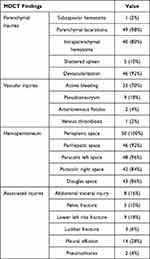
On abdominal contrast-enhanced MDCT, parenchymal lacerations and devascularized parenchymal lacerations were the most frequent parenchymal injuries reported (98% and 92%, respectively). Intraparenchymal hematoma occurred in 80% of patients. Active bleeding was the most frequent vascular injury reported (70%). In one of such cases, the patient had a thrombotic splenic vein (Figure 2). Hemoperitoneum was present in all patients with blunt splenic injuries and occupied large spaces in the abdomen. Regarding the MDCT reports, the 2018 AAST-OIS grade was grade III–V; grade IV accounted for 72% (Table 1).
 |
Table 1 MDCT Findings in Blunt Splenic Injury |
According to the 1994 AAST-OIS, 50 patients were classified as grade II, n = 2; III, n = 26; IV, n = 17; and V, n = 5. However, according to the 2018 AAST-OIS, 50 patients were classified as grade III, n = 5; IV, n = 36; and V, n = 9. Upon re-evaluation, the grades of two patients with grade II changed to grade IV (Figures 2 and 3). The grades of 21 patients with grade III increased to grade IV, and those of four patients with grade IV increased to grade V. Our study showed a difference between the 1994 AAST and 2018 AAST-OIS grades in the classification of blunt splenic injuries (p < 0.001) (Table 2).
 |
Table 2 Re-Classification According to the 2018-AAST Compared with the 1994-AAST Grading |
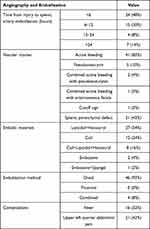
Splenic angiography and embolization were performed within 24 hours after admission for 86% of the patients (mean, 27.7 ± 68.5 hours). The angiography findings included active contrast extravasation (41, 82%), pseudoaneurysm (5, 10%), and cutoff sign (1, 2%). Permanent embolic agents were used in all cases in which Histoacryl and coils were the most frequent (70% and 40%, respectively). All patients underwent selective distal embolization (Figure 3), and four of these patients underwent combined distal and proximal embolization (8%) (Table 3).
 |
Table 3 Angiography and Embolization in Patients with Blunt Splenic Injury |
The primary clinical success rate was 100%, and the length of hospital stay was 11.8 ± 7.0 (range, 6–44) days. No differences among the 2018 AAST-OIS grades were observed (p > 0.05). Thirty (60%) patients received red blood cell transfusion, and the average volume of blood transfused was 1121 ± 1412.48 mL (Table 4).
 |
Table 4 Length of Stay and Blood Transfusion According to AAST Splenic Injury Grade |
Discussion
There are many studies describing outcomes of distal splenic artery embolization; however, this is the first study in Vietnam evaluating the outcome of splenic artery occlusion using the AAST-2018 classification of splenic injury.
Most patients with blunt splenic injury in this study were quite young, as reported in other studies.2,5 Therefore, splenic conservation is essential in these patients to prevent the need for antibiotic prophylaxis and overwhelming post-splenectomy infection.6
According to previous studies, age ≥55 years was a predictor of failure of conservative treatment.7 However, many recent studies have provided convincing evidence that age ≥55 years is not a contraindication to conservative treatment.8 Additionally, in this study, all elderly patients were successfully treated with embolization.
In this study, most patients with blunt splenic injury who underwent conservative management were conscious and hemodynamically stable at admission. Six patients who were hemodynamically unstable underwent successful conservative management, indicating that splenic artery embolization for blunt splenic trauma can be performed successfully even in hemodynamically unstable patients with a transient response to initial fluid resuscitation.9 Lee et al3 studied the clinical results of distal embolization in grade V splenic injury and reported that the clinical success rates and spleen salvage rates of patients who were hemodynamically stable showed no significant differences from those who were unstable. The classification of blunt splenic trauma by AAST-1994 has been widely used in Vietnam for nearly 30 years. The AAST-1994 grading is only based on the extent of anatomic disruption of the spleen, and previous studies have shown that AAST-1994 grades are poor predictors of outcome post-angiography and surgical intervention in patients with blunt splenic trauma. In fact, several patients had low-grade injuries and underwent observational treatment, but they to undergo surgical intervention because of the presence of hemoperitoneum.5 The new grading system by Marmery and the updated version of AAST-2018 proved to be more helpful than AAST-1994 for triaging patients who needed splenic artery embolization or surgery.3–5
All patients in this study were graded using both the AAST-1994 and AAST-2018 injury scales. According to AAST-1994, the classification of splenic injury focuses heavily on the extent of anatomic lesion, while vascular lesions in the splenic parenchyma, such as active bleeding, pseudoaneurysm, and arteriovenous fistula, are almost not mentioned. In contrast, in the updated version of AAST-2018, these vascular injury signs are given due attention, which is the basis for classifying the degree of injury and deciding on treatment choice. By default, any one of the above vascular injuries is classified as a severe splenic injury (IV–V) and should be considered for embolization. Compared with the AAST-1994 classification, the AAST-2018 grading system upgraded the grades of splenic injuries in 27 patients to grade IV or V. Statistically significant differences were found between both grading systems in grading patients with blunt splenic trauma (p < 0.001). Additionally, the AAST-2018 grading system was more effective than the AAST-1994 grading system in predicting the need for splenic arteriography.3,4 Notably, one patient graded by AAST-1994 as graded II was upgraded to grade IV by the AAST-2018 classification. This finding was important because when the patient was graded II. This patient underwent conservative management, which had a risk of failure. Obviously, the AAST-2018 guidelines demonstrated an increasing use of interventional radiologic treatment of blunt splenic trauma, which helped patients to undergo early embolization, contributing to an increase in the success rate of conservative treatment.10
From the angiography findings, 49 (98%) patients had at least one vascular lesions. Forty-six patients underwent distal embolization, and four patients underwent combined distal and proximal embolization. The primary clinical success rate was 100%, and successful hemostasis and symptom improvement were achieved by primary embolization. Two minor complications, including fever (32%) and upper left quadrant abdominal pain (42%), were reported. No patient developed major complications and required further intervention. In conclusion, embolization is a safe and feasible procedure that is effective for successful spleen salvage, regardless of the severity grade of the blunt splenic injury and morphology of the vascular injury.
The length of hospital stay was 11.8±7.0 (range, 6–44) days. No difference in the length of hospital stay was observed between the AAST-2018 and AAST-1994 grades (p > 0.05). Thirty patients underwent blood transfusion, and the volume of blood transfusion was 1121±1412.48 mL. No difference in the volume of blood transfusion was observed among the different grades of splenic injuries, as classified by AAST-1994 (p > 0.05). However, according to AAST-2018, a higher splenic injury grade resulted in a higher volume of blood transfused (p < 0.05). Therefore, the AAST-2018 classification more accurately reflects the vascular injury status of patients with splenic injury.
One of the limitations of the study, is the small number of patients included in the study. In our hospital, angiography and embolization were not performed in all patients with moderate-to-severe splenic trauma. Patients are only assigned to perform angiography and embolization after consultation with several specialties for each specific case. Therefore, the number of participants evaluated was still limited. However, with 50 cases of splenic trauma undergoing embolization over a period of about 5 years, we consider the sample size to be representative and quite common in medical facilities with vascular interventions in our country.
Conclusions
Our study findings showed that splenic artery embolization could be reserved for conservative treatment of high-grade splenic injuries and those presenting with active bleeding or a vascular pathology. The AAST-OIS 2018 is superior to the AAST-OIS 1994 in predicting embolization in the conservative treatment of blunt splenic trauma.
Ethics Approval and Consent to Participate
This study was conducted in accordance with the Declaration of Helsinki. The study conforms to the blunt splenic trauma angio guideline of Saint Paul hospital and was approved by the Ha Noi Medical University institutional ethics committee (number 633/GCN-HĐĐĐNCYSH-ĐHYHN/2022). All patients involved in the present study provided written informed consent.
Disclosure
All authors declare that they have no conflicts of interest.
References
1. Ruhnke H, Jehs B, Schwarz F, et al. Non-operative management of blunt splenic trauma: the role of splenic artery embolization depending on the severity of parenchymal injury. Eur J Radiol. 2021;137:109578. doi:10.1016/j.ejrad.2021.109578
2. Corn S, Reyes J, Helmer SD, Haan JM. Outcomes following blunt traumatic splenic injury treated with conservative or operative management. Kansas J Med. 2019;12(3):83–88. doi:10.17161/kjm.v12i3.11798
3. Lee R, Jeon CH, Kim CW, et al. clinical results of distal embolization in grade V splenic injury: four-year experience from a single regional trauma center. JVIR. 2020;31(10):1570–1577e1572. doi:10.1016/j.jvir.2020.01.029
4. Lin BC, Wu CH, Wong YC, et al. Splenic artery embolization changes the management of blunt splenic injury: an observational analysis of 680 patients graded by the revised 2018 AAST-OIS. Surg Endosc. 2023;37(1):371–381. doi:10.1007/s00464-022-09531-0
5. Marmery H, Shanmuganathan K, Alexander MT, Mirvis SE. Optimization of selection for nonoperative management of blunt splenic injury: comparison of MDCT grading systems. AJR. 2007;189(6):1421–1427. doi:10.2214/AJR.07.2152
6. Yiannoullou P, Hall C, Newton K, et al. A review of the management of blunt splenic trauma in England and Wales: have regional trauma networks influenced management strategies and outcomes? Ann Royal Coll Surg Eng. 2017;99(1):63–69. doi:10.1308/rcsann.2016.0325
7. Godley CD, Warren RL, Sheridan RL, McCabe CJ. Nonoperative management of blunt splenic injury in adults: age over 55 years as a powerful indicator for failure. J Am Coll Surg. 1996;183(2):133–139.
8. Stassen NA, Bhullar I, Cheng JD, et al. Selective nonoperative management of blunt splenic injury: an Eastern association for the surgery of trauma practice management guideline. J Trauma Acute Care Surg. 2012;73(5 Suppl 4):S294–300. doi:10.1097/TA.0b013e3182702afc
9. Hagiwara A, Fukushima H, Murata A, Matsuda H, Shimazaki S. Blunt splenic injury: usefulness of transcatheter arterial embolization in patients with a transient response to fluid resuscitation. Radiology. 2005;235(1):57–64. doi:10.1148/radiol.2351031132
10. Lee JT, Slade E, Uyeda J, et al. American society of emergency radiology multicenter blunt splenic trauma study: CT and clinical findings. Radiology. 2021;299(1):122–130. doi:10.1148/radiol.2021202917
 © 2023 The Author(s). This work is published and licensed by Dove Medical Press Limited. The full terms of this license are available at https://www.dovepress.com/terms.php and incorporate the Creative Commons Attribution - Non Commercial (unported, v3.0) License.
By accessing the work you hereby accept the Terms. Non-commercial uses of the work are permitted without any further permission from Dove Medical Press Limited, provided the work is properly attributed. For permission for commercial use of this work, please see paragraphs 4.2 and 5 of our Terms.


