
Back to Journals » Infection and Drug Resistance » Volume 19
Species Distribution and Antimicrobial Susceptibility Profiles of Non-Tuberculous Mycobacteria in Nanjing, Jiangsu Province, China: A Three-Year Retrospective Study
Authors Liu X, Wang T, Guo Y, Zeng Y, Gao W
Received 5 January 2026
Accepted for publication 23 March 2026
Published 1 April 2026 Volume 2026:19 593709
DOI https://doi.org/10.2147/IDR.S593709
Checked for plagiarism Yes
Review by Single anonymous peer review
Peer reviewer comments 3
Editor who approved publication: Dr Hazrat Bilal
Xingyu Liu,1,* Tianzhen Wang,1,* Yicheng Guo,2 Yi Zeng,1,* Weiwei Gao1,*
1Department of Tuberculosis, The Second Hospital of Nanjing, Affiliated to Nanjing University of Chinese Medicine, Nanjing, 211100, People’s Republic of China; 2Department of Tuberculosis, The School of Public Health of Nanjing Medical University, The Second Hospital of Nanjing, Nanjing, 211166, People’s Republic of China
*These authors contributed equally to this work
Correspondence: Yi Zeng; Weiwei Gao, Email [email protected]; [email protected]
Purpose: Non-tuberculous mycobacteria (NTM) are emerging pathogens with increasing clinical significance worldwide, yet regional epidemiological and drug susceptibility data remain limited. The purpose of this study is to analyze the distribution, demographic characteristics, and drug susceptibility of NTM infections in Nanjing, and to provide a basis for clinical diagnosis and treatment.
Patients and Methods: Confirmed cases of NTM infection were collected from the Tuberculosis Department of Nanjing Second Hospital between January 2023 and November 2025. Clinical isolates were cultured using the BACTEC MGIT960 system. Mycobacterium tuberculosis was ruled out by MPB64 antigen detection (immunochromatographic assay). Species identification was performed using either PCR-reverse dot blot hybridization or nanopore sequencing. Subsequently, in vitro drug susceptibility testing was conducted via the broth microdilution method.
Results: Among 249 cases of NTM detected, the most common bacterial strains are Mycobacterium intracellulare (M. intracellulare, 105/249, 42.2%) and Mycobacterium avium (M. avium, 82/249, 32.9%), with Mycobacterium abscessus accounting for 8.4% (M. abscessus, 21/249, 8.4%), 26 cases (26/249, 10.4%) were NTM mixed infection. The most common mixed infection pattern is M. abscessus and M. avium complex (MAB and MAC, 10/26, 38.5%). The age distribution of patients is mainly middle-aged and elderly, with females being the majority. The drug susceptibility results showed that amikacin (AMK) had a susceptibility rate of ≥ 93.3% to most strains, but only 61.9% to M. abscessus; The susceptibility rate of clarithromycin (CLR) to M. abscessus was 47.6%; moxifloxacin (MFX) has a susceptibility rate of over 97% to M. intracellulare and M. avium. In contrast, susceptibility to sulfamethoxazole (SMZ), imipenem/cilastatin (I/C), minocycline (MH), and doxycycline (DOX) was low, with rates below 50% for most species and 0% for I/C and MH across all isolates. Mixed infections exhibited distinct susceptibility profiles compared to single species. For example, M. abscessus-MAC mixed infections retained high susceptibility to MFX (90%) and AMK (90%), whereas M. abscessus-Mycobacterium chelonae mixed infection showed high macrolide resistance (> 80%) but 80% susceptibility to linezolid (LZD).
Conclusion: This study reveals distribution of NTM species in Nanjing Jiangsu Province China and distinct antimicrobial susceptibility profiles between single and mixed NTM infections, highlighting the clinical complexity of NTM disease, reinforce the necessity of species-level identification and antimicrobial susceptibility testing to guide individualized treatment and address the growing challenge of antimicrobial resistance in clinical practice.
Keywords: NTM mixed infection, minimum inhibitory concentration, MIC, antimicrobial susceptibility, antimicrobial resistance
Introduction
Non-tuberculous mycobacteria (NTM) are a type of mycobacteria widely present in the environment, with a wide variety of species that can cause various infections in human lungs, lymph nodes, skin, and soft tissues, among which the lungs are the most affected organ.1,2 In recent years, the incidence rate of NTM infection is on the rise and has become one of the global public health problems.3 Due to the wide variety of NTM types and significant differences in pathogenicity, epidemiological characteristics, and antimicrobial susceptibility among different strains, it poses challenges for clinical diagnosis and treatment.4,5 In China, NTM infections have emerged as an increasingly important public health concern, with a recent systematic review reporting a pooled NTM prevalence of 11.27% among suspected tuberculosis patients across mainland China and Taiwan, demonstrating a marked upward trend over the past decade.6 Substantial geographic heterogeneity exists, with the highest prevalence in northeast (24.18%) and southeast coastal regions (12.83%), and the lowest in the southwest (2.30%).6 Provincial studies further illustrate this variability: in Central China’s Jianghan Plain, NTM accounted for 7.47% of mycobacterial isolates (predominantly Mycobacterium intracellulare, 53.46%);7 In Jiangxi Province, the isolation rate was 1.60% (Mycobacterium intracellulare 51.7%, Mycobacterium abscessus 30.9%),8 and in Anhui Province, NTM comprised 15.11% (Mycobacterium intracellulare 75.36%, Mycobacterium abscessus 11.78%).9 A distinct north-south gradient is also observed, with rapidly growing mycobacteria (Mycobacterium abscessus, Mycobacterium chelonae) more frequent in the south, while Mycobacterium kansasii (M. kansasii) predominates in the north.8
In Nanjing, the capital of Jiangsu Province, China, several retrospective studies have investigated the epidemiological characteristics of NTM infections over the past decade. A study conducted at The Second Hospital of Nanjing from 2017 to 2018 found that NTM accounted for 15.5% of mycobacterial isolates, with Mycobacterium intracellulare (M. intracellulare, 70.1%) being the most prevalent species, followed by Mycobacterium abscessus (M. abscessus, 11.5%) and Mycobacterium avium (M. avium, 11.5%).10 More recent data from 2019 to 2021 revealed a dynamic shift in species distribution: although M. intracellulare, M. avium, and M. abscessus remained the top three species, the proportion of M. abscessus increased from 12% to 18% over the three-year period.11
At present, there are significant differences in the epidemiological characteristics of NTM infections among different regions and patients, and the issue of antibiotic resistance in NTMs is becoming increasingly prominent, especially in rapidly growing mycobacteria represented by M. abscessus, which often exhibit a multi drug resistance phenotype.12,13 In addition, the treatment strategies for NTM mixed infection still need to be clarified. Therefore, mastering the distribution of NTM infection strains, population characteristics, and drug susceptibility information in the local area is of great significance for guiding clinical precision medication and formulating effective prevention and control strategies.
This study aimed to retrospectively analyze confirmed cases of NTM infection diagnosed in the tuberculosis department of Nanjing Second Hospital between January 2023 and November 2025. Exploring their bacterial species distribution, patient demographic characteristics, immune function status and conducting in vitro drug susceptibility testing on prevalent NTM strains to inform the clinical diagnosis and management of NTM infections. As a renowned tertiary referral center for mycobacterial diseases, the cases diagnosed at this hospital are considered representative of the epidemiological characteristics of NTM infections in the Nanjing region in recent years.
Materials and Methods
Study Population and Sample Collection
This study was approved by the Institutional Review Board of the Second Hospital of Nanjing (ID: 2025-LS-ky112) and conducted in accordance with the Declaration of Helsinki. Confirmed cases of NTM infection were collected from the tuberculosis department of the Second Hospital of Nanjing from January 2023 to November 2025.
The diagnosis of NTM-PD was established in accordance with the 2020 guidelines of the American Thoracic Society/European Respiratory Society/European Society of Clinical Microbiology and Infectious Diseases/Infectious Diseases Society of America (ATS/ERS/ESCMID/IDSA).4 Confirmed cases required the presence of compatible clinical symptoms and radiographic abnormalities, combined with at least one of the following microbiological criteria: (1) Clinical Criteria: Presence of persistent or progressive respiratory symptoms (eg, chronic cough, sputum, hemoptysis), or relevant constitutional symptoms. (2) Radiological Criteria: Chest CT imaging revealing characteristic abnormalities, such as nodular/bronchiectatic changes (especially in the middle lobe and lingula) or cavitary lesions. (3) Microbiological Criteria: Isolation of the same NTM species from at least two separate expectorated sputum samples, or from one bronchial wash/lavage (BALF), or from a lung biopsy specimen. Demographic and clinical data, including age, sex, BMI, T-lymphocyte counts, sample types (eg, BALF, sputum, tissue), and sequencing reports, were collected from electronic medical records. The detailed screening process is outlined in the figure (Figure 1).
 |
Figure 1 Study flowchart. Workflow of the categorization of patients involved in the study. |
Mycobacterium Culture and Preliminary Identification
Mycobacterium culture and preliminary identification. All respiratory specimens were processed using the BACTEC MGIT960 system (BD, USA) according to the manufacturer’s instructions. Cultures were incubated at 37°C for up to 6 weeks, and growth was monitored automatically. Positive cultures were confirmed by Ziehl-Neelsen acid-fast staining. To exclude Mycobacterium tuberculosis, MPB64 antigen immunochromatography (Standard Diagnostics, Korea) was performed on positive culture supernatants. MPB64-negative samples were presumptively identified as NTM, and pure colonies were obtained by subculture on Middlebrook 7H10 agar (BD, USA) supplemented with oleic acid-albumin-dextrose-catalase (OADC) at 37°C for 2–4 weeks.
NTM Species Identification
Species identification was performed using a commercial mycobacterial identification gene detection kit (PCR-reverse dot blot hybridization, Yaneng BIO, China) according to the manufacturer’s instructions. This kit is designed to simultaneously identify 17 common NTM species and Mycobacterium tuberculosis complex by targeting species-specific 16S rRNA and 16S-23S rRNA internal transcribed spacer (ITS) regions. For isolates that could not be identified by this method or when mixed infection was suspected, targeted amplification combined with third-generation nanopore metagenomic sequencing (Oxford Nanopore Technologies) was used. Mixed infection was defined as detection of two or more distinct NTM species from the same specimen, confirmed by sequencing or separate colony morphotypes on solid media.
For nanopore targeted sequencing (Zhejiang ShengTing Biotech, China), respiratory samples (lung puncture fluid, sputum, or bronchoalveolar lavage fluid) were collected in DNAse-free tubes, stored at ≤4°C, and delivered to the laboratory within 24 h. Specimens were processed by dithiothreitol treatment, centrifugation, and homogenization with proteinase K, lysozyme, and zirconium beads. DNA was extracted using a magnetic bead-based method, and concentration was measured with a Qubit 4.0 fluorometer. PCR amplification was carried out with specific primers, and the products were purified, barcoded, and prepared for library construction using the Nanopore SQK-LSK110 kit. Sequencing was performed on the GridION (Mk1) platform. Raw data were filtered to remove low-quality reads (<200 bp) and host DNA sequences (aligned to the human reference genome GRCh38); the remaining reads were aligned to mycobacterial and drug resistance gene databases (TBDReaMDB, MUBII-TB-DB, RESEQTB.ORG) for final analysis.
In addition to the above laboratory evidence, the diagnosis of mixed infection was further supported by comprehensive clinical assessment, including compatible clinical symptoms and signs, typical radiological manifestations on chest imaging, and clinical response to subsequent targeted antimicrobial treatment. All mixed infection cases were confirmed by at least two independent methods or repeated testing to rule out laboratory contamination.
Antimicrobial Susceptibility Testing
Guidelines and AST Protocol
Antimicrobial susceptibility testing (AST) was performed using the broth microdilution method following the Clinical and Laboratory Standards Institute (CLSI) guidelines M24-A2.14 Custom 96-well plates containing lyophilized antibiotics were employed to evaluate a panel of 15 drugs; the abbreviations and concentration ranges for each antimicrobial agent are detailed in Table 1, in accordance with CLSI recommendations. Cation-adjusted Mueller-Hinton broth (CAMHB) served as the basal medium, and for slowly growing mycobacteria (SGM) it was supplemented with 5% oleic acid-albumin-dextrose-catalase (OADC).
 |
Table 1 Antimicrobial Agents Tested in This Study |
Inocula were prepared from fresh subcultures grown on solid medium. Rapidly growing mycobacteria (RGM, eg, Mycobacterium abscessus, Mycobacterium chelonae) were subcultured for 3–5 days, while slowly growing mycobacteria (SGM, eg, Mycobacterium avium complex, Mycobacterium kansasii) were subcultured for 7–14 days. Bacterial suspensions were adjusted to a 0.5 McFarland standard (approximately 1–5 × 10⁶ CFU/mL) using a calibrated densitometer and then diluted 1:100 in CAMHB to achieve a final inoculum of approximately 5 × 10⁵ CFU per well in the 96-well plates.
Antimicrobial Agents and Concentrations
The pre-prepared plates contained two-fold dilution series of each antibiotic, covering the following ranges: SMZ: 8.000–256 μg/mL, MFX: 0.125–16 μg/mL, RFB: 0.500–32 μg/mL, TOB: 0.500–64 μg/mL, CEF: 4.000–160 μg/mL, AMK: 1.000–64 μg/mL, DOX: 4.000–128 μg/mL, MH: 4.000–128 μg/mL, RFP: 0.500-16 μg/mL, CLR: 0.500–64 μg/mL, EMB: 2.500–20 μg/mL, LZD: 0.500–32 μg/mL, AZH: 1.000–32 μg/mL, I/C: 4.000–64 μg/mL, GAT: 0.060–8 μg/mL.
All plates were incubated at 37°C. RGM isolates were incubated in ambient air for 3–5 days, whereas SGM isolates were incubated in a 5% CO2 atmosphere for 7–14 days, until sufficient growth was observed in the drug-free control wells.
MIC Determination and Interpretation
The minimum inhibitory concentration (MIC) was defined as the lowest concentration of an antimicrobial agent that visibly inhibited bacterial growth. For isolates with unclear growth endpoints, the resazurin microtiter assay (REMA) was used as an adjunct colorimetric method;15 in such cases the MIC was recorded as the lowest concentration that prevented a color change from blue to pink. Susceptible (S), intermediate (I), and resistant (R) categories were assigned according to the clinical breakpoints published in CLSI document M24-A2.
Determination of MIC50 and MIC90: MIC values for each species-antimicrobial combination were arranged in ascending order. The MIC50 was defined as the concentration that inhibited at least 50% of the isolates (ie, the median MIC), and the MIC90 was defined as the concentration that inhibited at least 90% of the isolates. For an even number of isolates, the MIC50 was calculated as the geometric mean of the two middle values. Interpretive criteria for susceptibility categories (susceptible, intermediate, resistant) were based on clinical breakpoints or epidemiological cutoff values (ECOFFs) established by the Clinical and CLSI guidelines for nontuberculous mycobacteria.14,16
Analysis
The study used IBM SPSS Statistics27 software for all statistical analyses. Continuous variables that follow a normal distribution are represented as mean ± standard deviation (Mean ± SD), and inter group comparisons are conducted using one-way ANOVA; Continuous variables that do not follow a normal distribution are represented by the median (interquartile range) M (IQR), and inter group comparisons are conducted using the Kruskal Wallis H-test. Categorical variables (n, %) are represented by test or Fisher’s exact test for comparison between multiple groups. For antimicrobial susceptibility data (Tables 2 and 3), descriptive statistics were used to present susceptibility rates, and MIC50 and MIC90 values were calculated to summarize in vitro activity. A two-tailed P-value <0.05 was considered statistically significant. Pie charts and bar graphs were generated using GraphPad Prism, and radar charts were created using R software.
 |
Table 2 Antimicrobial Susceptibility Profiles of Single NTM Species |
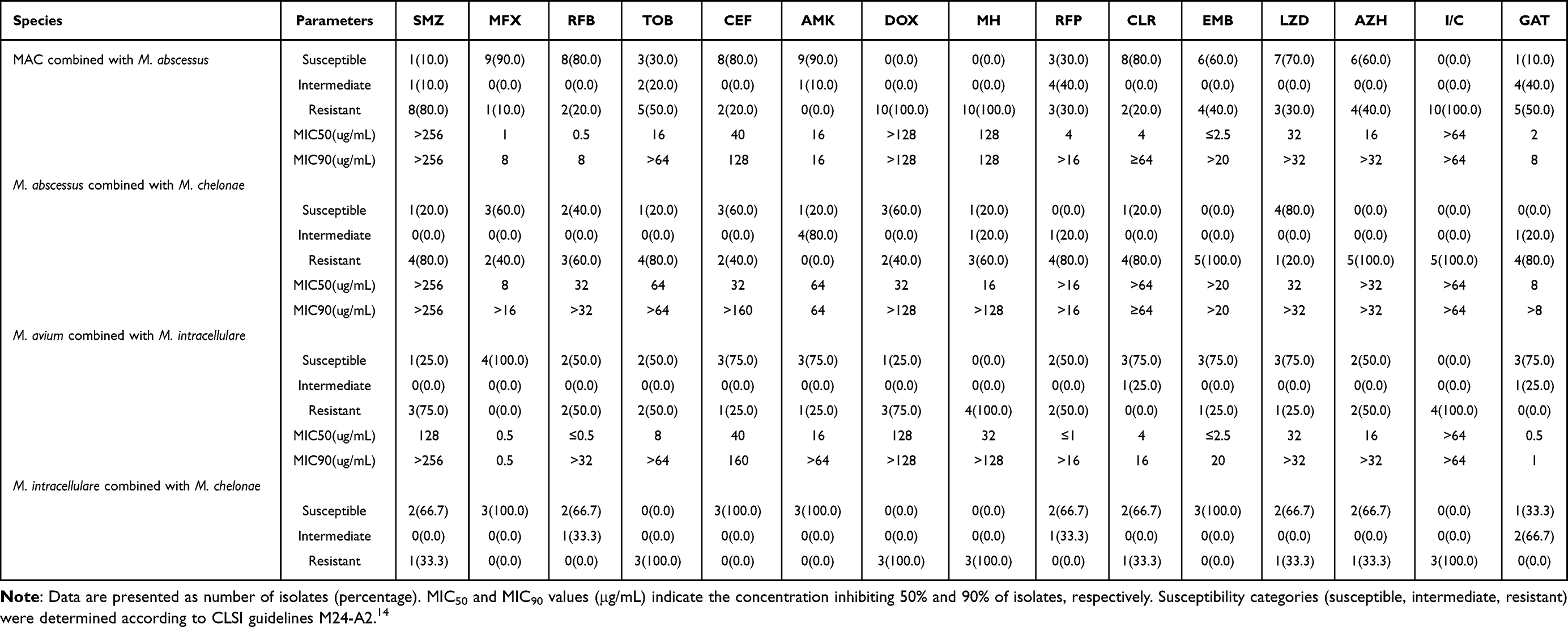 |
Table 3 Antimicrobial Susceptibility Profiles of NTM Mixed Infections |
Results
Distribution of NTM Species
Among 249 cases of NTM detected, Mycobacterium intracellulare (M. intracellulare) were the most common (105/249, 42.2%), followed by Mycobacterium avium (M. avium, 82/249, 32.9%) and Mycobacterium abscessus (M. abscessus, 21/249, 8.4%). Mycobacterium chelonae (M. chelonae) accounts for (6/249, 2.4%). Less common species included Mycobacterium kansasii (M. kansasii, 4/249, 1.6%) and Mycobacterium gordonae (M. gordonae, 2/249, 0.8%), and single isolates of Mycobacterium paragordonae (M. paragordonae), Mycobacterium scrofulaceum (M. scrofulaceum), and Mycobacterium xenopi (M. xenopi).
Notably, 26 cases (26/249, 10.4%) involved mixed infection with two or more NTM species. The most common mixed infection pattern is M. abscessus and Mycobacterium avium complex (MAB and MAC, 10/26, 38.5%), followed by M. abscessus and M. chelonae (MAB and MC, 5/26, 19.2%) and M. intracellulare combined with M. avium (MI and MA, 4/26, 15.4%). Other dual-species combinations included M. intracellulare combined with M. chelonae (MI and MC, 3/26, 11.5%), M. intracellulare combined with M. kansasii (MI and MK, 1 case). Three cases involved triple-species mixed infections: M. intracellulare combined with M. avium and M. chelonae (MI, MA and MC, 1 case), M. intracellulare combined with M. chelonae and M. abscessus (MI, MC and MAB, 1 case), M. abscessus combined with M. xenopi and M. avium (MAB, MX and MA, 1 case). The distribution of NTM species is illustrated in Figure 2.
 |
Figure 2 Distribution of NTM Species. Notes: Numbers inside the pie chart represent absolute case counts (n). Percentages were calculated based on the total number of NTM cases (N = 249). |
Among the 249 NTM-positive cases, the majority of respiratory specimens were sputum (146/249, 58.6%), followed by bronchoalveolar lavage fluid (BALF) (96/249, 38.6%) and tissue (7/249, 2.8%).
Clinical Characteristics According to NTM Species
Baseline demographic and clinical characteristics of patients stratified by infecting NTM species are summarized in Table 4. No significant differences were observed among groups regarding sex, body mass index (BMI), or presenting symptoms. However, significant differences were found in age and CD19 lymphocyte counts across groups. Patients infected with M. intracellulare, M. avium, and NTM mixed infection were predominantly older (mean age: 65.66 ± 9.98, 65.56 ± 9.34, and 64.35 ± 9.60 years, respectively), suggesting a predilection for middle-aged and elderly populations. In contrast, patients with M. abscessus or M. chelonae infections were significantly younger (mean age: 56.24 ± 10.88 and 56.83 ± 14.30 years, respectively), indicating a higher prevalence in middle-aged and younger adults.
 |
Table 4 Clinical Characteristics According to NTM Species |
Antimicrobial Susceptibility Profiles of Single NTM Species
The antimicrobial susceptibility profiles of the four most prevalent NTM species (M. intracellulare, M. avium, M. abscessus, and M. chelonae) are presented in Table 2 and Figure 3. As expected, the slow-growing MAC species (M. intracellulare and M. avium) exhibited similar susceptibility patterns, whereas the rapidly growing M. abscessus demonstrated broad resistance.
 |
Figure 3 Radar Diagram of Antibacterial Drugs for Single NTM Species. |
M. intracellulare showed high susceptibility to moxifloxacin (MFX; 98.1%, MIC50 = 0.5 μg/mL), rifabutin (RFB; 87.6%, MIC50 ≤ 0.05 μg/mL), and amikacin (AMK; 96.2%, MIC50 = 16 μg/mL). In contrast, resistance rates exceeded 92% for doxycycline (DOX), minocycline (MH), and imipenem/cilastatin (I/C). Similarly, M. avium was highly susceptible to MFX (98.8%, MIC50 = 0.5 μg/mL), RFB (92.7%, MIC50 ≤ 0.05 μg/mL), and AMK (93.9%, MIC50 = 16 μg/mL), while exhibiting nearly complete resistance to MH (96.3% resistance) and I/C (100% resistance).
M. abscessus displayed extensive resistance, particularly to sulfamethoxazole (SMZ; 76.2% resistance, MIC50 > 256 μg/mL), DOX (81.0% resistance, MIC50 > 128 μg/mL), MH (90.5% resistance, MIC50 = 128 μg/mL), rifampicin (RFP; 76.2% resistance), ethambutol (EMB; 85.7% resistance), I/C (100% resistance), and gatifloxacin (GAT; 90.5% resistance). Susceptibility to clarithromycin (CLR) was only 47.6%, and to AMK 61.9%, with a notable proportion of intermediate isolates (28.6%).
For M. chelonae, most agents showed moderate to high resistance; only AMK (83.3% susceptible, MIC50 = 4 μg/mL) and linezolid (LZD; 66.7% susceptible, MIC50 = 8 μg/mL) retained appreciable activity. Complete resistance was observed against I/C (100%) and MH (100%). Susceptibility to other drugs varied.
For less common NTM species susceptibility testing revealed that M. kansasii was universally susceptible to MFX, RFB, AMK, RFP, and LZD, but completely resistant to tobramycin (TOB), MH, and I/C. M. gordonae was fully susceptible to MFX and fully resistant to MH and I/C. M. xenopi and M. paragordonae remained susceptible to most agents, with complete resistance limited to TOB, MH, and I/C. In contrast, M. scrofulaceum exhibited a broader resistance profile, with full susceptibility only to MFX, RFB, CLR, and EMB, and complete resistance to multiple drugs including SMZ, TOB, AMK, DOX, MH, RFP, LZD, azithromycin (AZH), I/C, and GAT.
Notably, I/C showed 100% resistance across all NTM isolates and was therefore excluded from the radar chart analysis due to its lack of variability. The complete susceptibility data for I/C, along with all other tested agents, are presented in Table 2.
Antimicrobial Susceptibility Profiles of NTM Mixed Infections
The antimicrobial susceptibility profiles of the 26 NTM mixed infections are detailed in Table 3 and Figure 4. These profiles often differed from those of single-species infections. Antimicrobial susceptibility testing (AST) revealed that mixed infection with rapidly growing M. abscessus and slowly growing MAC showed the highest susceptibility to MFX and AMK, both reaching 90%, with MIC50 values of 1 μg/mL and 16 μg/mL, respectively. They also exhibited relatively high susceptibility (80% each) to RFB, CEF, and CLR. In contrast, susceptibility to EMB, AZM, and RFP was lower. Another common mixed infection involved M. abscessus and M. chelonae, which demonstrated a distinct susceptibility pattern: resistance to macrolides (CLR and AZM) exceeded 80%, while susceptibility to LZD remained at 80%. Susceptibility to MFX, CEF, and DOX was 60% each. In cases of M. avium combined with M. intracellulare, high susceptibility was observed to multiple agents, including MFX, CEF, AMK, CLR, EMB, LZD, and GAT. M. chelonae combined with M. intracellulare showed 100% susceptibility to MFX, RFB, CEF, and AMK. The remaining mixed infections, although limited in sample size, also displayed distinct AST profiles. For example, M. intracellulare combined with M. kansasii was fully susceptible to MFX, CEF, AMK, and EMB. Additionally, the three cases of triple-strain mixed infection each showed different susceptibility spectra: M. intracellulare combined with M. avium and M. chelonae was susceptible to CEF, AMK, CLR, EMB, LZD, AZM, and GAT; M. intracellulare combined with M. chelonae and M. abscessus was susceptible to SMZ, MFX, RFB, CEF, and LZD; and M. abscessus combined with M. xenopi and M. avium was susceptible to MFX, RFB, CEF, AMK, CLR, and EMB.
 |
Figure 4 Radar Diagram of Antibacterial Drugs of NTM Mixed Infections. |
A noteworthy finding was that I/C exhibited a 100% resistance rate among all tested NTM isolates. Consequently, this agent was excluded from the radar chart analysis, as its uniform resistance profile (lacking variation) would not contribute meaningfully to the visual comparison of susceptibility patterns across different antimicrobials. The complete susceptibility data for I/C, along with all other tested agents, are presented in Table 3.
Discussion
Accurate species identification is fundamental to the diagnosis and management of NTM infections. In this study, strain identification was performed using a mycobacterial identification gene detection kit or targeted amplification coupled with third-generation nanopore metagenomic sequencing. The results revealed that Mycobacterium intracellulare (M. intracellulare, 42.2%) and Mycobacterium avium (M. avium, 32.9%) were the most prevalent species, a finding consistent with multiple epidemiological reports from Asia.17 These were followed by Mycobacterium abscessus (M. abscessus, 8.4%) and Mycobacterium chelonae (M. chelonae 2.4%), while species such as Mycobacterium kansasii (M. kansasii), Mycobacterium gordonae (M. gordonae), and Mycobacterium paragordonae (M. paragordonae) were relatively less common. Additionally, two rare cases of infection caused by Mycobacterium scrofulaceum (M. scrofulaceum) and Mycobacterium xenopi (M. xenopi) were identified. Notably, 26 cases of NTM mixed infection were detected, involving Mycobacterium avium complex (MAC) and M. abscessus was the most common, underscoring the need for clinical vigilance regarding poly-microbial NTM disease. Significant regional variations in strain distribution exist, primarily influenced by geographical and climatic factors.18 Within China, the distribution of NTM species varies across provinces, with M. intracellulare predominating in eastern coastal areas and M. abscessus being more common in southern regions.4
Regarding demographic characteristics, patients infected with M. intracellulare and M. avium were typically older (mean age > 65 years), aligning with established epidemiological data indicating that NTM infections are more prevalent in middle-aged and elderly populations.19–21 In contrast, patients with M. abscessus and M. chelonae infections were relatively younger, suggesting distinct infection or colonization dynamics of rapidly growing mycobacteria in younger individuals. Furthermore, prior studies have identified low body mass and advanced age—particularly in postmenopausal women—as risk factors for NTM pulmonary disease (NTM-PD).22,23 The present study confirmed a significant association between NTM infection and age, but not with gender, a result consistent with the earlier observations by Hu et al.10 Given the global trends of population aging and increasing life expectancy, the rising incidence of NTM-PD represents an expected epidemiological shift attributable largely to the expanding elderly demographic.24 Furthermore, the present study observed significantly lower CD19+ B-cell counts in patients with NTM infection, which aligns with recent evidence highlighting the involvement of B-cell-mediated immunity in NTM disease.25
NTM-PD poses a considerable clinical challenge due to its prolonged treatment duration, frequent adverse drug reactions, extensive drug resistance, and high recurrence rate. According to the 2020 American Thoracic Society (ATS) guidelines, the recommended first-line regimen for macrolide-susceptible MAC infection consists of azithromycin (AZH) or (CLR) combined with rifampicin (RFP) and ethambutol (EMB).26 In this study, MAC isolates exhibited a high resistance rate to the core drug AZH, while maintaining favorable susceptibility to RFP, EMB, and CLR. Notably, a considerable proportion of isolates demonstrated intermediate susceptibility, with minimum inhibitory concentration (MIC) values close to or slightly above the current clinical breakpoints. These findings underscore the critical importance of drug susceptibility testing (DST) in guiding both initial and subsequent therapy and support the use of combination regimens to enhance efficacy and mitigate resistance development. Furthermore, this study revealed that MAC isolates showed relatively high in vitro susceptibility to MFX and AMK, suggesting these agents may serve as alternative therapeutic options.
M. abscessus and M. chelonae are clinically prevalent rapidly growing NTM. In vitro studies typically report potent activity of agents such as CLR, AZH, imipenem/cilastatin (I/C), and cefoxitin (CEF) against these species; linezolid (LZD) and MH also exhibit inhibitory effects. In contrast, moxifloxacin (MFX) generally shows weaker activity, and both species are intrinsically resistant to RFP and EMB.26 However, in the present study, the susceptibility rates of M. abscessus and M. chelonae to these commonly used antimicrobials were all below 50%. Conversely, MFX and LZD—agents often considered to have modest anti-mycobacterial activity—displayed comparatively higher susceptibility rates. Of particular concern, I/C, a key drug in the intensive phase of treatment for M. abscessus and M. chelonae infections showed a resistance rate of 100% among tested isolates. This finding highlights the necessity for caution and DST-guided selection of antimicrobial therapy in clinical practice.
Regarding the in vitro activity of antibiotics against different NTM species, CLR demonstrated good activity against M. intracellulare and M. avium but low susceptibility (47.6%) against M. abscessus. Acquired resistance of M. abscessus to CLR can arise from mutations in the rrl gene (encoding 23S rRNA), while inducible resistance is associated with the overexpression of the erm(41) gene, the Erm(41) methyltransferase, whose expression is induced by CLR, catalyzes methylation of specific nucleotides in the 23S rRNA, conferring resistance.5,27,28 AMK exhibited high in vitro activity (≥93.3%) against most NTM species, supporting its role as a key therapeutic agent.29 However, its activity against M. abscessus was notably lower, with a susceptibility rate of only 61.9% and a substantial proportion (28.6%) of isolates showing intermediate results, highlighting the emerging challenge of antibiotic resistance in rapidly growing mycobacteria (RGM).30,31 It should be noted that the sample size for M. abscessus in this study was limited (n = 21), which may affect the generalizability of these findings. Second-line agents such as MFX and LZD maintained high susceptibility against certain strains, but their clinical use must be integrated with DST results and patient-specific factors, including tolerance and toxicity.
The 2020 NTM treatment guidelines primarily offer recommendations based on clinical data from single-species infections, lacking direct guidance for the increasingly recognized challenge of NTM mixed infections, particularly those involving both RGM and slowly growing mycobacteria (SGM). A key finding of this study is that susceptibility patterns are closely associated with bacterial growth phenotype (rapid versus slow growth), extending beyond single-species comparisons. For mixed RGM infections (eg, M. abscessus plus M. chelonae), susceptibility to traditional anti-NTM drugs like macrolides and aminoglycosides was generally low, consistent with the intrinsic and inducible resistance mechanisms prevalent in RGM (eg, the widely present erm(41) gene). This study indicate that treatment strategies for such mixed infections should rely more on DST-guided, intensive combination regimens, potentially. In contrast, the susceptibility profiles of mixed SGM infections (eg, M. intracellulare plus M. avium) generally aligned with guideline expectations, showing higher susceptibility to core drugs including macrolides and EMB. However, an intriguing observation was noted: while single M. avium and M. intracellulare infections showed similar susceptibility to RFB and AMK, their mixed infection exhibited decreased susceptibility to these agents. Conversely, susceptibility to gatifloxacin (GAT) was increased in the mixed infection compared to single infections. Although limited by a small sample size, these findings suggest that mixed infections may alter the aggregate antimicrobial susceptibility profile, potentially impacting treatment response. This observation not only supports the standard MAC regimen but also emphasizes the necessity of DST verification in the context of mixed infections.
The most clinically complex scenario involves mixed RGM/SGM infections (eg, MAC plus M. abscessus). Core drugs effective against SGM (eg, macrolides for MAC) may be ineffective or even induce resistance in co-existing RGM. Conversely, intensive regimens targeting RGM (eg, certain parenteral β-lactams) may have limited activity against SGM and carry higher toxicity.32 Current NTM treatment guidelines, designed predominantly for single-species infections, face direct challenges when applied to mixed infections. This study demonstrates that the susceptibility profiles of NTM mixed infections, particularly those involving different growth phenotypes, can differ substantially from, the profiles of individual species. The most striking contradiction concerns macrolides: resistance rates as high as 80–100% were observed in M. chelonae combined with M. abscessus, directly challenging the basis of guideline-recommended regimens for these species. Concurrently, the study identified potential alternative agents, as some mixed infections retained susceptibility to fluoroquinolones (eg, MFX), aminoglycosides (eg, AMK), and LZD, albeit often with elevated MICs. These results suggest that empirically applying single-species guideline protocols to NTM mixed infections is inadvisable and may lead to initial treatment failure. It underscores the imperative for precise species identification and comprehensive AST in such patients and provides experimental evidence for constructing individualized, susceptibility-directed combination regimens.
Clinical Implications
The findings of this study carry several important implications for clinical practice. First, they align with IDSA/ATS guidelines that emphasize species identification and DST before initiating therapy for NTM infections.4 The predominance of M. intracellulare and M. avium in our setting supports the guideline-endorsed use of macrolide-based regimens for MAC infections, while the lower susceptibility of M. abscessus to CLR (47.6%) underscores the necessity of susceptibility-guided therapy, especially for this notoriously resistant species.
Second, the observed high prevalence of NTM mixed infection (10.4%) highlights a clinical scenario not extensively covered in current guidelines. In such cases, empiric therapy may fail if the regimen does not cover all co-infecting species. Therefore, we advocate for comprehensive microbiological assessment—including molecular identification and susceptibility profiling—in patients with suspected NTM disease, particularly those with poor initial treatment response.
Third, our data on antimicrobial susceptibility profiles provide a locally relevant framework for empiric therapy while awaiting culture results. For instance, AMK and CLR remain reasonable first-line options for suspected MAC infections, whereas agents like SMZ and I/C should be avoided due to high resistance rates. However, the considerable intermediate and resistant phenotypes observed, especially among M. abscessus isolates, stress the importance of de-escalation or regimen adjustment once susceptibility results are available.
Finally, this study underscores the emerging challenge of antimicrobial resistance in rapidly growing mycobacteria. Clinicians should be vigilant about the possibility of resistance even to traditionally active drugs like AMK in M. abscessus infections and consider combination therapy guided by in vitro testing.
In summary, our findings align with the core guideline principle of individualized treatment based on species identification and susceptibility testing, while also calling attention to local resistance trends and the complexity introduced by mixed infections. Implementing these practices can optimize therapeutic outcomes and mitigate the risk of amplifying drug resistance.
Limitations
This study has several limitations. First, its single-center, retrospective design may introduce selection bias and limit the generalizability of the findings to other populations or regions. Second, the sample size was relatively limited, particularly for rare NTM species (eg, M. paragordonae, M. scrofulaceum), which precludes robust statistical analysis and may not accurately reflect their true epidemiological or susceptibility profiles. Third, while the study provides detailed phenotypic identification and DST results, it did not systematically integrate clinical treatment response or long-term patient outcomes. As such, the correlation between in vitro susceptibility and in vivo therapeutic efficacy remains to be fully established. Fourth, the study design does not support longitudinal analysis; therefore, statements regarding increasing resistance trends over time are speculative and not substantiated by the data. Future prospective cohort studies are needed to evaluate the relationship between DST-guided therapy and clinical endpoints.
Conclusion
This study demonstrates that M. intracellulare and M. avium are the predominant NTM species in Nanjing, with mixed infections identified in 10.4% of cases. Age distribution patterns differed, with MAC infections more common in older adults and rapidly growing mycobacteria affecting younger individuals. AMK and MFX showed high in vitro activity against MAC species, while M. abscessus exhibited low susceptibility to CLR (47.6%) and AMK (61.9%), and high resistance to SMZ and I/C was observed across most species. Notably, susceptibility profiles in mixed infections often diverged from those in single-species infections. These findings underscore the critical need for species-level identification and antimicrobial susceptibility testing to guide individualized, effective treatment strategies and address the challenge of drug resistance.
Abbreviations
NTM, Non-tuberculous mycobacteria; NTM-PD, Non-tuberculous mycobacterial pulmonary disease; M. intracellulare, Mycobacterium intracellulare; M. avium, Mycobacterium avium; M. abscessus, Mycobacterium abscessus; M. chelonae, Mycobacterium chelonae; M. kansasii, Mycobacterium kansasii; M. gordonae, Mycobacterium gordonae; M. paragordonae, Mycobacterium paragordonae; M. scrofulaceum, Mycobacterium scrofulaceum; M. xenopi, Mycobacterium xenopi; MAC, M. avium complex; RGM, Rapidly growing mycobacteria; SGM, Slowly growing mycobacteria; AST, Antimicrobial susceptibility testing; DST, Drug susceptibility testing; MIC, Minimum inhibitory concentration; CLSI, Clinical and Laboratory Standards Institute; ATS, American Thoracic Society; IDSA, Infectious Diseases Society of America; ERS, European Respiratory Society; ESCMID, European Society of Clinical Microbiology and Infectious Diseases; PCR, Polymerase chain reaction; rRNA, Ribosomal RNA; ITS, Internal transcribed spacer; CAMHB, Cation-adjusted Mueller-Hinton broth; OADC, Oleic acid-albumin-dextrose-catalase; REMA, Resazurin microtiter assay; ECOFF, Epidemiological cutoff value; BMI, Body mass index; IQR, Interquartile range; SD, Standard deviation; SMZ, Sulfamethoxazole; MFX, Moxifloxacin; RFB, Rifabutin; TOB, Tobramycin; CEF, Cefoxitin; AMK, Amikacin; DOX, Doxycycline; MH, Minocycline; RFP, Rifampicin; CLR, Clarithromycin; EMB, Ethambutol; LZD, Linezolid; AZH, Azithromycin; I/C, Imipenem/Cilastatin; GAT, Gatifloxacin.
Data Sharing Statement
The datasets used and/or analyzed during the present study are available from the corresponding author Weiwei Gao upon reasonable request.
Ethical Approval
The study received approval from the Human Research Ethics and System Review Committee of the Second Hospital of Nanjing (ID: 2025-LS-ky112).
Consent to Participate
Informed consent was obtained from all the participants.
Consent for Publication
Informed consent was obtained from all the participants in this study as well as the co-authors.
Acknowledgments
We would like to express our gratitude to the staff at the medical record studio for their valuable help in collecting data and the technical assistance offered by Hangzhou Shengting Medical Technology Co., Ltd. in Zhejiang Province. Additionally, we would like to thank Professor Yi Zeng and Weiwei Gao for their invaluable guidance and support during the research.
Author Contributions
All authors made a significant contribution to the work reported, whether that is in the conception, study design, execution, acquisition of data, analysis and interpretation, or in all these areas; took part in drafting, revising or critically reviewing the article; gave final approval of the version to be published; have agreed on the journal to which the article has been submitted; and agree to be accountable for all aspects of the work.
Funding
This work was supported by the Nanjing Health Science and Technology Development Special Fund (grant number ZKX24048) from the Nanjing Health Commission and Reserve Talent Program of Nanjing Second Hospital (Application Research of Artificial Intelligence Computer-Aided Detection (AI-CAD) in Rapid Diagnosis of Tuberculosis in Schools [grant number: HBRCYL08]).
Disclosure
The authors report no conflicts of interest in this work.
References
1. Honda JR. Environmental sources and transmission of nontuberculous mycobacteria. Clin Chest Med. 2023;44(4):661–17. doi:10.1016/j.ccm.2023.07.001
2. Kumar K, Ponnuswamy A, Capstick TG, et al. Non-tuberculous mycobacterial pulmonary disease (NTM-PD): epidemiology, diagnosis and multidisciplinary management. Clin Med. 2024;24(1):100017. doi:10.1016/j.clinme.2024.100017
3. Kumar K, Loebinger MR. Nontuberculous mycobacterial pulmonary disease: clinical epidemiologic features, risk factors, and diagnosis: the nontuberculous mycobacterial series. Chest. 2022;161(3):637–646. doi:10.1016/j.chest.2021.10.003
4. Daley CL, Iaccarino JM, Lange C, et al. Treatment of nontuberculous mycobacterial pulmonary disease: an official ATS/ERS/ESCMID/IDSA clinical practice guideline. Eur Respir J. 2020;56(1):2000535. doi:10.1183/13993003.00535-2020
5. Johansen MD, Herrmann JL, Kremer L. Non-tuberculous mycobacteria and the rise of mycobacterium abscessus. Nat Rev Microbiol. 2020;18(7):392–407. doi:10.1038/s41579-020-0331-1
6. Xu X, Lei Y, Zheng L. Non-tuberculous mycobacterial infections in mainland China and Taiwan: a systematic review and meta-analysis of epidemiology, species distribution, and drug resistance (2013–2024). Systematic review. Front Public Health. 2025;13:2025. doi:10.3389/fpubh.2025.1676715
7. Li J, Chen H, Liu J, et al. Distribution and epidemiological characteristics of nontuberculous mycobacterium species in jianghan plain. Sci Rep. 2025;15(1):40730. doi:10.1038/s41598-025-24583-7
8. Xu N, Li L, Wu S. Epidemiology and laboratory detection of non-tuberculous mycobacteria. Heliyon. 2024;10(15):e35311. doi:10.1016/j.heliyon.2024.e35311
9. Lin WH, Yao C, Mei L, Wang DP, Bao XD, Liu SS. Screening, epidemic trends and drug sensitivity analysis of nontuberculous mycobacteria in a local area of China. Am J Transl Res. 2024;16(7):3298–3305. doi:10.62347/majy5046
10. Hu C, Huang L, Cai M, Wang W, Shi X, Chen W. Characterization of non-tuberculous mycobacterial pulmonary disease in Nanjing district of China. BMC Infect Dis. 2019;19(1):764. doi:10.1186/s12879-019-4412-6
11. Zhang R, Luo S, Wang N, Zhang H, Wu X. Epidemiology of nontuberculous mycobacteria in Nanjing and MAB_0540 mutations associated with clofazimine resistance in Mycobacterium abscessus. Infect Drug Resist. 2023;16:2751–2764. doi:10.2147/idr.S408986
12. Cristancho-Rojas C, Varley CD, Lara SC, Kherabi Y, Henkle E, Winthrop KL. Epidemiology of Mycobacterium abscessus. Clin Microbiol Infect. 2024;30(6):712–717. doi:10.1016/j.cmi.2023.08.035
13. Guo W, Shangguan Y, Ji Z, et al. Clinical characteristics and antimicrobial susceptibility profiles of Mycobacterium abscessus and mycobacterium massiliense pulmonary infection. J Glob Antimicrob Resist. 2024;38:83–89. doi:10.1016/j.jgar.2024.04.004
14. Woods GL, Brown-Elliott BA, Conville PS, et al. CLSI standards: guidelines for health care excellence. In: Susceptibility Testing of Mycobacteria, Nocardiae, and Other Aerobic Actinomycetes. Clinical and Laboratory Standards Institute; 2011.
15. Palomino JC, Martin A, Camacho M, Guerra H, Swings J, Portaels F. Resazurin microtiter assay plate: simple and inexpensive method for detection of drug resistance in Mycobacterium tuberculosis. Antimicrob Agents Chemother. 2002;46(8):2720–2722. doi:10.1128/aac.46.8.2720-2722.2002
16. Brown-Elliott BA, Woods GL. Antimycobacterial susceptibility testing of nontuberculous mycobacteria. J Clin Microbiol. 2019;57(10). doi:10.1128/jcm.00834-19
17. Ong LT. Epidemiology of nontuberculous mycobacteria infection in Asia: a narrative review. Indian J Tuberc. 2025;72(2):259–265. doi:10.1016/j.ijtb.2024.08.006
18. Falkinham JO. Ecology of nontuberculous mycobacteria. Microorganisms. 2021;9(11):2262. doi:10.3390/microorganisms9112262
19. Marras TK, Campitelli MA, Kwong JC, et al. Risk of nontuberculous mycobacterial pulmonary disease with obstructive lung disease. Eur Respir J. 2016;48(3):928–931. doi:10.1183/13993003.00033-2016
20. Wang PH, Pan SW, Wang SM, Shu CC, Chang CH. The impact of nontuberculous mycobacteria species on mortality in patients with nontuberculous mycobacterial lung disease. Front Microbiol. 2022;13:909274. doi:10.3389/fmicb.2022.909274
21. Huang HL, Cheng MH, Lu PL, et al. Epidemiology and predictors of NTM pulmonary infection in Taiwan - a retrospective, five-year multicenter study. Sci Rep. 2017;7(1):16300. doi:10.1038/s41598-017-16559-z
22. Park Y, Kim CY, Park MS, Kim YS, Chang J, Kang YA. Age- and sex-related characteristics of the increasing trend of nontuberculous mycobacteria pulmonary disease in a tertiary hospital in South Korea from 2006 to 2016. Korean J Intern Med. 2020;35(6):1424–1431. doi:10.3904/kjim.2019.395
23. Izumi K, Morimoto K, Hasegawa N, et al. Epidemiology of adults and children treated for nontuberculous mycobacterial pulmonary disease in Japan. Ann Am Thorac Soc. 2019;16(3):341–347. doi:10.1513/AnnalsATS.201806-366OC
24. Loebinger MR, Quint JK, van der Laan R, et al. Risk factors for nontuberculous mycobacterial pulmonary disease: a systematic literature review and meta-analysis. Chest. 2023;164(5):1115–1124. doi:10.1016/j.chest.2023.06.014
25. Wang L, Cheng LP, Shen XN, et al. Comparative analysis of lymphocyte subsets in tuberculosis, NTM infections, and other respiratory diseases. Tuberculosis. 2025;155:102696. doi:10.1016/j.tube.2025.102696
26. Daley CL, Iaccarino JM, Lange C, et al. Corrigendum to: treatment of nontuberculous mycobacterial pulmonary disease: an official ATS/ERS/ESCMID/IDSA clinical practice guideline. Clin Infect Dis. 2020;71(11):e31–e36. doi:10.1093/cid/ciaa1062
27. Wang X, Su B, Chen P, et al. Subspecies distribution and drug-resistance characteristics of Mycobacterium abscessus complex clinical isolates in South China. Microbiol Spectr. 2025;13(5):e0410323. doi:10.1128/spectrum.04103-23
28. Kumar K, Daley CL, Griffith DE, Loebinger MR. Management of Mycobacterium avium complex and Mycobacterium abscessus pulmonary disease: therapeutic advances and emerging treatments. Eur Respir Rev. 2022;31(163):210212. doi:10.1183/16000617.0212-2021
29. Lin S, Hua W, Wang S, et al. In vitro assessment of 17 antimicrobial agents against clinical Mycobacterium avium complex isolates. BMC Microbiol. 2022;22(1):175. doi:10.1186/s12866-022-02582-2
30. Jeong SH, Kim SY, Huh HJ, et al. Mycobacteriological characteristics and treatment outcomes in extrapulmonary Mycobacterium abscessus complex infections. Int J Infect Dis. 2017;60:49–56. doi:10.1016/j.ijid.2017.05.007
31. Raaijmakers J, Schildkraut JA, Hoefsloot W, van Ingen J. The role of amikacin in the treatment of nontuberculous mycobacterial disease. Expert Opin Pharmacother. 2021;22(15):1961–1974. doi:10.1080/14656566.2021.1953472
32. Kim S, Woo A, Yong SH, et al. Clinical characteristics and treatment outcomes of pulmonary diseases caused by coinfections with multiple nontuberculous mycobacterial species. J Korean Med Sci. 2024;39(20):e167. doi:10.3346/jkms.2024.39.e167
 © 2026 The Author(s). This work is published and licensed by Dove Medical Press Limited. The
full terms of this license are available at https://www.dovepress.com/terms
and incorporate the Creative Commons Attribution
- Non Commercial (unported, 4.0) License.
By accessing the work you hereby accept the Terms. Non-commercial uses of the work are permitted
without any further permission from Dove Medical Press Limited, provided the work is properly
attributed. For permission for commercial use of this work, please see paragraphs 4.2 and 5 of our Terms.
© 2026 The Author(s). This work is published and licensed by Dove Medical Press Limited. The
full terms of this license are available at https://www.dovepress.com/terms
and incorporate the Creative Commons Attribution
- Non Commercial (unported, 4.0) License.
By accessing the work you hereby accept the Terms. Non-commercial uses of the work are permitted
without any further permission from Dove Medical Press Limited, provided the work is properly
attributed. For permission for commercial use of this work, please see paragraphs 4.2 and 5 of our Terms.
Recommended articles
Fosfomycin Trometamol in the Management of Urinary Tract Infection: A Focus on Antimicrobial Susceptibility and Resistance
Stefani S, Tascini C
Infection and Drug Resistance 2026, 19:589847
Published Date: 15 July 2026