
Back to Journals » Journal of Inflammation Research » Volume 19
SIRT1 as a Key Regulator in Rheumatic Diseases: Integrating Molecular Insights with Traditional Chinese Medicine Approaches
Authors Wen J, Liu J  , Wan L, Wang F , Li Y
Received 3 September 2025
Accepted for publication 8 June 2026
Published 22 July 2026 Volume 2026:19 563557
DOI https://doi.org/10.2147/JIR.S563557
Checked for plagiarism Yes
Review by Single anonymous peer review
Peer reviewer comments 2
Editor who approved publication: Professor Yan Chen
Jianting Wen,1– 3 Jian Liu,1– 3 Lei Wan,1– 3 Fanfan Wang,1,3 Yang Li1,3
1Department of Rheumatology and Immunology, The First Affiliated Hospital of Anhui University of Chinese Medicine, Hefei, Anhui, 230031, People’s Republic of China; 2Institute of Rheumatology, Anhui Academy of Chinese Medicine, Hefei, Anhui, 230012, People’s Republic of China; 3Anhui Province Key Laboratory of Modern Chinese Medicine Department of Internal Medicine Application Foundation Research and Development, Hefei, Anhui, 230031, People’s Republic of China
Correspondence: Jian Liu, Department of Rheumatology and Immunology, The First Affiliated Hospital of Anhui University of Chinese Medicine, Hefei, Anhui, 230031, People’s Republic of China, Tel +86055162838582, Fax +86055162821605, Email [email protected]
Abstract: Rheumatic diseases are characterized by chronic inflammation and immune dysregulation that profoundly impair patients’ quality of life, yet their underlying pathogenic mechanisms remain incompletely elucidated. Sirtuin 1 (SIRT1), an NAD⁺-dependent deacetylase, has emerged as a pivotal regulatory hub that integrates inflammatory signaling, cellular stress responses, metabolic reprogramming, and epigenetic control. Through key pathways (e.g. NF-κB and AMPK), SIRT1 modulates immune activation, synovial pathology, cartilage degeneration, and systemic inflammatory phenotypes across a spectrum of major rheumatic diseases, including rheumatoid arthritis (RA), osteoarthritis (OA), systemic lupus erythematosus (SLE), and gout. To summarize the current body of evidence, we conducted a targeted literature search across major databases (e.g. PubMed and Web of Science) for publications published up to 2025. We included peer-reviewed original studies and reviews that investigated (a) SIRT1 expression/activity and its downstream mechanisms in rheumatic diseases, or (b) traditional Chinese medicine (TCM) compounds or formulations with experimental or clinical evidence of SIRT1 modulation. We synthesized mechanistic insights spanning immunometabolism, inflammasome regulation, autophagy/ferroptosis, and non-coding (nc)RNA-mediated epigenetic networks. Additionally, we critically discussed context-dependent—and at times conflicting—observations across experimental models and human studies. Furthermore, we evaluated the current landscape of TCM-based SIRT1 modulation and found that most evidence remains preclinical; translational efforts are constrained by bioavailability, standardization, dosing, and reproducibility. Taken together, this integrative review highlights SIRT1 as a biologically important but context-dependent regulatory node in rheumatic diseases. It also emphasizes that SIRT1-targeted interventions (including TCM-derived modulators) require biomarker-guided stratification, pharmacological validation, and rigorously designed clinical studies before they can be translated into clinical practice.
Keywords: SIRT1, rheumatic diseases, central regulator, traditional Chinese medicine
Introduction
Rheumatic diseases encompass a heterogeneous group of chronic inflammatory disorders that primarily affect the musculoskeletal system (eg., joints, tendons, ligaments, and bones) and frequently involve multisystem manifestations (eg., vasculature, kidneys, lungs).1,2 Clinically, these conditions present with persistent pain, debilitating stiffness, progressive joint swelling and destruction, functional impairment, and systemic symptoms (such as fatigue), all of which substantially diminish quality of life.3 Their pathogenesis arises from a complex interplay among genetic susceptibility, environmental triggers (eg., infections and smoking), and dysregulated immune responses; this results in loss of self-tolerance, aberrant activation of innate and adaptive immunity (including T cells, B cells, and macrophages), autoantibody production (eg., RF, ACPA, ANA), and sustained chronic inflammation.4,5 Epidemiologically, rheumatic diseases represent a substantial global health burden. Rheumatoid arthritis (RA) affects approximately 0.5–1% of the global population, with considerable geographic variation shaped by genetic, environmental, and socioeconomic factors.6 Osteoarthritis (OA) is the most prevalent form of arthritis worldwide, affecting hundreds of millions of individuals and constituting a leading cause of chronic pain and disability, particularly among aging populations.7 Although systemic lupus erythematosus (SLE) and gout are less common, both are associated with substantial morbidity, multisystem involvement, and higher healthcare utilization.8 Collectively, these conditions place a profound burden on patients and healthcare systems, underscoring the urgent need for improved disease-modifying strategies.
Sirtuin 1 (SIRT1), an evolutionarily conserved NAD⁺-dependent class III histone deacetylase (HDAC), has emerged as a pivotal regulator of multiple biological processes implicated in rheumatic disease pathogenesis.9 As a crucial metabolic sensor and stress adaptor, SIRT1 exhibits enzymatic activity tightly linked to cellular NAD⁺ availability. Accumulating evidence indicates that SIRT1 dysfunction profoundly disrupts pathways central to rheumatic disease onset and progression, including inflammatory cascades, oxidative stress, mitochondrial dynamics, and key cell fate processes such as apoptosis, autophagy, and senescence.10,11 Rather than functioning solely as a general metabolic regulator, SIRT1 is now recognized as a context-dependent regulatory node across rheumatic diseases. In RA, altered SIRT1 activity links to immune dysregulation. This includes abnormal T cell activation, macrophage polarization, and fibroblast-like synoviocyte (FLS) behavior, consequently driving cytokine production, synovial hyperplasia, pannus formation, and tissue remodeling. In OA, evidence supports the involvement of SIRT1 in chondrocyte survival, autophagy, senescence, and extracellular matrix (ECM) homeostasis. In contrast, in SLE and gout, SIRT1 is closely associated with immune activation, inflammasome signaling, oxidative stress, and macrophage-mediated inflammation.12–16 These findings suggest that SIRT1 participates in both shared pathogenic pathways and disease-specific processes. Its biological consequences depend on disease type, tissue compartment, cell type, and disease stage.
In addition, several bioactive compounds derived from traditional Chinese medicine (TCM), including resveratrol and curcumin, modulate SIRT1 expression, activity, or SIRT1-mediated downstream signaling pathways.17,18 These compounds exert anti-inflammatory and immunoregulatory effects in experimental models of rheumatic diseases, partially through SIRT1-dependent regulation of nuclear factor kappa-B (NF-κB), AMP-activated protein kinase (AMPK), and oxidative stress.19,20 However, most evidence linking TCM-derived compounds to SIRT1 modulation remains preclinical, with direct clinical validation limited. Therefore, these findings provide a mechanistic rationale for exploring TCM-based SIRT1 modulation, rather than definitive evidence for clinical application.
Current therapeutic strategies for rheumatic diseases aim to suppress inflammation and modulate immunity. These include non-steroidal anti-inflammatory drugs (NSAIDs), glucocorticoids, conventional synthetic disease-modifying antirheumatic drugs (csDMARDs), and targeted biologic DMARDs (bDMARDs) —such as tumor necrosis factor (TNF) inhibitors and interleukin-6 receptor (IL-6R) blockers—as well as Janus kinase inhibitors (JAKi). Despite these advances, achieving sustained remission without significant toxicity and preventing irreversible tissue damage remain substantial unmet clinical needs, highlighting the need for deeper mechanistic insights and novel therapeutic approaches.21,22 In this context, TCM-derived compounds and formulations may provide complementary value due to their multi-component and multi-target properties, but their translational potential requires careful evaluation of pharmacokinetics, standardization, reproducibility, safety, and efficacy.23 Accordingly, this review synthesizes current evidence on the roles and mechanisms of SIRT1 across major rheumatic diseases, including RA, OA, SLE, and gout, with particular attention to epigenetic regulation and context-dependent biological effects. We further integrate evidence on TCM-mediated modulation of SIRT1 signaling, while emphasizing its limitations and associated translational challenges.
Literature Search and Study Selection
We conducted a systematic literature search to identify studies on SIRT1’s role in rheumatic diseases and its modulation by TCM. PubMed and Web of Science were searched from inception to December 2025.
The search strategy combined Medical Subject Headings (MeSH) terms and free-text keywords. The primary search string used was “SIRT1” OR “sirtuin 1” in combination with terms for rheumatic diseases (“rheumatic diseases”, “rheumatoid arthritis”, “osteoarthritis”, “systemic lupus erythematosus,” and “gout”) and “traditional Chinese medicine.” Reference lists of relevant articles and reviews were additionally hand-searched for additional eligible studies.
Studies were included if they met one or more of the following criteria: (1) investigation of SIRT1 expression, regulation, or function in rheumatic diseases; (2) evaluation of TCM-derived compounds or herbal formulations with experimental or clinical evidence of SIRT1 modulation; and (3) peer-reviewed original research or reviews written in English. The exclusion criteria were: (1) studies unrelated to rheumatic diseases; (2) studies lacking mechanistic relevance to SIRT1 signaling; and (3) conference abstracts without full-text, editorials, and duplicate publications.
We screened title and abstracts, followed by full-text assessment of eligibility. Discrepancies regarding study relevance were resolved by consensus based on mechanistic content and relevance to the review scope.
The SIRT Family and SIRT1
The SIRT proteins are a phylogenetically conserved family of class III HDACs and mono-ADP-ribosyltransferases.24 Defined by their unique NAD⁺-dependent enzymatic mechanism, these proteins integrate metabolic status with epigenetic and post-translational regulation. In mammals, the SIRT family comprises seven SIRT paralogs (SIRT1-7), which exhibit distinct localizations (nucleus, cytoplasm, and mitochondria), substrate specificities, and physiological functions. Collectively, they regulate critical cellular processes, including metabolism, stress response, genomic stability, and senescence.25
The human SIRT1 gene is localized on chromosome 10q22.1 and comprises nine exons interspersed with eight introns, encoding a protein of 747 amino acids (aa). The murine SIRT1 ortholog encodes a slightly shorter protein of 737 aa.26 SIRT1 is ubiquitously expressed across human tissues and cell types. However, its localization is highly dynamic and context-dependent, influenced by factors including tissue and cell type, stress levels, and dynamic interactions with binding partners. Structurally, SIRT1 comprises three principal domains: an N-terminal domain, a catalytic core domain, and a C-terminal domain. At the tertiary level, SIRT1 features a highly conserved central Rossmann fold domain characteristic of NAD⁺-binding proteins.27 This is coupled with a smaller domain consisting of a zinc-binding module and a helical module. The catalytic reaction—involving deacetylation of target substrates—initiates within a cleft formed between these two major structural domains. This cleft accommodates the binding of both the acetylated lysine residue of the substrate protein and the essential cofactor NAD⁺.28
SIRT1’s functions are primarily mediated by its NAD⁺-dependent deacetylase activity, which targets many proteins, including both histones and non-histone substrates.29 Histones, the fundamental chromatin-associated proteins in eukaryotic cells, undergo dynamic post-translational modifications; notably, acetylation of specific lysine residues within their N-terminal tails directly modulates chromatin accessibility and transcriptional activity. Evidence demonstrates that SIRT1 functions as an HDAC; for example, it deacetylates lysine 26 on histone H1 (H1K26ac), a modification implicated in chromatin compaction. Critically, the influence of SIRT1 extends far beyond chromatin remodeling. Through deacetylation of many non-histone proteins, it exerts profound effects on key pathophysiological processes. It directly targets and deacetylates key transcriptional factors and co-regulators, thereby modulating their activity, stability, localization, and ultimately, the transcription of their downstream target genes. Prominent substrates include the tumor suppressor p53, forkhead box transcription factors 1/3/4, HSF1, hypoxia-inducible factor-1 alpha (HIF-1α), NF-κB, p300, and TIP60. Through this targeted deacetylation, SIRT1 orchestrates complex signaling networks central to maintaining cellular homeostasis and driving disease states,13,30 as illustrated in Figure 1.
 |
Figure 1 SIRT1-mediated regulation of key signaling pathways. SIRT1 integrates multiple cellular signaling pathways involved in inflammation, metabolism, stress responses, and cell fate decisions. Through deacetylation of histone and non-histone substrates, SIRT1 modulates key signaling molecules (including NF-κB, p53, FOXO transcription factors, PGC-1α, and Nrf2), thereby regulating inflammatory responses, oxidative stress, apoptosis, autophagy, mitochondrial function, and cellular senescence. In addition, SIRT1 negatively regulates NLRP3 inflammasome activation and ROS production, contributing to the maintenance of cellular homeostasis under both physiological and pathological conditions. |
SIRT1 and RA
RA is a prototypic chronic systemic autoimmune disease characterized by persistent synovial inflammation, pannus formation, and progressive joint destruction, which leads to functional disability and reduced quality of life.31–33 Beyond its established role as a metabolic sensor, SIRT1 has emerged as a central, yet context-dependent, regulator in RA, linking key pathological processes such as inflammation, immune dysregulation, synovial hyperplasia, and tissue destruction.34 The major SIRT1-related pathways in RA are summarized in Table 1 and Figure 2.
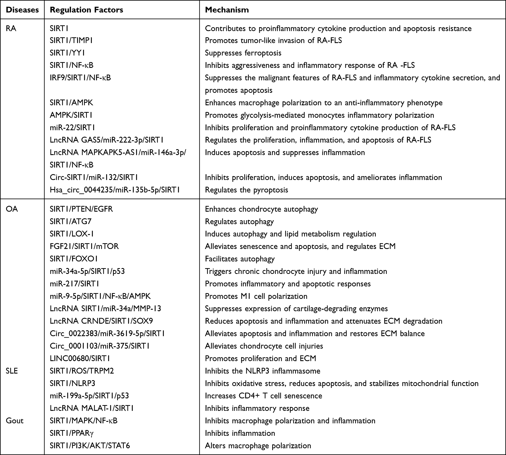 |
Table 1 SIRT1-Associated Regulatory Pathways in Rheumatic Diseases |
 |
Figure 2 SIRT1-associated regulatory pathways in rheumatic diseases. This schematic illustrates the context-dependent roles of SIRT1 across major rheumatic diseases, including RA, OA, SLE, and gout. In RA, altered SIRT1 expression is associated with clinical diagnostic markers, inflammatory responses, macrophage metabolism, ferroptosis, and disease aggressiveness, involving key pathways (such as NF-κB, AMPK, and YY1). In OA, SIRT1 regulates chondrocyte autophagy, lipid metabolism, senescence, and apoptosis through interactions with FOXO1, mTOR, LC3, and PTEN. Its expression and activity are extensively modulated by epigenetic mechanisms, including miRNAs, lncRNAs, and circRNAs. In SLE and gout, SIRT1 influences inflammatory signaling, apoptosis, and inflammasome activation via pathways such as NLRP3, MAPK, PPARγ, and PI3K/AKT, with additional regulatory input at the epigenetic level. Collectively, this figure highlights the multifaceted and disease-specific regulatory network through which SIRT1 links clinical features, cellular functions, and epigenetic modulation in rheumatic diseases. |
SIRT1 as a Potential Diagnostic Biomarker for RA
Accumulating evidence suggests that SIRT1 may serve as a biomarker reflecting RA disease activity. Elevated serum SIRT1 levels have been reported in RA patients and show positive correlations with anti-CCP titers and DAS28 scores, indicating potential diagnostic and prognostic utility.35 Conversely, reduced SIRT1 protein expression has been observed in peripheral blood mononuclear cells (PBMCs), where it correlates inversely with IL-23 levels.36 These conflicting findings highlight that circulating SIRT1 levels may not directly mirror intracellular or tissue-specific SIRT1 activity. This underscores the need to interpret SIRT1 as a compartment-specific biomarker rather than a uniform indicator of RA severity.
SIRT1-Mediated Regulation of Biological Functions in RA
SIRT1 regulates multiple processes relevant to RA, including inflammation, apoptosis, angiogenesis, and ferroptosis. However, its expression and functional outcomes appear to be highly context-dependent, varying markedly across different cell types and disease states. Although SIRT1 is upregulated in RA synovial tissues, its overexpression in specific cellular contexts may exacerbate chronic inflammation by promoting pro-inflammatory cytokine production and suppressing apoptosis.37 Moreover, SIRT1 enhances RA-FLS invasiveness and cartilage degradation by inhibiting TIMP1 transcription via disruption of SP1 binding to the TIMP1 promoter.38
However, SIRT1 also exerts protective effects in RA, particularly through inhibiting ferroptosis, a form of lipid peroxidation-driven cell death recognized as a central player in RA pathophysiology.39 By regulating reactive oxygen species (ROS) generation and maintaining iron homeostasis, SIRT1 may attenuate ferroptotic damage and limit inflammatory responses, exerting both pathogenic and protective effects in RA, with its net impact determined by disease stage and the specific cellular environment.
SIRT1-Mediated Regulation of NF-κB in RA
SIRT1 serves as an epigenetic regulator of NF-κB signaling, primarily by deacetylating the RelA/p65 subunit at Lys310, thereby suppressing its transcriptional activity. Additionally, SIRT1 indirectly modulates NF-κB signaling by stabilizing IκBα and regulating upstream kinases such as TAK1.40
In RA-FLS, SIRT1 expression is often reduced compared to healthy controls (HCs), and its overexpression suppresses cell proliferation, migration, and invasion, while concurrently promoting apoptosis. This effect involves coordinated inhibition of NF-κB phosphorylation and acetylation.41 Furthermore, TNF-α-induced upregulation of IRF9 enhances NF-κB signaling while suppressing SIRT1 expression; silencing IRF9 restores SIRT1 expression and markedly attenuates the inflammatory and invasive phenotypes of RA-FLS.42
Collectively, these findings support an anti-inflammatory role of SIRT1-mediated NF-κB regulation at the cellular level, despite contradictory systemic observations in clinical samples.
SIRT1-Mediated Regulation of AMPK in RA
SIRT1 and AMPK form a bidirectional regulatory loop that integrates energy metabolism with inflammatory signaling. SIRT1 activates AMPK by deacetylating LKB1, while AMPK activation enhances NAD⁺ availability, thereby reinforcing SIRT1 activity.43 Disruption of this SIRT1/AMPK axis drives immunometabolic imbalance in RA, manifesting as enhanced glycolysis and pro-inflammatory macrophage polarization.
Activation of the SIRT1/AMPK pathway promotes anti-inflammatory M2 macrophage polarization while suppressing M1-associated inflammatory responses.44 Consistently, AMPK/SIRT1 deficiency exacerbates adjuvant-induced arthritis (AIA) by enhancing glycolytic flux and driving inflammatory monocyte differentiation; however, pharmacological or genetic activation of SIRT1 attenuates disease severity and restores metabolic homeostasis.45
These data highlight that the SIRT1/AMPK axis may be a critical immunometabolic checkpoint in RA.
SIRT1-Mediated Regulation of Epigenetic Alterations in RA
SIRT1 plays a central role in RA-associated epigenetic remodeling through its interaction with non-coding RNA (ncRNA) networks. Specifically, microRNAs (miRNAs), lncRNAs, and circular RNAs (circRNAs) form competing endogenous RNA (ceRNA) circuits that fine-tune SIRT1 expression and downstream inflammatory responses. For instance, reduced miR-22 expression enhances SIRT1 activity but contributes to aberrant cytokine secretion and RA-FLS proliferation.46 Additionally, lncRNA GAS5 alleviates inflammatory and apoptotic dysregulation by sequestering miR-222-3p and restoring SIRT1 expression.47 Conversely, WTAP-dependent upregulation of lncRNA MK5-AS1 suppresses SIRT1, exacerbating inflammation and apoptosis resistance in RA.48
Emerging evidence implicates circRNAs in modulation of SIRT1 signaling. Notably, circ-SIRT1 overexpression suppresses inflammation and proliferation while promoting apoptosis in RA-FLS through miR-132-mediated SIRT1 activation.49 Similarly, hsa_circ_0044235 regulates NOD-like receptor family pyrin domain containing 3 (NLRP3)-dependent pyroptosis via the miR-135b-5p/SIRT1 axis.50
Taken together, these epigenetic mechanisms highlight a dynamic and multilayered regulation of SIRT1 expression in RA, offering opportunities for precision epigenetic intervention.
Overall, evidence from RA studies suggests that SIRT1 is not uniformly protective or pathogenic. In cellular and animal models, SIRT1 activation often suppresses NF-κB signaling, inflammasome activation, glycolytic reprogramming, and synovial inflammation. However, human studies show divergent patterns, including elevated serum SIRT1 levels associated with disease activity and reduced intracellular SIRT1 expression in PBMCs or synovial tissues. These discrepancies may reflect differences between circulating biomarkers and tissue-specific enzymatic activity. Therefore, SIRT1 in RA should be regarded as a compartment-specific regulator and potential biomarker rather than an anti-inflammatory target.
SIRT1 and OA
OA is the most prevalent degenerative joint disease, characterized by progressive cartilage degradation, subchondral bone remodeling, synovial inflammation, and chronic pain.51–53 Unlike RA, the pathogenesis of OA is dominated by chondrocyte dysfunction, impaired autophagy, oxidative stress, and metabolic dysregulation. This distinct pathological landscape positions SIRT1 as an intracellular regulator, rather than a systemic inflammatory mediator.
SIRT1-Mediated Regulation of Biological Functions in OA
Autophagy dysfunction is a hallmark of OA progression. SIRT1 enhances chondrocyte autophagy through two complementary mechanisms: promoting PTEN-mediated EGFR ubiquitination and directly interacting with autophagy proteins such as autophagy-related 7 (ATG7), microtubule-associated protein 1 light chain 3 (LC3), and Beclin-1.54,55 This relationship is bidirectional: SIRT1 is subject to autophagic degradation, revealing a reciprocal regulatory loop that links SIRT1 activity to autophagic flux.56 SIRT1 also modulates lipid metabolism and cellular senescence. Mechanistically, activation of the SIRT1-mTOR-TFEB axis restores lysosomal biogenesis and autophagy flux, thereby suppressing chondrocyte apoptosis and ECM degradation, as demonstrated in FGF21-mediated OA models.57
SIRT1-Mediated Regulation of Forkhead Box O1 (FOXO1) in OA
FOXO1 is a direct deacetylation substrate of SIRT1. SIRT1-mediated FOXO1 activation enhances its nuclear retention and transcription of autophagy- and ECM-related genes (including Atg7 and SIRT1 itself).58 This feed-forward loop is critical for maintaining cartilage homeostasis and resistance to OA-related degenerative stress.
SIRT1-Mediated Regulation of Epigenetic Alterations in OA
Epigenetic mechanisms critically shape SIRT1 activity in OA. Multiple studies show that several miRNAs (such as miR-34a-5p, miR-217, and miR-9-5p) suppress SIRT1 expression, thereby promoting inflammatory signaling, oxidative stress, and M1 macrophage polarization.59–61 LncRNAs and circRNAs further refine SIRT1 regulation. For example, SIRT1-AS lncRNA modulates cartilage catabolism by sponging miR-34a,62 whereas CRNDE promotes chondrogenic differentiation via the SIRT1/SOX9 axis.63 Protective circRNAs (such as circ_0022383 and circ_0001103) attenuate IL-1β-induced cartilage injury by restoring SIRT1 expression.64,65
Importantly, N6-methyladenosine (m6A)-dependent epigenetic regulation has emerged as a higher-order regulator of SIRT1 stability in OA. METTL3-mediated m6A modification stabilizes lncRNA LINC00680, which then enhances SIRT1 mRNA stability via IGF2BP2, thereby accelerating OA progression.66 Targeting this epigenetic hierarchy may offer therapeutic opportunities.
Compared with RA, the role of SIRT1 in OA appears more consistently linked to chondrocyte homeostasis, autophagy, senescence, and ECM metabolism. Most experimental studies support a protective function of SIRT1 in maintaining cartilage integrity. However, some epigenetic findings suggest that SIRT1-related regulatory networks may contribute to disease progression depending on the upstream RNA modification or cellular context. Importantly, most evidence in OA derives from chondrocyte models or surgical animal models, whereas direct validation in human OA tissues and longitudinal clinical cohorts remains limited. Thus, SIRT1 should be considered a promising mechanistic node in OA, but its clinical utility as a biomarker or therapeutic target remains unproven.
SIRT1 and SLE
SLE is a chronic and heterogeneous autoimmune disease characterized by loss of immune tolerance. This loss leads to pathogenic autoantibody production against nuclear and self-antigens, driving widespread inflammation and multi-organ tissue damage.67 Its pathogenesis is complex, involving intricate interactions among genetic susceptibility, epigenetic modifications, hormonal influences, and environmental triggers, all of which contribute to aberrant activation of both innate and adaptive immunity.68 Current therapeutic strategies for SLE employ a tiered approach combining antimalarials for immunomodulation, corticosteroids for rapid anti-inflammation, immunosuppressants for broad immune suppression, and biologic agents for targeted intervention.69 However, managing disease heterogeneity, minimizing treatment toxicity, and achieving sustained remission remain clinical challenges.
SIRT1 as a Potential Diagnostic Biomarker for SLE
Emerging evidence suggests that dysregulated SIRT1 expression may serve as a biomarker in SLE. Recent studies highlight elevated levels of SIRT1 in SLE patients, suggesting its potential as a diagnostic marker for SLE and its involvement in disease activity and progression.70 Further analyses show that SIRT1 level is significantly higher in SLE patients compared to that in HCs, and these levels strongly correlate with SLEDAI scores. Additionally, SIRT1 level is associated with specific clinical manifestations (such as keratoconjunctivitis sicca), suggesting that SIRT1 could serve as a biomarker for SLE disease phenotypes.71
SIRT1-Mediated Regulation of NLRP3 in SLE
Given the inflammasome’s role in driving inflammation and SIRT1’s therapeutic potential, the interplay between SIRT1 and the NLRP3 inflammasome in SLE is a key research area that may provide insights into new therapeutic strategies. Research has demonstrated that SIRT1 can modulate the NLRP3 inflammasome through various pathways. For instance, SIRT1 inhibits the NLRP3 inflammasome by reducing ROS production and modulating calcium influx through the TRPM2 channel; these mechanisms are critical in the pathogenesis of lupus nephritis, a severe SLE manifestation.72 Moreover, SIRT1’s ability to inhibit NF-κB expression and transcription further underscores its role in dampening inflammatory responses. Another study supports this anti-inflammatory mechanism of SIRT1, showing that upregulating SIRT1 alleviates lupus nephritis symptoms by inhibiting the NLRP3 inflammasome in human glomerular mesangial cells.73 In brief, SIRT1-mediated regulation of the NLRP3 inflammasome may be a promising approach for SLE treatment.74
SIRT1-Mediated Regulation of Epigenetic Alterations in SLE
SIRT1 is an important epigenetic modifier, and epigenetic dysregulation has recently been reported to contribute to SLE pathogenesis. Cheng et al have revealed that human umbilical cord (hUC)-mesenchymal stem cell (MSC) transplantation alleviates SLE pathology in MRL/lpr mice by promoting the senescence of pathogenic splenic CD4⁺ T cells.74 This therapeutic effect involves transfer of miR-199a-5p from hUC-MSCs to CD4⁺ T cells; miR-199a-5p directly targets and downregulates SIRT1 expression, leading to increased p53 acetylation and subsequent upregulation of senescence markers (p21 and p16). This study confirmed the pivotal role of the miR-199a-5p/SIRT1/p53 axis in the immunomodulatory function of hUC-MSCs for SLE. Yang et al have also noted that lncRNA MALAT-1 functions as a critical inflammatory regulator in SLE. It exerts its pathogenic effects by positively regulating the IL-21/SIRT1 signaling pathway, as shown by reduced SIRT1 protein levels following MALAT-1 silencing in THP-1 cells and patient monocytes.75
SIRT1 Polymorphisms and SLE
The pathogenesis of SLE is complex and involves genetic polymorphisms, including SIRT1. These genetic variations influence not only disease susceptibility but also clinical manifestations and severity. In-depth investigation of these genetic polymorphisms may provide new insights into SLE etiology and identify therapeutic targets for personalized therapy. For example, Consiglio et al have investigated the role of SIRT1 promoter polymorphisms rs12778366 and rs3758391 in SLE susceptibility and clinical manifestations in a Southern Brazilian cohort (367 patients, 290 controls).76 Although no significant association emerged between either polymorphism and overall SLE susceptibility (allelic, genotypic, or haplotypic frequencies), rs3758391 was a critical modifier of disease severity. Carriers of the rs3758391 T allele (TT/CT genotypes) exhibited a markedly higher risk of lupus nephritis and demonstrated elevated disease activity scores compared to CC homozygotes. These findings suggest that the SIRT1 rs3758391 T allele, though not a SLE susceptibility factor for SLE, acts as a clinical modifier associated with more severe SLE phenotypes (particularly nephritis) by dysregulating SIRT1 expression and its downstream immune pathways. The functional mechanism linking this polymorphism to SLE severity remains to be elucidated.
In SLE, SIRT1 has dual implications. On the one hand, elevated SIRT1 levels in patients may serve as a biomarker of disease activity. On the other hand, experimental studies suggest that modulation of SIRT1 affects inflammasome activation, T-cell senescence, and inflammatory signaling. These findings indicate that SIRT1 may function as a disease-associated biomarker in some human settings, while acting as a mechanistic regulator in experimental models. The apparent inconsistency may arise from differences among immune-cell subsets, renal compartments, disease stages, and treatment backgrounds. Further studies are needed to determine whether SIRT1 is a driver, a compensatory response, or a downstream marker in different SLE phenotypes.
SIRT1 and Gout
Gout is a prevalent metabolic disorder characterized by hyperuricemia and deposition of monosodium urate (MSU) crystals in joints and periarticular tissues.77 Its pathogenesis stems from elevated serum urate levels, primarily due to impaired renal excretion. Less commonly, it results from uric acid overproduction, leading to MSU crystal formation upon supersaturation.78 These crystals trigger acute inflammation by activating the NLRP3 inflammasome in resident macrophages and neutrophils, leading to release of pro-inflammatory cytokines (notably IL-1β).
SIRT1-Mediated Regulation of NF-κB in Gout
Evidence indicates that SIRT1 inhibits macrophage polarization and inflammation. For instance, Zhao et al have shown that SIRT1 exerts anti-inflammatory effects on gouty arthritis by modulating macrophage polarization and suppressing inflammatory responses.79 In murine models and THP-1 macrophages, SIRT1 activation significantly reduces M1 macrophage polarization, ROS production, and pro-inflammatory cytokine release (IL-1β, IL-6, and TNF-α). Mechanistically, SIRT1 inhibits the MAPK/NF-κB/AP-1 pathway by suppressing p38/JNK phosphorylation and activates the Nrf2/HO-1 antioxidant pathway. Conversely, SIRT1 inhibition exacerbates inflammation. These findings highlight SIRT1 as a potential therapeutic target for gouty arthritis through its modulation of key inflammatory pathways.
SIRT1-Mediated Regulation of PPARγ in Gout
SIRT1-mediated regulation of PPARγ is complex. One key mechanism involves direct deacetylation of PPARγ, which modulates its transcriptional activity. Wang et al have indicated that SIRT1 may control the acute onset of gouty arthritis in mice by inhibiting inflammatory cell infiltration and secreting pro-inflammatory molecules through PPARγ.80 This study underscored the SIRT1-PPARγ interaction as a regulator of inflammatory pathways in gout.
SIRT1-Mediated Regulation of PI3K/AKT in Gout
As an upstream regulator of PI3K/AKT, SIRT1 directly engages this pathway. Studies have illustrated the protective role of SIRT1 against MSU crystal-induced inflammation in acute gout. For example, Zhou et al have demonstrated that SIRT1 expression is elevated in PBMCs from acute gout patients but not in chronic tophus tissues.81 Their investigation reveals that SIRT1 activation significantly attenuates gouty inflammation. Mechanistically, SIRT1 mitigates MSU-induced inflammation by modulating macrophage polarization toward an anti-inflammatory phenotype. This protective effect is mediated, in part, through the PI3K/AKT/STAT6 axis. Specifically, inhibiting PI3K/AKT abolishes SIRT1’s anti-inflammatory actions and blocks STAT6 phosphorylation and nuclear translocation. Collectively, this study identified SIRT1 as a regulator in acute gout pathogenesis, exerting its anti-inflammatory effects primarily via the PI3K/AKT/STAT6 axis to reprogram macrophage responses.
Current evidence mainly supports an anti-inflammatory role of SIRT1 in MSU crystal-induced macrophage activation, notably through suppression of NF-κB/NLRP3-related inflammatory signaling and regulation of macrophage polarization. However, the evidence remains limited compared with RA and OA, and human data are insufficient. Notably, SIRT1 expression differs between acute gout PBMCs and chronic tophus tissues, suggesting that its function may vary across disease stages and tissue compartments. Therefore, SIRT1 may be more relevant to acute inflammatory regulation than to all aspects of chronic gout pathology. Future studies should distinguish its role as a protective regulator, compensatory response, or disease-stage-specific biomarker in gout.
TCM-Based Modulation of SIRT1 in Rheumatic Diseases: Preclinical Evidence and Translational Limitations
TCM-derived compounds and formulations have been increasingly explored for regulating inflammatory, metabolic, oxidative-stress, angiogenic, and epigenetic pathways in rheumatic diseases. Unlike conventional therapies with defined molecular targets, TCM formulations often contain multiple bioactive constituents and may exert regulatory effects through multi-component and multi-target mechanisms.82 Some experimental studies suggest that these effects may involve SIRT1-related signaling, including modulation of NF-κB, AMPK, oxidative stress, macrophage polarization, angiogenesis, and synovial and cartilage homeostasis.
However, the current evidence base remains heterogeneous and predominantly preclinical. Most mechanistic findings are derived from in vitro experiments, animal models, molecular docking, or network pharmacology analyses, whereas direct clinical evidence for SIRT1-dependent effects in rheumatic diseases remains limited. Some clinical studies have reported that adjunctive TCM therapy improves symptoms such as pain, stiffness, and fatigue. However, its effects on disease progression, radiographic outcomes, glucocorticoid/DMARD-related toxicity, and long-term safety require further validation in rigorously designed clinical trials.83 Therefore, TCM-based modulation of SIRT1 should be interpreted as an emerging mechanistic hypothesis and research direction, rather than an established clinical therapeutic strategy. The reported mechanisms by which TCM-derived compounds and formulations regulate SIRT1-related pathways in rheumatic diseases are summarized in Table 2.
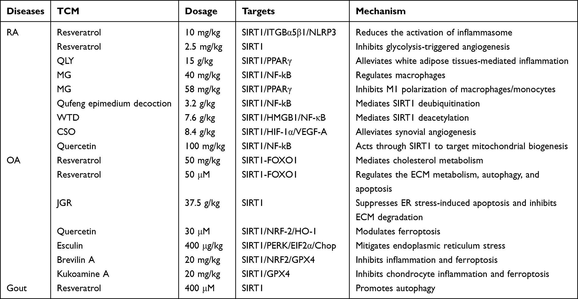 |
Table 2 SIRT1-Targeted Mechanisms of TCM in Rheumatic Diseases |
RA
Resveratrol
Resveratrol, a natural polyphenolic compound, has been extensively investigated for its therapeutic potential in RA by modulating SIRT1-related signaling pathways. For instance, Wang et al have demonstrated that resveratrol remarkably alleviates arthritis severity in RA model rats by activating the SIRT1/Nrf2 axis, thereby reducing oxidative stress, inflammatory cell infiltration, joint swelling, and synovial hyperplasia.84 Consistently, Cai et al have reported that resveratrol suppresses NLRP3 inflammasome activation through dual mechanisms: (1) enhancing SIRT1-mediated deacetylation of NLRP3, which reduces caspase-1 cleavage and IL-1β maturation; and (2) competitively inhibiting integrin α5β1 signaling.85 Additionally, SIRT1 activation inhibits angiogenesis in RA by suppressing pro-angiogenic cytokine production, glycolysis, and endothelial angiogenic capacity under inflammatory conditions.86
Qing-Luo-Yin (QLY)
QLY is a traditional Chinese herbal formula, exerting anti-rheumatic effects partially by modulating SIRT1 activity. Ye et al have reported that QLY treatment effectively alleviates joint inflammation in AIA while promoting adipocyte differentiation and suppressing pro-inflammatory adipokine release from white adipose tissues (WAT).87 Mechanistically, QLY normalizes aberrant SIRT1 hyperactivity in AIA, thereby reducing extracellular NAMPT release. Molecular docking and in vitro assays further suggest that key QLY components bind SIRT1 with high affinity, stabilizing the protein while inhibiting its deacetylase activity.
α-Mangostin (MG)
MG, an isoprenyl-substituted xanthone, exhibits immunomodulatory effects in RA. Chen et al have revealed that MG activates the cholinergic anti-inflammatory (CAP)-SIRT1 pathway, suppresses NF-κB signaling, and inhibits M1 macrophage polarization, thereby ameliorating immune dysregulation in early AIA rats.88 Similarly, Wu et al have demonstrated that MG concurrently upregulates SIRT1 and PPARγ, leading to reduced inflammatory cytokine production and attenuation of synovial inflammation.89 Pharmacological inhibition of either SIRT1 or PPARγ markedly weakens MG-mediated protection, suggesting that SIRT1/PPARγ co-activation may be a mechanism underlying MG’s immunometabolic regulation in RA.
Qufeng Epimedium Decoction
Qufeng epimedium decoction has shown therapeutic efficacy in RA by modulating inflammatory signaling. For example, Wu et al have reported that this formula alleviates RA symptoms by stabilizing SIRT1 via deubiquitination, thereby suppressing NF-κB/GSDMD signaling and inflammatory cell death.90
Wutou Decoction (WTD)
WTD is a classical Chinese prescription long used in RA treatment that shows potent anti-inflammatory activity in experimental models. According to experimental evidence, WTD alleviates arthritis severity in collagen-induced arthritis (CIA) rat models by suppressing TNF-α, IL-6, and IL-1β production and inhibiting M1 macrophage infiltration.91 Mechanistically, these effects are mediated by SIRT1-dependent deacetylation and inhibition of the HMGB1/NF-κB pathway, highlighting SIRT1’s role in controlling RA inflammation.
Coix Seed Oil (CSO)
Research shows that CSO mitigates synovial angiogenesis, a hallmark pathological feature of RA. Recent studies have demonstrated that CSO reduces HIF-1α and vascular endothelial growth factor A (VEGF-A) expression in CIA rats, and these effects are closely associated with SIRT1 upregulation.92 SIRT1 activation may suppress angiogenesis by deacetylating and inactivating HIF-1α, thereby limiting VEGF-A-driven neovascularization.
Quercetin
Quercetin is a dietary flavonoid that exhibits potent anti-arthritic activity in RA models. In CIA mice, quercetin notably reduces clinical arthritis scores, synovial inflammation, and bone and cartilage destruction.93 Mechanistically, quercetin enhances mitochondrial biogenesis through the SIRT1/PGC-1α/NRF1/TFAM axis while suppressing inflammation through the HMGB1/TLR4/p38/ERK1/2/NF-κB pathway.
OA
Resveratrol
In OA, resveratrol exerts chondroprotective effects primarily by activating the SIRT1/FOXO1 axis. Evidence indicates that resveratrol reduces cholesterol accumulation in osteoarthritic cartilage by enhancing SIRT1 expression and FOXO1 phosphorylation; this leads to LXRα upregulation and inhibition of SREBP2-mediated cholesterol synthesis.94 Consistently, resveratrol protects IL-1β-stimulated chondrocytes by restoring autophagy, maintaining ECM homeostasis, and inhibiting apoptosis through SIRT1/FOXO1 activation.95
Jiangu Recipe (JGR)
JGR demonstrates significant chondroprotective effects in OA through SIRT1-dependent mechanisms. Studies have shown that JGR alleviates tert-butyl hydroperoxide (TBHP)-induced endoplasmic reticulum stress, apoptosis, and ECM degradation in chondrocytes by upregulating SIRT1.96 Importantly, SIRT1 knockdown abolishes the protective effects of JGR, confirming that SIRT1 is essential for JGR-induced chondroprotection.
Quercetin
Quercetin has been shown to attenuate OA progression by suppressing inflammation, oxidative stress, and ferroptosis. In ACLT-induced OA models, quercetin reduces cartilage degeneration, inflammatory mediator expression, and pain behaviors; these effects involve activation of the SIRT1/Nrf2/HO-1 pathway and inhibition of chondrocyte ferroptosis.97
Esculin
Esculin exhibits therapeutic potential in OA by targeting SIRT1-dependent stress responses. In TBHP-induced chondrocyte injury and ACLT-induced OA models, esculin upregulates SIRT1 expression, suppresses PERK-eIF2α-ATF4-CHOP signaling, and reduces ER stress-induced chondrocyte apoptosis.98
Brevilin A
Brevilin A is a bioactive compound derived from Centipeda minima and has been shown to attenuate OA-associated cartilage destruction. For instance, Ruan et al have reported that Brevilin A inhibits inflammation and ferroptosis in OA by activating the SIRT1/Nrf2/GPX4 signaling pathway.99
Kukoamine A (KuKA)
KuKA, isolated from Lycium chinense, exhibits potent anti-inflammatory and chondroprotective properties in OA. Sun et al have demonstrated that KuKA suppresses inflammation, matrix degradation, and ferroptosis in chondrocytes via SIRT1-dependent activation of Nrf2 and GPX4 signaling; these effects are reversed upon SIRT1 inhibition.100
Gout
Resveratrol
Studies have examined the role of resveratrol in gout based on its SIRT1-driven anti-inflammatory effects. Analysis of PBMCs reveals remarkably reduced SIRT1 expression in acute gout and intercritical gout patients compared to HCs. Mechanistically, resveratrol treatment induces enhanced autophagic activity, shown by increased Beclin-1 and LC3 expressions and downregulated key inflammatory mediators (including NLRP3 and NF-κB p65) at the transcriptional level. These findings suggest that resveratrol may attenuate gout-associated inflammation partly by restoring SIRT1 expression, promoting autophagy, and modulating the NLRP3/NF-κB axis in response to monosodium urate (MSU) crystals.101
Notably, although these observations offer clinically mechanistic insights, the limited sample size and observational design warrant caution in interpreting therapeutic efficacy.
Translational Considerations and Limitations of TCM-Based SIRT1 Modulation
TCM-derived compounds have established regulatory effects on SIRT1 signaling in rheumatic diseases. However, most data come from in vitro systems and animal models, with limited high-quality clinical evidence. While these preclinical studies offer valuable mechanistic insights, their translational relevance requires caution, as the therapeutic efficacy and long-term safety in humans remain insufficiently validated.102
Several challenges currently hinder the clinical translation of TCM-based SIRT1-targeting strategies. First, poor bioavailability and pharmacokinetic variability limit efficacy, as many natural compounds exhibit low absorption, rapid metabolism, or inadequate tissue penetration. Second, standardization and quality control of herbal formulations remain challenging due to batch-to-batch variability, complex multi-component compositions, and differences in preparation methods. Third, inadequate dose optimization and poor reproducibility across studies complicate direct comparison of outcomes and hinder reliable dose extrapolation to clinical settings.103
Translational combination of SIRT1-targeted TCM interventions with conventional treatments (eg., NSAIDs, csDMARDs, biologics, or JAK inhibitors) represents a promising future direction. SIRT1 modulation may provide complementary benefits by targeting immunometabolic and stress-response pathways that remain unaffected by current anti-inflammatory or immunosuppressive agents. However, this combination strategy also raises safety concerns regarding drug-drug interactions. Many TCM-derived compounds (including polyphenols and flavonoids) can influence drug-metabolizing enzymes or transporters, altering the pharmacokinetics of co-administered therapies. Moreover, overlapping immunomodulatory effects may increase the risk of infection or organ toxicity when added to existing immunosuppressive regimens.104
Furthermore, most existing clinical investigations are observational or small-scale, lacking rigorous randomized controlled trial design, standardized endpoints, and comprehensive safety assessment. Therefore, while TCM-based modulation of SIRT1 represents a promising therapeutic concept, the current evidence remains largely exploratory or preclinical rather than for immediate clinical translation. Future studies should integrate pharmacokinetic analyses, systematic safety evaluation, and trial designs focused on combination therapy to determine the translational potential of SIRT1-targeted TCM interventions.
Conclusions and Perspective
This review synthesized current evidence identifying SIRT1 as an immunometabolic and epigenetic regulator in major rheumatic diseases, including RA, OA, SLE, and gout. SIRT1 integrates inflammatory signaling pathways, such as NF-κB and the NLRP3 inflammasome, metabolic regulation, including AMPK and PPARγ signaling, autophagy, ferroptosis, apoptosis, cellular senescence, and multilayered epigenetic mechanisms. It thus appears to function as a context-dependent molecular node linking chronic inflammation, immune dysregulation, tissue remodeling, and structural damage. However, evidence also indicates that the biological effects of SIRT1 are not uniform across diseases, tissues, cell types, or disease stages. Therefore, SIRT1 should not be interpreted simply as a universally protective or pathogenic factor, but rather as a dynamic regulator whose functional significance depends on the pathological context. Meanwhile, multiple TCM-derived compounds and formulations modulate these pathogenic processes, at least in part, through regulation of SIRT1-related pathways. However, the supporting evidence remains preclinical or exploratory, and robust clinical validation is limited.
To advance the field, future research should prioritize the following five directions:
First, the context-dependent roles of SIRT1 should be more precisely defined. Conflicting observations—such as divergent serum vs. tissue SIRT1 levels and opposing protective vs. pathogenic effects—highlight the need to clarify cell-type, tissue-compartment, and disease-stage-specific functions of SIRT1, as summarized in Table 3. This is key to avoiding oversimplified therapeutic interpretations and developing biomarker-guided intervention strategies.
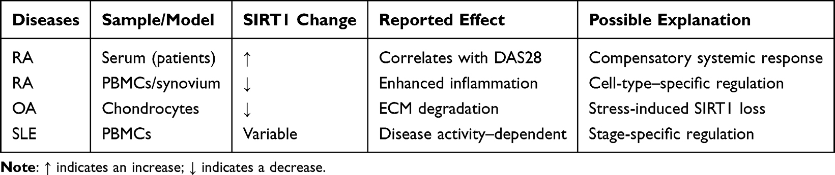 |
Table 3 Context-Dependent and Conflicting Roles of SIRT1 in Rheumatic Diseases |
Second, hierarchical epigenetic regulatory networks involving SIRT1 require further investigation. Although interactions between SIRT1 and miRNAs, lncRNAs, circRNAs, and m6A modifications are increasingly recognized, the dominant regulatory nodes, feedback loops, and causal hierarchies within these networks remain poorly defined. Systematic dissection of these epigenetic circuits will be critical for identifying clinically actionable targets.
Third, the translational rigor of TCM-based SIRT1 modulation needs to be strengthened. Future studies should address the bioavailability, pharmacokinetics, standardization, dosing, reproducibility, and quality-control challenges of TCM-derived compounds and formulations. Importantly, claims regarding clinical translation should be supported by pharmacokinetic, pharmacodynamic, safety, and mechanistic validation, especially given the complex composition of multi-herbal formulations.
Fourth, high-quality clinical evidence is required. Rigorous randomized controlled trials are needed to evaluate the efficacy, safety, and patient-stratification value of SIRT1-targeted interventions, including TCM-based strategies. Such studies should incorporate standardized endpoints, biomarker-guided stratification (eg., SIRT1 activity or downstream molecular signatures), and long-term safety monitoring. At present, available clinical evidence is insufficient to support SIRT1-targeted TCM interventions as therapeutic options.
Fifth, precision strategies for SIRT1 targeting should be developed. Innovative approaches, such as tissue-specific delivery systems, context-selective SIRT1 activators or inhibitors, and SIRT1 modulators, may enhance therapeutic specificity while reducing off-target effects. These strategies should be guided by disease-specific mechanisms and biomarker-defined patient subsets, rather than by generalized activation or inhibition of SIRT1.
From a clinical and regulatory perspective, SIRT1-targeted strategies are more likely to be evaluated as adjunctive approaches rather than stand-alone therapies, particularly in combination with established anti-inflammatory or immunomodulatory agents. However, combination use raises concerns about drug-drug interactions, additive immunosuppression, organ toxicity, and long-term safety. In addition, regulatory challenges—including standardization of active components, quality control, batch-to-batch consistency, and alignment with botanical drug approval frameworks—must be addressed before clinical translation can be pursued.
In conclusion, SIRT1 represents an important but context-dependent regulatory node in rheumatic diseases. Evidence supports SIRT1’s involvement in multiple processes, including inflammation, immunometabolism, oxidative stress, cell death, senescence, and epigenetic regulation. However, its value as a biomarker or therapeutic target remains an emerging hypothesis that requires validation. Integrating mechanistic insights from immunometabolism and epigenetics with clinical research is essential, particularly for TCM-based strategies, to determine whether SIRT1-targeted interventions can become meaningful adjunctive therapies.
Abbreviations
AMPK, AMP-activated protein kinase; ACPA, Anti-citrullinated protein antibody; ATG, Autophagy-related gene; bDMARDs, Biologic disease-modifying antirheumatic drugs; CAP, Cholinergic anti-inflammatory pathway; ceRNA, Competing endogenous RNA; CIA, Collagen-induced arthritis; circRNA, Circular RNA; csDMARDs, Conventional synthetic disease-modifying antirheumatic drugs; ECM, Extracellular matrix; ER, Endoplasmic reticulum; FLS, Fibroblast-like synoviocytes; FoxO1, Histone deacetylase; HMGB1, High mobility group box 1; HIF-1α, Hypoxia-inducible factor-1 alpha; IL, Interleukin; IG, Intercritical gout; IRF9, Interferon regulatory factor 9; JAKi, Janus kinase inhibitors; JGR, Jiangu Recipe; LC3, Microtubule-associated protein 1 light chain 3; lncRNA, Long non-coding RNA; LKB1, Liver kinase B1; METTL3, Methyltransferase-like 3; miRNA, MicroRNA; MMP,Matrix metalloproteinase; MSU, Monosodium urate; m6A, N6-methyladenosine; NAMPT, Nicotinamide phosphoribosyltransferase; NF-κB, Nuclear factor kappa-B; NLRP3, NOD-like receptor family pyrin domain containing 3; NSAIDs, Non-steroidal anti-inflammatory drugs; OA, Osteoarthritis; PBMCs, Peripheral blood mononuclear cells; PERK, Protein kinase R-like endoplasmic reticulum kinase; PGC-1α, Peroxisome proliferator-activated receptor gamma coactivator-1 alpha; PPARγ, Peroxisome proliferator-activated receptor gamma; RA, Rheumatoid arthritis; RD, Rheumatic diseases; RF, Rheumatoid factor; ROS, Reactive oxygen species; SIRT1, Sirtuin 1; SLE, Systemic lupus erythematosus; TCM, Traditional Chinese medicine; TBHP, tert-Butyl hydroperoxide; TFEB, Transcription factor EB; TLR4, Toll-like receptor 4; TNF-α, Tumor necrosis factor-alpha; VEGF-A, Vascular endothelial growth factor A; WAT, White adipose tissue.
Data Sharing Statement
Data sharing is not applicable to this article as no data were created or analysed in this study.
Author Contributions
Jianting Wen: Writing-original draft preparation, Conceptualization, and Funding acquisition. Jian Liu: Conceptualization, Supervision, Project administration, Funding acquisition, Writing – Review & Editing. Lei Wan: Writing – Review & Editing, Methodology, and Validation. Fanfan Wang: Data curation and writing-review & editing. Yang Li: Data curation and Writing-review & editing.
All authors have approved the final version for publication and agreed on the journal to which the article was submitted. They agree to be accountable for all aspects of the work.
Funding
This work was supported by Clinical Research Project of Anhui University of Traditional Chinese Medicine in 2024 (2024YFYLCZX09), Anhui University of Chinese Medicine 2024 Annual University-Level Exploratory Research Project (AHUCM2024TS099), Traditional Chinese Medicine Treatment Advantage Disease (Clinical Evidence based Ability Enhancement) Project (Wan Cai She [2024] No. 1359), and National Key Discipline of Traditional Chinese Medicine - Traditional Chinese Medicine Bi Disease ([2023] No. 85).
Disclosure
The authors declare that they have no known competing financial interests or personal relationships that could have appeared to influence the work reported in this paper.
References
1. Umar S, Tyagi AM, Singh AK, Haseeb A. Molecular mechanism of rheumatic diseases and efficacy of current therapies. Biomed Res Int. 2017;2017:2401329. doi:10.1155/2017/2401329
2. Anyfanti P, Triantafyllou A, Panagopoulos P, et al. Predictors of impaired quality of life in patients with rheumatic diseases. Clin Rheumatol. 2016;35(7):1705–18. doi:10.1007/s10067-015-3155-z
3. Mei X, Li Y, Yeung WF, et al. Effectiveness of ecological momentary interventions on pain, mental health, and quality of life in individuals with rheumatic diseases: a systematic review and meta-analysis of randomized controlled trials. J Nurs Manag. 2025;2025:9923240. doi:10.1155/jonm/9923240
4. Cutolo M, Soldano S, Smith V, Gotelli E, Hysa E. Dynamic macrophage phenotypes in autoimmune and inflammatory rheumatic diseases. Nat Rev Rheumatol. 2025;21:546–565. doi:10.1038/s41584-025-01279-w
5. Wen J, Liu J, Wan L, Wang F. Mechanistic and therapeutic insights into the function of different cell death modalities in rheumatoid arthritis: emphasis on the crosstalk with non-coding RNAs. Front Immunol. 2025;16:1620209. doi:10.3389/fimmu.2025.1620209
6. Finckh A, Gilbert B, Hodkinson B, et al. Global epidemiology of rheumatoid arthritis. Nat Rev Rheumatol. 2022;18(10):591–602. doi:10.1038/s41584-022-00827-y
7. Collins KH, Haugen IK, Neogi T, et al. Osteoarthritis as a systemic disease. Nat Rev Rheumatol. 2025;1–13.
8. Fu X, Liu H, Huang G, et al. The emerging role of neutrophils in autoimmune-associated disorders: effector, predictor, and therapeutic targets. MedComm. 2021;2(3):402–413. doi:10.1002/mco2.69
9. Kong S, Yeung P, Fang D. The class III histone deacetylase sirtuin 1 in immune suppression and its therapeutic potential in rheumatoid arthritis. J Genet Genomics. 2013;40(7):347–354. doi:10.1016/j.jgg.2013.04.001
10. Imai S, Guarente L. NAD+ and sirtuins in aging and disease. Trends Cell Biol. 2014;24(8):464–471. doi:10.1016/j.tcb.2014.04.002
11. Chang HC, Guarente L. SIRT1 and other sirtuins in metabolism. Trends Endocrinol Metab. 2014;25(3):138–145. doi:10.1016/j.tem.2013.12.001
12. Shen P, Deng X, Chen Z, et al. SIRT1: a potential therapeutic target in autoimmune diseases. Front Immunol. 2021;12:779177. doi:10.3389/fimmu.2021.779177
13. Singh V, Ubaid S. Role of silent information regulator 1 (SIRT1) in regulating oxidative stress and inflammation. Inflammation. 2020;43(5):1589–1598. doi:10.1007/s10753-020-01242-9
14. Li G, Xia Z, Liu Y, et al. SIRT1 inhibits rheumatoid arthritis fibroblast-like synoviocyte aggressiveness and inflammatory response via suppressing NF-κB pathway. Biosci. Rep. 2018;38(3):BSR20180541. doi:10.1042/BSR20180541
15. Wu Y, Fang W, Pan S, et al. Regulation of Sirt1 on energy metabolism and immune response in rheumatoid arthritis. Int Immunopharmacol. 2021;101:108175. doi:10.1016/j.intimp.2021.108175
16. Poniewierska-Baran A, Bochniak O, Warias P, et al. Role of sirtuins in the pathogenesis of rheumatoid arthritis. Int J Mole Sci. 2023;24(2):1532. doi:10.3390/ijms24021532
17. Sheng S, Wang X, Liu X, et al. The role of resveratrol on rheumatoid arthritis: from bench to bedside. Front Pharmacol. 2022;13:829677. doi:10.3389/fphar.2022.829677
18. Cai L, Zhang K, Gao J, et al. Resveratrol reduces the activation of NLRP3 inflammasomes in rheumatoid arthritis through SIRT1 and ITGB α5β1, especially in patients with high expression of ACPA. Phytomedicine. 2025;144:156897. doi:10.1016/j.phymed.2025.156897
19. Zhao X, Li M, Lu Y, et al. Sirt1 inhibits macrophage polarization and inflammation in gouty arthritis by inhibiting the MAPK/NF-κB/AP-1 pathway and activating the Nrf2/HO-1 pathway. Inflamm Res. 2024;73(7):1173–1184. doi:10.1007/s00011-024-01890-9
20. Wang G, Xie X, Yuan L, et al. Resveratrol ameliorates rheumatoid arthritis via activation of SIRT1-Nrf2 signaling pathway. Biofactors. 2020;46(3):441–453. doi:10.1002/biof.1599
21. Smolen JS, Landewé RBM, Bergstra SA, et al. EULAR recommendations for the management of rheumatoid arthritis with synthetic and biological disease-modifying antirheumatic drugs: 2022 update. Ann Rheum Dis. 2023;82(1):3–18. doi:10.1136/ard-2022-223356
22. Fanouriakis A, Kostopoulou M, Andersen J, et al. EULAR recommendations for the management of systemic lupus erythematosus: 2023 update. Ann Rheum Dis. 2024;83(1):15–29. doi:10.1136/ard-2023-224762
23. Wang Y, Chen S, Du K, et al. Traditional herbal medicine: therapeutic potential in rheumatoid arthritis. J Ethnopharmacol. 2021;279:114368. doi:10.1016/j.jep.2021.114368
24. Sauve AA, Wolberger C, Schramm VL, Boeke JD. The biochemistry of sirtuins. Annu Rev Biochem. 2006;75:435–465. doi:10.1146/annurev.biochem.74.082803.133500
25. Bonkowski MS, Sinclair DA. Slowing ageing by design: the rise of NAD+ and sirtuin-activating compounds. Nat Rev Mol Cell Biol. 2016;17(11):679–690. doi:10.1038/nrm.2016.93
26. Wu QJ, Zhang TN, Chen HH, et al. The sirtuin family in health and disease. Signal Transduct Target Ther. 2022;7(1):402. doi:10.1038/s41392-022-01257-8
27. Yuan H, Marmorstein R. Structural basis for sirtuin activity and inhibition. J Biol Chem. 2012;287(51):42428–42435. doi:10.1074/jbc.R112.372300
28. Michan S, Sinclair D. Sirtuins in mammals: insights into their biological function. Biochem J. 2007;404(1):1–13. doi:10.1042/BJ20070140
29. Sanders BD, Jackson B, Marmorstein R. Structural basis for sirtuin function: what we know and what we don’t. Biochim Biophys Acta. 2010;1804(8):1604–1616. doi:10.1016/j.bbapap.2009.09.009
30. Xu C, Wang L, Fozouni P, et al. SIRT1 is downregulated by autophagy in senescence and ageing. Nat Cell Biol. 2020;22(10):1170–1179. doi:10.1038/s41556-020-00579-5
31. Smolen JS, Aletaha D, McInnes IB. Rheumatoid arthritis. Lancet. 2016;388(10055):2023–2038. doi:10.1016/S0140-6736(16)30173-8
32. Nagafusa T, Mizushima T, Suzuki M, Yamauchi K. Comprehensive relationship between disease activity indices, mTSS, and mHAQ and physical function evaluation and QOL in females with rheumatoid arthritis. Sci Rep. 2023;13(1):21905. doi:10.1038/s41598-023-49380-y
33. Huang J, Fu X, Chen X, Li Z, Huang Y, Liang C. Promising therapeutic targets for treatment of rheumatoid arthritis. Front Immunol. 2021;12:686155. doi:10.3389/fimmu.2021.686155
34. Wu YJ, Fang WJ, Pan S, et al. Regulation of Sirt1 on energy metabolism and immune response in rheumatoid arthritis. Int Immunopharmacol. 2021;101(Pt A):108175.
35. Tan L, Ouyang T, Li X, et al. Serum sirtuin-1 a potential marker in the diagnosis of rheumatoid arthritis. Autoimmunity. 2023;56(1):2181234. doi:10.1080/08916934.2023.2181234
36. Wendling D, Abbas W, Godfrin-Valnet M, et al. Dysregulated serum IL-23 and SIRT1 activity in peripheral blood mononuclear cells of patients with rheumatoid arthritis. PLoS One. 2015;10(3):e0119981. doi:10.1371/journal.pone.0119981
37. Niederer F, Ospelt C, Brentano F, et al. SIRT1 overexpression in the rheumatoid arthritis synovium contributes to proinflammatory cytokine production and apoptosis resistance. Ann Rheum Dis. 2011;70(10):1866–1873. doi:10.1136/ard.2010.148957
38. Guo J, Zhao W, Cao X, et al. SIRT1 promotes tumor-like invasion of fibroblast-like synoviocytes in rheumatoid arthritis via targeting TIMP1. Oncotarget. 2017;8(51):88965–88973. doi:10.18632/oncotarget.21628
39. Zhan Y, Yang Z, Zhan F, Huang Y, Lin S. SIRT1 is transcriptionally repressed by YY1 and suppresses ferroptosis in rheumatoid arthritis. Adv Rheumatol. 2023;63(1):9. doi:10.1186/s42358-023-00289-0
40. Kauppinen A, Suuronen T, Ojala J, Kaarniranta K, Salminen A. Antagonistic crosstalk between NF-κB and SIRT1 in the regulation of inflammation and metabolic disorders. Cell Signal. 2013;25(10):1939–1948. doi:10.1016/j.cellsig.2013.06.007
41. Li G, Xia Z, Liu Y, et al. SIRT1 inhibits rheumatoid arthritis fibroblast-like synoviocyte aggressiveness and inflammatory response via suppressing NF-κB pathway. Biosci Rep. 2018;38(3):BSR20180541.
42. Jiang F, Zhou HY, Zhou LF, Zeng W, Zhao LH. IRF9 affects the TNF-induced phenotype of rheumatoid-arthritis fibroblast-like synoviocytes via regulation of the SIRT-1/NF-κB signaling pathway. Cells Tissues Organs. 2020;209(2–3):110–119. doi:10.1159/000508405
43. Anggreini P, Kuncoro H, Sumiwi SA, Levita J. Role of the AMPK/SIRT1 pathway in non-alcoholic fatty liver disease. Mol Med Rep. 2023;27(2):35. doi:10.3892/mmr.2022.12922
44. Park SY, Lee SW, Lee SY, et al. SIRT1/adenosine monophosphate-activated protein kinase α signaling enhances macrophage polarization to an anti-inflammatory phenotype in rheumatoid arthritis. Front Immunol. 2017;8:1135. doi:10.3389/fimmu.2017.01135
45. Wang DD, He CY, Wu YJ, et al. AMPK/SIRT1 deficiency drives adjuvant-induced arthritis in rats by promoting glycolysis-mediated monocytes inflammatory polarization. J Inflamm Res. 2022;15:4663–4675. doi:10.2147/JIR.S378090
46. Zhang C, Fang L, Liu X, et al. miR-22 inhibits synovial fibroblasts proliferation and proinflammatory cytokine production in RASF via targeting SIRT1. Gene. 2020;724:144144. doi:10.1016/j.gene.2019.144144
47. Yang Z, Lin SD, Zhan F, Liu Y, Zhan YW. LncRNA GAS5 alleviates rheumatoid arthritis through regulating miR-222-3p/Sirt1 signalling axis. Autoimmunity. 2021;54(1):13–22. doi:10.1080/08916934.2020.1846183
48. Wen J, Liu J, Wan L, et al. m6A-mediated lncRNA MAPKAPK5-AS1 induces apoptosis and suppresses inflammation via regulating miR-146a-3p/SIRT1/NF-κB axis in rheumatoid arthritis. Cell Cycle. 2023;22(23–24):2602–2621. doi:10.1080/15384101.2024.2302281
49. Zhang S, Zhao J, Ma W. Circ-Sirt1 inhibits proliferation, induces apoptosis, and ameliorates inflammation in human rheumatoid arthritis fibroblast-like synoviocytes. Autoimmunity. 2021;54(8):514–525. doi:10.1080/08916934.2021.1969550
50. Chen S, Luo Z, Chen X. Hsa_circ_0044235 regulates the pyroptosis of rheumatoid arthritis via MiR-135b-5p-SIRT1 axis. Cell Cycle. 2021;20(12):1107–1121. doi:10.1080/15384101.2021.1916272
51. Abramoff B, Caldera FE. Osteoarthritis: pathology, diagnosis, and treatment options. Med Clin North Am. 2020;104(2):293–311. doi:10.1016/j.mcna.2019.10.007
52. Huang K, Cai H. The role of chondrocyte senescence in osteoarthritis pathogenesis and therapeutic implications. Exp Gerontol. 2025;208:112828. doi:10.1016/j.exger.2025.112828
53. He M, Liu J, Sun Y, Fang Y, Wang F. Novel insights into the regulatory role of N6-methyladenosine in the pathogenesis and clinical treatment of osteoarthritis: research status and prospect. J Inflamm Res. 2025;18:6749–6766. doi:10.2147/JIR.S508973
54. Lu Q, Liu P, Miao Z, Luo D, Li S, Lu M. SIRT1 restoration enhances chondrocyte autophagy in osteoarthritis through PTEN-mediated EGFR ubiquitination. Cell Death Discov. 2022;8(1):203. doi:10.1038/s41420-022-00896-8
55. Liao FX, Huang F, Ma WG, et al. The new role of sirtuin1 in human osteoarthritis chondrocytes by regulating autophagy. Cartilage. 2021;13(2_suppl):1237S–1248S. doi:10.1177/1947603519847736
56. Papageorgiou AA, Goutas A, Trachana V, Tsezou A. Dual role of SIRT1 in autophagy and lipid metabolism regulation in osteoarthritic chondrocytes. Medicina. 2021;57(11):1203. doi:10.3390/medicina57111203
57. Lu H, Jia C, Wu D, et al. Fibroblast growth factor 21 (FGF21) alleviates senescence, apoptosis, and extracellular matrix degradation in osteoarthritis via the SIRT1-mTOR signaling pathway. Cell Death Dis. 2021;12(10):865. doi:10.1038/s41419-021-04157-x
58. Xu M, Qian Z, Zhang Y, et al. Sirt1 alleviates osteoarthritis via promoting FoxO1 nucleo-cytoplasm shuttling to facilitate autophagy. Int Immunopharmacol. 2024;131:111893. doi:10.1016/j.intimp.2024.111893
59. Zhou M, Liu B, Ye HM, et al. ROS-induced imbalance of the miR-34a-5p/SIRT1/p53 axis triggers chronic chondrocyte injury and inflammation. Heliyon. 2024;10(11):e31654. doi:10.1016/j.heliyon.2024.e31654
60. Papageorgiou AA, Roussos A, Papathanasiou I, et al. MiR-217 regulates SIRT1 expression and promotes inflammatory and apoptotic responses in osteoarthritis. Genes. 2023;14(12):2155. doi:10.3390/genes14122155
61. Wang J, Ma S, Yu J, et al. MiR-9-5p promotes M1 cell polarization in osteoarthritis progression by regulating NF-κB and AMPK signaling pathways by targeting SIRT1. Int Immunopharmacol. 2021;101(Pt A):108207. doi:10.1016/j.intimp.2021.108207
62. Tokura T, Matsushita T, Nishida K, et al. SIRT1 antisense long noncoding RNA attenuates interleukin-1β-induced osteoarthritic gene expression in human chondrocytes through its mRNA interaction. Sci Rep. 2025;15(1):23338. doi:10.1038/s41598-025-06565-x
63. Shi C, Zheng W, Wang J. lncRNA-CRNDE regulates BMSC chondrogenic differentiation and promotes cartilage repair in osteoarthritis through SIRT1/SOX9. Mol Cell Biochem. 2021;476(4):1881–1890. doi:10.1007/s11010-020-04047-4
64. Qian L, Yu B, Chen T, et al. Circ_0022383 alleviates IL-1β-induced apoptosis, inflammation and extracellular matrix degeneration in osteoarthritis cell model by miR-3619-5p/SIRT1 axis. Int Immunopharmacol. 2022;112:109289. doi:10.1016/j.intimp.2022.109289
65. Zhang M, Mou L, Liu S, Sun F, Gong M. Circ_0001103 alleviates IL-1β-induced chondrocyte cell injuries by upregulating SIRT1 via targeting miR-375. Clin Immunol. 2021;227:108718. doi:10.1016/j.clim.2021.108718
66. Ren J, Li Y, Wuermanbieke S, Hu S, Huang G. N6-methyladenosine (m6A) methyltransferase METTL3-mediated LINC00680 accelerates osteoarthritis through m6A/SIRT1 manner. Cell Death Discov. 2022;8(1):240. doi:10.1038/s41420-022-00890-0
67. Kiriakidou M, Ching CL. Systemic lupus erythematosus. Ann Intern Med. 2020;172(11):ITC81–ITC96. doi:10.7326/AITC202006020
68. Miceli-Richard C. Epigenetics and lupus. Joint Bone Spine. 2015;82(2):90–93. doi:10.1016/j.jbspin.2014.03.004
69. Fortuna G, Brennan MT. Systemic lupus erythematosus: epidemiology, pathophysiology, manifestations, and management. Dent Clin North Am. 2013;57(4):631–655. doi:10.1016/j.cden.2013.06.003
70. Yang C, Li R, Xu WD, Huang AF. Increased levels of sirtuin-1 in systemic lupus erythematosus. Int J Rheum Dis. 2022;25(8):869–876. doi:10.1111/1756-185X.14360
71. Abdallah EA, Elzorkany KMA, Salem D, et al. Sirtuin-1 in systemic lupus erythematosus: frequency of keratoconjunctivitis sicca and relation to disease activity. Lupus. 2025;34(9):932–942. doi:10.1177/09612033251349458
72. Tian J, Huang T, Chen J, et al. SIRT1 slows the progression of lupus nephritis by regulating the NLRP3 inflammasome through ROS/TRPM2/Ca2+ channel. Clin Exp Med. 2023;23(7):3465–3478. doi:10.1007/s10238-023-01093-2
73. Zhao Y, Zhang AP, Bao BY, Fan H, Yang XY. Sirt1 protects lupus nephritis by inhibiting the NLRP3 signaling pathway in human glomerular mesangial cells. Open Life Sci. 2025;20(1):20221038. doi:10.1515/biol-2022-1038
74. Cheng T, Ding S, Liu S, Li Y, Sun L. Human umbilical cord-derived mesenchymal stem cell therapy ameliorates lupus through increasing CD4+ T cell senescence via MiR-199a-5p/Sirt1/p53 axis. Theranostics. 2021;11(2):893–905. doi:10.7150/thno.48080
75. Yang H, Liang N, Wang M, et al. Long noncoding RNA MALAT-1 is a novel inflammatory regulator in human systemic lupus erythematosus. Oncotarget. 2017;8(44):77400–77406. doi:10.18632/oncotarget.20490
76. Consiglio CR, Juliana da Silveira S, Monticielo OA, Xavier RM, Brenol JC, Chies JA. SIRT1 promoter polymorphisms as clinical modifiers on systemic lupus erythematosus. Mol Biol Rep. 2014;41(7):4233–4239. doi:10.1007/s11033-014-3294-3
77. Dalbeth N, Gosling AL, Gaffo A, Abhishek A. Gout. Lancet. 2021;397(10287):1843–1855. doi:10.1016/S0140-6736(21)00569-9
78. Sivera F, Andrés M, Quilis N. Gout: diagnosis and treatment. Med Clin. 2017;148(6):271–276. doi:10.1016/j.medcli.2016.10.019
79. Zhao X, Li M, Lu Y, et al. Sirt1 inhibits macrophage polarization and inflammation in gouty arthritis by inhibiting the MAPK/NF-κB/AP-1 pathway and activating the Nrf2/HO-1 pathway. Inflamm Res. 2024;73(7):1173–1184.
80. Wang J, Chen G, Lu L, Zou H. Sirt1 inhibits gouty arthritis via activating PPARγ. Clin Rheumatol. 2019;38(11):3235–3242. doi:10.1007/s10067-019-04697-w
81. Liu L, Zhu X, Zhao T, Yu Y, Xue Y, Zou H. Sirt1 ameliorates monosodium urate crystal-induced inflammation by altering macrophage polarization via the PI3K/Akt/STAT6 pathway. Rheumatology. 2019;58(9):1674–1683. doi:10.1093/rheumatology/kez165
82. Wang X, Kong Y, Li Z. Advantages of Chinese herbal medicine in treating rheumatoid arthritis: a focus on its anti-inflammatory and anti-oxidative effects. Front Med. 2024;11:1371461. doi:10.3389/fmed.2024.1371461
83. Lu P, Li L, Liu B, et al. Efficacy and safety of integrated traditional Chinese and Western medicine for rheumatoid arthritis-interstitial lung disease: a systematic review and meta-analysis. Heliyon. 2024;10(21):e38771. doi:10.1016/j.heliyon.2024.e38771
84. Wang G, Xie X, Yuan L, et al. Resveratrol ameliorates rheumatoid arthritis via activation of SIRT1-Nrf2 signaling pathway. Biofactors. 2020;46(3):441–453.
85. Cai L, Zhang K, Gao J, et al. Resveratrol reduces the activation of NLRP3 inflammasomes in rheumatoid arthritis through SIRT1 and ITGB α5β1, especially in patients with high expression of ACPA. Phytomedicine. 2025;144:156897.
86. Jiang TT, Ji CL, Yu LJ, et al. Resveratrol-induced SIRT1 activation inhibits glycolysis-fueled angiogenesis under rheumatoid arthritis conditions independent of HIF-1α. Inflamm Res. 2023;72(5):1021–1035. doi:10.1007/s00011-023-01728-w
87. Ye P, Wang QH, Liu CS, et al. SIRT1 inhibitors within Qing-Luo-Yin alleviated white adipose tissues-mediated inflammation in antigen-induced arthritis mice. Phytomedicine. 2024;122:155132. doi:10.1016/j.phymed.2023.155132
88. Chen WG, Zhang SS, Pan S, et al. α-mangostin treats early-stage adjuvant-induced arthritis of rat by regulating the CAP-SIRT1 pathway in macrophages. Drug Des Devel Ther. 2022;16:509–520. doi:10.2147/DDDT.S348836
89. Wu YJ, Zhang SS, Yin Q, et al. α-mangostin inhibited M1 polarization of macrophages/monocytes in antigen-induced arthritis mice by up-regulating silent information regulator 1 and peroxisome proliferators-activated receptor γ simultaneously. Drug Des Devel Ther. 2023;17:563–577. doi:10.2147/DDDT.S397914
90. Wu Z, Zhang P, Huang W, Zhou Y, Cao Z, Wu C. Qufeng epimedium decoction alleviates rheumatoid arthritis through CYLD-antagonized NF-kB activation by deubiquitinating Sirt1. Immunobiology. 2025;230(2):152875. doi:10.1016/j.imbio.2025.152875
91. Shen P, Lin W, Huang Y, et al. Wutou decoction attenuates rheumatoid arthritis in rats through SIRT1-mediated deacetylation of the HMGB1/NF-κB pathway. J Ethnopharmacol. 2025;337(Pt 3):118921. doi:10.1016/j.jep.2024.118921
92. Xu Q, Kong H, Ren S, et al. Coix seed oil alleviates synovial angiogenesis through suppressing HIF-1α/VEGF-A signaling pathways via SIRT1 in collagen-induced arthritis rats. Chin Med. 2023;18(1):119. doi:10.1186/s13020-023-00833-6
93. Shen P, Lin W, Ba X, et al. Quercetin-mediated SIRT1 activation attenuates collagen-induced mice arthritis. J Ethnopharmacol. 2021;279:114213. doi:10.1016/j.jep.2021.114213
94. Liang C, Xing H, Wang C, Xu X, Hao Y, Qiu B. Resveratrol improves the progression of osteoarthritis by regulating the SIRT1-FoxO1 pathway-mediated cholesterol metabolism. Mediators Inflamm. 2023;2936236. doi:10.1155/2023/2936236
95. Liang C, Xing H, Wang C, Xu X, Hao Y, Qiu B. Resveratrol protection against IL-1β-induced chondrocyte damage via the SIRT1/FOXO1 signaling pathway. J Orthop Surg Res. 2022;17(1):406. doi:10.1186/s13018-022-03306-y
96. Qiao J, Cheng C, Yang G, Zhong C, Jin J, Wu B. Jiangu recipe suppresses ER stress-induced apoptosis and inhibits extracellular matrix degradation in chondrocytes through upregulating SIRT1 expression. Curr Pharm Biotechnol. 2024.
97. Ruan H, Zhu T, Wang T, Guo Y, Liu Y, Zheng J. Quercetin modulates ferroptosis via the SIRT1/Nrf-2/HO-1 pathway and attenuates cartilage destruction in an osteoarthritis rat model. Int J Mol Sci. 2024;25(13):7461. doi:10.3390/ijms25137461
98. Cheng Z, Liu Z, Liu C, Yang A, Miao H, Bai X. Esculin suppresses the PERK-eIF2α-CHOP pathway by enhancing SIRT1 expression in oxidative stress-induced rat chondrocytes, mitigating osteoarthritis progression in a rat model. Int Immunopharmacol. 2024;132:112061. doi:10.1016/j.intimp.2024.112061
99. Ruan Q, Wang C, Zhang Y, Sun J. Brevilin A attenuates cartilage destruction in osteoarthritis mouse model by inhibiting inflammation and ferroptosis via SIRT1/Nrf2/GPX4 signaling pathway. Int Immunopharmacol. 2023;124(Pt B):110924. doi:10.1016/j.intimp.2023.110924
100. Sun J, Zhang Y, Wang C, Ruan Q. Kukoamine A protects mice against osteoarthritis by inhibiting chondrocyte inflammation and ferroptosis via SIRT1/GPX4 signaling pathway. Life Sci. 2023;332:122117. doi:10.1016/j.lfs.2023.122117
101. Yang QB, He YL, Zhong XW, Xie WG, Zhou JG. Resveratrol ameliorates gouty inflammation via upregulation of sirtuin 1 to promote autophagy in gout patients. Inflammopharmacology. 2019;27(1):47–56. doi:10.1007/s10787-018-00555-4
102. Tao Z, Jin Z, Wu J, et al. Sirtuin family in autoimmune diseases. Front immunol. 2023;14:1186231. doi:10.3389/fimmu.2023.1186231
103. Schultz F, Garbe LA. How to approach a study in ethnopharmacology? Providing an example of the different research stages for newcomers to the field today. Pharmacol Res Perspect. 2023;11(4):e01109. doi:10.1002/prp2.1109
104. Wolf CPJG, Rachow T, Ernst T, et al. Interactions in cancer treatment considering cancer therapy, concomitant medications, food, herbal medicine and other supplements. J Cancer Res Clin Oncol. 2022;148(2):461–473. doi:10.1007/s00432-021-03625-3
 © 2026 The Author(s). This work is published and licensed by Dove Medical Press Limited. The
full terms of this license are available at https://www.dovepress.com/terms
and incorporate the Creative Commons Attribution
- Non Commercial (unported, 4.0) License.
By accessing the work you hereby accept the Terms. Non-commercial uses of the work are permitted
without any further permission from Dove Medical Press Limited, provided the work is properly
attributed. For permission for commercial use of this work, please see paragraphs 4.2 and 5 of our Terms.
© 2026 The Author(s). This work is published and licensed by Dove Medical Press Limited. The
full terms of this license are available at https://www.dovepress.com/terms
and incorporate the Creative Commons Attribution
- Non Commercial (unported, 4.0) License.
By accessing the work you hereby accept the Terms. Non-commercial uses of the work are permitted
without any further permission from Dove Medical Press Limited, provided the work is properly
attributed. For permission for commercial use of this work, please see paragraphs 4.2 and 5 of our Terms.