
Back to Journals » Medical Devices: Evidence and Research » Volume 19
Simple-to-Fabricate and Water-Stable Instrument Markers for Preclinical Magnetic Particle Imaging and Magnetic Resonance Imaging
Authors Wegner F  , Bielenberg PR , Friedrich T , Aderhold E , Stagge P, Schumacher J , Ackers J, Ahlborg M, Dell AC, Malhotra A, Graeser M , Buhné MJ , Sieren MM , Koch MA, Haegele J, Kloeckner R , Buzug TM, Barkhausen J , Lüdtke-Buzug K
Received 14 October 2025
Accepted for publication 30 January 2026
Published 10 February 2026 Volume 2026:19 574399
DOI https://doi.org/10.2147/MDER.S574399
Checked for plagiarism Yes
Review by Single anonymous peer review
Peer reviewer comments 2
Editor who approved publication: Professor Mohamad Bashir
Franz Wegner,1,2 Paul Rasmus Bielenberg,1 Thomas Friedrich,2 Eric Aderhold,2 Pascal Stagge,2 Jonas Schumacher,2 Justin Ackers,2 Mandy Ahlborg,2 Annika C Dell,2 Ankit Malhotra,2 Matthias Graeser,2,3 Maria-Josephina Buhné,4 Malte Maria Sieren,1,4 Martin A Koch,5 Julian Haegele,6 Roman Kloeckner,1 Thorsten M Buzug,2,5 Jörg Barkhausen,4 Kerstin Lüdtke-Buzug2,5
1Institute of Interventional Radiology, University Hospital Schleswig-Holstein, Lübeck, Germany; 2Fraunhofer IMTE, Fraunhofer Research Institution for Individualized Medical Technology and Engineering, Lübeck, Germany; 3Department of Metrology, University of Rostock, Rostock, Germany; 4Institute of Radiology and Nuclear Medicine, University Hospital Schleswig-Holstein, Lübeck, Germany; 5Institute of Medical Engineering, University of Lübeck, Lübeck, Germany; 6Zentrum für Radiologie und Nuklearmedizin, Grevenbroich, Germany
Correspondence: Franz Wegner, University Hospital Schleswig-Holstein, Campus Lübeck, Institute of Interventional Radiology, Ratzeburger Allee 160, Lübeck, 23538, Germany, Email [email protected]
Purpose: Magnetic Particle Imaging (MPI) is a tracer-based imaging modality with high spatial and temporal resolution, very promising for cardiovascular imaging and peri-interventional guidance. However, most interventional instruments are invisible in MPI due to a lack of signal generation. Existing instrument marking approaches do not address water durability, which is essential for maintaining temporal consistency during experiments. This study aims to develop a simple and water-stable marking technique with smooth surfaces for interventional devices for preclinical MPI and MRI research.
Material and Methods: Commercial superparamagnetic iron-oxide nanoparticles (SPIONs) were mixed with a transparent varnish and applied to nitinol stents via dip-coating. An impregnation solution was used to seal the markers. The coating was optically evaluated by microscopy. The water-stability of the markers was assessed after 24 hours in water by Magnetic Particle Spectroscopy (MPS) of the surrounding fluid. The imaging performance of the markers was tested in preclinical MPI and MRI systems.
Results: Microscopy showed homogeneous SPION distribution and smooth marker surfaces after the sealing. MPS revealed no detectable signal of the water which surrounded the sealed markers, indicating their water-stability. The MPI-scans demonstrated sufficient visualization of individual markers at the stent ends (SNR=11.5). In MRI, susceptibility artefacts of the marked stents were 4.6 times larger compared to unmarked references.
Conclusion: The presented technique allows for easy fabrication of instrument markers with commercially available components for preclinical experiments with the magnetic imaging modalities MPI and MRI.
Keywords: instrument markers, interventional instruments, stents, nanoparticles
Introduction
Magnetic Particle Imaging (MPI) is a three-dimensional tomographic tracer-based modality.1 The main principle is the visualization of Superparamagnetic Iron Oxide Nanoparticles (SPIONs) by static and oscillating magnetic fields. MPI yields a high spatial resolution and the capability of real-time imaging.2 Additionally, the lack of ionizing radiation and the use of non-nephrotoxic contrast media make MPI a highly promising option for cardiovascular imaging, particularly in guiding endovascular interventions. Multiple proof-of-principle studies have illustrated this potential intensively over the last years.3–11
As MPI is a tracer-based technology, only the particle signal without any morphological information can be displayed. Most of the commercially available interventional devices are not visible in MPI, as they generate neither a sufficient MPI signal nor artefacts.9,12 Consequently, there is a need to make devices selectively visible for their application in MPI. Three different instrument marking approaches have been introduced thus far. First, the filling of instruments, especially balloon catheters with SPIONs make inflation and deflation visible.3,7 This approach is limited to hollow devices of a sufficiently large volume to be filled with SPIONs. Second, SPIONs have been integrated into the material of balloon catheters. This method offers sufficient tracking of the inflation, but is currently limited to polymer-based instruments.13 Third, SPIONs have been mixed with varnishes and applied to the surface of interventional instruments. The application of this technique, which requires chemical expertise, was successfully tested for guidewires, catheters, and stents.5,7,10 The requirements of a preclinical SPION-varnish are sufficient signal generation, mechanical resistance and water stability. The last aspect, which is especially important for quantification experiments and reconstruction issues, has not been addressed so far. Due to the direct correlation between SPION concentration and MPI signal intensity, a leaching of SPIONs could influence the accuracy and calibration stability during experiments with marked instruments. Furthermore, SPION detachments could cause dynamic changes in the MPI signal that compromise longitudinal and intra-experimental comparisons. In addition, irregular surface morphologies in existing marking concepts may compromise biocompatibility,5 while the role of a sealing layer remains unexplored. Another aspect which has not been prioritized so far is the straightforward realization of the marking approach without chemical expertise and by using commercially available components.
The purpose of this work was to develop simple-to-fabricate SPION-markers for interventional instruments with smooth surfaces and water-stability, which are eligible for pre-clinical visualization and quantification experiments in MPI. Furthermore, the transferability of the marking-concept to MRI was tested.
Materials and Methods
SPION-Varnish
Commercial Resovist particles (I’rom Pharmaceuticals, Tokyo, Japan, CFe=27.875 mg/mL) were selected for the manufacturing of the markers. The used varnish is a commercially available transparent water-based clear coat with the following ingredients: polyacrylate-polyurethane dispersion, water, glycol ether, zinc pyrithione and benzisothiazolinone (Schöner Wohnen Home Möbel-Klarlack seidenmatt, Schöner-Wohnen-Farbe, Hamburg, Germany). The SPIONs and the varnish were mixed in a ratio of 1:5 and vortexed three times for three seconds each. For the experiments, commercially available self-expendable nitinol stents (Absolute Pro, Abbott, Chicago, Illinois, USA) with a diameter of 6 mm and a length of 40 mm were used. The stents have six radiopaque nitinol markers at each end. For the dip-coating of the stent ends, beaker lids were filled with a varnish volume of 400 µL resulting in a filling height of 1 mm after 6 minutes (delay due to surface tension). The stents were dipped three times for 2 seconds each in the center of the beaker lids and then dried for 30 minutes. Then, the dipping procedure was repeated and the stents dried for 24 hours after the last dipping. To increase the SPION-marker size, the coating technique can be repeated as described above. In a second step, some of the marked stents furthermore were dip-coated in an undiluted commercial impregnation solution (Nanoseal 180W, JELN Imprägnierung GmbH, Schwalmtal, Germany) three times for 2 seconds, with a drying period of 5 minutes in between. This procedure was repeated two times with a following drying period of 24 hours. After 24 hours of drying the impregnation procedure was repeated as described before. The stent markers were completely covered by the impregnation solution. To achieve a homogenous distribution of the Nanoseal, the stents were placed vertically for the first 5 minutes of drying time, and horizontally afterwards. All manufacturing steps, including air drying, were performed at room temperature (21°C).
Microscopy
For the evaluation of the coating results, a SPION-marked stent and an unmarked reference stent were analyzed under an optical microscope (VK-X3100, Keyence, Osaka, Japan). A 50x magnification was chosen to achieve the images.
Water Solubility Experiments
To investigate the water solubility of the sealed markers, a SPION-marked stent with Nanoseal-coating and a SPION-marked stent without Nanoseal-coating were placed in separate beakers filled with 1 mL of demineralised water for a duration of 24 hours at room temperature. The markers were completely covered by water. A 24 hour timeframe was chosen to represent a long experimental session during which the markers should be stable in water. After the markers had been in water for 24 hours, the surrounding fluid was extracted, vortexed three times for 1 second, and a volume of 500 µL was pipetted and filled in glass tubes for Magnetic Particle Spectroscopy (MPS). A potential MPS-signal would be caused by SPIONs which were detached from the markers.
Magnetic Particle Spectroscopy
To analyze the signal characteristics, 500 µL samples of the water which surrounded the marked stents with and without Nanoseal-coating (after contact time of 24 h) were measured five times each in a 1D MPS developed in-house.14 A sample of demineralised water was chosen as a control for the MPS-data. The following MPS-parameters were applied: excitation frequency = 25 kHz, magnetic field amplitude = 20 mT, data averaging for 0.4 sec.
Magnetic Particle Imaging
The marking concept was evaluated by scanning a SPION-marked stent in a custom-built permanent magnet based preclinical MPI-system with a field-free line (FFL).15 The following scan parameters were applied: excitation frequency: 25 kHz; excitation field strength: 20 mT; gradient strength: ~2,3 T/m. By using focus fields with a magnetic field strength of 10 mT the circular FOV was extended to a diameter of ~26.1 mm. For image reconstruction a system matrix with a voxel size of 0.45 mm and a FOV of 16.7 mm x 16.7 mm on a grid of 37 x 37 pixels was acquired with a nanoparticle sample (Perimag, micromod, Rostock, Germany). The reconstruction parameters for the L2 regularized Kaczmarz solver, were set as follows: lambda = 0.1 (dimensionless regularization factor16), number of iterations = 10. The SNR of the reconstructed axial MPI image (signal attributed to the six SPION markers vs standard deviation of signal inside the stent lumen) was determined by using ImageJ (NIH, USA).
Magnetic Resonance Imaging
For MRI experiments, a preclinical 1 T MRI-system (Icon, Bruker, Ettlingen, Germany) was used. The SPION-marked stent and an unmarked reference stent, respectively, were fixed centrally in acrylic glass tubes (inner diameter: 26 mm), which were filled with water-diluted gadolinium-based MRI contrast agent (1:200, Gadovist, Bayer, Berlin, Germany). The phantom symmetry axis was perpendicular to the vertical static magnetic field. T1-weighted FLASH sequences were applied. The chosen sequence parameters were the following: TR = 98.9 ms, TE = 8.0 ms, flip angle = 20°, FOV = 6.0 cm x 4.0 cm, matrix size = 128 x 128, slice thickness = 0.5 mm. The resulting artefact sizes of the nitinol stent markers with and without SPION-markers were calculated based on manual segmentations in the axial image which were performed with ImageJ software (NIH, USA).
Results
Coating Results
Due to the uniform coloration of the originally transparent varnish, the microscope images show a homogeneous distribution of the nanoparticles in the dried suspension (Figure 1). The images also show complete coverage of the SPION-markers with the Nanoseal-coating with smooth surface properties. For one SPION marker, the size and layer thickness values were determined based on microscopy images: nitinol marker dimensions before preparation 1.30 mm x 0.78 mm, with SPION-varnish before Nanoseal-coating 2.21 mm x 1.65 mm, max. Nanoseal-coating layer thickness 0.19 mm.
 |
Figure 1 (A) Coating of all six radiopaque nitinol markers of the stent. A microscopic image shows a single marker with the SPION-coating in the center, encased with a transparent layer of Nano-Seal 180W. (B) Plain stent with microscopic image of one of the six radiopaque nitinol markers at the end of the stent before coating. |
MPS Measurements
In Figure 2, the MPS-data of “demineralised water” is used as reference, as it did not contain any SPIONs. The data labeled “Surrounding Water – Marker without Nanoseal” showed mean amplitudes (226 ± 38 pAm2 at the third harmonic) slightly above the mean level of the reference (148 ± 46 pAm2). The MPS-data labeled “Surrounding Water – Marker with Nanoseal” cannot be safely distinguished from the noise level of the MPS as its amplitudes were smaller than the signal components of demineralised water.
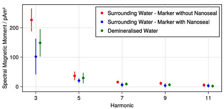 |
Figure 2 MPS measurements of samples taken from the water which had surrounded the SPION-marked stents with and without Nanoseal coating for a duration of 24 h, and a reference sample of demineralised water. |
MPI and MRI Measurements
The coated stents were sufficiently visualized with MPI (Figure 3A and B). The six individual markers at the ends of the stents were clearly distinguishable from each other, and the distance between two opposite markers represented the nominal stent diameter of 6 mm. The resulting SNR (markers vs stent lumen) was 11.5. Furthermore, the stent lumen was clearly visible. Comparative measurements with uncoated reference stents confirmed that they did not produce any signal in MPI.
 |
Figure 3 (A and B) show reconstructed MPI images of the marked stent. The dotted line represents the selected MPI FOV size. In (C and D), MRI images of the SPION-marked stent with resulting susceptibility artefacts are displayed. In (E and F), MRI images of an unmarked reference stent are shown. (The static field direction was vertical in images (C and E) and perpendicular to the image plane in (D and F)). |
In MRI, the unmarked reference stent was clearly detectable by signal extinctions due to susceptibility artefacts caused by the stent material (Figure 3E and F). The SPION-markers resulted in 4.6 times larger artefacts at the stent ends compared to the unmarked stent (Figure 3C and D). Both the unmarked and the SPION-marked stent showed V-shaped artefacts in the transversal plane and circular artefacts in the coronal plane, which differed considerably in size. Furthermore, the stent lumina showed homogenous hypointense signal, probably due to radiofrequency shielding by the stent mesh, as compared to the surrounding of the stent.
Discussion
In this study, we present an uncomplicated technique for the fabrication of stent markers designed for preclinical MPI and MRI. We prove the water insolubility of the introduced marking concept, which is a prerequisite for a broad range of preclinical cardiovascular research and especially quantification experiments with interventional instruments in MPI.
Due to the tracer-based nature of MPI, most cardiovascular instruments do not generate sufficient MPI signal.8,9,12 However, it has been described that some commercial metallic stents cause MPI-signals which can be used for tracking without any nanomodification.17 For devices that are invisible to MPI, the application of dedicated marking technologies is essential. The so far introduced SPION-varnishes produced sufficient MPI-signals and were mechanically stable.5,7 But the marker fabrication requires chemical expertise, and the water-stability of those markers has not been addressed so far. Especially regarding the direct correlation between signal intensity and particle concentration of MPI, this might be an important issue.18 Hence, the amount of iron of the markers should be stable, so as not to influence the quantification of stent lumina, for example. Furthermore, a loss of marker substance could lead to varying image reconstruction requirements, especially in coexistence of higher SPION-concentrations. Here, the loss of iron amount could lead to extinction artefacts.19 The SPIONs used for MPI are water soluble due to their dextran coating, which is essential for preventing agglomeration of the iron-oxide cores.20 Due to this characteristic, the varnish must prevent the contact of particles with water. In case of only mixing the particles with varnish, particles which are not fully embedded in the lacquer on the surface could dissolve. Following the MPS measurements of the water which surrounded the stent for 24 hours, the water solubility of the particles embedded in the varnish could be prevented by the applied sealing. Nevertheless, the detection limit of the MPS that we used must be considered.21 Possibly, a very low number of particles could be dissolved in the water but was not detectable. Here, vibrating sample magnetometry (VSM) could provide added value in overcoming the detection limit of MPS in future studies. Concerning potential degradation mechanisms, the properties of the marker material appear to be crucial. In polymers, hydrolysis can cause two mechanisms: superficial erosion and bulk erosion.22 For instance, Elfers et al recently reported degradable uncoated MPI markers based on polylactic acid, which developed cavities over time (bulk erosion).23 Thus, the application of a sealant layer seems to prevent marker degradation, probably irrespective of the underlying matrix material and degradation mechanism. Additionally, the irregular surface of varnish-based markers was described in previous work.5 In our experiments, the marker surface was very smooth due to the applied sealant.
When using MPI in hybrid-imaging settings together with MRI or CT, there is the need for fiducials.24,25 In contrast to liquid fiducials, the presented solid markers are eligible for experiments in water or body fluids and guarantee a stable signal intensity during the experiments in both MPI and MRI. The performance of the markers in MRI allows for reliable localization of the marked instrument. Despite the tested stents being clearly visible without the SPION-markers by material-induced susceptibility artefacts, the introduced concept seems to be transferable to non-metallic objects, eg catheters. Furthermore, deep learning-based automatic marker detection may be realized by the use of dedicated SPION marking technologies in MRI even when using visible metallic stents.26
Our proof-of-concept study has several limitations. We focused on the development of water-stable markers for in vitro research. Thus, any aspect of biocompatibility, eg cytotoxicity, was not addressed and should be part of future studies. Furthermore, we only investigated nitinol stents. Other commonly used metals as well as other devices should be tested. The conditions during the manufacturing process have not been monitored regarding humidity, coating viscosity and particle-to-varnish homogeneity, which thus should be considered in future work. In regard of the perspective to use these markers in in vivo experiments, stability of the markers under more realistic conditions with flow, a temperature of 37° C, blood as surrounding medium and prolonged experimental periods must be tested. Furthermore, systematic tests with multiple marked stents should be performed to allow for robust statistical analyses. Building upon the coating thickness values reported by Haegele et al for guidewires and catheters (30 and 35 µm),5 future investigations should focus on further reducing the stent coating thickness through the application of our method.
Conclusion
This work presents a simple and cost-effective instrument marking technique for MPI and MRI, using commercially available varnish and sealant to ensure water-stability of the markers. Although biocompatibility, long-term stability, and in vivo performance of the presented markers have not yet been addressed, this approach enables reliable visualization of interventional instruments in preclinical settings and thus contributes to the translational progress of MPI toward clinical application.
Ethical Approval
This article does not contain any studies with human participants or animals performed by any of the authors.
Informed Consent
For this type of study informed consent is not required.
Consent for Publication
For this type of study consent for publication is not required.
Funding
Fraunhofer IMTE and this work are supported by the EU, the State Schleswig-Holstein, Germany, and by internal programs (grants 12420002/LPWE1.1.1/1536 and 139-600251). Furthermore, this work was supported in part by the Clinician Scientist Program and Advanced Clinician Scientist Program of the University of Luebeck under grants CS10-2021, LACS04-2025.
Disclosure
The authors declare that they have no conflicts of interest in this work.
References
1. Gleich B, Weizenecker J. Tomographic imaging using the nonlinear response of magnetic particles. Nature. 2005;435(7046):1214–8. doi:10.1038/nature03808
2. Weizenecker J, Gleich B, Rahmer J, Dahnke H, Borgert J. Three-dimensional real-time in vivo magnetic particle imaging. Phys Med Biol. 2009;54(5):L1–L10. doi:10.1088/0031-9155/54/5/L01
3. Haegele J, Rahmer J, Gleich B, et al. Magnetic particle imaging: visualization of instruments for cardiovascular intervention. Radiology. 2012;265(3):933–938. doi:10.1148/radiol.12120424
4. Haegele J, Vaalma S, Panagiotopoulos N, et al. Multi-color magnetic particle imaging for cardiovascular interventions. Phys Med Biol. 2016;61(16):N415–426. doi:10.1088/0031-9155/61/16/N415
5. Haegele J, Panagiotopoulos N, Cremers S, et al. Magnetic particle imaging: a resovist based marking technology for guide wires and catheters for vascular interventions. IEEE Trans Med Imag. 2016;35(10):2312–2318. doi:10.1109/TMI.2016.2559538
6. Vaalma S, Rahmer J, Panagiotopoulos N, et al. Magnetic particle imaging (MPI): experimental quantification of vascular stenosis using stationary stenosis phantoms. PLoS One. 2017;12(1):e0168902. doi:10.1371/journal.pone.0168902
7. Salamon J, Hofmann M, Jung C, et al. Magnetic particle / magnetic resonance imaging: in-vitro MPI-guided real time catheter tracking and 4D angioplasty using a road map and blood pool tracer approach. PLoS One. 2016;11(6):e0156899. doi:10.1371/journal.pone.0156899
8. Wegner F, Friedrich T, von Gladiss A, et al. Magnetic particle imaging: artifact-free metallic stent lumen imaging in a phantom study. Cardiovasc Intervent Radiol. 2020;43(2):331–338. doi:10.1007/s00270-019-02347-x
9. Wegner F, von Gladiss A, Haegele J, et al. Magnetic particle imaging: in vitro signal analysis and lumen quantification of 21 endovascular stents. Int J Nanomed. 2021;16:213–221. doi:10.2147/IJN.S284694
10. Herz S, Vogel P, Kampf T, et al. Magnetic particle imaging–guided stenting. J Endovasc Ther. 2019;26(4):512–519. doi:10.1177/1526602819851202
11. Herz S, Vogel P, Dietrich P, et al. Magnetic particle imaging guided real-time percutaneous transluminal angioplasty in a phantom model. Cardiovasc Intervent Radiol. 2018;41(7):1100–1105. doi:10.1007/s00270-018-1955-7
12. Haegele J, Biederer S, Wojtczyk H, et al. Toward cardiovascular interventions guided by magnetic particle imaging: first instrument characterization. Magn Reson Med. 2013;69(6):1761–1767. doi:10.1002/mrm.24421
13. Ahlborg M, Friedrich T, Gottsche T, et al. First dedicated balloon catheter for magnetic particle imaging. IEEE Trans Med Imag. 2022;41(11):3301–3308. doi:10.1109/TMI.2022.3183948
14. Aderhold E, Ackers J, Stagge P, et al. Development and Assessment of a 1D-MPS. Int J Mag Part Imag. 2025;11(1 Suppl 1). doi:10.18416/IJMPI.2025.2503056
15. Aderhold E, Schumacher J, Stagge P, Ahlborg M, Buzug TM, Graeser M. Towards a fully integrated preclinical field-free line MPI scanner. Int J Mag Part Imag. 2023;9(1 Suppl 1).
16. Scheffler K, Boberg M, Knopp T. Solving the MPI reconstruction problem with automatically tuned regularization parameters. Phys Med Biol. 2024;69(4):045024. doi:10.1088/1361-6560/ad2231
17. Wegner F, Friedrich T, Wattenberg M, et al. Bare-metal stent tracking with magnetic particle imaging. Int J Nanomed. 2024;19:2137–2148. doi:10.2147/IJN.S447823
18. Saritas EU, Goodwill PW, Croft LR, et al. Magnetic particle imaging (MPI) for NMR and MRI researchers. J Magn Reson. 2013;229:116–126. doi:10.1016/j.jmr.2012.11.029
19. Boberg M, Gdaniec N, Szwargulski P, Werner F, Möddel M, Knopp T. Simultaneous imaging of widely differing particle concentrations in MPI: problem statement and algorithmic proposal for improvement. Phys Med Biol. 2021;66(9):095004. doi:10.1088/1361-6560/abf202
20. Panagiotopoulos N, Duschka RL, Ahlborg M, et al. Magnetic particle imaging: current developments and future directions. Int J Nanomed. 2015;10:3097–3114. doi:10.2147/IJN.S70488
21. Biederer S, Knopp T, Sattel TF, et al. Magnetization response spectroscopy of superparamagnetic nanoparticles for magnetic particle imaging. J Phy D App Phy. 2009;42(20):205007. doi:10.1088/0022-3727/42/20/205007
22. von Burkersroda F, Schedl L, Göpferich A. Why degradable polymers undergo surface erosion or bulk erosion. Biomaterials. 2002;23(21):4221–4231. doi:10.1016/s0142-9612(02)00170-9
23. Elfers PN, Lüdtke-Buzug K, Malhotra A, et al. Development and degradation study of PLA-based medical implant markers for magnetic particle imaging. Macromol Biosci. 2025;25(4):e2400495. doi:10.1002/mabi.202400495
24. Vogel P, Markert J, Rückert MA, et al. Magnetic particle imaging meets computed tomography: first simultaneous imaging. Sci Rep. 2019;9(1):12627. doi:10.1038/s41598-019-48960-1
25. Franke J, Heinen U, Lehr H, et al. System characterization of a highly integrated preclinical hybrid MPI-MRI scanner. IEEE Trans Med Imag. 2016;35(9):1993–2004. doi:10.1109/TMI.2016.2542041
26. Nijsink H, Overduin CG, Brand P, et al. Optimised passive marker device visibility and automatic marker detection for 3-T MRI-guided endovascular interventions: a pulsatile flow phantom study. Eur Radiol Exp. 2022;6(1):11. doi:10.1186/s41747-022-00262-4
 © 2026 The Author(s). This work is published and licensed by Dove Medical Press Limited. The
full terms of this license are available at https://www.dovepress.com/terms
and incorporate the Creative Commons Attribution
- Non Commercial (unported, 4.0) License.
By accessing the work you hereby accept the Terms. Non-commercial uses of the work are permitted
without any further permission from Dove Medical Press Limited, provided the work is properly
attributed. For permission for commercial use of this work, please see paragraphs 4.2 and 5 of our Terms.
© 2026 The Author(s). This work is published and licensed by Dove Medical Press Limited. The
full terms of this license are available at https://www.dovepress.com/terms
and incorporate the Creative Commons Attribution
- Non Commercial (unported, 4.0) License.
By accessing the work you hereby accept the Terms. Non-commercial uses of the work are permitted
without any further permission from Dove Medical Press Limited, provided the work is properly
attributed. For permission for commercial use of this work, please see paragraphs 4.2 and 5 of our Terms.