
Back to Journals » International Journal of Nanomedicine » Volume 20
Silicon-Based Nanomaterials in Chronic Wound Healing: Mechanisms, Therapeutic Applications, and Clinical Prospects
Authors Zhao X, Xu Z, Wang D  , Li T, Li Z, Bai X, Zhu H, Liu Y, Wang Y
Received 15 March 2025
Accepted for publication 18 September 2025
Published 30 September 2025 Volume 2025:20 Pages 11959—11988
DOI https://doi.org/10.2147/IJN.S528531
Checked for plagiarism Yes
Review by Single anonymous peer review
Peer reviewer comments 2
Editor who approved publication: Prof. Dr. RDK Misra
Xuan Zhao,1– 4 Zhikai Xu,1– 4 Dongfang Wang,1– 4 Tonghan Li,1– 4 Zhanfei Li,1– 4 Xiangjun Bai,1– 4 Hao Zhu,5 Yukun Liu,6 Yuchang Wang1– 4
1Division of Trauma Surgery, Emergency Surgery & Surgical Critical, Tongji Hospital, Tongji Medical College, Huazhong University of Science and Technology, Wuhan, 430030, People’s Republic of China; 2Trauma Center, Tongji Hospital, Tongji Medical College, Huazhong University of Science and Technology, Wuhan, 430030, People’s Republic of China; 3Department of Emergency and Critical Care Medicine, Tongji Hospital, Tongji Medical College, Huazhong University of Science and Technology, Wuhan, 430030, People’s Republic of China; 4Sino-German Research Institute of Disaster Medicine, Huazhong University of Science and Technology, Wuhan, 430030, People’s Republic of China; 5Department of Orthopedic Surgery, Tongji Hospital, Tongji Medical College, Huazhong University of Science and Technology, Wuhan, 430030, People’s Republic of China; 6Department of Plastic and Aesthetic Surgery, Tongji Hospital, Tongji Medical College, Huazhong University of Science and Technology, Wuhan, 430030, People’s Republic of China
Correspondence: Yukun Liu; Yuchang Wang, Email [email protected]; [email protected]
Abstract: Silicon-based nanosystems are emerging as promising nanotherapeutic platforms in the biomedical field, particularly for the treatment of chronic wounds. These materials possess several advantageous features. They offer excellent drug-loading capacity, controlled and stimuli-responsive drug release, and highly customizable structures and functions. These characteristics make them well-suited for personalized therapeutic approaches.This review provides a comprehensive summary of recent advances in the application of silicon-based nanosystems in wound healing. It highlights their mechanisms of action and discusses future development directions. We begin by outlining the clinical significance and complex pathophysiological characteristics of chronic wounds. A detailed classification of silicon-based nanomaterials is then provided, including mesoporous silica nanoparticles and silicon-based composites. The review emphasizes their key roles in modulating inflammation, reducing oxidative stress, and promoting angiogenesis and tissue regeneration. In addition, we summarize recent findings from in vitro and in vivo studies, as well as updates from relevant clinical research. The biocompatibility and safety profiles of these systems are also comprehensively evaluated. Future research should focus on optimizing the synthesis of these materials and improving their long-term biosafety. Efforts should also aim to integrate multifunctional therapeutic strategies to enhance efficacy and translational potential. Moreover, large-scale, rigorously designed clinical trials are urgently needed. These studies will help build robust clinical evidence and support the practical application of silicon-based nanosystems in advanced wound care. In conclusion, silicon-based nanosystems represent a next-generation approach for wound therapy. However, further interdisciplinary research is essential to fully realize their clinical value.
Keywords: silicon, nanomaterial, chronic wound healing, wound healing, silicon-based nanosystems
Graphical Abstract:

Introduction
The skin is an important organ that covers the human body and interacts directly with the outside world. It plays a key role in immunity, feeling, and protection.1,2 In recent years, the prevalence of chronic wounds has sharply increased, primarily due to the growing population of individuals with diabetes and obesity, in addition to external physical damage.3,4 Under normal conditions, wound healing is a complex, dynamic process supported by numerous cellular events, tightly coordinated to repair damaged tissues effectively.5 However, in certain situations such as diabetes, wound infection, inflammation, and vascular or neural dysfunction, the normal wound healing process becomes disrupted, leading to the development of chronic wounds.6,7 In the United States, chronic wounds affect approximately 10.5 million Medicare beneficiaries (an increase of 2.3 million since 2014), with annual treatment costs exceeding $25 billion.8,9 Despite advancements, promoting rapid and high-quality skin wound healing remains a significant challenge.
In recent years, silica nanoparticles (SNPs) and their multifunctional carriers have gained considerable attention due to their significant advantages in drug delivery.10–12 These advantages include their hydrophilic surface, diverse surface functionalization options, tunable shapes, and sizes, biocompatibility, ease of large-scale synthesis, and low production costs,10,13–15 making them highly promising therapeutic carriers. Among these, mesoporous silica nanoparticles (MSNs), with pore sizes ranging from 2 to 50 nanometers, have been extensively studied. MSNs have been applied in targeted drug delivery and tissue engineering,11,16 and their surfaces can be modified with stimuli-responsive molecules and various therapeutic macromolecules, enabling on-demand and localized controlled drug release.17 Additionally, capping strategies endow MSNs with intelligent drug delivery properties, allowing them to respond to various stimuli (eg, PH changes, photothermal, and photodynamic effects) and release therapeutic cargo for targeted applications.18 The development of silica-based nanosystems has shown significant potential in biomedicine, demonstrating their utility in clinical practice19–23 (Figure 1).
 |
Figure 1 Applications of polymer-coated MSNs in various therapeutic fields, including photodynamic therapy (PDT), photothermal therapy (PTT), chemotherapy, RNA delivery, wound healing, tissue engineering, food packaging, and the treatment of neurodegenerative diseases. Reprinted from Nair A, Chandrashekhar H R, Day CM, et al. Polymeric functionalization of mesoporous silica nanoparticles: biomedical insights. Int J Pharm. 2024;660:124314. Creative Commons.19 |
Over the past few decades, advancements in nanotechnology have led to the emergence of various nanomaterials and nanomaterial-based drug delivery systems, which have been applied to wound repair and regeneration.24 The quantum size and surface effects of different nanomaterials impart unique physicochemical properties and functionalities, enabling them to carry and release bioactive drugs in controlled and sustained manners.25 These functionalized nanoparticles promote wound healing by modulating the microenvironment through antibacterial, anti-inflammatory, antioxidant, and pro-angiogenic properties.26–31 This review discusses different types of SNPs and their surface engineering, elucidating their mechanisms and potential roles in improving wound healing, while also exploring their biocompatibility and safety profiles.
Wound Classification and Pathophysiology of Injury Repair
Skin wounds are defined as disruptions or damage to the structure and function of the skin caused by various factors, including trauma, burns, and physiological or medical conditions.32,33 In such cases, the anatomical structure of the skin is compromised, leading to a loss of its physiological functions. Based on the healing timeline, wounds are generally classified into two categories: acute wounds and chronic wounds. Acute wounds are often caused by mechanical injury or exposure to extreme temperatures, radiation, electrical shocks, or corrosive chemicals.34 With proper wound management, acute wounds restore skin integrity within weeks or a month through the normal and orderly stages of tissue repair.35,36 Chronic wounds, however, are often complications of specific conditions such as diabetes, vascular diseases, or pressure ulcers, although wound-specific factors like infection, inflammation, and radiation also contribute.37 Systemic factors, including malnutrition, immunosuppression, aging, and other complications, can further delay wound healing.38,39 Wounds can also be classified based on their depth: superficial wounds (involving only partial epidermal loss), partial-thickness wounds (affecting the epidermis and deeper dermis), and full-thickness wounds (damaging subcutaneous fat and deeper tissues).40
Following injury, damaged skin initiates a complex repair process involving the interplay of various cell types, cytokines, and biological mediators. Under normal conditions, this process progresses through distinct stages, including hemostasis, inflammation, proliferation, and remodeling (Figure 2).33,41–43
 |
Figure 2 Damaged skin initiates a complex repair process involving the interaction of multiple cell types, cytokines, and biological mediators. Under normal conditions, this process undergoes a series of stages, including hemostasis, inflammation, proliferation, and remodeling. Adapted from from Cioce A, Cavani A, Cattani C, Scopelliti F. Role of the Skin Immune System in Wound Healing. Cells. 2024; 13(7):624. Creative Commons.44 |
Hemostasis
Immediately after injury, the body triggers hemostatic responses to minimize blood loss. Local blood vessels constrict via smooth muscle contraction to limit blood flow, and both intrinsic and extrinsic coagulation pathways are activated.45,46 The fibrin clot formed by platelets and coagulation factors not only prevents bleeding but also provides a temporary scaffold for cell adhesion and migration.47–49
Inflammation
The inflammatory phase typically occurs from day 2 to day 5 post-injury. During this phase, platelets activated by thrombin release various growth factors, including epidermal growth factor (EGF), insulin-like growth factor-1 (IGF-1), platelet-derived growth factor (PDGF), fibroblast growth factor (FGF), and transforming growth factors (TGF-α and TGF-β).50,51 These factors serve as biological signals that attract neutrophils, monocytes, leukocytes, and macrophages to the wound site, mediating inflammation, protecting the skin from infection, and secreting additional growth factors to accelerate healing.40,52,53
Proliferation
From 3 days to 2 weeks post-injury, the wound enters the proliferative phase, characterized by cell proliferation and migration.40 Angiogenesis and capillary formation occur under the influence of pro-angiogenic factors such as PDGF released by platelets and inflammatory cells.54 Simultaneously, fibroblast migration is stimulated by PDGF and FGF, resulting in granulation tissue formation.55,56 Fibroblasts accumulate and proliferate, producing a new extracellular matrix (ECM) composed of collagen, proteoglycans, and elastin. Some fibroblasts differentiate into myofibroblasts, contributing to wound contraction.57 Additionally, activated keratinocytes at the wound edges migrate to the injured area, completing re-epithelialization.58
Remodeling
The remodeling phase involves structural reorganization and cellular reduction. Type III collagen in the granulation tissue is gradually replaced by type I collagen, the primary matrix component of the dermis. The rearrangement, cross-linking, and alignment of collagen fibers involve the dynamic synthesis of collagen and matrix metalloproteinases (MMPs), leading to ECM strengthening and contraction, and ultimately the formation of mature scar tissue. This phase typically lasts from 3 weeks to 2 years.59,60 Wound repair is a highly organized and complex pathological process. Disruptions at any stage can lead to wound pathologies, such as hypertrophic scars, keloids, or chronic wounds.61,62
If the normal healing process is disrupted, wounds may become chronic. Chronic wounds fail to heal in a timely and orderly manner. Clinically, diabetic foot ulcers (DFUs), pressure ulcers (PUs), and venous leg ulcers (VLUs) are common types of chronic wounds.37,39,63 Despite varying etiologies, these wounds share common characteristics, including excessive exudate, infection, tissue necrosis, insufficient re-epithelialization, reduced angiogenesis, and elevated levels of reactive oxygen species (ROS).64,65 Diabetes and hyperglycemia also contribute to peripheral neuropathy and peripheral vascular disease, significantly increasing the risk of DFUs and VLUs.66,67 Vascular damage leads to hypoxia, promoting the formation of avascular, non-viable tissue that creates an environment conducive to bacterial growth and biofilm formation. Biofilms exacerbate inflammation, hinder ECM deposition, and impair tissue repair.68 Unlike acute wounds, chronic wounds are influenced by multifactorial elements, including dysfunction of inflammatory cells; limited bioavailability of growth factors and cytokines; overexpression of proteases, persistent infection, reduced angiogenesis, and inadequate nutrient supply.53,69 These factors often interrupt the wound healing process, leaving it stalled in the inflammatory phase.
Diabetes significantly impacts all stages of wound repair, creating a pro-inflammatory environment characterized by elevated levels of pro-inflammatory cytokines such as TNF-α and reduced concentrations of healing-promoting mediators like IL-10 and TGF-β. This imbalance can lead to tissue necrosis and chronic wound formation.70 Additionally, the wound microenvironment after infection becomes pro-inflammatory, with prolonged presence of myeloid cells (eg, macrophages and neutrophils) and reduced levels of skin dendritic cells (DCs), Langerhans cells (LCs), and eosinophils, perpetuating the inflammatory state.64,71,72
Systemic or localized pathological factors often impair immune cells’ ability to eliminate pathogens, creating a vicious cycle that exacerbates the hostile wound microenvironment.73 Moreover, failure in signal transduction, weakened keratinocyte migration, and proliferation, and delayed re-epithelialization further contribute to chronic wound progression.71,74 Excessive ECM degradation, partly induced by the imbalance of MMPs and their inhibitors, hinders proper ECM deposition.75,76 In wounds with neuropathy, reduced neuropeptides and neurotrophic factors also impede critical healing mechanisms.76
Classification and Structural Characteristics of SNPs
The main synthesis methods of silicon-based nanomaterials include chemical vapor deposition (CVD), sol-gel method, solvothermal method, hydrothermal method, reverse microemulsion method, and self-assembly method.77–79 Due to their versatility in synthesis, size and shape regulation, surface functionalization, and biocompatibility, various types of silica nanoparticles (SNPs) have been developed for biomedical applications,77–80 including non-porous, mesoporous, hollow, core-shell, yolk-shell, and Janus architectures.81 Typically, SNPs are classified into two classic types—non-porous and mesoporous—based on their porosity (Figure 3). The term “mesoporous” generally refers to materials with pore sizes ranging from 2 to 100 nm.82 Non-porous SNPs lack special surface structures, while mesoporous silica nanoparticles (MSNs) feature tunable mesopores. The most notable difference between the two is the orderly, porous structure of MSNs.83 Characterization analysis shows that both types exhibit an amorphous, near-spherical morphology.
 |
Figure 3 Silica MSNs-based nanocomposites developed in the biomedical field. Various nanostructured MSN-based nanocomposites. Depending on the assembly process, the functional nanostructures can be introduced as the shell (Type (I) or core (Type IV), loaded in the pore channels (Type II) or surface (Type III), or form Janus-type hierarchical structures (Type V). Reprinted from Xu B, Li S, Shi R, et al. Multifunctional mesoporous silica nanoparticles for biomedical applications. Signal Transduct Target Ther. 2023;8(1):435. Creative Commons.16 |
Amorphous SNPs not only provide drug-loading capabilities and enable precise regulation of pharmacokinetics through porous channels but also possess two critical functional surfaces: cylindrical pore surfaces and external particle surfaces. Both surfaces can be functionalized according to specific requirements. On the one hand, pore surfaces can be specifically functionalized to precisely control drug release rates and mechanisms; on the other hand, external particle surfaces can bind to targeting ligands to achieve specific drug delivery.84 According to assembly strategies, specific nanostructured composites can be obtained, and MSNs are categorized into five types16 (Figure 3):
- Type I: Core-shell structures where MSNs act as the core, and functional components serve as the shell. Functional nanocoatings of specific sizes can be easily achieved by manipulating MSNs as hard templates.
- Type II: Small functional components are directly loaded into MSN pores. Common functional components, such as carbon quantum dots and black phosphorus quantum dots, are often encapsulated in this form. In such structures, MSNs enable the slow and controlled release of small functional components.
- Type III: Functional components are loaded onto MSN surfaces or pore peripheries through covalent bonds or electrostatic adsorption. This approach avoids masking the active sites of functional components, thereby ensuring catalytic stability.
- Type IV: Core-shell structures where MSNs act as the shell, and functional components serve as the core. This structure prevents the aggregation of exposed inorganic functional components, enhancing the stability of nanocomposites and reducing physiological toxicity.
- Type V: Janus-type architecture. Janus-type nanocomposites feature biphasic geometric shapes with distinct compositions or anisotropic structures. Unlike the Type I–IV nanocomposites, the physicochemical properties of individual components in Janus structures remain largely unaffected.
Using these unique structures, researchers have developed numerous silicon-based hybrid nanomaterials with distinctive properties through surface functionalization, such as silicon-drug polymers, silicon-nucleic acid hybrids, silicon-protein hybrids, and silicon-magnetic composites. Additionally, silica can hybridize with peptides, amino acids, gold nanomaterials, and quantum dots to meet the demands of complex biomedical applications.85,86
In recent years, experts have continually proposed new insights into the design principles of silica nanostructures,87 such as engineering pore geometry, surface topology, and asymmetry to enhance the efficiency of drug, gene, and protein delivery.87,88 Innovations include altering surface roughness to improve cellular uptake and adhesion, as well as hollow MSNs (HMSNs) with mesoporous shells and hollow interiors. Another class of innovative mesoporous silica materials has been designed to overcome the limitations of traditional MSNs in delivering large-volume drugs like proteins by exhibiting advanced mass diffusion properties and high storage capacities.88–90 Additionally, novel SNPs have been designed as biological modulators to regulate intracellular microenvironments and cell signaling, such as oxidative stress and glutathione levels, thereby enhancing therapeutic anticancer effects and mRNA transfection in specific cell lines91 (Figure 4). Thus, silicon-based nanomaterials are becoming increasingly diverse in terms of morphology and functionality, finding widespread applications in biomedical fields, including targeted drug delivery, tissue engineering, biosensing, bioimaging, and more.92
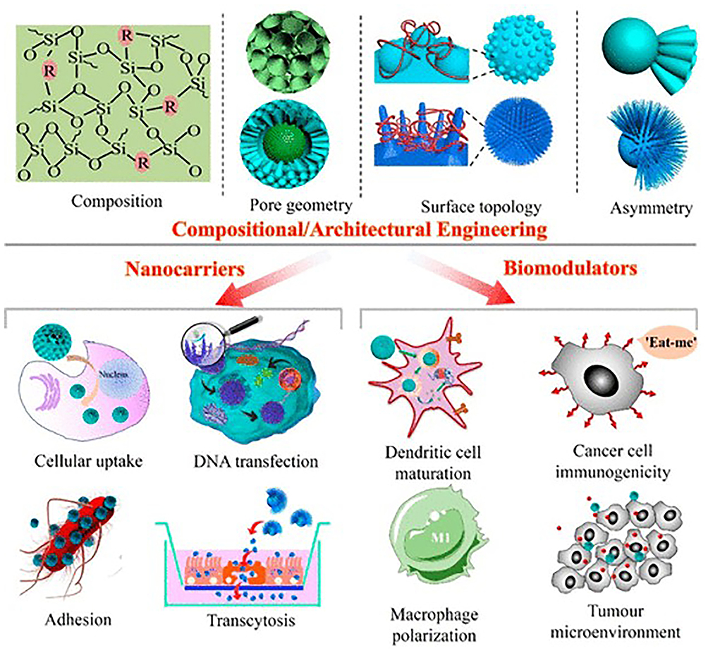 |
Figure 4 Schematic illustration of the composition and architecture of engineered silica-based nanoparticles with diverse appealing properties as nanocarriers and biomodulators for biomedical applications. Reprinted from Fu JY, Gu Z, Liu Y, et al. Bottom-up self-assembly of heterotrimeric nanoparticles and their secondary Janus generations. Chem Sci. 2019;10(44):10388–10394. Creative Commons.91 |
Potential Regulatory Mechanisms of Silicon-Based Nanomaterials in Wound Healing
Silicon-based nanoparticles (SNPs) have shown immense potential in tissue regeneration, particularly in wound healing. Silicon-based nanostructured composites exhibit promising roles, either individually or synergistically, in promoting wound healing through antibacterial/anti-inflammatory effects, antioxidation, and tissue regeneration.93,94 Below, we elucidate their potential regulatory mechanisms with illustrative examples.
Antibacterial Effects
After skin injury, bacteria easily migrate from the surface to non-resident areas, leading to wound infection and significantly disrupting the healing process.37 Wound exudates, as a hallmark of chronic wounds, contain corrosive components that exacerbate extracellular matrix (ECM) degradation. Matrix metalloproteinases (particularly MMP-9) have been identified as key destructive factors, and the increase in bacterial load is closely associated with elevated MMP-9 levels.95 Furthermore, biofilm formation provides bacteria with protection, enhances their proliferation, and increases their resistance to antibacterial treatments, which is a major cause of chronic wound infections.96 With the growing prevalence of bacterial antibiotic resistance, effectively treating chronic infections and promoting wound healing has become increasingly challenging. Silicon-based nanocomposites have garnered significant attention in wound healing due to their excellent antibacterial properties. Their antibacterial and anti-infection effects are primarily attributed to two mechanisms: serving as physical barriers and releasing antibacterial active substances.97,98
 |
Figure 5 (I) TEM images (a and b), with the nano-Ag particle size distribution shown in the inset of (a), dark field image (c), corresponding elemental mapping for silicon (d), oxygen (e), silver (f), Si-Ag-O merged image (g), nitrogen sorption isotherm (h), and the inset showing pore size distribution for SiNPs-Ag. (II) SEM images of E. coli (a–d) and S. epidermidis (e–h) after treatment with vancomycin (a, e), SiNPs-Van (b, f), SiNPs-Ag (c, g), and SiNPs-Ag-Van (d, h), along with a schematic illustration of antibacterial activity (i). Reprinted from Ni C, Zhong Y, Wu W, et al. Co-Delivery of Nano-Silver and Vancomycin via Silica Nanopollens for Enhanced Antibacterial Functions. Antibiotics. 2022;11(5):685. Creative Commons.98 (III) Schematic description of the preparation of Ag-Bi@SiO2 NPs and their synergistic antibacterial effects. Reprinted from Cao CY, Ge W, Yin J, et al. Mesoporous Silica Supported Silver-Bismuth Nanoparticles as Photothermal Agents for Skin Infection Synergistic Antibacterial Therapy. Small. 2020;16(24). © 2020 WILEY-VCH Verlag GmbH & Co. KGaA, Weinheim.99 Note: The original figure contains a spelling error “quantaty”, which is retained as published. |
Silicon-based nanosystems are commonly used as multifunctional delivery platforms to load and release antibacterial active substances to inhibit pathogen growth. For example, Ni et al successfully loaded vancomycin and nanosilver onto pollen-shaped silica nanoparticles. The unique spiked morphology of the silica carrier enhanced nanoparticle adhesion to bacterial surfaces, thereby promoting local drug release for bacterial eradication. This dual delivery of vancomycin and nanosilver demonstrated enhanced bactericidal activity98 (Figure 5I and II). Additionally, Cao et al constructed mesoporous silica nanoparticles loaded with silver-bismuth nanoparticles (Ag-Bi@SiO2NP). The high-temperature effect of bismuth nanoparticles accelerated the release of Ag ions, exhibiting excellent antibacterial performance against methicillin-resistant Staphylococcus aureus (MRSA)99 (Figure 5III). Rasool et al developed functionalized silica-ceria nanocomposites (FSC), which combined mesoporous silica nanoparticles with ceria’s inherent antibacterial activity, effectively inhibiting biofilm formation.100
 |
Figure 6 (I) Silicon-based Ag dendritic nanoforests for light-assisted bacterial inhibition. Reprinted from Li Y, Xu T, Tu Z, et al. Bioactive antibacterial silica-based nanocomposites hydrogel scaffolds with high angiogenesis for promoting diabetic wound healing and skin repair. Theranostics. 2020;10(11):4929–4943. Creative Commons.22 (II) Top- and side-view SEM images of Ag-DNFs/Si synthesized at various times. (III) Schematic of plasmonic-assisted bacterial inhibition. Reprinted from Huang HJ, Chang H-W, Lin Y-W, et al. Silicon-Based Ag Dendritic Nanoforests for Light-Assisted Bacterial Inhibition. Nanomaterials. 2020;10(11):2244. Creative Commons.101 |
The smart-responsive properties of silicon-based nanomaterials further enhance their antibacterial efficacy. For instance, Li et al developed a silicon-based nanocomposite hydrogel scaffold with biomimetic elastic mechanics, self-healing behavior, and broad-spectrum antibacterial activity. This scaffold significantly enhanced the vitality and angiogenic capability of endothelial progenitor cells (EPCs) in vitro and promoted collagen deposition and vascular network reconstruction by enhancing HIF-1α/VEGF expression in vivo, thereby accelerating full-thickness wound healing22 (Figure 6I). Moreover, specific nanostructure designs of silicon-based materials, such as sharp nanoblades, microspheres, and nanoflower-like structures, further improve their antibacterial effects. Huang et al synthesized silver dendritic nanoforests (Ag-DNFs/Si) on silicon substrates. The sharp nanoblade structures exhibited superior antibacterial efficiency, showing significant bactericidal effects against Escherichia coli and Staphylococcus aureus101 (Figure 6II and III).
Silicon-based nanoparticles can also be specifically designed to meet diverse needs. Khan et al prepared silica microspheres loaded with nitrofurazone (NFZ) and lidocaine (LD). The successful incorporation of drugs into the microspheres avoided drug-drug interactions and effectively addressed pathological skin infections. In-vitro and in-vivo experiments demonstrated good antibacterial activity and biocompatibility.102 Hashemikia et al prepared hybrid nanofibers of chitosan/polyethylene oxide/silica, incorporating ciprofloxacin into the electrospun mixture. The nanofibers absorbed moisture during degradation and gradually released ciprofloxacin, exhibiting excellent antibacterial activity against E. coli and S. aureus.103 Gwon et al synthesized antibacterial silicon-based nickel nanoflowers (Si@Ni) using microfluidics and photopolymerization techniques and encapsulated them in methacryloyl gelatin (GelMA) to construct uniform microscale hydrogel spheres (Si@Ni-GelMA). Due to nickel’s antibacterial effects against Pseudomonas aeruginosa, Klebsiella pneumoniae, and MRSA, the injectable Si@Ni-GelMA demonstrated superior antibacterial activity with negligible cytotoxicity.104 By acting as physical barriers, releasing antibacterial active substances, and leveraging smart-responsive properties, silicon-based nanomaterials not only address antibiotic resistance but also significantly accelerate wound healing. Additionally, their customizable designs for various drug metabolism needs make them ideal materials for wound repair applications (Table 1).
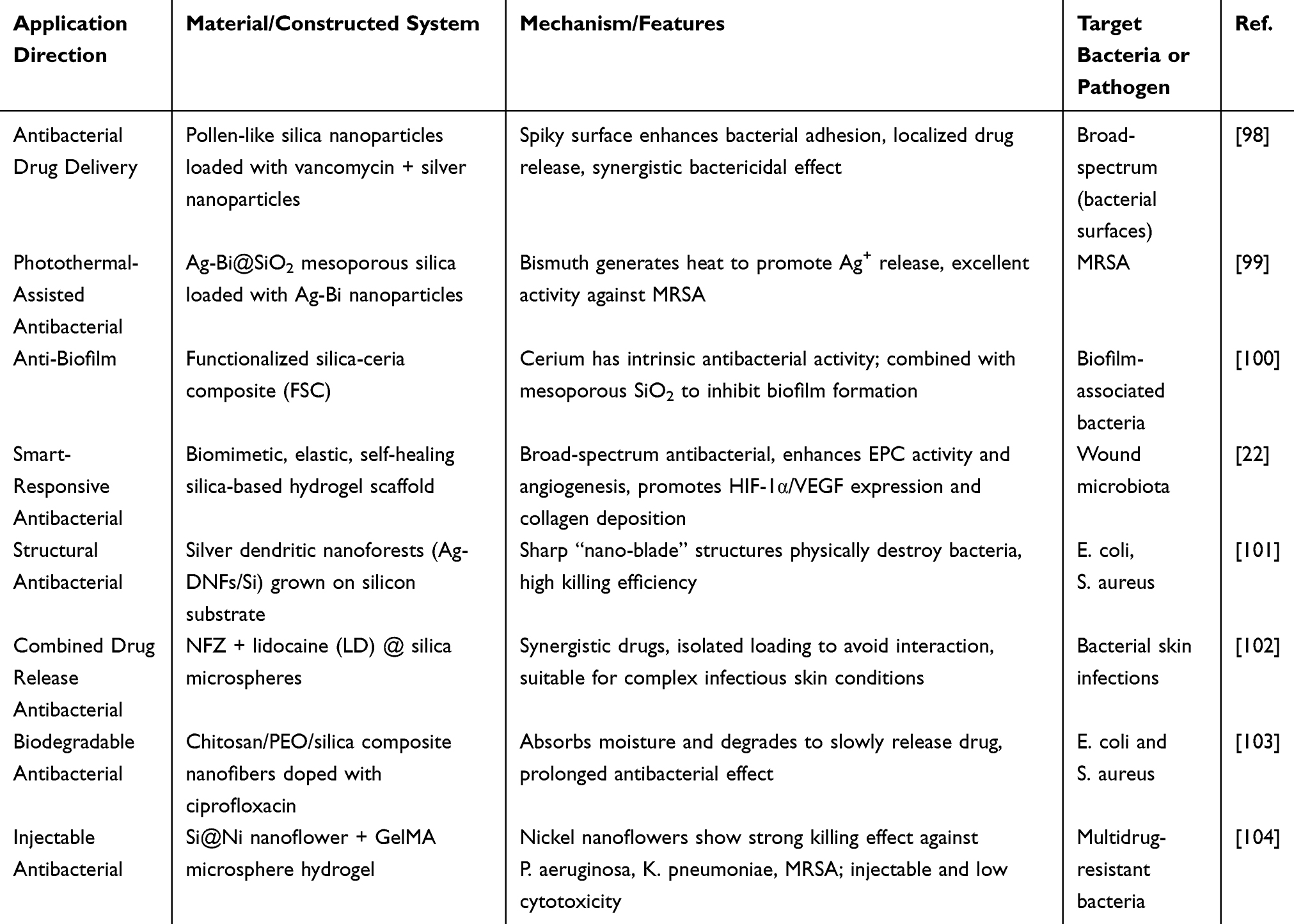 |
Table 1 Application of Silicon-Based Materials in Antibacterial Therapy |
Inflammation Regulation
Inflammation is a critical stage of wound healing; however, excessive or persistent inflammation, often accompanied by microbial recruitment and biofilm formation, leads to chronic wound formation. Regulating inflammatory responses to maintain immune balance is one of the key strategies for promoting wound healing.105,106 In recent years, silicon-based nanomaterials have demonstrated significant advantages in regulating chronic wound inflammation due to their unique structural properties and functionalization potential as drug carriers for inflammatory modulators (Table 2).
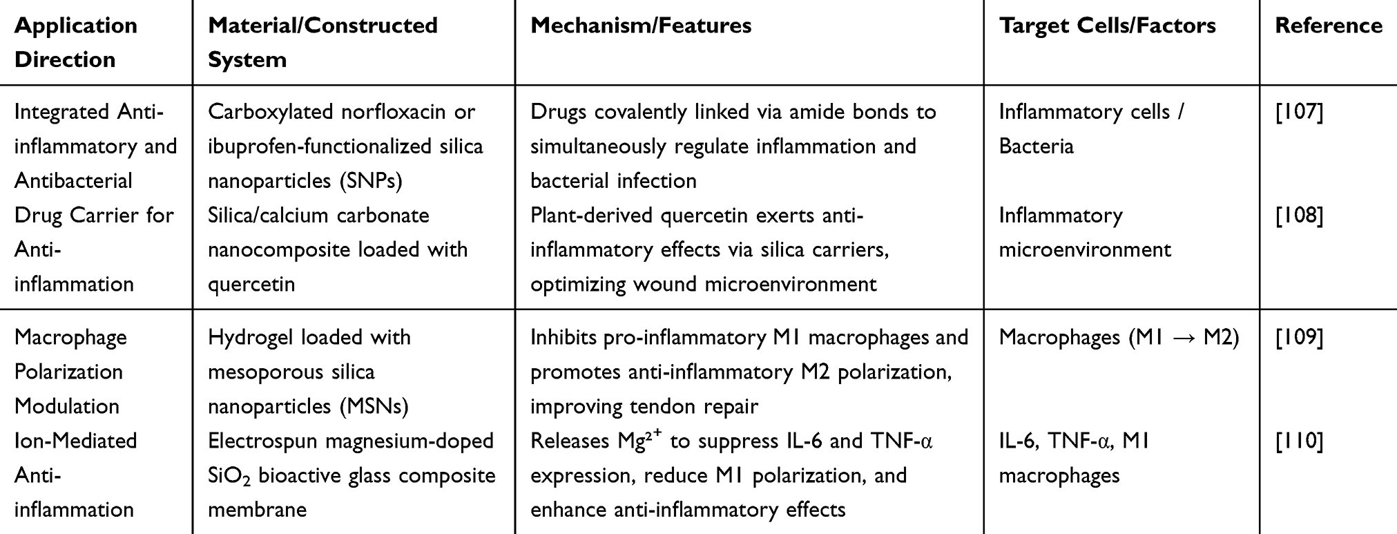 |
Table 2 Application of Silicon-Based Materials in Anti-Inflammatory Therapy |
Single silicon-based monomers containing carboxylated antibiotics, specifically norfloxacin, and anti-inflammatory agents, such as ibuprofen, were developed. Liu et al covalently linked silicon nanoparticles (SNPs) derived from these single silicon-based monomers with the anti-inflammatory and antibacterial drugs through amide bonds. This process resulted in the synthesis of bifunctional SNPs that exhibit both inflammation-regulating and antibacterial activities.107 Additionally, quercetin, a plant-derived anti-inflammatory drug, was loaded onto calcium carbonate/silica nanocomposites to exert anti-inflammatory effects and improve wound healing rates.108 These silicon-based nanosystems function as drug carriers to modulate inflammatory responses, thereby optimizing the wound healing environment.
Moreover, macrophages play a crucial role in wound healing, with their polarization states determining the progression of inflammation and the efficiency of tissue repair. During the early inflammatory stage, M1 macrophages function to clear pathogens, but their overactivation can delay healing. In the later repair stage, M2 macrophages accelerate tissue regeneration by secreting anti-inflammatory factors (eg, IL-10) and pro-repair factors (eg, VEGF).111 Scholars have designed various silicon-based composites to effectively promote macrophage polarization from M1 to M2. For example, MSN-loaded hydrogels significantly inhibited pro-inflammatory M1 macrophages while promoting anti-inflammatory M2 macrophages, accelerating tendon healing and reducing inflammatory responses109 (Figure 7I). Liu et al fabricated magnesium-doped SiO2 bioactive glass composite membranes using electrospinning technology. The release of magnesium ions significantly suppressed the expression of the pro-inflammatory factors IL-6 and TNF-α while inhibiting M1 macrophage polarization, thereby enhancing anti-inflammatory effects110 (Figure 7II).
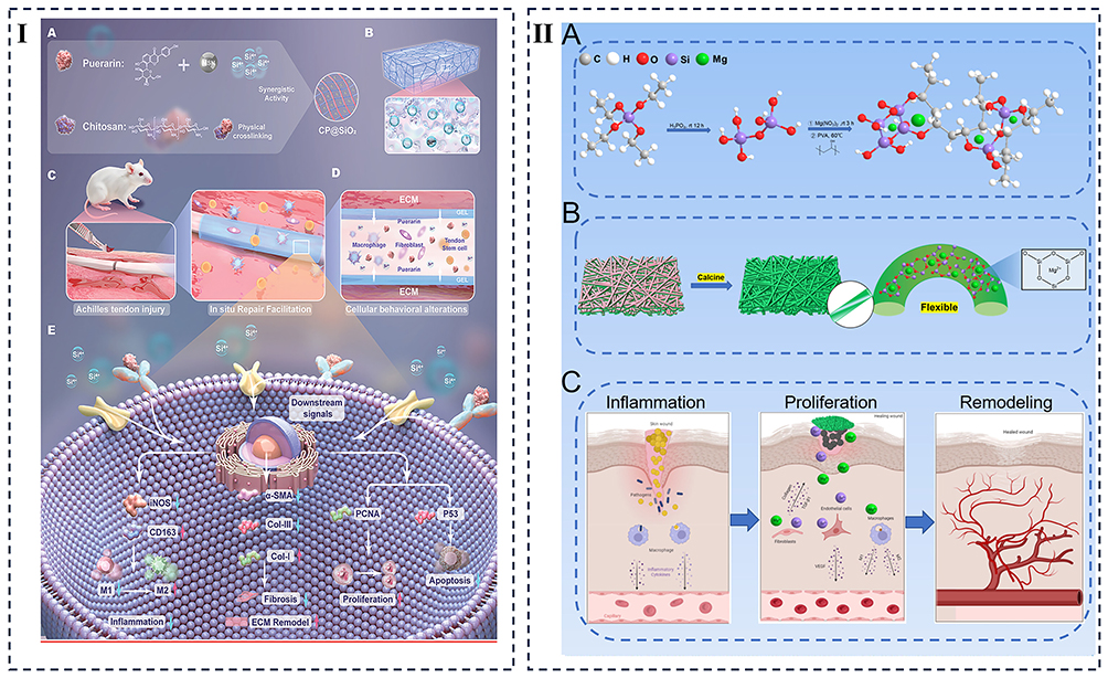 |
Figure 7 (I) (A) Preparation of CP@SiO2 composite by combining puerarin, chitosan, and SiO2 nanoparticles. (B) Structure of CP@SiO2 hydrogel showing uniform distribution of SiO2 within the network. (C) Application in Achilles tendon injury to achieve in situ repair. (D) Regulation of extracellular matrix (ECM) and cellular behaviors, including macrophage polarization and fibroblast activity. (E) Schematic mechanism showing CP@SiO2-mediated signaling pathways that regulate inflammation, fibrosis, proliferation, and apoptosis. Reprinted from Wan R, Luo Z, Nie X, et al. A Mesoporous Silica-Loaded Multi-Functional Hydrogel Enhanced Tendon Healing via Immunomodulatory and Pro-Regenerative Effects. Adv Healthc Mater. 2024;13(26):e2400968. © 2024 Wiley-VCH GmbH.109 (II) (A) Synthesis scheme of precursor solutions for electrospinning. (B) Synthesis and chemical structure of flexible inorganic SiO2/MgO nanofiber membranes. (C) Electrospun flexible SiO2/MgO nanofiber membranes inhibit S. aureus infection and promote the healing of infected wounds. Reprinted from Liu MY, Wang X, Cui J, et al. Electrospun flexible magnesium-doped silica bioactive glass nanofiber membranes with anti-inflammatory and pro-angiogenic effects for infected wounds. J Mat Chem B. 2023;11(2):359–376. © Royal Society of Chemistry 2023.110 |
Through the release of anti-inflammatory factors, inhibition of pro-inflammatory signals, regulation of macrophage polarization, or synergistic mechanisms, silicon-based nanomaterials demonstrate significant potential in inflammation regulation, making them promising candidates for research and applications in chronic wound healing.
Antioxidant Effects
Reactive oxygen species (ROS) play a dual role in the wound healing process. At physiological levels, ROS act as signaling molecules to regulate cell migration, angiogenesis, and immune responses. However, in chronic wounds or infected wounds, excessive ROS can induce oxidative stress, damage cellular structures and functions, and lead to the oxidative modification of proteins, lipids, and DNA, further exacerbating tissue inflammation and damage.112–114 This persistent oxidative stress inhibits the proliferation of fibroblasts and keratinocytes, thereby delaying wound healing.115 Thus, eliminating excessive ROS and providing sustained antioxidant protection are critical for accelerating wound repair.
 |
Table 3 Application of Silicon-Based Materials in Combating Oxidative Stress |
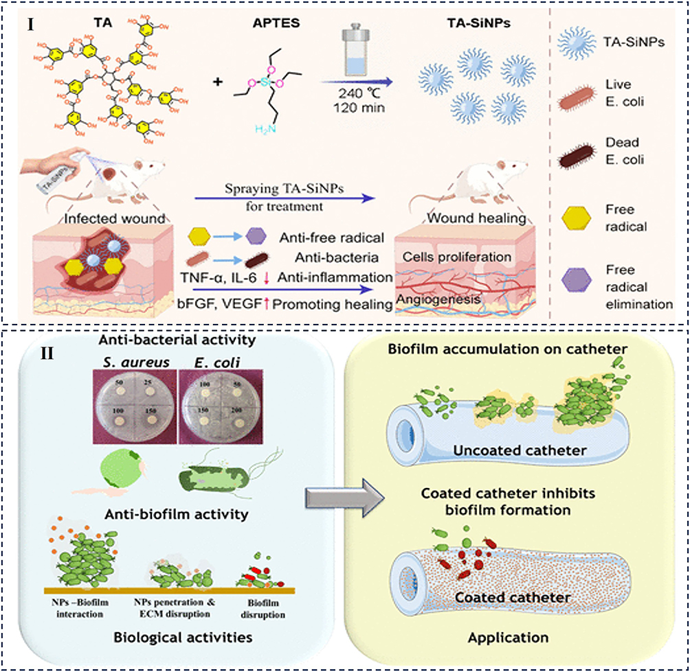 |
Figure 8 (I) Nanoparticles demonstrated multifunctional therapeutic effects by disrupting biofilms, eradicating E. coli, neutralizing reactive oxygen species (ROS), stimulating fibroblast growth, and enhancing angiogenesis. Additionally, they suppressed inflammatory cytokines while upregulating factors involved in cell proliferation and vascular formation, ultimately facilitating wound repair in experimental models. Reprinted from Shen Y, Jia T, Zeng J, et al. Broad-Spectrum Bactericidal Multifunctional Tiny Silicon-Based Nanoparticles Modified with Tannic Acid for Healing Infected Diabetic Wounds. ACS Appl Mater Interfaces. 2024;16(46):63241–63254. Copyright © 2024 American Chemical Society.117 (II) Res_GSH@SNP exerts antibacterial effects by binding to bacterial surface receptors, disrupting membrane potential, inducing ROS production, causing membrane damage, and leading to DNA degradation. Reprinted from Verma M, Nisha A, Bathla M, et al. Resveratrol-Encapsulated Glutathione-Modified Robust Mesoporous Silica Nanoparticles as an Antibacterial and Antibiofilm Coating Agent for Medical Devices. ACS Appl Mater Interfaces. 2023;15(50):58212–58229. Copyright © 2023 American Chemical Society.119 |
Silicon-based nanomaterials, owing to their unique structures and bioactivity, are widely applied in ROS scavenging (Table 3). On one hand, silicon-based nanomaterials can act as drug carriers with antioxidant effects. For instance, tannic acid-modified silicon nanoparticles (TA-SNPs) have demonstrated significant free radical scavenging potential in antioxidant experiments. Additionally, TA-SNPs exhibit efficient intracellular ROS scavenging ability117 (Figure 8I). On the other hand, silicon ions themselves possess antioxidant properties. For example, silicon ions at optimal concentrations enhance ROS metabolism and scavenging under oxidative stress conditions in C2C12 cells, protecting them from oxidative damage and promoting skeletal muscle cell regeneration.116 Furthermore, silicon ions released from amorphous silicon nitride surfaces can enhance superoxide dismutase (SOD1) activity and significantly promote collagen matrix formation by increasing antioxidant expression.120
Moreover, the functionalization of silicon-based nanomaterials can further enhance their antioxidant activity. Ashraf et al synthesized MnS2-SiO2 nano-heterojunction photocatalysts with varying MnS2 contents, demonstrating antioxidant activity through DPPH radical scavenging.118 Additionally, silicon-based nanocomposites with combined antioxidant and antibacterial properties have been reported to promote wound healing more effectively. For instance, functionalized silicon nanoparticles conjugated with antioxidants such as glutathione or vitamin C enhance their antioxidant effects, creating an optimal microenvironment for wound healing. Researchers have used glutathione (GSH) to modify biosilica nanoparticles, forming GSH@SNPs, which can target bacterial surfaces and biofilms. By loading resveratrol onto GSH@SNPs, they developed ResGSH@SNPs, which bind to bacterial surface receptors, disrupt membrane potential, induce excessive ROS production, cause membrane damage and DNA disruption, and ultimately exhibit antibacterial activity119 (Figure 8II).
The application of silicon-based materials in oxidative stress management for wound healing is progressing toward multifunctionality and intelligence. Enhancing material stability and achieving sustained release of antioxidant components through structural optimization remain key challenges to address.
Promoting Angiogenesis
Angiogenesis is an indispensable biological process in wound healing. In chronic wounds, insufficient local angiogenesis often leads to hypoxia and limited nutrient supply, thereby delaying tissue repair and regeneration. Thus, stimulating angiogenesis at the wound site is considered a crucial strategy for treating chronic wounds. Silicon-based composite nanomaterials, with their unique bioactivity and tunable physicochemical properties, exhibit significant potential in promoting angiogenesis and accelerating wound healing (Table 4).
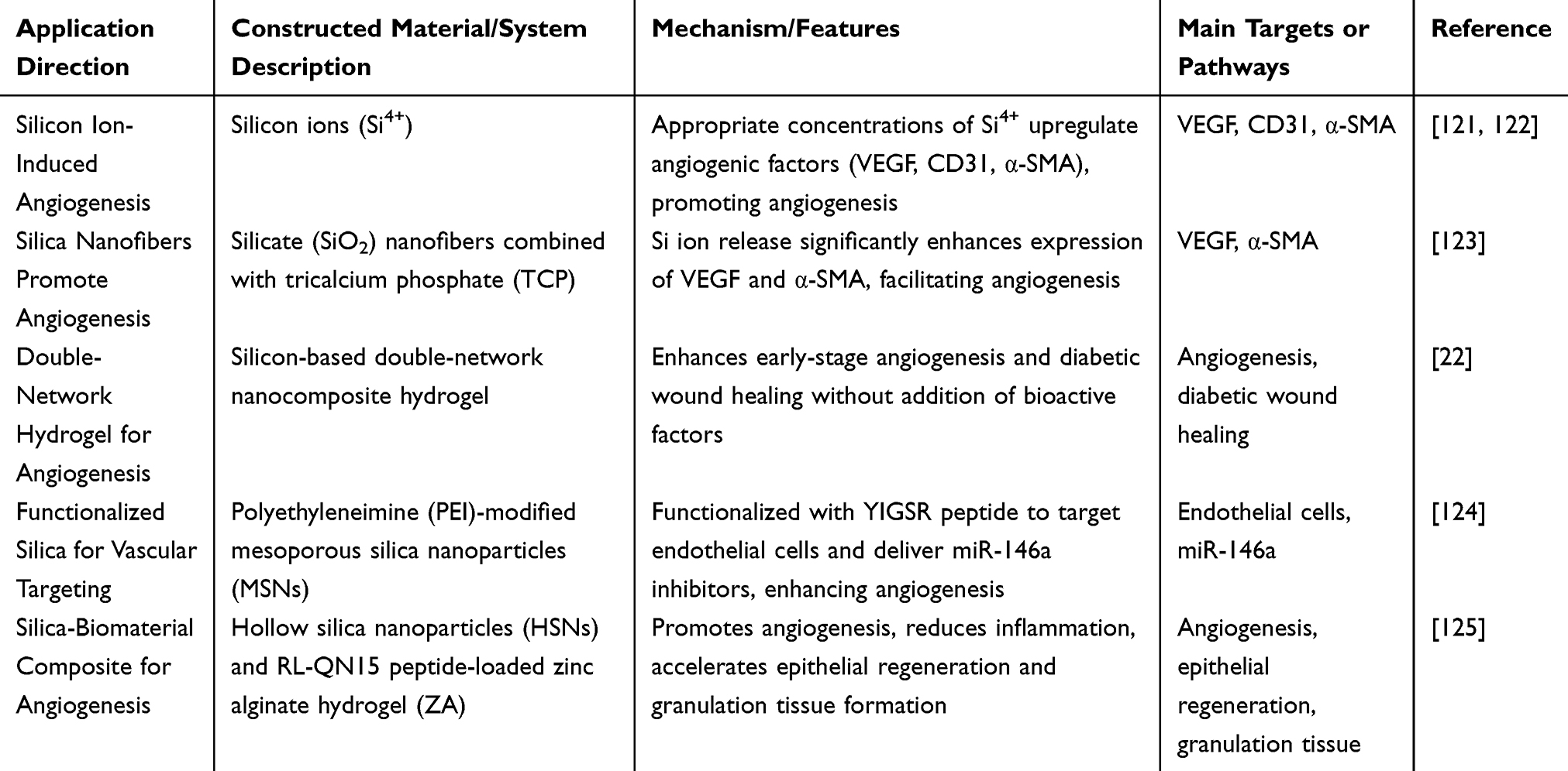 |
Table 4 The Role of Silicon-Based Materials in Promoting Angiogenesis |
Studies have shown that appropriate concentrations of silicon ions (Si4+) can significantly enhance angiogenesis by upregulating pro-angiogenic factors such as vascular endothelial growth factor (VEGF), CD31, and α-smooth muscle actin (α-SMA).121,122 Wang et al prepared short SiO2 nanofibers via electrospinning and blended them with varying proportions of tricalcium phosphate (TCP) to fabricate TCPx@SSF aerogel scaffolds. These scaffolds released Si4+, significantly upregulating VEGF and α-SMA expression, thereby promoting angiogenesis123 (Figure 9I). Li et al designed a dual-network silica-based nanocomposite hydrogel scaffold that, without the addition of any bioactive factors, significantly enhanced early angiogenesis and promoted diabetic wound healing.22
 |
Figure 9 (I) Schematic representation of the interactive and dynamic healing mechanisms of TCPx@SSF dressings during wound repair. Reprinted from Wang XY, Yuan Z, Shafiq M, et al. Composite Aerogel Scaffolds Containing Flexible Silica Nanofiber and Tricalcium Phosphate Enable Skin Regeneration. ACS Appl Mater Interfaces. 2024;16(20):25843–25855. Copyright © 2024 American Chemical Society.123 (II) Mesoporous silica nanoparticles (MSNs) modified with polyethyleneimine (PEI) and functionalized with the endothelial-recognition pentapeptide YIGSR enable precise delivery of miR-146a inhibitors, significantly boosting angiogenesis. Reprinted from Wang Y, Wu J, Feng J, et al. From Bone Remodeling to Wound Healing: an miR-146a-5p-Loaded Nanocarrier Targets Endothelial Cells to Promote Angiogenesis. ACS Appl Mater Interfaces. 2024;16(26):32992–33004. Copyright © 2024 American Chemical Society.124 |
Furthermore, the functionalized design of silicon-based materials is particularly notable Functionalized silicon-based materials can carry pro-angiogenic factors (eg, VEGF, FGF) or nucleic acids (eg, miRNA) for precise release. Wang et al functionalized mesoporous silica nanoparticles (MSNs) with polyethyleneimine (PEI) and modified their surface with a pentapeptide (YIGSR) capable of recognizing endothelial cells. This system delivered miR-146a inhibitors with high precision, significantly enhancing angiogenesis124 (Figure 9II). Combining silicon nanoparticles with other biomaterials can further enhance their pro-angiogenic properties. For example, hollow silica nanoparticles (HSNs) loaded with RL-QN15 peptide and incorporated into a zinc alginate (ZA) hydrogel formed HSN@RL-QN15/ZA hydrogels. These hydrogels effectively regulated angiogenesis, significantly reduced inflammation, and accelerated epithelial regeneration and granulation tissue formation, thereby promoting rapid healing of chronic wounds.125
In summary, silicon-based nanomaterials demonstrate great potential in promoting angiogenesis and accelerating wound healing through silicon ion release, loading of pro-angiogenic factors, and optimizing material microstructures. Their multifunctional design not only improves therapeutic efficiency but also provides new strategies for the comprehensive treatment of chronic wounds.
Promoting Cell Adhesion, Migration, and Proliferation
The proliferation phase of wound healing is characterized by cell proliferation and migration. During this phase, angiogenic factors actively promote new blood vessel formation, which further supports the proliferation and migration of fibroblasts. These fibroblasts accumulate and act at the wound site, producing new extracellular matrix (ECM) that eventually forms granulation tissue.54,126 A subset of fibroblasts differentiates into myofibroblasts, playing a crucial role in wound contraction.57 Additionally, keratinocyte proliferation and migration are essential for epidermal regeneration.127,128
Cell adhesion is a prerequisite for proliferation and migration, directly impacting wound healing efficiency. Studies have shown that functionalized silicon-based nanomaterials significantly enhance cell adhesion. For example, Zhang et al investigated the effects of glycine-aspartic acid (RGD)-functionalized mesoporous silica nanoparticles (MSNs-RGD) on stem cell adhesion and differentiation. Results indicated that when the total RGD density increased from 1.06 to 5.32 nmol/cm², cell adhesion and spreading significantly improved, suggesting that MSNs-RGD could serve as drug carriers to promote cell adhesion and proliferation129 (Figure 10I). Furthermore, Motealleh et al incorporated bifunctional nanomaterials into glass and polydimethylsiloxane surfaces, creating a hybrid nanocomposite material. This material significantly enhanced cell adhesion and proliferation while inhibiting bacterial biofilm formation, demonstrating excellent antibacterial properties and biocompatibility.130
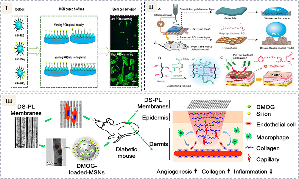 |
Figure 10 (I) Fabrication of mesoporous silica nanoparticle-based films with adjustable densities and clustering levels of arginine–glycine–aspartate peptides to investigate stem cell adhesion and differentiation. Reprinted from Zhang X, Karagöz Z, Swapnasrita S, et al. Development of Mesoporous Silica Nanoparticle-Based Films with Tunable Arginine–Glycine–Aspartate Peptide Global Density and Clustering Levels to Study Stem Cell Adhesion and Differentiation. ACS Appl Mater Interfaces. 2023;15(32):38171–38184. Creative Commons.129 (II) (A) Illustration of the preparation process for PCL/Gel-Pio nanofiber membranes. (B) Mechanistic depiction of the genipin-induced cross-linking reaction in the fiber membrane. (C) Mechanism by which the fiber membrane enhances diabetic wound healing. Reproduced from Gao ZJ, Wang Q, Yao Q, et al. Application of Electrospun Nanofiber Membrane in the Treatment of Diabetic Wounds. Pharmaceutics. 2022;14(1):6. Creative Commons.131 (III) A directional porous composite membrane (DS-PL) was developed using polylactic acid (PLLA) electrospun fibers incorporating mesoporous silica nanoparticles (DS) loaded with dimethyloxalylglycine (DMOG). The system facilitates controlled DMOG and silicon ion release, enhancing HUVEC proliferation, migration, and angiogenesis-related gene expression, which accelerates vascularization in diabetic wound environments. Reprinted from Acta Biomater. Volume 70. Ren XZ, Han Y, Wang J, et al. An aligned porous electrospun fibrous membrane with controlled drug delivery - An efficient strategy to accelerate diabetic wound healing with improved angiogenesis. 140–153, copyright 2018, with permission from Elsevier.132 |
In tissue engineering research, cell migration ability is a critical indicator of cell proliferation and its role in wound repair. Silicon-based nanomaterials have been shown to promote cell migration, thereby supporting wound healing. For instance, TA-SNP exhibited outstanding efficacy in treating full-thickness wounds in diabetic mice infected with E. coli. At a TA-SNP concentration of 100 μg/mL, the cell migration rate increased from 9.19% to 18.21%, further rising to 30.98% at a concentration of 500 μg/mL. This significant effect is attributed to the positive impact of silicon nanoparticles on cell proliferation, migration, and wound repair.117 Silicon-based materials also support cell proliferation through various pathways. Shie et al found that appropriate concentrations of Si ions significantly promoted the proliferation of osteoblast-like cells while inducing specific biological responses through bone-specific protein synthesis in MG63 cells. Cells maintained normal morphology and proliferated well under these conditions, further validating the excellent biocompatibility of silicon-based materials.133,134
The structure and micro-morphology of scaffolds are critical for cell adhesion, migration, and proliferation. Gao et al successfully developed silica-SFO-P scaffolds using extrusion-based 3D printing and validated their performance through direct cell biocompatibility tests. The porous structure of the scaffold significantly supported L929 cell adhesion and proliferation, with cells demonstrating robust growth on the scaffold surface and within its pores after three weeks of culture131 (Figure 10II). Ren et al fabricated a directional porous composite membrane (DS-PL), a polylactic acid (PLLA) directional electrospun fiber membrane containing mesoporous silica nanoparticles (DS). By loading the drug dimethyloxalylglycine (DMOG), controlled release of DMOG and Si ions was achieved. The combined directional porous structure and functional components synergistically promoted the proliferation, migration, and expression of angiogenesis-related genes in human umbilical vein endothelial cells (HUVECs), rapidly stimulating angiogenesis in diabetic wound beds, offering a novel therapeutic strategy for efficient diabetic wound healing132 (Figure 10III). In summary, silicon-based nanomaterials systematically enhance cell adhesion, migration, and proliferation by optimizing surface chemical properties, functional design, and regulating biological signals in the local microenvironment, thereby promoting wound healing (Table 5).
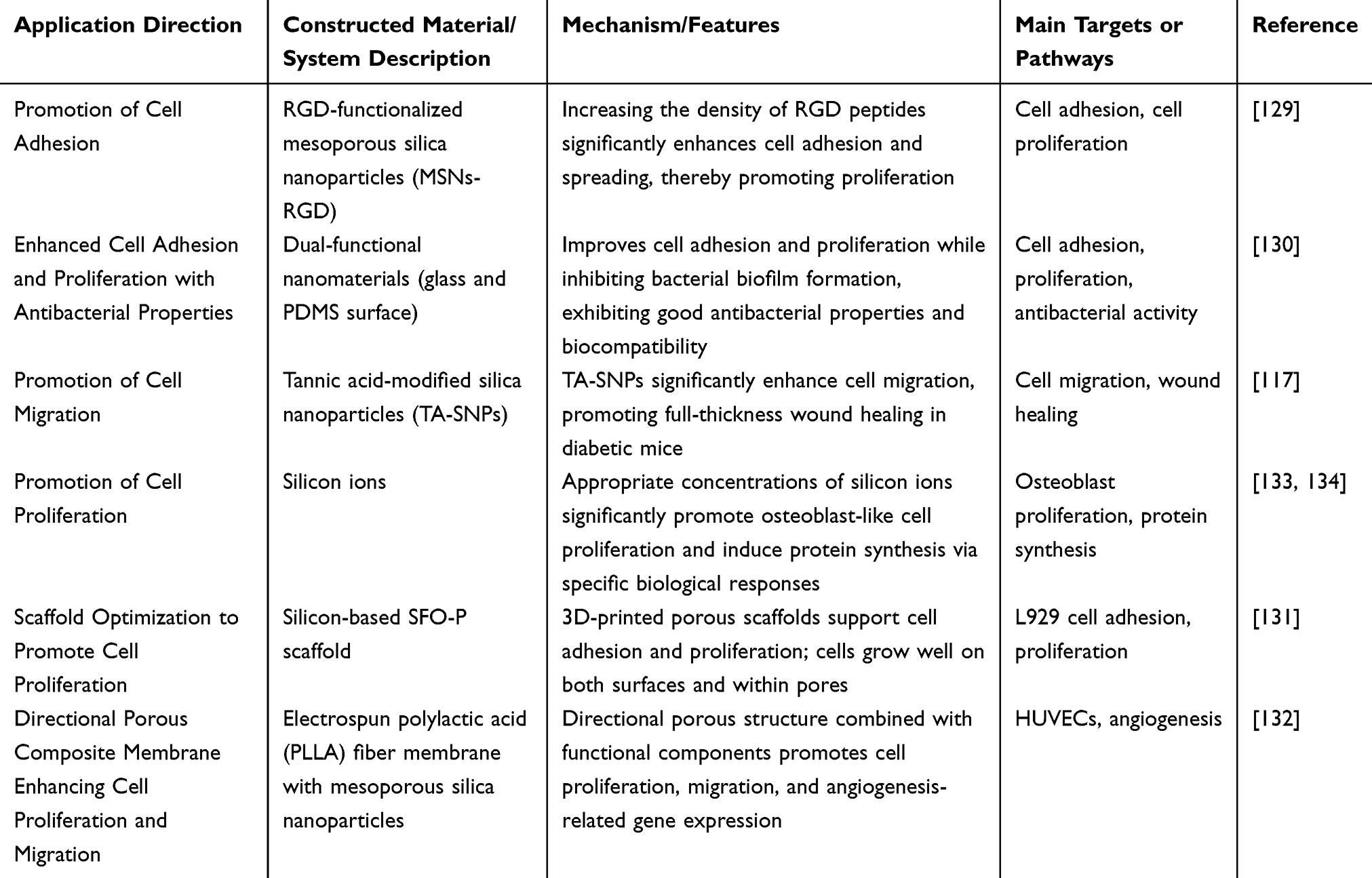 |
Table 5 The Role of Silicon-Based Materials in Promoting Cell Adhesion, Migration, and Proliferation |
Promoting Extracellular Matrix Deposition
The extracellular matrix (ECM) is an indispensable structural and functional unit in wound healing, primarily produced and organized by myofibroblasts. ECM serves as both a structural scaffold for cells and a reservoir of cytokines and growth factors, interacting with surrounding cells to regulate critical behaviors during their life cycle, including migration, growth, proliferation, differentiation, and morphogenesis.135,136 Major ECM components include interstitial collagen and elastic fibers, non-collagenous proteins (eg, fibronectin and laminin families), glycosaminoglycans (GAGs), and proteoglycans (PGs). These molecules collectively provide mechanical support to cells while regulating cellular behavior. Silicon, as a component of certain GAGs and PGs, participates in GAG synthesis. By binding to polysaccharide matrices, silicon acts as a biological crosslinker, enhancing the structure and elasticity of connective tissues and further promoting ECM deposition.137
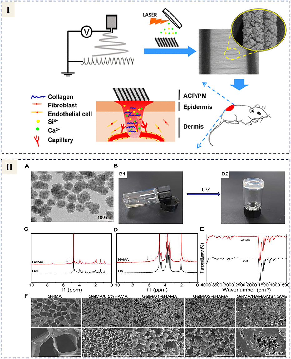 |
Figure 11 (I) Jiang et al designed an electrospun scaffold featuring uniformly distributed silicon-doped amorphous calcium phosphate nanoparticles (Si-ACP/PM, ~40 nm diameter). The scaffold released silicon ions gradually, enhancing collagen deposition and re-epithelialization in diabetic wound models. Reprinted from Jiang Y, Han Y, Wang J, et al. Space-Oriented Nanofibrous Scaffold with Silicon-Doped Amorphous Calcium Phosphate Nanocoating for Diabetic Wound Healing. ACS Appl Bio Mater. 2019;2(2):787–795. Copyright © 2019 American Chemical Society.138 (II) (A) TEM visualization of MSNs. (B) Photograph showing the UV cross-linked GelMA/HAMA. Hydrogel.flowing liquid (B1) before cross-linking and gradually changed into a solid phase after the UV curing (B2). (C) ¹H NMR analysis of Gel and GelMA. The distinctive double peaks (δ = 5.4 and 5.6 ppm), marked by the black arrows, were observed in GelMA. (D) ¹H NMR analysis of HA and HAMA. The distinctive double peaks (δ = 5.7 and 6.1 ppm), marked by the black arrows, were observed in HAMA. (E) FTIR spectra comparing Gel and GelMA. (F) SEM images of GelMA/HAMA hydrogels prepared with varying HAMA concentrations. Reprinted from Xue LY, Deng T, Guo R, et al. A Composite Hydrogel Containing Mesoporous Silica Nanoparticles Loaded With Extract for Improving Chronic Wound Healing. Front Bioeng Biotechnol. 2022;10:825339. Creative Commons.139 |
During the remodeling phase of wound healing, collagen deposition plays a pivotal role in enhancing tissue strength and supporting wound contraction. Studies have shown that silicon-based materials significantly promote collagen deposition, thereby accelerating wound repair. For example, Jiang et al designed an electrospun scaffold with uniformly distributed silicon-doped amorphous calcium phosphate nanoparticles (Si-ACP/PM) approximately 40 nm in size. This scaffold continuously released silicon ions, promoting collagen deposition and re-epithelialization in diabetic wound beds, effectively accelerating wound healing138 (Figure 11I). Through functionalization, silicon-based nanocomposites demonstrate multifunctionality in promoting ECM deposition and wound repair. For instance, Masson staining revealed that diabetic wounds treated with double-network silica-based nanocomposite hydrogel scaffolds exhibited excellent collagen deposition and tissue remodeling, along with effective vascular network repair.22 Additionally, Xue et al developed a hydrogel based on gelatin methacrylate (GelMA)/hyaluronic acid methacrylate (HAMA) and mesoporous silica nanoparticles (MSNs), with artemisia extract (AE) loaded for sustained release. This GelMA/HAMA/MSNs@AE hydrogel accelerated wound healing by promoting re-epithelialization and collagen deposition139 (Figure 11II). In summary, silicon-based materials offer significant advantages in promoting collagen deposition and improving chronic wound repair, providing innovative ideas for developing efficient wound repair materials (Table 6).
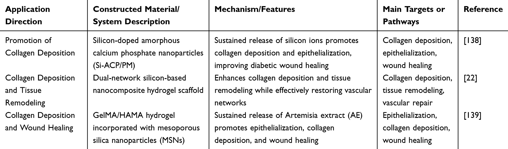 |
Table 6 The Role of Silicon-Based Materials in Extracellular Matrix Deposition |
Multimechanism Synergy
The pathological causes of refractory wounds are often complex and diverse. Thus, leveraging the overlapping and synergistic effects of two or more functions of silicon-based nanomaterials—including antibacterial properties, antioxidation and anti-inflammation, angiogenesis promotion, cell proliferation and migration promotion, and tissue remodeling promotion—can significantly accelerate the wound healing process. For instance, Xi et al developed biodegradable bioactive elastic multifunctional PPCP nanofiber scaffolds. The optimized assembly and combination of the multifunctional elastomer polylactic acid/polysiloxane citrate/curcumin endowed PPCP scaffolds with inherent multifunctionality. PPCP significantly promoted normal and infection-induced wound healing through infection prevention, reduction of proinflammatory factors, upregulation of CD31 and VEGF growth factors, stimulation of collagen deposition, and promotion of dermal and skin appendage formation.103
 |
Figure 12 (I) A multifunctional MN@Ag@MSN@CeO2 patch was developed to promote healing in infected diabetic wounds (DW) through its antibacterial, antioxidant, anti-inflammatory, and angiogenic properties. Reprinted from Yu D, Chen L, Yan T, et al. Enhancing Infected Diabetic Wound Healing through Multifunctional Nanocomposite-Loaded Microneedle Patch: inducing Multiple Regenerative Sites. Adv Healthc Mater. 2024;13(20):e2301985. © 2024 Wiley-VCH GmbH.140 (II) A conductive nanofibrous composite scaffold incorporating silicate-based bioceramic particles (Nagelschmidtite, NAGEL, Ca7P2Si2O16) was fabricated via co-electrospinning. Biological assessments confirmed that NAGEL particles activated epithelial-to-mesenchymal transition (EMT) and endothelial-to-mesenchymal transition (EndMT) pathways, both in vitro and in vivo. Reprinted from Acta Biomater. Volume 60. Lv F, Wang J, Xu P, et al. A conducive bioceramic/polymer composite biomaterial for diabetic wound healing. 128–143, copyright 2017, with permission from Elsevier.141 |
Yu et al designed soluble microneedle (MN) patches made of γ-PGA with multiple arrays loaded with core-shell-structured nanoparticles (NPs) called Ag@MSNs@CeO2. These MN tips induced the formation of multiple regeneration sites at different points, enabling antibacterial effects, reduced reactive oxygen species, macrophage niche regulation, enhanced angiogenesis, and promoted collagen deposition, thereby significantly accelerating the healing of infectious diabetic wounds (DW) (Figure 12I).140 Additionally, Lv et al successfully prepared a nanofiber composite scaffold (NAG-PL) containing partial silicate bioceramic particles using co-electrospinning technology. In-vivo and in-vitro studies showed that this scaffold significantly activated epithelial-mesenchymal transition (EMT) and endothelial-mesenchymal transition (EndMT) pathways, inducing angiogenesis, collagen deposition, and epithelial regeneration in diabetic mouse models while suppressing inflammation, thereby accelerating the repair of diabetic wounds (Figure 12II).141 Through the synergistic effects of multiple mechanisms, silicon-based nanomaterial systems demonstrate greater potential and application prospects in wound repair (Table 7).
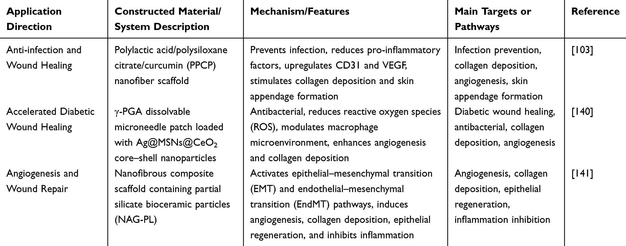 |
Table 7 Multimechanism Synergy of Silicon-Based Materials |
Clinical Trials of Silicon-Based Nanomaterials
Silicon-based nanosystems have demonstrated significant potential in wound treatment due to their excellent biocompatibility, tunable porosity, and multifunctional properties. Colloidal silica, as a material, has been used as a flow aid in tablet production for decades and is recognized as safe by the US Food and Drug Administration (FDA).142 Currently, several silicon-based nano-products have reached clinical or preclinical stages of application143 (Table 8).
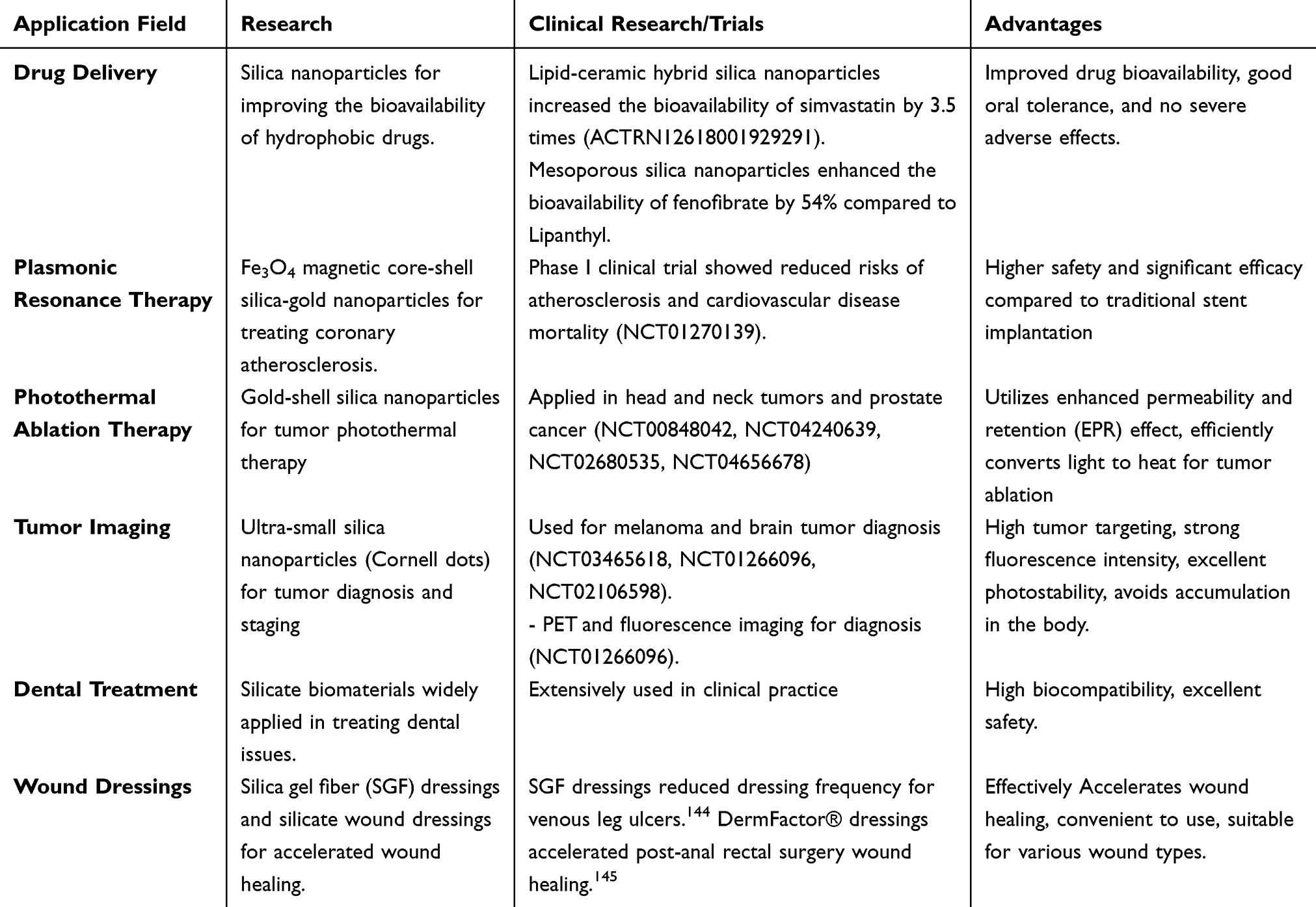 |
Table 8 A Summary of the Current Clinical Research and Applications of Silica-Based Nanomaterials |
To date, more than ten clinical trials on silica nanoparticles have highlighted their favorable safety profiles. Oral delivery systems exhibited good tolerance, with no severe adverse effects, and significantly improved the bioavailability of hydrophobic drugs. In a clinical trial involving 12 adults, lipid-ceramic hybrid silica nanoparticles were used to enhance the pharmacokinetics of simvastatin. Compared to a commercial simvastatin formulation (Sandoz), these nanoparticles improved bioavailability by 3.5 times (ACTRN12618001929291). Similarly, in another clinical study involving 12 healthy adults, mesoporous silica nanoparticles increased the bioavailability of fenofibrate by 54% compared to the commercial formulation (Lipanthyl), demonstrating their advantage in drug delivery and bioavailability enhancement.
In addition to drug delivery applications, silica nanoparticles have been explored in fields such as plasmonic therapy and thermal ablation treatment. For instance, in a Phase I clinical trial, Fe3O4 magnetic core-shell silica-gold nanoparticles (90–150 nm in diameter) were used in plasmonic therapy to significantly reduce coronary atherosclerosis (NCT01270139). Compared to traditional stent implantation, this treatment method reduced the risk of atherosclerosis and cardiovascular disease-related mortality with acceptable safety. In photothermal ablation therapy, gold-shell silica nanoparticles (eg, Aurolase and Auroshell) have been applied in clinical trials for malignant tumors of the head, neck, and prostate (NCT00848042, NCT04240639, NCT02680535, NCT04656678). These nanoparticles preferentially accumulate at tumor sites through enhanced permeability and retention (EPR) effects, converting near-infrared light into heat to achieve tumor ablation. In a pilot study involving 16 prostate cancer patients, gold-shell silica nanoparticles successfully performed photothermal ablation with fewer side effects compared to traditional local ablation therapies.
Ultrasmall silica nanoparticles (Cornell dots, 6–10 nm in diameter) have shown remarkable potential in tumor imaging. These nanoparticles have been utilized for the diagnosis and staging of melanoma and malignant brain tumors (NCT03465618, NCT01266096, NCT02106598). Due to their size being below the renal clearance threshold of 10 nm, Cornell dots can be excreted through the kidneys, avoiding the risk of accumulation in the body. Functionalized with RGDY peptides and fluorescent dye Cy5.5, Cornell dots exhibit enhanced tumor targeting and act as efficient imaging agents. In a first-in-human clinical trial, 124I-labeled Cornell dots were used for PET and fluorescence-guided tumor diagnosis and staging (NCT01266096). Results showed that these particles were stable, well-tolerated, with no significant side effects, and had a plasma half-life of 8.7 hours. Furthermore, Cornell dots have been employed to detect and locate sentinel lymph nodes in head and neck melanoma patients (NCT02106598), significantly improving biopsy accuracy. Compared to traditional radio-guided methods, they demonstrated higher sensitivity, enhanced fluorescence intensity, and improved photostability.
Currently, commercial products derived from silicate biomaterials are widely used in clinical settings to address dental-related issues.144–146 Silica gel fiber (SGF) dressings, a bioabsorbable inorganic silica gel fiber patch, consist of a network of hydroxyethoxy-siloxane polymers with the molecular structure H[Si8O12O(OH) x(OC2H5)6-x]nOH. In a study involving 130 patients with leg venous ulcers, participants were randomly divided into two groups to receive four weeks of local care with either SGF dressings or alginate dressings, followed by follow-up until the 8th week. Results showed that SGF dressings reduced dressing change frequency while maintaining equivalent therapeutic efficacy.147
In a randomized study, the effects of silicate wound dressings (DermFactor®) were evaluated in post-anorectal surgery wound treatment. A total of 328 patients were randomly assigned to a control group (routine dressing changes) and an observation group (routine dressing changes + DermFactor®). The observation group exhibited significantly shorter average wound healing times (mixed hemorrhoids: 19.04 days; anal fistula: 23.72 days; anal fissure: 21.14 days) compared to the control group (23.25 days, 27.76 days, and 24.32 days, respectively), along with a higher effective rate (80.4% vs 70.4%). These findings indicate that DermFactor® dressings effectively accelerate wound healing, making them a valuable adjunctive tool for post-anorectal surgery treatment.148
Although some silicon-based nano-products have reached clinical trial stages, challenges such as long-term biosafety, production feasibility, and individualized treatment remain to be addressed. With the integration of multidisciplinary technologies, silicon-based nanosystems are expected to become key technologies in wound treatment, driving the development of personalized and efficient therapies.
Prospects and Challenges
Silicon-based nanosystems hold immense promise in wound treatment, with development directions including personalized and intelligent therapies, combination therapy strategies, novel material development, and multicenter clinical studies. In personalized therapy, intelligent dressings integrated with sensor technology can monitor wound environments in real time and dynamically release drugs as needed, achieving efficient and precise treatment. Combined with wireless technologies, these systems support remote management. For instance, infected sites and bacterial biofilms exhibit microenvironments distinct from normal tissues, such as low PH, elevated local temperatures, and altered redox potentials, which can be exploited for nanosystem targeting.149 Endogenous stimulus-responsive nanosystems successfully deliver lower doses of antimicrobials to infection sites, reducing systemic distribution and minimizing side effects on normal organs and tissues.150
In combination therapies, silicon-based nanomaterials can carry photosensitizers, antimicrobials, and tissue regeneration promoters, synergizing with photothermal or photodynamic therapy to effectively control infections and accelerate wound healing. For example, polydopamine and curcumin exhibit excellent near-infrared photothermal and anticancer properties. Researchers have synthesized multifunctional nanofiber matrices through surface functionalization, achieving photothermal chemotherapy for skin tumors and wound healing induced by infections.93
The size of nanoparticles plays a critical role in their in-vivo transport due to physiological size thresholds and size-dependent biological effects. It has been widely observed that nanoparticle size significantly influences their cellular uptake efficiency and mechanisms. Additionally, protein corona adsorption varies significantly with particle size.151 Therefore, optimizing the physicochemical properties of nanoparticles, such as pore size, can enhance drug delivery and therapeutic performance.
Multicenter clinical studies validate the efficacy and safety of silicon-based nanosystems through large-scale trials, promoting their standardization and commercialization. These efforts lay a solid foundation for the broad application of silicon-based nanosystems in complex wound treatment, facilitating their transition from laboratory research to clinical practice.
However, clinical translation of silicon-based nanosystems for wound treatment faces multiple challenges, including safety, biocompatibility, applicability to complex wound models, multifunctional integration, and scalable production. Although their preliminary safety has been validated, long-term toxicity and potential impacts of chronic exposure require further investigation, especially under high-dose or repeated-use conditions. The diverse characteristics of complex wounds, such as chronic inflammation and impaired angiogenesis in diabetic ulcers or antimicrobial needs in burns, necessitate tailored functional strategies for specific pathologies, validated through clinically relevant 3D wound models or organ-on-chip technologies.
Materials with single functions are insufficient to meet the diverse needs of complex wounds, making multifunctional integration a research trend. However, verifying the combined efficacy of such systems remains challenging. Additionally, the complex fabrication processes of silicon-based nanosystems lead to significant batch-to-batch variability, affecting quality stability and clinical efficacy consistency. To achieve scalable production, industrial technologies must be developed, and stringent quality control standards established to ensure reproducibility and material performance consistency. Addressing these issues will pave the way for clinical application of silicon-based nanosystems.
Conclusion
Silicon-based nanomaterials, with their unique physicochemical properties and biological advantages, exhibit tremendous potential in wound healing applications. These materials feature high specific surface area, excellent mechanical and chemical stability, low toxicity, and superior biocompatibility, enabling precise drug delivery. In wound healing, silicon-based nanosystems synergistically enhance repair through multiple mechanisms, including antimicrobial, anti-inflammatory, and antioxidant activities, as well as promoting angiogenesis and cell proliferation.
Although studies have demonstrated their good biocompatibility, only a few silicon-based nanoproducts have entered clinical trials. Challenges such as long-term biosafety, production feasibility, and personalized treatment strategies remain unresolved. Future research on silicon-based nanosystems should focus on personalized and intelligent therapies, combination treatment strategies, novel material development, and multicenter clinical studies. These efforts aim to provide more efficient, safe, and comprehensive solutions for wound treatment, ultimately improving patient experiences and clinical outcomes.
Data Sharing Statement
Data sharing is not applicable to this article as no new data were created or analyzed in this study.
Acknowledgments
The authors would like to thank Qinxin Liu for her assistance during the revision stage of the manuscript.
Funding
This study was supported by grants from Hubei Provincial Natural Science Foundation of China (No. 2023AFB825,2023AFB216) and Tongji Hospital, Tongji Medical College, Huazhong University of Science and Technology (No. 2023A15).
Disclosure
The authors declare that the research was conducted without any commercial or financial relationships that could be construed as a potential conflict of interest.
References
1. Dabrowska AK, Spano F, Derler S, et al. The relationship between skin function, barrier properties, and body-dependent factors. Skin Res Technol. 2018;24(2):165–174. doi:10.1111/srt.12424
2. Nethi SK, Das S, Patra CR, et al. Recent advances in inorganic nanomaterials for wound-healing applications. Biomater Sci. 2019;7(7):2652–2674. doi:10.1039/C9BM00423H
3. Kuehn BM. Chronic wound care guidelines issued. JAMA. 2007;297(9):938–939.
4. Spampinato SF, Caruso GI, De Pasquale R, et al. The Treatment of Impaired Wound Healing in Diabetes: looking among Old Drugs. Pharmaceuticals. 2020;13(4):60. doi:10.3390/ph13040060
5. Wilkinson HN, Hardman MJ. Wound healing: cellular mechanisms and pathological outcomes. Open Biol. 2020;10(9):200223. doi:10.1098/rsob.200223
6. Hoversten KP, Kiemele LJ, Stolp AM, et al. Prevention, Diagnosis, and Management of Chronic Wounds in Older Adults. Mayo Clin Proc. 2020;95(9):2021–2034. doi:10.1016/j.mayocp.2019.10.014
7. Mu R, Campos de Souza S, Liao Z, et al. Reprograming the immune niche for skin tissue regeneration - From cellular mechanisms to biomaterials applications. Adv Drug Deliv Rev. 2022;185:114298. doi:10.1016/j.addr.2022.114298
8. Sen CK, Gordillo GM, Roy S, et al. Human skin wounds: a major and snowballing threat to public health and the economy. Wound Repair Regen. 2009;17(6):763–771. doi:10.1111/j.1524-475X.2009.00543.x
9. Sen CK. Human Wound and Its Burden: updated 2022 Compendium of Estimates. Adv Wound Care. 2023;12(12):657–670. doi:10.1089/wound.2023.0150
10. Tang F, Li L, Chen D. Mesoporous silica nanoparticles: synthesis, biocompatibility and drug delivery. Adv Mater. 2012;24(12):1504–1534. doi:10.1002/adma.201104763
11. Chen L, Zhou X, He C. Mesoporous silica nanoparticles for tissue-engineering applications. Wiley Interdiscip Rev Nanomed Nanobiotechnol. 2019;11(6):e1573. doi:10.1002/wnan.1573
12. Gisbert-Garzaran M, Lozano D, Vallet-Regi M. Mesoporous Silica Nanoparticles for Targeting Subcellular Organelles. Int J Mol Sci. 2020;21(24):9696. doi:10.3390/ijms21249696
13. Tang L, Cheng J. Nonporous Silica Nanoparticles for Nanomedicine Application. Nano Today. 2013;8(3):290–312. doi:10.1016/j.nantod.2013.04.007
14. Li Z, Zhang Y, Feng N. Mesoporous silica nanoparticles: synthesis, classification, drug loading, pharmacokinetics, biocompatibility, and application in drug delivery. Expert Opin Drug Deliv. 2019;16(3):219–237. doi:10.1080/17425247.2019.1575806
15. Abu-Dief AM, Alsehli M, Al-Enizi A, et al. Recent Advances in Mesoporous Silica Nanoparticles for Targeted Drug Delivery Applications. Curr Drug Deliv. 2022;19(4):436–450. doi:10.2174/1567201818666210708123007
16. Xu B, Li S, Shi R, et al. Multifunctional mesoporous silica nanoparticles for biomedical applications. Signal Transduct Target Ther. 2023;8(1):435. doi:10.1038/s41392-023-01654-7
17. Gisbert-Garzaran M, Vallet-Regi M. Influence of the Surface Functionalization on the Fate and Performance of Mesoporous Silica Nanoparticles. Nanomaterials. 2020;10(5). doi:10.3390/nano10050916
18. Yan T, He J, Liu R, et al. Chitosan capped pH-responsive hollow mesoporous silica nanoparticles for targeted chemo-photo combination therapy. Carbohydr Polym. 2020;231:115706. doi:10.1016/j.carbpol.2019.115706
19. Nair A, Chandrashekhar H R, Day CM, et al. Polymeric functionalization of mesoporous silica nanoparticles: biomedical insights. Int J Pharm. 2024;660:124314. doi:10.1016/j.ijpharm.2024.124314
20. Zhang Y, Chang M, Bao F, et al. Multifunctional Zn doped hollow mesoporous silica/polycaprolactone electrospun membranes with enhanced hair follicle regeneration and antibacterial activity for wound healing. Nanoscale. 2019;11(13):6315–6333. doi:10.1039/C8NR09818B
21. Hooshmand S, Mollazadeh S, Akrami N, et al. Mesoporous Silica Nanoparticles and Mesoporous Bioactive Glasses for Wound Management: from Skin Regeneration to Cancer Therapy. Materials. 2021;14(12):3337. doi:10.3390/ma14123337
22. Li Y, Xu T, Tu Z, et al. Bioactive antibacterial silica-based nanocomposites hydrogel scaffolds with high angiogenesis for promoting diabetic wound healing and skin repair. Theranostics. 2020;10(11):4929–4943. doi:10.7150/thno.41839
23. Mirzahosseinipour M, Khorsandi K, Hosseinzadeh R, et al. Antimicrobial photodynamic and wound healing activity of curcumin encapsulated in silica nanoparticles. Photodiagnosis Photodyn Ther. 2020;29:101639. doi:10.1016/j.pdpdt.2019.101639
24. Wang M, Huang X, Zheng H, et al. Nanomaterials applied in wound healing: mechanisms, limitations and perspectives. J Control Release. 2021;337:236–247. doi:10.1016/j.jconrel.2021.07.017
25. Berthet M, Gauthier Y, Lacroix C, et al. Nanoparticle-Based Dressing: the Future of Wound Treatment? Trends Biotechnol. 2017;35(8):770–784. doi:10.1016/j.tibtech.2017.05.005
26. Mirrezaei N, Yazdian-Robati R, Oroojalian F, et al. Recent Developments in Nano-Drug Delivery Systems Loaded by Phytochemicals for Wound Healing. Mini Rev Med Chem. 2020;20(18):1867–1878. doi:10.2174/1389557520666200807133022
27. Bernal-Chavez S, Nava-Arzaluz MG, Quiroz-Segoviano RIY, et al. Nanocarrier-based systems for wound healing. Drug Dev Ind Pharm. 2019;45(9):1389–1402. doi:10.1080/03639045.2019.1620270
28. Mihai MM, Dima MB, Dima B, et al. Nanomaterials for Wound Healing and Infection Control. Materials. 2019;12(13):2176. doi:10.3390/ma12132176
29. Pormohammad A, Monych NK, Ghosh S, et al. Nanomaterials in Wound Healing and Infection Control. Antibiotics. 2021;10(5):473. doi:10.3390/antibiotics10050473
30. Parani M, Lokhande G, Singh A, et al. Engineered Nanomaterials for Infection Control and Healing Acute and Chronic Wounds. ACS Appl Mater Interfaces. 2016;8(16):10049–10069. doi:10.1021/acsami.6b00291
31. Rahman MA, Abul Barkat H, Harwansh RK, et al. Carbon-based Nanomaterials: carbon Nanotubes, Graphene, and Fullerenes for the Control of Burn Infections and Wound Healing. Curr Pharm Biotechnol. 2022;23(12):1483–1496. doi:10.2174/1389201023666220309152340
32. Eming SA, Martin P, Tomic-Canic M. Wound repair and regeneration: mechanisms, signaling, and translation. Sci Transl Med. 2014;6(265):265sr6. doi:10.1126/scitranslmed.3009337
33. Gurtner GC, Werner S, Barrandon Y, et al. Wound repair and regeneration. Nature. 2008;453(7193):314–321. doi:10.1038/nature07039
34. Sorg H, Tilkorn DJ, Hager S, et al. Skin Wound Healing: an Update on the Current Knowledge and Concepts. Eur Surg Res. 2017;58(1–2):81–94. doi:10.1159/000454919
35. Whitney JD. Overview: acute and chronic wound. Nurs Clin North Am. 2005;40(2):191–205. doi:10.1016/j.cnur.2004.09.002
36. Demidova-Rice TN, Hamblin MR, Herman IM. Acute and impaired wound healing: pathophysiology and current methods for drug delivery, part 1: normal and chronic wounds: biology, causes, and approaches to care. Adv Skin Wound Care. 2012;25(7):304–314. doi:10.1097/01.ASW.0000416006.55218.d0
37. Falanga V, Isseroff RR, Soulika AM, et al. Chronic wounds. Nat Rev Dis Primers. 2022;8(1):50. doi:10.1038/s41572-022-00377-3
38. Jarbrink K, Ni G, Sönnergren H, et al. Prevalence and incidence of chronic wounds and related complications: a protocol for a systematic review. Syst Rev. 2016;5(1):152. doi:10.1186/s13643-016-0329-y
39. Jones RE, Foster DS, Longaker MT. Management of Chronic Wounds-2018. JAMA. 2018;320(14):1481–1482. doi:10.1001/jama.2018.12426
40. Singer AJ, Clark RA. Cutaneous wound healing. N Engl J Med. 1999;341(10):738–746. doi:10.1056/NEJM199909023411006
41. Rodrigues M, Kosaric N, Bonham CA, et al. Wound Healing: a Cellular Perspective. Physiol Rev. 2019;99(1):665–706. doi:10.1152/physrev.00067.2017
42. Talbott HE, Mascharak S, Griffin M, et al. Wound healing, fibroblast heterogeneity, and fibrosis. Cell Stem Cell. 2022;29(8):1161–1180. doi:10.1016/j.stem.2022.07.006
43. Peña OA, Martin P. Cellular and molecular mechanisms of skin wound healing. Nat Rev Mol Cell Biol. 2024;25(8):599–616. doi:10.1038/s41580-024-00715-1
44. Cioce A, Cavani A, Cattani C, Scopelliti F. Role of the Skin Immune System in Wound Healing. Cells. 2024; 13(7):624. Creative Commons
45. Berk BC, Alexander RW, Brock TA, et al. Vasoconstriction: a new activity for platelet-derived growth factor. Science. 1986;232(4746):87–90. doi:10.1126/science.3485309
46. Pool JG. Normal hemostatic mechanisms: a review. Am J Med Technol. 1977;43(8):776–780.
47. Pierce GF, Mustoe TA, Altrock BW, et al. Role of platelet-derived growth factor in wound healing. J Cell Biochem. 1991;45(4):319–326. doi:10.1002/jcb.240450403
48. Furie B, Furie BC. Mechanisms of thrombus formation. N Engl J Med. 2008;359(9):938–949. doi:10.1056/NEJMra0801082
49. Clark RA. Fibrin and wound healing. Ann N Y Acad Sci. 2001;936(1):355–367. doi:10.1111/j.1749-6632.2001.tb03522.x
50. Braund R, Hook S, Medlicott NJ. The role of topical growth factors in chronic wounds. Curr Drug Deliv. 2007;4(3):195–204. doi:10.2174/156720107781023857
51. Gainza G, Villullas S, Pedraz JL, et al. Advances in drug delivery systems (DDSs) to release growth factors for wound healing and skin regeneration. Nanomedicine. 2015;11(6):1551–1573. doi:10.1016/j.nano.2015.03.002
52. Kiritsy CP, Lynch AB, Lynch SE. Role of growth factors in cutaneous wound healing: a review. Crit Rev Oral Biol Med. 1993;4(5):729–760. doi:10.1177/10454411930040050401
53. Eming SA, Krieg T, Davidson JM. Inflammation in wound repair: molecular and cellular mechanisms. J Invest Dermatol. 2007;127(3):514–525. doi:10.1038/sj.jid.5700701
54. Velnar T, Bailey T, Smrkolj V. The wound healing process: an overview of the cellular and molecular mechanisms. J Int Med Res. 2009;37(5):1528–1542. doi:10.1177/147323000903700531
55. Malinda KM, Sidhu GS, Banaudha KK, et al. Thymosin alpha 1 stimulates endothelial cell migration, angiogenesis, and wound healing. J Immunol. 1998;160(2):1001–1006. doi:10.4049/jimmunol.160.2.1001
56. Tettamanti G, Grimaldi A, Rinaldi L, et al. The multifunctional role of fibroblasts during wound healing in Hirudo medicinalis (Annelida, Hirudinea). Biol Cell. 2004;96(6):443–455. doi:10.1016/j.biolcel.2004.04.008
57. Li B, Wang JH. Fibroblasts and myofibroblasts in wound healing: force generation and measurement. J Tissue Viability. 2011;20(4):108–120. doi:10.1016/j.jtv.2009.11.004
58. Montesinos MC, Gadangi P, Longaker M, et al. Wound healing is accelerated by agonists of adenosine A2 (G alpha s-linked) receptors. J Exp Med. 1997;186(9):1615–1620. doi:10.1084/jem.186.9.1615
59. Gill SE, Parks WC. Metalloproteinases and their inhibitors: regulators of wound healing. Int J Biochem Cell Biol. 2008;40(6–7):1334–1347. doi:10.1016/j.biocel.2007.10.024
60. Ehrlich HP, Hunt TK. Collagen Organization Critical Role in Wound Contraction. Adv Wound Care. 2012;1(1):3–9. doi:10.1089/wound.2011.0311
61. Mascharak S, Talbott HE, Januszyk M, et al. Multi-omic analysis reveals divergent molecular events in scarring and regenerative wound healing. Cell Stem Cell. 2022;29(2):315–327e6. doi:10.1016/j.stem.2021.12.011
62. Broughton II G, Janis JE, C.e A. Wound healing: an overview. Plast Reconstr Surg. 2006;117(7 Suppl):1e–S–32e–S. doi:10.1097/01.prs.0000222562.60260.f9
63. Tomic-Canic M, Burgess JL, O’Neill KE, et al. Skin Microbiota and its Interplay with Wound Healing. Am J Clin Dermatol. 2020;21(Suppl 1):36–43. doi:10.1007/s40257-020-00536-w
64. Matoori S, Veves A, Mooney DJ. Advanced bandages for diabetic wound healing. Sci Transl Med. 2021;13(585). doi:10.1126/scitranslmed.abe4839
65. Huang YZ, Gou M, Da L-C, et al. Mesenchymal Stem Cells for Chronic Wound Healing: current Status of Preclinical and Clinical Studies. Tissue Eng Part B Rev. 2020;26(6):555–570. doi:10.1089/ten.teb.2019.0351
66. Nunan R, Harding KG, Martin P. Clinical challenges of chronic wounds: searching for an optimal animal model to recapitulate their complexity. Dis Model Mech. 2014;7(11):1205–1213. doi:10.1242/dmm.016782
67. da Silva LP, Reis RL, Correlo VM, et al. Hydrogel-Based Strategies to Advance Therapies for Chronic Skin Wounds. Annu Rev Biomed Eng. 2019;21(1):145–169. doi:10.1146/annurev-bioeng-060418-052422
68. Vestby LK, Grønseth T, Simm R, et al. Bacterial Biofilm and its Role in the Pathogenesis of Disease. Antibiotics. 2020;9(2):59. doi:10.3390/antibiotics9020059
69. Bergan JJ, Schmid-Schönbein GW, Smith PDC, et al. Chronic venous disease. N Engl J Med. 2006;355(5):488–498. doi:10.1056/NEJMra055289
70. Seraphim PM, Leal EC, Moura J, et al. Lack of lymphocytes impairs macrophage polarization and angiogenesis in diabetic wound healing. Life Sci. 2020;254:117813. doi:10.1016/j.lfs.2020.117813
71. Naghibi M, Smith RP, Baltch AL, et al. The effect of diabetes mellitus on chemotactic and bactericidal activity of human polymorphonuclear leukocytes. Diabet Res Clin Pract. 1987;4(1):27–35. doi:10.1016/S0168-8227(87)80030-X
72. Wang Y, Wang Z, Dong Y. Collagen-Based Biomaterials for Tissue Engineering. ACS Biomater Sci Eng. 2023;9(3):1132–1150. doi:10.1021/acsbiomaterials.2c00730
73. Wynn TA, Vannella KM. Macrophages in Tissue Repair, Regeneration, and Fibrosis. IMMUNITY. 2016;44(3):450–462. doi:10.1016/j.immuni.2016.02.015
74. Barrientos S, Stojadinovic O, Golinko MS, et al. Growth factors and cytokines in wound healing. Wound Repair Regen. 2008;16(5):585–601. doi:10.1111/j.1524-475X.2008.00410.x
75. Ladwig GP, ROBSON MC, LIU R, et al. Ratios of activated matrix metalloproteinase-9 to tissue inhibitor of matrix metalloproteinase-1 in wound fluids are inversely correlated with healing of pressure ulcers. Wound Repair Regen. 2002;10(1):26–37. doi:10.1046/j.1524-475X.2002.10903.x
76. Laverdet B, Danigo A, Girard D, et al. Skin innervation: important roles during normal and pathological cutaneous repair. Histol Histopathol. 2015;30(8):875–892. doi:10.14670/HH-11-610
77. Mebert AM, Baglole CJ, Desimone MF, et al. Nanoengineered silica: properties, applications and toxicity. Food Chem Toxicol. 2017;109(Pt 1):753–770. doi:10.1016/j.fct.2017.05.054
78. Mochizuki C, Nakamura J, Nakamura M. Development of Non-Porous Silica Nanoparticles towards Cancer Photo-Theranostics. Biomedicines. 2021;9(1):73. doi:10.3390/biomedicines9010073
79. Additives EPOF, Younes M, Aggett P, et al. Re-evaluation of silicon dioxide (E 551) as a food additive. EFSA J. 2018;16(1):e05088. doi:10.2903/j.efsa.2018.5088
80. Okoturo-Evans O, Dybowska A, Valsami-Jones E, et al. Elucidation of toxicity pathways in lung epithelial cells induced by silicon dioxide nanoparticles. PLoS One. 2013;8(9):e72363. doi:10.1371/journal.pone.0072363
81. Arriagada F, Nonell S, Morales J. Silica-based nanosystems for therapeutic applications in the skin. Nanomedicine. 2019;14(16):2243–2267. doi:10.2217/nnm-2019-0052
82. Qian KK, Bogner RH. Application of mesoporous silicon dioxide and silicate in oral amorphous drug delivery systems. J Pharm Sci. 2012;101(2):444–463. doi:10.1002/jps.22779
83. Ding R, Li Y, Yu Y, et al. Prospects and hazards of silica nanoparticles: biological impacts and implicated mechanisms. Biotechnol Adv. 2023;69:108277. doi:10.1016/j.biotechadv.2023.108277
84. Zhou Y, Quan G, Wu Q, et al. Mesoporous silica nanoparticles for drug and gene delivery. Acta Pharm Sin B. 2018;8(2):165–177. doi:10.1016/j.apsb.2018.01.007
85. Bagheri E, Ansari L, Abnous K, et al. Silica based hybrid materials for drug delivery and bioimaging. J Control Release. 2018;277:57–76. doi:10.1016/j.jconrel.2018.03.014
86. Zhu J, Niu Y, Li Y, et al. Stimuli-responsive delivery vehicles based on mesoporous silica nanoparticles: recent advances and challenges. J Mater Chem B. 2017;5(7):1339–1352. doi:10.1039/C6TB03066A
87. Yang Y, Zhang M, Song H, et al. Silica-Based Nanoparticles for Biomedical Applications: from Nanocarriers to Biomodulators. Acc Chem Res. 2020;53(8):1545–1556. doi:10.1021/acs.accounts.0c00280
88. Zhang Y, Yu Y, Yang Y, et al. Engineered Silica Nanoparticles for Nucleic Acid Delivery. Small Methods. 2024;8(3):e2300812. doi:10.1002/smtd.202300812
89. Song H, Ahmad Nor Y, Yu M, et al. Silica Nanopollens Enhance Adhesion for Long-Term Bacterial Inhibition. J Am Chem Soc. 2016;138(20):6455–6462. doi:10.1021/jacs.6b00243
90. Song H, Yu M, Lu Y, et al. Plasmid DNA Delivery: nanotopography Matters. J Am Chem Soc. 2017;139(50):18247–18254. doi:10.1021/jacs.7b08974
91. Fu JY, Gu Z, Liu Y, et al. Bottom-up self-assembly of heterotrimeric nanoparticles and their secondary Janus generations. Chem Sci. 2019;10(44):10388–10394. doi:10.1039/C9SC02961C
92. Singh P, Srivastava S, Singh SK. Nanosilica: recent Progress in Synthesis, Functionalization, Biocompatibility, and Biomedical Applications. ACS Biomater Sci Eng. 2019;5(10):4882–4898. doi:10.1021/acsbiomaterials.9b00464
93. Xi Y, Ge J, Wang M, et al. Bioactive Anti-inflammatory, Antibacterial, Antioxidative Silicon-Based Nanofibrous Dressing Enables Cutaneous Tumor Photothermo-Chemo Therapy and Infection-Induced Wound Healing. ACS Nano. 2020;14(3):2904–2916. doi:10.1021/acsnano.9b07173
94. Yao Y, Feng J, Ao N, et al. Natural agents derived Pickering emulsion enabled by silica nanoparticles with enhanced antibacterial activity against drug-resistant bacteria. J Colloid Interface Sci. 2025;678(Pt B):1158–1168. doi:10.1016/j.jcis.2024.09.066
95. Jahromi MAM, Sahandi Zangabad P, Moosavi Basri SM, et al. Nanomedicine and advanced technologies for burns: preventing infection and facilitating wound healing. Adv. Drug Delivery Rev. 2018;123:33–64. doi:10.1016/j.addr.2017.08.001
96. Roy R, Tiwari M, Donelli G, et al. Strategies for combating bacterial biofilms: a focus on anti-biofilm agents and their mechanisms of action. Virulence. 2018;9(1):522–554. doi:10.1080/21505594.2017.1313372
97. Wang Y, Yang Y, Shi Y, et al. Antibiotic-Free Antibacterial Strategies Enabled by Nanomaterials: progress and Perspectives. Adv Mater. 2020;32(18):e1904106. doi:10.1002/adma.201904106
98. Ni C, Zhong Y, Wu W, et al. Co-Delivery of Nano-Silver and Vancomycin via Silica Nanopollens for Enhanced Antibacterial Functions. Antibiotics. 2022;11(5):685. doi:10.3390/antibiotics11050685
99. Cao CY, Ge W, Yin J, et al. Mesoporous Silica Supported Silver-Bismuth Nanoparticles as Photothermal Agents for Skin Infection Synergistic Antibacterial Therapy. Small. 2020;16(24). doi:10.1002/smll.202000436.
100. Rasool N, Srivastava R, Singh Y. Cationized silica ceria nanocomposites to target biofilms in chronic wounds. Biomater Adv. 2022;138:212939. doi:10.1016/j.bioadv.2022.212939
101. Huang HJ, Chang H-W, Lin Y-W, et al. Silicon-Based Ag Dendritic Nanoforests for Light-Assisted Bacterial Inhibition. Nanomaterials. 2020;10(11):2244. doi:10.3390/nano10112244
102. Khan HU, Nasir F, Maheen S, et al. Antibacterial and Wound-Healing Activities of Statistically Optimized Nitrofurazone- and Lidocaine-Loaded Silica Microspheres by the Box-Behnken Design. Molecules. 2022;27(8):2532.
103. Hashemikia S, Farhangpazhouh F, Parsa M, et al. Fabrication of ciprofloxacin-loaded chitosan/polyethylene oxide/silica nanofibers for wound dressing application: in vitro and in vivo evaluations. Int J Pharm. 2021;597:120313. doi:10.1016/j.ijpharm.2021.120313
104. Gwon K, Park J-D, Lee S, et al. Fabrication of silicon-based nickel nanoflower-encapsulated gelatin microspheres as an active antimicrobial carrier. Int J Biol Macromol. 2024;264:130617. doi:10.1016/j.ijbiomac.2024.130617
105. McCarty SM, Percival SL. Proteases and Delayed Wound Healing. Adv Wound Care. 2013;2(8):438–447. doi:10.1089/wound.2012.0370
106. James GA, Swogger E, Wolcott R, et al. Biofilms in chronic wounds. Wound Repair Regen. 2008;16(1):37–44. doi:10.1111/j.1524-475X.2007.00321.x
107. Liu M, Guinart A, Granados A, et al. Coated Cotton Fabrics with Antibacterial and Anti-Inflammatory Silica Nanoparticles for Improving Wound Healing. ACS Appl Mater Interfaces. 2024;16(12):14595–14604. doi:10.1021/acsami.4c00383
108. Kumar AS, Prema D, Rao RG, et al. Fabrication of poly (lactic-co-glycolic acid)/gelatin electro spun nanofiber patch containing CaCO3/SiO2 nanocomposite and quercetin for accelerated diabetic wound healing. Int J Biol Macromol. 2024;254(Pt 3):128060. doi:10.1016/j.ijbiomac.2023.128060
109. Wan R, Luo Z, Nie X, et al. A Mesoporous Silica-Loaded Multi-Functional Hydrogel Enhanced Tendon Healing via Immunomodulatory and Pro-Regenerative Effects. Adv Healthc Mater. 2024;13(26):e2400968. doi:10.1002/adhm.202400968
110. Liu MY, Wang X, Cui J, et al. Electrospun flexible magnesium-doped silica bioactive glass nanofiber membranes with anti-inflammatory and pro-angiogenic effects for infected wounds. J Mat Chem B. 2023;11(2):359–376. doi:10.1039/D2TB02002E
111. Zhao XD, Pei D, Yang Y, et al. Green Tea Derivative Driven Smart Hydrogels with Desired Functions for Chronic Diabetic Wound Treatment. Adv Funct Mater. 2021;31(18):2009442.
112. Kyriakis JM, Avruch J. Mammalian mitogen-activated protein kinase signal transduction pathways activated by stress and inflammation. Physiol Rev. 2001;81(2):807–869. doi:10.1152/physrev.2001.81.2.807
113. Ben-Porath I, Weinberg RA. The signals and pathways activating cellular senescence. Int J Biochem Cell Biol. 2005;37(5):961–976. doi:10.1016/j.biocel.2004.10.013
114. Sitte N, Merker K, Zglinicki T, et al. Protein oxidation and degradation during cellular senescence of human BJ fibroblasts: part I--effects of proliferative senescence. FASEB J. 2000;14(15):2495–2502. doi:10.1096/fj.00-0209com
115. Wu J, Liu Q, Zhang X, et al. STING-dependent induction of lipid peroxidation mediates intestinal ischemia-reperfusion injury. Free Radic Biol Med. 2021;163:135–140. doi:10.1016/j.freeradbiomed.2020.12.010
116. Awad K, Ahuja N, Fiedler M, et al. Ionic Silicon Protects Oxidative Damage and Promotes Skeletal Muscle Cell Regeneration. Int J Mol Sci. 2021;22(2):497. doi:10.3390/ijms22020497
117. Shen Y, Jia T, Zeng J, et al. Broad-Spectrum Bactericidal Multifunctional Tiny Silicon-Based Nanoparticles Modified with Tannic Acid for Healing Infected Diabetic Wounds. ACS Appl Mater Interfaces. 2024;16(46):63241–63254. doi:10.1021/acsami.4c13360
118. Ashraf MA, Peng WX, Fakhri A, et al. Manganese disulfide-silicon dioxide nano-material: synthesis, characterization, photocatalytic, antioxidant and antimicrobial studies. J Photochem Photobiol B. 2019;198:111579.
119. Verma M, Nisha A, Bathla M, et al. Resveratrol-Encapsulated Glutathione-Modified Robust Mesoporous Silica Nanoparticles as an Antibacterial and Antibiofilm Coating Agent for Medical Devices. ACS Appl Mater Interfaces. 2023;15(50):58212–58229. doi:10.1021/acsami.3c13733
120. Ilyas A, Odatsu T, Shah A, et al. Amorphous Silica: a New Antioxidant Role for Rapid Critical-Sized Bone Defect Healing. Adv Healthc Mater. 2016;5(17):2199–2213. doi:10.1002/adhm.201600203
121. Zhai W, Lu H, Chen L, et al. Silicate bioceramics induce angiogenesis during bone regeneration. Acta Biomater. 2012;8(1):341–349. doi:10.1016/j.actbio.2011.09.008
122. Day RM. Bioactive Glass Stimulates the Secretion of Angiogenic Growth Factors and Angiogenesis in Vitro. Tissue Eng. 2005;11(5–6):768–777. doi:10.1089/ten.2005.11.768
123. Wang XY, Yuan Z, Shafiq M, et al. Composite Aerogel Scaffolds Containing Flexible Silica Nanofiber and Tricalcium Phosphate Enable Skin Regeneration. ACS Appl Mater Interfaces. 2024;16(20):25843–25855. doi:10.1021/acsami.4c03744
124. Wang Y, Wu J, Feng J, et al. From Bone Remodeling to Wound Healing: an miR-146a-5p-Loaded Nanocarrier Targets Endothelial Cells to Promote Angiogenesis. ACS Appl Mater Interfaces. 2024;16(26):32992–33004. doi:10.1021/acsami.4c03598
125. Qin P, Tang J, Sun D, et al. Zn 2+ Cross-Linked Alginate Carrying Hollow Silica Nanoparticles Loaded with RL-QN15 Peptides Provides Promising Treatment for Chronic Skin Wounds. ACS Appl Mater Interfaces. 2022;14(26):29491–29505. doi:10.1021/acsami.2c03583
126. Lin P, Zhang G, Li H. The Role of Extracellular Matrix in Wound Healing. Dermatol Surg. 2023;49(5S):S41–S48. doi:10.1097/DSS.0000000000003779
127. Broughton II G, Janis JE, Attinger CE. The basic science of wound healing. Plast Reconstr Surg. 2006;117(7 Suppl):12S–34S. doi:10.1097/01.prs.0000225430.42531.c2
128. Dekoninck S, Blanpain C. Stem cell dynamics, migration and plasticity during wound healing. Nat Cell Biol. 2019;21(1):18–24. doi:10.1038/s41556-018-0237-6
129. Zhang X, Karagöz Z, Swapnasrita S, et al. Development of Mesoporous Silica Nanoparticle-Based Films with Tunable Arginine–Glycine–Aspartate Peptide Global Density and Clustering Levels to Study Stem Cell Adhesion and Differentiation. ACS Appl Mater Interfaces. 2023;15(32):38171–38184. doi:10.1021/acsami.3c04249
130. Motealleh A, Dorri P, Czieborowski M, et al. Bifunctional nanomaterials for simultaneously improving cell adhesion and affecting bacterial biofilm formation on silicon-based surfaces. Biomed Mater. 2021;16(2):025013. doi:10.1088/1748-605X/abd872
131. Gao ZJ, Wang Q, Yao Q, et al. Application of Electrospun Nanofiber Membrane in the Treatment of Diabetic Wounds. Pharmaceutics. 2022;14(1):6.
132. Ren XZ, Han Y, Wang J, et al. An aligned porous electrospun fibrous membrane with controlled drug delivery - An efficient strategy to accelerate diabetic wound healing with improved angiogenesis. Acta Biomater. 2018;70:140–153. doi:10.1016/j.actbio.2018.02.010
133. Shie MY, Ding SJ, Chang HC. The role of silicon in osteoblast-like cell proliferation and apoptosis. Acta Biomater. 2011;7(6):2604–2614. doi:10.1016/j.actbio.2011.02.023
134. Bhuyan MK, Rodriguez-Devora JI, Fraser K, et al. Silicon substrate as a novel cell culture device for myoblast cells. J Biomed Sci. 2014;21(1). doi:10.1186/1423-0127-21-47.
135. Rozario T, DeSimone DW. The extracellular matrix in development and morphogenesis: a dynamic view. Dev Biol. 2010;341(1):126–140. doi:10.1016/j.ydbio.2009.10.026
136. Godwin J, Kuraitis D, Rosenthal N. Extracellular matrix considerations for scar-free repair and regeneration: insights from regenerative diversity among vertebrates. Int J Biochem Biotechnol. 2014;56:47–55. doi:10.1016/j.biocel.2014.10.011
137. Schwarz K. A bound form of silicon in glycosaminoglycans and polyuronides. Proc Natl Acad Sci U S A. 1973;70(5):1608–1612. doi:10.1073/pnas.70.5.1608
138. Jiang Y, Han Y, Wang J, et al. Space-Oriented Nanofibrous Scaffold with Silicon-Doped Amorphous Calcium Phosphate Nanocoating for Diabetic Wound Healing. ACS Appl Bio Mater. 2019;2(2):787–795. doi:10.1021/acsabm.8b00657
139. Xue LY, Deng T, Guo R, et al. A Composite Hydrogel Containing Mesoporous Silica Nanoparticles Loaded With Extract for Improving Chronic Wound Healing. Front Bioeng Biotechnol. 2022;10:825339.
140. Yu D, Chen L, Yan T, et al. Enhancing Infected Diabetic Wound Healing through Multifunctional Nanocomposite-Loaded Microneedle Patch: inducing Multiple Regenerative Sites. Adv Healthc Mater. 2024;13(20):e2301985. doi:10.1002/adhm.202301985
141. Lv F, Wang J, Xu P, et al. A conducive bioceramic/polymer composite biomaterial for diabetic wound healing. Acta Biomater. 2017;60:128–143. doi:10.1016/j.actbio.2017.07.020
142. Janjua TI, Cao Y, Kleitz F, et al. Silica nanoparticles: a review of their safety and current strategies to overcome biological barriers. Adv Drug Deliv Rev. 2023;203:115115. doi:10.1016/j.addr.2023.115115
143. Janjua TI, Cao Y, Yu C, et al. Clinical translation of silica nanoparticles. Nat Rev Mater. 2021;6(12):1072–1074. doi:10.1038/s41578-021-00385-x
144. Casarrubios L, Gómez-Cerezo N, Sánchez-Salcedo S, et al. Silicon substituted hydroxyapatite/VEGF scaffolds stimulate bone regeneration in osteoporotic sheep. Acta Biomater. 2020;101:544–553. doi:10.1016/j.actbio.2019.10.033
145. Shakibaie MB. Comparison of the effectiveness of two different bone substitute materials for socket preservation after tooth extraction: a controlled clinical study. Int J Periodontics Restorative Dent. 2013;33(2):223–228. doi:10.11607/prd.0734
146. Turkyilmaz A, Baris SD, Hancerliogullari D, et al. Postobturation Pain of three Novel Calcium Silicate-based sealers with asymptomatic irreversible pulpitis or necrotic pulp with chronic apical periodontitis: prospective clinical trial. BMC Oral Health. 2024;24(1):1366. doi:10.1186/s12903-024-05161-1
147. Zhang L, Yang J, Wang H, et al. A non-inferiority study to compare the effect of silica gel fiber dressing with alginate dressing on healing of venous leg ulcers. Wound Manag Prev. 2023;69(4). doi:10.25270/wmp.22091.
148. Chen S, Huan Z, Zhang L, et al. The clinical application of a silicate-based wound dressing (DermFactor((R))) for wound healing after anal surgery: a randomized study. Int J Surg. 2018;52:229–232. doi:10.1016/j.ijsu.2018.02.036
149. Yang N, Sun M, Wang H, et al. Progress of stimulus responsive nanosystems for targeting treatment of bacterial infectious diseases. Adv Colloid Interface Sci. 2024;324:103078. doi:10.1016/j.cis.2024.103078
150. Chen H, Jin Y, Wang J, et al. Design of smart targeted and responsive drug delivery systems with enhanced antibacterial properties. Nanoscale. 2018;10(45):20946–20962. doi:10.1039/C8NR07146B
151. Xu M, Qi Y, Liu G, et al. Size-Dependent In Vivo Transport of Nanoparticles: implications for Delivery, Targeting, and Clearance. ACS Nano. 2023;17(21):20825–20849. doi:10.1021/acsnano.3c05853
 © 2025 The Author(s). This work is published and licensed by Dove Medical Press Limited. The
full terms of this license are available at https://www.dovepress.com/terms
and incorporate the Creative Commons Attribution
- Non Commercial (unported, 4.0) License.
By accessing the work you hereby accept the Terms. Non-commercial uses of the work are permitted
without any further permission from Dove Medical Press Limited, provided the work is properly
attributed. For permission for commercial use of this work, please see paragraphs 4.2 and 5 of our Terms.
© 2025 The Author(s). This work is published and licensed by Dove Medical Press Limited. The
full terms of this license are available at https://www.dovepress.com/terms
and incorporate the Creative Commons Attribution
- Non Commercial (unported, 4.0) License.
By accessing the work you hereby accept the Terms. Non-commercial uses of the work are permitted
without any further permission from Dove Medical Press Limited, provided the work is properly
attributed. For permission for commercial use of this work, please see paragraphs 4.2 and 5 of our Terms.
Recommended articles
Cell Membrane-Coated Nanoparticles: Advanced Drug Delivery Systems for Chronic Wound Healing Therapy
Zhao X, Liu Q, Xu Z, Yang F, Li Z, Bai X, Zhu H, Liu Y, Wang Y
International Journal of Nanomedicine 2026, 21:563555
Published Date: 26 February 2026