
Back to Journals » Biologics: Targets and Therapy » Volume 18
Silencing AREG Enhances Sensitivity to Irradiation by Suppressing the PI3K/AKT Signaling Pathway in Colorectal Cancer Cells
Authors Zhang W, Zhang W, Tang C  , Hu Y, Yi K, Xu X, Chen Z
Received 29 May 2024
Accepted for publication 18 September 2024
Published 28 September 2024 Volume 2024:18 Pages 273—284
DOI https://doi.org/10.2147/BTT.S480361
Checked for plagiarism Yes
Review by Single anonymous peer review
Peer reviewer comments 2
Editor who approved publication: Dr Shein-Chung Chow
Wenbing Zhang,1,* Wenjuan Zhang,2,* Chenling Tang,3,4,* Yan Hu,5 Ke Yi,5 Xiaohui Xu,3– 5 Zhihua Chen3,4
1Department of Gastrointestinal Surgery, Anqing First People’s Hospital Affiliated to Anhui Medical University, Anqing, Anhui, 246000, People’s Republic of China; 2Department of Anesthesiology, QingPu Branch of Zhongshan Hospital Affiliated to Fudan University, Shanghai, People’s Republic of China; 3The First People’s Hospital of Taicang City, Taicang Affiliated Hospital of Soochow University, Suzhou, Jiangsu, People’s Republic of China; 4Suzhou Medical College of Soochow University, Suzhou, Jiangsu, People’s Republic of China; 5Central Laboratory, The First People’s Hospital of Taicang, Taicang Affiliated Hospital of Soochow University, Suzhou, Jiangsu, 215400, People’s Republic of China
*These authors contributed equally to this work
Correspondence: Zhihua Chen; Xiaohui Xu, The First People’s Hospital of Taicang, Taicang Affiliated Hospital of Soochow University, No. 58 Changsheng South Road, Taicang, 215400, Jiangsu, People’s Republic of China, Email [email protected]; [email protected]
Background: It has been established that Spalt-Like Transcription Factor 4 (SALL4) promotes Colorectal Cancer (CRC) cell proliferation. Furthermore, Amphiregulin (AREG) is crucially involved in cancer cell proliferation and therapeutic resistance regulation. In this regard, this study aimed to establish whether SALL4 affects the radiosensitization of CRC cells via AREG expression regulation.
Methods: Transcriptome sequencing and the Human Transcription Factor Database (HumanTFDB) were used to identify the potential SALL4 targets. The dual-luciferase reporter analysis was used to confirm the SALL4-induced AREG activation. Western Blot (WB) and Reverse Transcription quantitative Polymerase Chain Reaction (RT-qPCR) assays were used to examine the effect of X-ray irradiation on SALL4 and AREG expression. The AREG-KD (Knockdown) stable cell lines were created through lentiviral infection. Cell proliferation was tracked using Cell Counting Kit 8 (CCK-8) and 5-Ethynyl-2′-deoxyuridine (EdU)-incorporation assays. Cell cycle and apoptosis were examined through flow cytometry. The cells were exposed to a controlled X-ray radiation dose (6 Gy) for imaging purposes.
Results: SALL4 could bound to the AREG promoter, enhancing AREG expression. Furthermore, irradiation upregulated SALL4 and AREG in CRC cells. Additionally, AREG knockdown in CRC cells led to reduced DNA replication efficiency, suppressed cell proliferation, increased DNA damage, and enhanced G1 phase arrest and apoptosis following irradiation. On the other hand, AREG overexpression reversed the inhibitory effects of SALL4 downregulation on AREG expression.
Conclusion: In CRC cells, SALL4 downregulation suppressed AREG expression, regulating CRC cell radiosensitivity via the PI3K-AKT pathway, thus presenting a potential therapeutic pathway for CRC treatment using Radiotherapy (RT).
Keywords: colorectal cancer, SALL4, AREG, PI3K-AKT, radiosensitivity
Introduction
Colorectal Cancer is a multifactorial malignant disease that progresses through multiple stages. It has been established that the prognosis of advanced CRC patients, especially those with distant metastases, remains poor.1 Surgery and Radiotherapy are the first-line treatments for such patients, with the latter being a key component in CRC management.2 A key element of the comprehensive treatment for locally advanced rectal cancer is radiotherapy, which greatly enhances the CRC prognosis.3,4 However, radiation resistance often diminishes treatment efficacy, making improved patient responsiveness to RT a key research objective.5 A deeper understanding of the molecular mechanisms that govern CRC cell survival following irradiation is essential for identifying new molecular targets to enhance the sensitivity of CRC to radiotherapy.
Spalt-Like Transcription Factor 4 (SALL4), a Transcription Factor (TF) belonging to the SALL gene family, has been strongly associated with various biological tumor behaviors. For example, SALL4 overexpression in cancers promotes tumor development via the PI3K/AKT and Wnt/β-Catenin pathways.6–8 Additionally, SALL4 upregulation induced RT resistance in Nasopharyngeal Carcinoma (NPC).9 Therefore, besides promoting tumor development, SALL4 also plays a crucial role in radiation resistance incidence.
Amphiregulin (AREG) is an Epidermal Growth Factor Receptor (EGFR) ligand often released in an autocrine or paracrine manner. According to research, multiple stimuli could activate AREG expression, including inflammation, ischemia and hypoxia, growth factors, and radiation.10–12 Notably, AREG was first identified in Breast Cancer cells and has since been shown to promote the proliferation of numerous cell types, primarily by binding to and stimulating EGFR.13 Furthermore, AREG is overexpressed in most cancers, and its oncogenic activity has been observed in human epithelial malignancies, such as lung, breast, ovarian, and colorectal cancers. Furthermore, AREG controls cancer treatment resistance,14,15 tumor escape, and radioresistance and is an essential regulatory factor in radiation response in intracranial tumors.16,17 On this basis, AREG has emerged as a potential therapeutic target in RT interventions. For example, interference with AREG expression significantly lowered the severity of radiation-induced kidney fibrosis, minimizing RT’s harmful consequences.18 AREG can serve as a promising predictive factor after radiotherapy for locally advanced rectal cancer.19 Nevertheless, the involvement of AREG in governing CRC cells’ susceptibility to irradiation is yet to be elucidated.
One of our recent investigations revealed that SALL4 promoted CRC cell proliferation in vitro.20 Herein, SALL4 promoted CRC cell proliferation via AREG expression regulation, presenting an avenue for anti-cancer actions and enhancing radiation efficacy through the PI3K/AKT signaling pathway. In other words, the SALL4-driven AREG expression regulation may serve as a pathway for improving CRC cells’ radiosensitivity.
Materials and Methods
Cell Culture, Transfection, and Infection
The HCT116, HT29, and 293T cell lines used in this study were purchased from the Chinese Academy of Sciences Cell Bank (Shanghai, China). The CRC cells were cultured in McCoy’s 5A medium (Hyclone, Thermo Fisher Scientific), whereas 293T cells were sustained in a DMEM medium (Hyclone, Thermo Fisher Scientific) supplemented with 10% Fetal Bovine Serum (FBS; Thermo Fisher Scientific) and antibiotics [penicillin (100 units/mL) and streptomycin (100 units/mL)]. A conventional incubator was used to create a humid environment with 5% CO2 and a constant temperature of 37°C for cell culture purposes. The shRNA-SALL4 plasmid (GCTAGACACATCCAAGAAAGGTTCAAGAGACCTTTCTTGGATGTGTCTAG CTTTTTT, sequencing primers: U6 promoter universal primers GGACTATCATATGCTTACCG) (Weizhen Biological Technology, China) was used to downregulate SALL4, with the negative control (shRNA-NC) plasmid as a reference. During plasmid transfection, cells were cultivated in a 6-well plate to 70–80% confluence and then transfected with 2.5 μg plasmids using Lipofectamine 3000 (Invitrogen, Grand Island, NY, USA) for 15 min, per the manufacturer’s guidelines. The cells were collected for further assays after 48 h of transfection.
Weizhen Biologicals (Shandong, China) supplied the shRNA-targeting AREG lentivirus vector, which was then used to infect HCT116 and HT29 cells. Stable transfected cells were cultured in a medium containing 3 μg/mL puromycin until uninfected cells were eliminated. Western Blotting (WB) and Reverse Transcription quantitative Polymerase Chain Reaction (RT-qPCR) were used to verify the AREG-KD stable cell lines.
An X-ray linear accelerator (Varian Medical Systems) was used to irradiate CRC cells. The cells were exposed to high-energy X-rays at a rate of 300 mu/minute. The irradiation dose was 6 Gy, administered at a rate of 3 Gy/minute.
RNA Sequencing Analysis
The HCT116 cell line was transfected with SALL4 knockdown plasmids and cultured for 48 hours. Total RNA was then extracted using an RNA Isolation kit (Ambion; Thermo Fisher Scientific). RNA purity and concentration were assessed with a Thermo Fisher Scientific NanoDrop 2000 spectrophotometer. Using 4 µg of RNA as the starting material, sequencing libraries were prepared with the TruSeq Stranded mRNA LT Sample Prep kit (Illumina, Inc). Libraries were sequenced on the Illumina HiSeq X Ten platform, producing 150 bp paired-end reads. Clean reads were aligned to the human genome using HISAT2.21 Cufflinks22 was used to compute each gene’s FPKM value, whereas HTSeq-count23 provided the read counts for each gene. Differentially Expressed Genes (DEGs) were identified based on a p-value < 0.05 and an absolute log2FoldChange > 1. All sequencing and data analyses were performed by OE Biotech Co., Ltd.
The Dual-Luciferase Report Assay and AREG Expression Analysis
The Human Transcription Factor Database (HumanTFDB; http://bioinfo.life.hust.edu.cn/HumanTFDB#!/tfbs_predict) was used to identify the SALL4 binding sites in the AREG gene promoter. The dual-luciferase assay was then conducted using a luciferase reporter system (Promega) to demonstrate the interaction of SALL4 with AREG promoters. The AREG wild-type (wt) promoter reporter plasmid (RiboBio, China) and the SALL4 plasmid were co-transfected into 293T cells using Lipofectamine 3000. The cells were then divided into two groups: Normal Control (NC) and SALL4 overexpression. The activities of firefly luciferase and sea kidney luciferase were quantified using the Dual-Luciferase Reporter Assay System, with the firefly luciferase to renilla luciferase activity ratios indicating the comparative luciferase activities. AREG and SALL4 mRNA expression data were retrieved from the UCSC Xena database (https://xenabrowser.net/datapages/) and plotted as expression scatterplots using R software (Version 4.1.1).
The Cell Proliferation Assay
The AREG-KD and control groups of CRC cells were resuspended in 96-well plates at a density of 1 × 10³/well and exposed to 6Gy X-ray after 24 h. The Cell Counting Kit 8 (CCK-8; MedChemExpress, USA) assay was used to assess cell viability at 0, 24, 48, and 72 h post-irradiation. The OD value at 450 nm was measured using a versatile microplate reader (Thermo Scientific, USA), and the results were analyzed and plotted using GraphPad 9.1.1.
The 5‐Ethynyl‐2′‐deoxyuridine (EdU)-Incorporated Assay
The cells were plated in 96-well plates at 1×10⁴ cells/well density and incubated for 24 h before adding EdU (RiboBio, China). The cells were incubated with EdU for 30 min at room temperature in the dark after adding 1× ApolloR reaction mix, per the manufacturer’s protocol. The cells were then cultured with Hoechst 33342 for 30 min for nuclei visualization purposes. Positive cells were observed using a fluorescence microscope (Leica, Wetzlar, Germany).
Flow Cytometric Analysis of Cell Apoptosis
The cells in the logarithmic growth phase were selected and evenly seeded in 6-well plates. The cells were cultured for an additional 48 h after x-ray irradiation. The experimental groups were designated as follows: shNC, shAREG#1, shAREG#2, shNC-6 Gy, shAREG#1-6 Gy, and shAREG#2-6 Gy. After gently resuspending the cells in the binding buffer, Annexin V-FITC and the PI Staining Solution (Yeasen Biotechnology, Shanghai, China) were added, gently mixed, and incubated for 15 min at RT in the dark, and then the on-board assay (BD Biosciences, San Jose, California, USA) was completed within 1 h. The results were analyzed using FlowJo software (version 10.8.1).
Flow Cytometric Analysis of the Cell Cycle
The cells were cultured for an additional 48 h following X-ray irradiation. The experimental groups were designated as follows: shNC, shAREG#1, shAREG#2, shNC-6 Gy, shAREG#1-6 Gy, and shAREG#2-6 Gy. The cells were gently resuspended in pre-cooled 70% ethanol and fixed overnight at 4°C. Ten volumes of dye PI and ten volumes of RNase A were added separately to 500 volumes of the staining buffer (Yeasen Biotechnology, Shanghai, China) to prepare single-cell suspensions. Subsequently, the cells were incubated for 30 min at 37°C, in the dark, and assessed through flow cytometry (BD Biosciences, San Jose, California, USA). The FlowJo software (version 10.8.1) was employed to analyze data and prepare graphs.
Western Blot Analysis
Proteins within cells were separated using Sodium Dodecyl Sulfate-Polyacrylamide Gel Electrophoresis (SDS-PAGE) and transferred to cellulose nitrate membranes, which were then blocked with PBST. Subsequently, the membranes were incubated with the SALL4 (1:1000, ab29112, Abcam, Cambridge, UK), AREG (1:500, Proteintech, USA), PI3K [1:1000, ab2302, Cell Signaling Technology (CST), UK], AKT (1:500, ab2324, CST, UK), P-AKT (1:1000, CST, USA), γ-H2Ax (1:1000, ab2893, Abcam, Cambridge, UK), Bcl-2 (1:1000, ab692, Abcam, Cambridge, UK), Cleaved Caspase 3 (1:1000, ab2302, Abcam, Cambridge, UK), Bax (1:1000, CST, USA), and β-actin (1:1000, GB1201, Servicebio, Woburn, MA, USA) primary antibodies, with GAPDH (1:1000, ab176560, Abcam, Cambridge, UK) as the internal reference. Subsequently, the membranes were incubated with goat-targeting secondary antibodies (1:1000), which were acquired from CWBIO (China).
Statistical Analysis
All experiments were replicated at least three times, and data were expressed as Mean ± Standard Deviation (SD) unless otherwise specified. Differences between groups were compared using t-tests. Statistical analyses were performed using GraphPad Prism version 9.1.1 and R version 4.1.1. Differences were considered statistically significant at *p < 0.05, **p < 0.01, ***p < 0.001, and ****p < 0.0001.
Results
The Radioresistance Factor, AREG, as a Regulatory SALL4 Target
Our previous study revealed that SALL4 promotes CRC development.20 However, whether SALL4 is involved in CRC radiosensitivity regulation remains unknown. Consequently, we downregulated SALL4 in HCT116 cells which was identified as treat group and performed transcriptome sequencing analysis (Figure 1A). According to a previous study,12 we selected 20 representative radiation-associated CRC genes, including those that were upregulated (CXCR4, HBE1, RAC2, LCN2, KLK10, PTGDS, KRT20, CD52, AREG, and GZMB) and those that were downregulated (ICAM4, PRKCQ, UBE2L6, RGS10, FAM35A, REC8, PRTFDC1, SNORD93, SMO, and CRMP4). We then compared the sequencing results with the previously identified set of radioresistance-related genes, ultimately pinpointing AREG as a potential target regulated by SALL4 (Figure 1B). Notably, AREG expression was markedly higher in CRC tissues than normal tissues (Figure 1C), confirming the regulatory effect of SALL4 upregulation on AREG expression in the context of CRC suppression or promotion (Figure 1D).
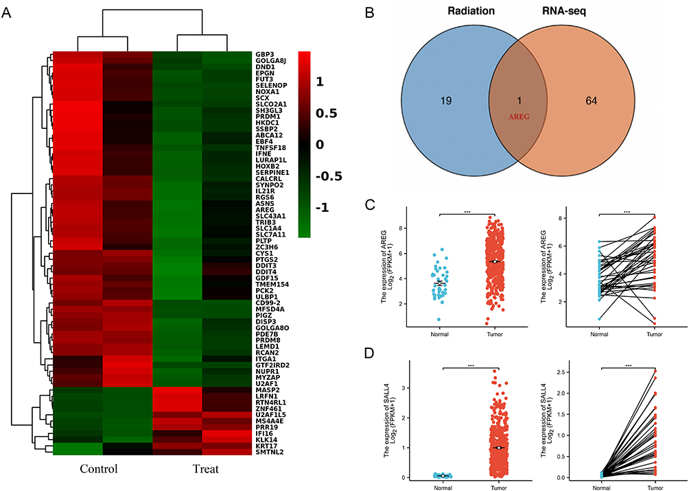 |
Figure 1 AREG, as a potential regulatory target for SALL4. (A) Heat map of DEGs between the control and treatment groups after SALL4 downregulation in HCT116 cells; (B) Venn plots of the intersection of DEGs and CRC radiation-associated gene sets; and (C) AREG and (D) SALL4 expression in unpaired and paired sample tissues of CRC. *P < 0.05, **P < 0.01, ***P < 0.001, ****P < 0.0001. |
The SALL4 binding site in the AREG promoter was predicted using the HumanTFDB database (Figure 2A). Notably, the dual-luciferase assay supported the SALL4-AREG association (Figure 2B), and SALL4 overexpression significantly increased the luciferase activity of the AREG promoter (P < 0.001). Compared to other CRC cell lines, AREG expression, was higher at the mRNA or protein level in HCT116 and HT29 cells (Figure 2C and D). Consequently, we used the HCT116 and HT29 cell lines to examine the effect of RT on CRC cells. Compared to the shNC group, AREG expression was correspondingly lower after transfection of the SALL4-KD plasmid (Figure 2E and F).
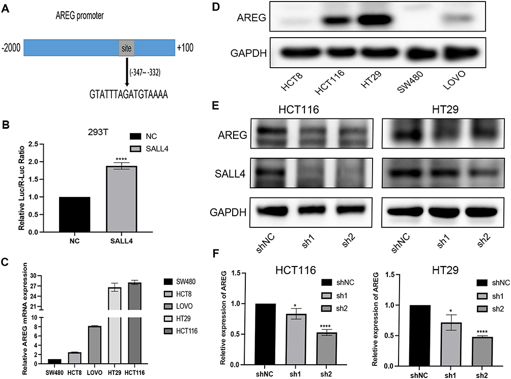 |
Figure 2 SALL4 binds to AREG and regulates its expression. (A) The SALL4 binding site in the AREG promoter; (B) Detection of luciferase activity using the dual luciferase reporter gene assay; AREG mRNA (C) and protein (D) expression in CRC cell lines; and changes in AREG mRNA (E) and protein (F) levels after SALL4 downregulation. *P < 0.05, **P < 0.01, ***P < 0.001, ****P < 0.0001. |
X-Ray Irradiation Enhances SALL4 and AREG Expression
To assess the role of AREG in CRC radiosensitivity, we analyzed the changes in SALL4 and AREG expression over a short period after a single irradiation of CRC cells. After 48 h of cell irradiation, SALL4 and AREG proteins were significantly upregulated (Figure 3A), and the mRNA level of AREG also increased (Figure 3B).
 |
Figure 3 Effect of irradiation on SALL4 and AREG expression and establishment of AREG-KD stable cell lines. Changes in SALL4 and AREG protein (A) and mRNA (B) levels after X-ray irradiation; GFP fluorogram (C); and Validation of AREG mRNA (D) and protein (E) levels after AREG shRNA lentiviral infection. *P < 0.05, **P < 0.01, ***P < 0.001, ****P < 0.0001. |
To further explore the role AREG played in CRC radioresistance, we infected CRC cells with an AREG shRNA lentiviral vector, selected cells that were successfully infected, and created a stable strain of CRC cells. Cellular Green Fluorescent Protein (GFP) detection was conducted to verify the infection efficiency of AREG-KD (Figure 3C), The results were also confirmed by RT-qPCR (Figure 3D) and WB (Figure 3E) experiments.
AREG Knockdown Inhibits Cell Proliferation and Promotes Irradiation-Induced DNA Damage
We performed an EdU assay to assess the impact of AREG on cell viability and proliferation. The results demonstrated that AREG knockdown significantly inhibited DNA replication in the cells (Figure 4A). The CCK-8 assay was also performed after x-ray irradiation, and the results indicated that cell viability was significantly lower in AREG-KD cell lines than in controls. Furthermore, AREG knockdown significantly increased the radiosensitivity of CRC cells (Figure 4B). These findings suggest that the targeted inhibition of AREG expression sensitizes CRC cells to RT.
 |
Figure 4 Influence of AREG on CRC cell proliferation. (A) Immunofluorescence plots of DNA replication capacity tests and statistical plots of differences in DNA replication efficiency between groups; (B) CRC cell proliferation after AREG knockdown in combination with X-rays; and γ-H2AX expression at different time points after irradiation (C) and AREG knockdown combined with X-ray irradiation (D) in CRC cells. *P < 0.05, **P < 0.01, ***P < 0.001, ****P < 0.0001. |
Radiation confers lethality on cells by inducing DNA Double-Strand Breaks (DSB), in which γ-H2AX is a sensitive marker. Herein, γ-H2AX expression peaked 30 min after irradiation of CRC cells (Figure 4C). Furthermore, after combining X-ray irradiation with AREG knockdown, we found that γ-H2AX expression was significantly higher in the treatment group compared to the control group (Figure 4D). These findings suggest that AREG knockdown enhances the sensitivity of CRC cells to irradiation, probably via augmenting irradiation-induced DNA DSBs.
AREG Knockdown Increases Radiation-Induced Apoptosis and G1 Phase Block in CRC Cells
Radiation-induced DNA damage triggers the G1 or G2 arrest of the cell cycle, allowing the cell time to repair the DNA damage.24 Herein, DNA levels were assessed through PI staining and flow cytometry. The results indicated that AREG knockdown led to a moderate increase in G1 phase arrest in CRC cells, both in the presence and absence of radiation, compared to the control group. This finding further supports the notion that reducing AREG expression enhances radiosensitivity in CRC cells (Figure 5A–D).
 |
Figure 5 AREG knockdown promoted X-ray-induced G1 arrest and apoptosis. Histogram of cell cycle distribution of HCT116 (A, B) and HT29 (C, D) cell lines after AREG knockdown combined with irradiation; (E) Proportion of apoptotic cells in non-transfected and AREG shRNA-transfected cells subjected to 0 and 6-Gy irradiation; and (F) Expression levels of apoptosis-related proteins (Bax, Bcl-2, and Cleaved Caspase 3). *P < 0.05, **P < 0.01, ***P < 0.001, ****P < 0.0001. |
Cell apoptosis was also evaluated using flow cytometry and WB. According to the results, AREG knockdown significantly enhanced radiation-induced apoptosis compared to the control group (Figure 5E). Furthermore, AREG knockdown upregulated Cleaved Caspase 3 and Bax and downregulated Bcl-2, which are essential apoptosis indicators (Figure 5F). Collectively, these findings suggest that AREG knockdown enhanced radiation-induced apoptosis.
AREG Knockdown Suppresses PI3K/AKT Signaling in CRC Cells
The PI3K/AKT signaling pathway is often implicated in tumor radiosensitivity, with its disruption enhancing radiosensitivity by hindering DNA double-strand break (DSB) repair.25 In this study, we observed that AREG knockdown suppressed PI3K/AKT protein expression (Figure 6A), indicating that reduced AREG levels may enhance radiosensitivity in colorectal cancer (CRC). Additionally, AREG overexpression counteracted the suppressive effect of SALL4 downregulation on AREG expression (Figure 6B), highlighting the regulatory interplay between SALL4 and AREG. Overall, our findings suggest that the PI3K/AKT pathway is involved in modulating radiosensitivity in CRC through SALL4 and AREG interactions.
 |
Figure 6 SALL4 downregulation targets AREG and inhibits the PI3K/AKT pathway. (A) A corresponding inhibition of the PI3K/AKT pathway protein expression followed AREG knockdown; and (B) AREG overexpression reversed the inhibitory effects of SALL4 downregulation on AREG expression. |
Discussion
According to research, SALL4 is a major malignancy driver and a potential target for cancer treatment.6,26,27 In our previous study, we observed that SALL4 expression was significantly elevated in CRC tissues compared to normal tissues. Furthermore, reducing SALL4 levels led to decreased proliferation, migration, and invasion of CRC cells.20 Furthermore, higher levels of SALL4 expression induced RT resistance in NPC,9 implying that in addition to its involvement in tumor development, SALL4 may also play a key role in tumor radiosensitivity. Herein, we discovered that the SALL4-targeted AREG modulated radiosensitivity in CRC cells by regulating the PI3K-AKT pathway. Kong et al reported that SALL4 relies on zinc finger structures to function as a gene regulator by binding to AT-rich sequences.28 In our case, SALL4 could bound to the AREG promoter to regulate its expression, indicating a positive correlation between the expression of the two. Furthermore, AREG overexpression reversed the inhibitory effects of SALL4 downregulation on AREG expression. Additionally, X-ray irradiation upregulated SALL4 and AREG in CRC cells. This phenomenon may be associated with CRC radioresistance, an assumption supported by the fact that AREG knockdown promoted X-ray-induced DNA damage. Furthermore, AREG knockdown reduced the DNA replication efficiency of CRC cells and, when combined with X-ray irradiation, resulted in a notable reduction in cell growth. Additionally, AREG knockdown increased the G1 phase arrest and apoptosis in CRC cells after irradiation.
As an external stimulus, ionizing radiation alters the Tumor Microenvironment (TME), resulting in gene expression alterations. According to research, DNA DSBs is the most common and severe form of ionizing radiation-induced damage, posing a great threat to genome integrity and stability.29 Furthermore, DNA DSBs are always accompanied by histone H2AX phosphorylation and rapidγ-H2AX foci formation. It has been established thatγ-H2AX correlates positively with DNA DSBs, highlighting it as a potential biomarker of DNA damage.30 Herein, we found thatγ-H2AX expression in CRC cells peaked 30 min post-irradiation and gradually recovered. Consistent with previous observations, which revealed that impaired DNA repair could lead to increased radiosensitivity,31,32 our findings revealed that AREG knockdown significantly upregulatedγ-H2AX post-irradiation and promoted X-ray-induced DNA damage.
Although AREG was initially detected in BC cells, it has been found to promote the proliferation of most cell types, primarily by binding to and activating EGFR.13 According to research, AREG is overexpressed in patients with different types of cancer, and its cancer-causing properties have been observed in typical human epithelial malignancies, including lung, breast, colorectal, ovarian, and prostate cancers.14,33–36 Furthermore, the involvement of AREG in the regulation of cancer treatment resistance has been reported.37–39 For example, AREG induced Epithelial-Mesenchymal Transition (EMT) in pancreatic cancer cells via the EGFR/ERK/NF-kB signaling pathway.40 Moreover, miR-34c-5p inhibited stemness and drug resistance and blocked ovarian cancer progression by targeting the AREG-EGFR-ERK pathway.41 Furthermore, AREG is considered a key gene involved in the regulation of tumor escape and radioresistance, which is critical to radiation response regulation in intracranial tumors.16,42
In many tumors, PI3K, an essential signaling protein that operates downstream of various protein kinases, particularly those from the EGFR family, is often overactive.43 Protein Kinase B (AKT), a major PI3K kinase, is involved in the regulation of multiple tumor cell processes, including proliferation, metabolism, invasion, metastasis, angiogenesis, and therapeutic resistance, by switching intracellular signals and activating the downstream target proteins.25,44–46 The oncogenic activation of AKT or stress, such as ionization-induced activation of the PI3K / AKT pathway, can accelerate DNA DSB repair and reduce ionization-induced cell death, causing treatment resistance.47–49 Our findings revealed that AREG knockdown downregulated the phosphorylated AKT and inhibited PI3K/AKT pathway activation, implying that the targeted regulation of AREG expression by SALL4 promotes the development of radioresistance in CRC cells via PI3K/AKT signaling pathway activation.
In summary, our results indicate that SALL4 transcriptionally activates AREG in CRC cells. This activation suggests that the SALL4/AREG/PI3K-AKT signaling pathway may contribute to the development of radioresistance in CRC cells. These insights contribute to our understanding of CRC treatment with RT.
Abbreviations
SALL4, Spalt-Like Transcription Factor 4; CRC, Colorectal Cancer; AREG, Amphiregulin; HumanTFDB, Human Transcription Factor Database; WB, Western Blot; RT-qPCR, Reverse Transcription quantitative Polymerase Chain Reaction; KD, Knockdown; CCK-8, Cell Counting Kit 8; EdU, 5-Ethynyl-2′-deoxyuridine; RT, Radiotherapy; TF, Transcription Factor; NPC, Nasopharyngeal Carcinoma; EGFR, Epidermal Growth Factor Receptor; DEGs, Differentially Expressed Genes; SDS-PAGE, Sodium Dodecyl Sulfate-Polyacrylamide Gel Electrophoresis; SD, Standard Deviation; GFP, Green Fluorescent Protein; DSB, DNA Double-Strand Breaks; TME, Tumor Microenvironment; EMT, Epithelial-Mesenchymal Transition; AKT, Protein Kinase B.
Data Sharing Statement
The data used to support the findings of this study are included within the article. More information can be accessed from correspondence authors.
Ethics Statement
The data used to support the findings of this study are included within the article. More information can be accessed from correspondence authors. This study was authorized by the Taicang Affiliated Hospital of Soochow University Human Research Ethics Committee (KY-2019-020).
Acknowledgments
This study was supported by the Science and Technology Project of Taicang (Grant Number TC2019JCYL02), Gusu Health Talents Training Project (Grant Number GSWS2019078 and GSWS2019079), and the Youth Science Foundation Project of AnHui Medical University (Grant Number 2023xk j098).
Author Contributions
All authors made a significant contribution to the work reported, whether that is in the conception, study design, execution, acquisition of data, analysis and interpretation, or in all these areas; took part in drafting, revising or critically reviewing the article; gave final approval of the version to be published; have agreed on the journal to which the article has been submitted; and agree to be accountable for all aspects of the work.
Disclosure
The author reports no conflicts of interest in this work.
References
1. Long F, Lin Z, Li L, et al. Comprehensive landscape and future perspectives of circular RNAs in colorectal cancer. Mol Cancer. 2021;20(1):26. doi:10.1186/s12943-021-01318-6
2. Kuipers EJ, Grady WM, Lieberman D, et al. Colorectal cancer. Nat Rev Dis Primers. 2015;1:15065. doi:10.1038/nrdp.2015.65
3. Glynne-Jones R, Hall M. Radiotherapy and locally advanced rectal cancer. Br J Surg. 2015;102(12):1443–1445. doi:10.1002/bjs.9930
4. Benson AB, Venook AP, Al-Hawary MM, et al. Rectal cancer, version 2.2022, NCCN clinical practice guidelines in oncology. J Natl Compr Canc Netw. 2022;20(10):1139–1167. doi:10.6004/jnccn.2022.0051
5. Baptistella AR, Landemberger MC, Dias MVS, et al. Rab5C enhances resistance to ionizing radiation in rectal cancer. J Mol Med (Berl). 2019;97(6):855–869. doi:10.1007/s00109-019-01760-6
6. Sun B, Xu L, Bi W, et al. SALL4 oncogenic function in cancers: mechanisms and therapeutic relevance. Int J Mol Sci. 2022;23(4):1.
7. Jiang G, Liu CT. Knockdown of SALL4 overcomes cisplatin-resistance through AKT/mTOR signaling in lung cancer cells. Int J Clin Exp Pathol. 2018;11(2):634–641.
8. Liu C, Wu H, Li Y, et al. SALL4 suppresses PTEN expression to promote glioma cell proliferation via PI3K/AKT signaling pathway. J Neurooncol. 2017;135(2):263–272. doi:10.1007/s11060-017-2589-3
9. Nie X, Guo E, Wu C, et al. SALL 4 induces radioresistance in nasopharyngeal carcinoma via the ATM /Chk2/p53 pathway. Cancer Med. 2019;8(4):1779–1792. doi:10.1002/cam4.2056
10. Berasain C, Avila MA. Amphiregulin. Semin Cell Dev Biol. 2014;28:31–41. doi:10.1016/j.semcdb.2014.01.005
11. Singh B, Carpenter G, Coffey RJ. EGF receptor ligands: recent advances. F1000Res. 2016;5:2270. doi:10.12688/f1000research.9025.1
12. Park SY, Kim J-T, Park E, et al. Collapsin response mediator protein 4 enhances the radiosensitivity of colon cancer cells through calcium‑mediated cell signaling. Oncol Rep. 2021;45(4). doi:10.3892/or.2021.7957
13. Shoyab M, McDonald VL, Bradley JG, et al. Amphiregulin: a bifunctional growth-modulating glycoprotein produced by the phorbol 12-myristate 13-acetate-treated human breast adenocarcinoma cell line MCF-7. Proc Natl Acad Sci U S A. 1988;85(17):6528–6532. doi:10.1073/pnas.85.17.6528
14. Busser B, Sancey L, Brambilla E, et al. The multiple roles of amphiregulin in human cancer. Biochim Biophys Acta. 2011;1816(2):119–131. doi:10.1016/j.bbcan.2011.05.003
15. Jiang J, Zhao W, Tang Q, et al. Over expression of amphiregulin promoted malignant progression in gastric cancer. Pathol Res Pract. 2019;215(10):152576. doi:10.1016/j.prp.2019.152576
16. Bouchet A, Sakakini N, Atifi ME, et al. Identification of AREG and PLK1 pathway modulation as a potential key of the response of intracranial 9L tumor to microbeam radiation therapy. Int, J, Cancer. 2015;136(11):2705–2716. doi:10.1002/ijc.29318
17. Toulany M, Dittmann K, Krüger M, et al. Radioresistance of K-Ras mutated human tumor cells is mediated through EGFR-dependent activation of PI3K-AKT pathway. Radiother Oncol. 2005;76(2):143–150. doi:10.1016/j.radonc.2005.06.024
18. Son B, Kim TR, Park JH, et al. SAMiRNA targeting amphiregulin alleviate total-body-irradiation-induced renal fibrosis. Radiat Res. 2022;197(5):471–479. doi:10.1667/RADE-21-00220.1
19. Sebio A, Salazar J, Páez D, et al. EGFR ligands and DNA repair genes: genomic predictors of complete response after capecitabine-based chemoradiotherapy in locally advanced rectal cancer. Pharmacogenomics J. 2015;15(1):77–83. doi:10.1038/tpj.2014.33
20. Zhang W, Hu Y, Zhang W, et al. The invasion and metastasis of colon adenocarcinoma (COAD) Induced by SALL4. J Immunol Res. 2022;2022:9385820. doi:10.1155/2022/9385820
21. Kim D, Langmead B, Salzberg SL. HISAT: a fast spliced aligner with low memory requirements. Nat Methods. 2015;12(4):357–360. doi:10.1038/nmeth.3317
22. Trapnell C, Williams BA, Pertea G, et al. Transcript assembly and quantification by RNA-Seq reveals unannotated transcripts and isoform switching during cell differentiation. Nat Biotechnol. 2010;28(5):511–515. doi:10.1038/nbt.1621
23. Anders S, Pyl PT, Huber W. HTSeq--A Python framework to work with high-throughput sequencing data. Bioinformatics. 2015;31(2):166–169. doi:10.1093/bioinformatics/btu638
24. Sitko JC, Yeh B, Kim M, et al. SOCS3 regulates p21 expression and cell cycle arrest in response to DNA damage. Cell Signal. 2008;20(12):2221–2230. doi:10.1016/j.cellsig.2008.08.011
25. Toulany M, Rodemann HP. Phosphatidylinositol 3-kinase/Akt signaling as a key mediator of tumor cell responsiveness to radiation. Semin Cancer Biol. 2015;35:180–190. doi:10.1016/j.semcancer.2015.07.003
26. Moein S, Tenen DG, Amabile G, et al. SALL4: an intriguing therapeutic target in cancer treatment. Cells. 2022;11(16). doi:10.3390/cells11162601
27. Yang J. SALL4 as a transcriptional and epigenetic regulator in normal and leukemic hematopoiesis. Biomark Res. 2018;6:1. doi:10.1186/s40364-017-0115-6
28. Kong NR, Bassal MA, Tan HK, et al. Zinc Finger Protein SALL4 Functions through an AT-rich motif to regulate gene expression. Cell Rep. 2021;34(1):108574. doi:10.1016/j.celrep.2020.108574
29. Khanna KK, Jackson SP. DNA double-strand breaks: signaling, repair and the cancer connection. Nat Genet. 2001;27(3):247–254. doi:10.1038/85798
30. Kuo LJ, Yang LX. Gamma-H2AX - A novel biomarker for DNA double-strand breaks. Vivo. 2008;22(3):305–309.
31. Taylor EM, Lehmann AR. Conservation of eukaryotic DNA repair mechanisms. Int J Radiat Biol. 1998;74(3):277–286. doi:10.1080/095530098141429
32. Krenning L, van den Berg J, Medema RH. Life or death after a break: what determines the choice? Mol Cell. 2019;76(2):346–358. doi:10.1016/j.molcel.2019.08.023
33. Baillo A, Giroux C, Ethier SP. Knock-down of amphiregulin inhibits cellular invasion in inflammatory breast cancer. J Cell Physiol. 2011;226(10):2691–2701. doi:10.1002/jcp.22620
34. Hsu YL, Huang M-S, Cheng D-E, et al. Lung tumor-associated dendritic cell-derived amphiregulin increased cancer progression. J Immunol. 2011;187(4):1733–1744. doi:10.4049/jimmunol.1100996
35. Sehgal I, Bailey J, Hitzemann K, et al. Epidermal growth factor receptor-dependent stimulation of amphiregulin expression in androgen-stimulated human prostate cancer cells. Mol Biol Cell. 1994;5(3):339–347. doi:10.1091/mbc.5.3.339
36. Bormann F, Stinzing S, Tierling S, et al. Epigenetic regulation of amphiregulin and epiregulin in colorectal cancer. Int, J, Cancer. 2019;144(3):569–581. doi:10.1002/ijc.31892
37. Pommer M, Kuphal S, Bosserhoff AK. Amphiregulin Regulates Melanocytic Senescence. Cells. 2021;10(2):326. doi:10.3390/cells10020326
38. Xu Q, Long Q, Zhu D, et al. Targeting amphiregulin (AREG) derived from senescent stromal cells diminishes cancer resistance and averts programmed cell death 1 ligand (PD-L1)-mediated immunosuppression. Aging Cell. 2019;18(6):e13027. doi:10.1111/acel.13027
39. Kim SA, Park H, Kim K-J, et al. Amphiregulin can predict treatment resistance to palliative first-line cetuximab plus FOLFIRI chemotherapy in patients with RAS wild-type metastatic colorectal cancer. Sci Rep. 2021;11(1):23803. doi:10.1038/s41598-021-03197-9
40. Wang L, Wang L, Zhang H, et al. AREG mediates the epithelial‑mesenchymal transition in pancreatic cancer cells via the EGFR/ERK/NF‑κB signalling pathway. Oncol Rep. 2020;43(5):1558–1568. doi:10.3892/or.2020.7523
41. Tung SL, Huang W-C, Hsu F-C, et al. miRNA-34c-5p inhibits amphiregulin-induced ovarian cancer stemness and drug resistance via downregulation of the AREG-EGFR-ERK pathway. Oncogenesis. 2017;6(5):e326. doi:10.1038/oncsis.2017.25
42. Minjgee M, Toulany M, Kehlbach R, et al. K-RAS(V12) induces autocrine production of EGFR ligands and mediates radioresistance through EGFR-dependent Akt signaling and activation of DNA-PKcs. Int J Radiat Oncol Biol Phys. 2011;81(5):1506–1514. doi:10.1016/j.ijrobp.2011.05.057
43. Liu P, Cheng H, Roberts TM, et al. Targeting the phosphoinositide 3-kinase pathway in cancer. Nat Rev Drug Discov. 2009;8(8):627–644. doi:10.1038/nrd2926
44. Manning BD, Toker A. AKT/PKB Signaling: navigating the Network. Cell. 2017;169(3):381–405. doi:10.1016/j.cell.2017.04.001
45. Shariati M, Meric-Bernstam F. Targeting AKT for cancer therapy. Expert Opin Investig Drugs. 2019;28(11):977–988. doi:10.1080/13543784.2019.1676726
46. Tsai PJ, Lai Y-H, Manne RK, et al. Akt: a key transducer in cancer. J Biomed Sci. 2022;29(1):76. doi:10.1186/s12929-022-00860-9
47. Sutcu HH, Montagne B, Ricchetti M. DNA-PKcs regulates myogenesis in an Akt-dependent manner independent of induced DNA damage. Cell Death Differ. 2023;30(8):1900–1915. doi:10.1038/s41418-023-01177-2
48. Toulany M, Kehlbach R, Rodemann HP, et al. Radiocontrast media affect radiation-induced DNA damage repair in vitro and in vivo by affecting Akt signalling. Radiother Oncol. 2010;94(1):110–116. doi:10.1016/j.radonc.2009.11.006
49. Wang Y, Su P, Zhuo Z, et al. Ginsenoside Rk1 attenuates radiation-induced intestinal injury through the PI3K/AKT/mTOR pathway. Biochem Biophys Res Commun. 2023;643:111–120. doi:10.1016/j.bbrc.2022.12.072
 © 2024 The Author(s). This work is published and licensed by Dove Medical Press Limited. The
full terms of this license are available at https://www.dovepress.com/terms
and incorporate the Creative Commons Attribution
- Non Commercial (unported, 3.0) License.
By accessing the work you hereby accept the Terms. Non-commercial uses of the work are permitted
without any further permission from Dove Medical Press Limited, provided the work is properly
attributed. For permission for commercial use of this work, please see paragraphs 4.2 and 5 of our Terms.
© 2024 The Author(s). This work is published and licensed by Dove Medical Press Limited. The
full terms of this license are available at https://www.dovepress.com/terms
and incorporate the Creative Commons Attribution
- Non Commercial (unported, 3.0) License.
By accessing the work you hereby accept the Terms. Non-commercial uses of the work are permitted
without any further permission from Dove Medical Press Limited, provided the work is properly
attributed. For permission for commercial use of this work, please see paragraphs 4.2 and 5 of our Terms.