
Back to Journals » International Journal of General Medicine » Volume 19
Serum Aberrant Expression of miR-377-3p and Its Diagnostic Value in Carotid Artery Stenosis
Authors Qie G  , Han H , Ning J , Wang W , Lei Y
Received 31 October 2025
Accepted for publication 16 December 2025
Published 8 January 2026 Volume 2026:19 573541
DOI https://doi.org/10.2147/IJGM.S573541
Checked for plagiarism Yes
Review by Single anonymous peer review
Peer reviewer comments 3
Editor who approved publication: Dr Redoy Ranjan
Guohua Qie,* Huixian Han,* Jianfeng Ning, Wenqiang Wang, Yan Lei
The Laboratory Medicine Department, The First Hospital of Zhangjiakou City, Zhangjiakou, Hebei, 075000, People’s Republic of China
*These authors contributed equally to this work
Correspondence: Yan Lei, The Laboratory Medicine Department, The First Hospital of Zhangjiakou City, No. 6 Jianguo Road, Qiaodong District, Zhangjiakou, Hebei, 075000, People’s Republic of China, Tel +86 0313 2102972, Email [email protected]
Purpose: To determine the serum expression profile of miR-377-3p in carotid artery stenosis (CAS) patients and to assess its diagnostic potential in a clinical setting.
Patients and Methods: Using qRT-PCR, miR-377-3p expression was measured in serum samples from 78 CAS patients and 78 matched healthy controls. We used the Pearson correlation to analyze inter-indicator relationships and the ROC curve to assess the diagnostic significance of miR-377-3p. The cellular functions of human aortic smooth muscle cells (HASMCs) were assessed. Bioinformatics predictions of target associations were experimentally verified with a luciferase activity assay.
Results: Patients diagnosed with CAS had a lower serum level of miR-377-3p. ROC curve-based analysis showed that miR-377-3p exerted high diagnostic value, where the area under the curve (AUC) was 0.9597. Serum miR-377-3p was negatively correlated with systolic blood pressure (SBP), diastolic blood pressure (DBP), fasting blood glucose (FBG), and low-density lipoprotein cholesterol (LDL), while positively correlated with high-density lipoprotein cholesterol (HDL). miR-377-3p inhibited the proliferation and migration of HASMCs, but this effect was counteracted by EDIL3.
Conclusion: These results suggest the dual utility of miR-377-3p as a promising biomarker and a potential therapeutic target in the management of CAS. miR-377-3p can inhibit the proliferation and migration of HASMCs by targeting EDIL3.
Keywords: carotid artery stenosis, miR-377-3p, clinical diagnostic value, human aortic smooth muscle cells, endothelial cell-specific molecule 3
Introduction
Carotid Artery Stenosis (CAS) is a major risk factor for ischemic stroke, with atherosclerotic plaque deposition as its core pathological basis.1 While imaging techniques (eg, carotid ultrasound, CTA) are the mainstay of diagnosis, they have limitations in early subclinical lesion detection.2 Therefore, it is of great necessity to find reliable molecular biomarkers for accurate early diagnosis of CAS patients.
Over the past few years, MicroRNAs (miRNAs) have emerged as crucial players in orchestrating epigenetic gene regulation and modulating diverse metabolic and physiological pathways involved in disease.3 MicroRNAs (miRNAs) have emerged as promising non-invasive biomarkers for CAS, with several miRNAs (eg, miR-28-5p, miR-92a) implicated in diagnosis and prognosis.4–10 These findings highlight a promising avenue for improving CAS management through non-invasive molecular diagnostics.
Vascular smooth muscle cells (VSMCs) are key cellular components of the vascular media, and their abnormal proliferation and migration are core pathological events in CAS progression. During atherosclerosis, VSMCs migrate from the media to the intima, undergo excessive proliferation, and secrete extracellular matrix components such as collagen to form fibrous caps. Uncontrolled VSMC proliferation and migration further increase plaque volume, narrow the vascular lumen, and promote plaque instability, ultimately contributing to CAS development and ischemic stroke risk.11–13 Therefore, exploring the regulatory mechanisms of HASMCs function is crucial for understanding CAS pathogenesis and identifying potential therapeutic targets. Although previous studies have implicated miR-377-3p in vascular pathophysiology—such as its role in blocking HASMCs proliferation and migration in atherosclerosis via NRP2 and its regulation by Circ_0010283 under ox-LDL stimulation—their focus remained on atherosclerotic mechanisms.14,15 Consequently, the diagnostic value of miR-377-3p in CAS and its specific functional impact on HASMCs within the CAS context remain undetermined.
This study aimed to: ① evaluate the diagnostic potential of serum miR-377-3p in CAS; ② investigate its effect on HASMC proliferation/migration; ③ identify its target gene and clarify the underlying mechanism.
Methods
Study Participants
This is a cross-sectional observational study conducted at the First Hospital of Zhangjiakou City from January 2022 to December 2023. A total of 78 CAS patients and 78 age- and gender-matched healthy controls were enrolled consecutively. During the same period, 78 healthy individuals who received physical check-ups at the same hospital were selected as the control group.
Inclusion Criteria for the Case Group (CAS Patients): ① Diagnosis of CAS confirmed by carotid ultrasound, CTA, or MRA, with a stenosis degree of ≥ 50% (moderate or severe stenosis, to avoid interference from mild stenosis); ② Aged 50–75 years (covering the high-incidence population); ③ Complete clinical data (eg, stenosis degree, plaque properties, history of transient ischemic attack (TIA) or stroke).
Exclusion Criteria for the Case Group: ① Comorbidity with acute cardiovascular and cerebrovascular events; ② Comorbidity with malignant tumors, chronic liver disease (liver cirrhosis), kidney disease (uremia), or autoimmune diseases (eg, rheumatoid arthritis); ③ Use of lipid-lowering drugs (eg, statins) or anti-inflammatory drugs (eg, aspirin) within the past 3 months; ④ Pregnant or lactating women; ⑤ History of coronary artery disease (confirmed by coronary angiography, CT coronary angiography, or myocardial infarction) or peripheral artery disease (confirmed by lower extremity arterial ultrasound, angiography, or clinical symptoms such as intermittent claudication); ⑥ Abnormal findings in electrocardiogram (ECG) or cardiac ultrasound suggesting myocardial ischemia or structural heart disease; ⑦ Ankle-brachial index (ABI) < 0.9 (a recognized indicator of lower extremity arterial stenosis).
Inclusion Criteria for the Control Group: ① Carotid ultrasound showing a stenosis degree of < 10% (essentially no stenosis); ② Age and gender matched with the case group (1:1 matching, to reduce age/gender bias); ③ No risk factors for carotid artery stenosis, such as hypertension, diabetes mellitus, or hyperlipidemia.
Exclusion Criteria for the Control Group: ① Comorbidity with acute cardiovascular and cerebrovascular events; ② Comorbidity with malignant tumors, chronic liver disease (liver cirrhosis), kidney disease (uremia), or autoimmune diseases (eg, rheumatoid arthritis); ③ Pregnant or lactating women; ④ No family history of cardiovascular diseases.
The procedures used in this study complied with the principles of the Declaration of Helsinki. This study was approved by the Ethics Committee of the First Hospital of Zhangjiakou City. All participants were informed of the study procedures and signed an informed consent form.
Collection of Clinical Experimental Data
Serum samples were collected using sterile blood collection tubes without anticoagulants. All samples were allowed to clot at room temperature for 30 minutes and then centrifuged at 3000 rpm for 20 minutes within 2 hours of blood draw to avoid miRNA degradation. All serum samples from CAS patients were collected within 1 week after imaging-based diagnosis (carotid ultrasound/CTA/MRA) and before any treatment (eg, lipid-lowering drugs, antiplatelet therapy) was initiated. To exclude the influence of acute clinical events, patients with acute cerebrovascular events (eg, transient ischemic attack, stroke) within 3 months were excluded from the CAS group (as specified in exclusion criteria).
Cell Culture and Transfection
To investigate the functional role of miR-377-3p, we manipulated its expression in HASMCs (3H Biomedical, shanghai, China; Catalog No.: 3H-001) by transfecting with specific mimics or inhibitors. miR-377-3p mimic (50 nM), inhibitor (100 nM), and their negative controls (NC, 50 nM/100 nM) were purchased from RiboBio (Guangzhou, China; Catalog Nos.: miR10000421-1-5, miR20000421-1-5). The transfected cells were then stimulated with 20 μg/mL ox-LDL (Sigma-Aldrich, St. Louis, USA; Catalog No.: L4149, Lot No.: SLCM8832V) for 48 hours to mimic a disease-relevant environment before being harvested for further experiments.
RT-qPCR Assay
Total RNA was isolated from cells using Trizol reagent. Subsequently, mRNA and miRNA were reverse-transcribed into cDNA using the SuperRT cDNA Synthesis Kit (Kangwei Shiji), followed by RT-qPCR analysis with the SuperFast Lyo Probe One Step RT-qPCR Mix on a Bio-Rad CFX96 system. U6 and GAPDH served as internal controls for miR-377-3p and EDIL3, respectively. Relative expression levels were calculated using the 2^(-ΔΔCt) method, and all primers (sequences in Table 1) were synthesized by Sangon Biotech.
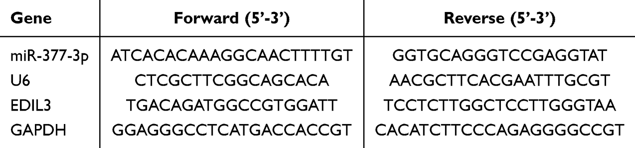 |
Table 1 Primer Sequence |
Cell Counting Kit-8 (CCK-8) Assay
To evaluate cell proliferation, HASMCs were plated in 96-well plates (2×10⁴ cells/well) and analyzed with a CCK-8 kit at 0, 24, 48, and 72 hours. Absorbance at 450 nm was recorded following a 3-hour incubation period. Background correction was performed by setting blank wells (containing only medium and CCK-8 reagent without cells) to subtract non-specific absorbance, ensuring accurate quantification of cell proliferation.
Transwell Assay
A Transwell migration assay was performed in 24-well plates using Transwell inserts (Corning, USA; Catalog No.: 3422) with an 8 μm pore size. The upper chamber was seeded with 1×10⁵ cells suspended in serum-free DMEM, and the lower chamber was filled with DMEM supplemented with 10% FBS. Following a 24-hour incubation, non-migratory cells were removed by swabbing, followed by fixation, staining, and quantification of the migrated cells on the lower side of the membrane across five random microscopic fields.
Luciferase Reporter Gene Assay
To validate the predicted interaction, we constructed pmirGLO vectors harboring either the wild-type (WT) or mutant (MUT) sequence of the EDIL3 3’-UTR encompassing the putative miR-377-3p binding site. We co-transfected the reporter vectors into HASMCs with the miR-377-3p mimic, inhibitor, or their negative controls (NC) using Lipofectamine 3000. We measured luciferase activity following a 48-hour transfection period.
Statistical Analysis
Data from three independent experiments (mean ± SD) were analyzed with SPSS 27.0 and GraphPad Prism 8.0.2. Inter-group comparisons used Student’s t-test, while correlations and diagnostic values (via ROC curves) of miR-377-3p and EDIL3 were assessed by Pearson correlation analysis. A P-value < 0.05 was considered statistically significant.
Results
Clinical Indicators of the Study Groups
The baseline characteristics of the study participants are summarized in Table 2. Briefly, both the CAS group and the control group comprised 78 subjects, with comparable mean ages (61.82 ± 5.14 vs 61.05 ± 5.53 years, respectively). The CAS group had an equal sex distribution (39 males/39 females), while the control group consisted of 38 males and 40 females. There were no statistically significant differences in age, gender, body mass index (BMI), smoking status, or alcohol consumption status between the CAS group and the control group (P > 0.05). Compared with the control group, the CAS group had higher values of SBP, DBP, FBG, and LDL, while the level of HDL was lower (P < 0.05).
 |
Table 2 Comparison of General Data Between the Two Study Groups |
Abnormal miR-377-3p Expression in CAS Patients and Its Diagnostic Efficacy Analysis
As shown in Figure 1a, the serum level of miR-377-3p in the CAS group was significantly lower than that in the control group (P < 0.001). To evaluate serum miR-377-3p’s CAS diagnostic value, a ROC curve (Figure 1b) was plotted using CAS and control groups’ levels; it distinguished CAS patients from healthy people, with AUC=0.9597 (95% CI: 0.9319–0.9876), 97.44% sensitivity, 83.33% specificity, and cut-off 0.865. These results indicate that miR-377-3p has high diagnostic value in CAS patients.
 |
Figure 1 Expression level of miR-377-3p in CAS. (a) Serum miR-377-3p levels: CAS vs Control. (b) ROC curve of serum miR-377-3p used to distinguish CAS patients from healthy individual. ***: P < 0.001, compared with the control group. |
Correlation Between Serum miR-377-3p and Clinical Indicators
Pearson correlation analysis revealed that serum miR-377-3p levels were significantly negatively correlated with SBP (r = −0.6710, P < 0.001), DBP (r = −0.5577, P < 0.001), FBG (r = −0.6801, P < 0.001), and LDL (r = −0.5022, P < 0.001), but showed a significant positive correlation with HDL (r = 0.5192, P < 0.001) (Figure 2). It is important to note that the observed correlations, while statistically significant, derive from univariate analysis. Given that SBP, DBP, FBG, and LDL are themselves established risk factors for CAS and were imbalanced between our groups, these correlations do not establish the independence of miR-377-3p as a biomarker. Future studies with larger cohorts are warranted to perform multivariate logistic regression analyses to determine whether the association between low miR-377-3p levels and CAS persists after adjustment for these and other potential confounders.
 |
Figure 2 Correlation between miR-377-3p and clinical indicators. a-e Correlations between serum miR-377-3p and SBP (a), DBP (b), FBG (c), HDL (d), and LDL (e). |
miR-377-3p Inhibits the Proliferation and Migration of HASMCs
Figure 3a reflects the transfection efficiency. miR-377-3p expression in HASMCs was effectively upregulated by the mimic and downregulated by the inhibitor, confirming the efficiency of the transfection. Results from the CCK-8 assay confirmed that miR-377-3p overexpression inhibited the proliferation of HASMCs, while miR-377-3p inhibitor stimulated HASMC proliferation (Figure 3b). Transwell migration assays revealed that miR-377-3p plays functionally opposing roles: its overexpression impeded HASMC migration, while its inhibition conversely enhanced migratory capacity (Figure 3c).
 |
Figure 3 miR-377-3p suppresses HASMC proliferation and migration. (a) Level of miR-377-3p in cells transfected with miR-377-3p mimic (upregulation) or inhibitor (downregulation) and treated with ox-LDL. (b) Effects of miR-377-3p mimic and inhibitor on HASMC proliferation. (c) Migration of miR-377-3p mimic/inhibitor-transfected cells. ***: P < 0.001, a,b,c: different letters indicate significant differences. |
EDIL3 Serves as a Target Gene of miR-377-3p
We utilized StarBase, miRWalk, miRBD, and miRTarBase to screen for putative mRNA targets of miR-377-3p, and the overlapping predictions were visualized via the jvenn online tool (Figure 4a). A total of 17 overlapping genes were determined to be candidate target genes of miR-377-3p related to CAS. Among them, epidermal growth factor-like repeats and discoidin I-like domain 3 (EDIL3) directly participates in atherosclerosis, a core pathological process of carotid artery stenosis, by promoting vascular inflammation, endothelial cell adhesion and angiogenesis. It can activate the integrin signaling pathway, driving phenotypic transformation, abnormal migration and proliferation of vascular smooth muscle cells, leading to the formation of new intima, thereby directly triggering and exacerbating lumen stenosis. Therefore, EDIL3 is a key molecule that connects vascular inflammation, extracellular matrix signaling and vascular smooth muscle cell dysfunction. As shown in Figure 4b, the putative binding site for miR-377-3p on the EDIL3 transcript was identified through bioinformatic analysis using the StarBase database. The target gene was verified using a dual-luciferase reporter gene assay. miR-377-3p directly regulated EDIL3 expression, as evidenced by the increase in luciferase activity with its mimic and the decrease with its inhibitor in HASMCs transfected with the WT reporter vector (Figure 4c).
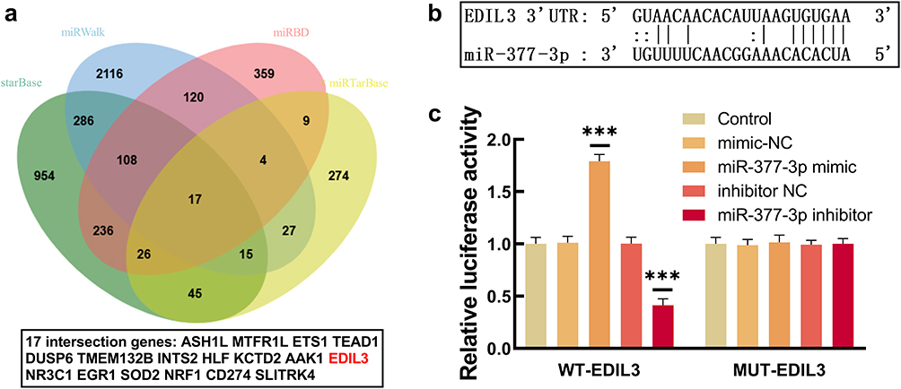 |
Figure 4 Validation of EDIL3 as a direct target of miR-377-3p. (a) Venn diagram showing overlapping genes from StarBase, miRWalk, miRBD, and miRTarBase datasets. EDIL3 (red font) was selected as the target gene. (b) Binding site between miR-377-3p and EDIL3 predicted by StarBase. (c) Luciferase activity: miR-377-3p mimic vs inhibitor. ***: P < 0.001. |
Discussion
Currently, the clinical diagnosis of CAS relies on imaging techniques such as carotid ultrasound and CTA. However, these methods have limitations, including dependence on equipment, invasiveness (eg, CTA requires a contrast agent), and difficulty in early identification of subclinical lesions. In contrast, traditional serum markers (such as LDL and high-sensitivity C-reactive protein) have low diagnostic specificity, as they are often interfered by factors like infection and metabolic abnormalities. By comparison, miRNAs as circulating markers offer advantages of high stability (less affected by RNase degradation), convenient detection (requiring only a small amount of serum), and capability for dynamic monitoring. In this study, the AUC of miR-377-3p was close to 1.0, indicating an extremely strong ability to distinguish CAS patients from healthy individuals. In similar studies, the AUC values of miR-483-5p, miRNA-146a, miR-361-5p, and miR-503-5p in CAS were mostly between 0.70 and 0.85.7,9,16,17 The diagnostic efficacy of miR-377-3p is significantly superior, which may be attributed to its direct involvement in the pathological regulation of CAS. Although the ROC curve analysis showed that miR-377-3p had high diagnostic value for CAS (AUC = 0.9597), this result should be interpreted with caution. First, the sample size of this single-center study is relatively small (78 cases/78 controls), which may lead to overfitting of the ROC model. Second, the control group was selected to exclude all atherosclerotic risk factors, which is inconsistent with the real-world population of CAS patients (who often have comorbid cardiometabolic diseases). This “extremely healthy” control group may inflate the discriminative ability of miR-377-3p. Future multi-center studies with larger sample sizes, including controls with cardiometabolic risk factors but no CAS, are needed to validate the true diagnostic accuracy of miR-377-3p.
Abnormal proliferation and migration of HASMCs are key pathological events in CAS. Under normal conditions, HASMCs are located in the media of blood vessels and maintain the stability of vascular structure. During the development of atherosclerosis (AS), HASMCs migrate to the intima, proliferate excessively, secrete collagen to form a fibrous cap, and may cause an increase in plaque volume and narrowing of the vascular lumen due to uncontrolled proliferation.11 All CAS patients and controls in this study were strictly excluded from coronary artery disease and peripheral artery disease through detailed medical history collection, physical examination, ECG, ABI measurement, and imaging confirmation. Therefore, the observed abnormal proliferation and migration of HASMCs, as well as the aberrant expression of miR-377-3p, are primarily associated with carotid artery stenosis rather than systemic atherosclerotic diseases, reducing the confounding effmiR of other vascular lesions on the results. There have been plenty of studies reporting the effect of miRNAs on the proliferation and migration of HASMCs. Regarding our clinical observation that miR-377-3p is aberrantly expressed in patient sera, this study confirmed through rescue experiments that overexpression of miR-377-3p could significantly inhibit the proliferation and migration of HASMCs, while its inhibitor exerted the opposite effect. This result is consistent with previous research findings.14
The translational potential of our findings is further supported by recent studies highlighting the complex interplay between molecular biomarkers and traditional risk factors in CAS progression. Francesca’s research indicates that hypertension and diabetes can regulate plaque composition by modulating vascular cell signaling pathways, which is consistent with our observation that serum miR-377-3p levels are negatively correlated with SBP, DBP, and FBG.18 Combining miR-377-3p with these traditional risk factors may improve the accuracy of CAS early diagnosis and risk stratification. Additionally, targeting the miR-377-3p/EDIL3 pathway could provide a novel therapeutic strategy for CAS, especially in patients with comorbid metabolic disorders, as it addresses the core pathological process of HASMC dysfunction. Future clinical trials are needed to validate the utility of miR-377-3p as a diagnostic tool and evaluate the efficacy of targeting this pathway in CAS treatment.
Studies have indicated that miRNAs exert their biological roles by involving themselves in the regulation of the translational process of their downstream genes.19 Our results demonstrate that miR-377-3p inhibits VSMC proliferation and migration by targeting EDIL3, which aligns with the core pathological process of CAS. EDIL3 is known to activate the PI3K/Akt signaling pathway, which promotes VSMC proliferation and migration—key events in atherosclerotic plaque formation.20 Existing studies have confirmed that EDIL3 is involved in regulating the proliferation and migration of HASMCs.21–23 Compared with previous studies on CAS-related miRNAs, our study uniquely identifies the miR-377-3p/EDIL3 axis as a novel regulatory pathway in CAS. While most previous studies focused solely on diagnostic value, we further elucidated the underlying mechanism, providing a theoretical basis for miR-377-3p as a therapeutic target.
There are still several limitations in this research. First, this study adopted a cross-sectional design with a relatively small sample size (approximately 78 cases in each group), which cannot verify the causal relationship between miR-377-3p and the progression of CAS. It is necessary to expand the sample size in subsequent studies to further confirm its predictive value. Second, the levels of SBP, DBP, LDL and FBG in the CAS group were significantly higher than those in the control group. It is difficult to determine whether the observed decrease in miRNA was caused by CAS itself or was due to the significant baseline differences between the two groups. Therefore, not performing multivariate adjustment is an important flaw. Although EDIL3 has been confirmed as a target gene, miR-377-3p may have other target genes (such as other members among the 17 candidate genes) that jointly participate in the regulation of CAS; furthermore, the specific downstream pathways through which EDIL3 affects the function of HASMCs still need further exploration.
Conclusion
In conclusion, this study identifies serum miR-377-3p as a promising diagnostic biomarker with high discriminatory power for CAS and delineates a novel mechanistic pathway through which it regulates HASMC function by directly targeting EDIL3. These findings have several translational implications. First, the robust AUC value, though requiring validation in more heterogeneous populations, suggests the potential of miR-377-3p as a complementary, non-invasive tool for early CAS screening, particularly in high-risk individuals where imaging may not be routinely justified. Second, the miR-377-3p/EDIL3 axis presents a potential therapeutic target; strategies to restore miR-377-3p levels (eg, using miRNA mimics or agomirs) or inhibit EDIL3 function could be explored to modulate the pathological behavior of VSMCs and stabilize plaques.
Abbreviations
CAS, Carotid Artery Stenosis; miRNA, MicroRNA; VSMC, Vascular Smooth Muscle Cell; HASMC, Human Aortic Smooth Muscle Cell; EDIL3, Epidermal Growth Factor-Like Repeats and Discoidin I-Like Domain 3; SBP, Systolic Blood Pressure; DBP, Diastolic Blood Pressure; FBG, Fasting Blood Glucose; LDL, Low-Density Lipoprotein Cholesterol; HDL, High-Density Lipoprotein Cholesterol; qRT-PCR, Quantitative Reverse Transcription Polymerase Chain Reaction; ROC, Receiver Operating Characteristic; AUC, Area Under the Curve; CCK-8, Cell Counting Kit-8; NC, Negative Control; WT, Wild-Type; MUT, Mutant.
Data Sharing Statement
All data generated or analysed during this study are included in this published article.
Ethics Approval and Consent to Participate
This study was performed in line with the principles of the Declaration of Helsinki. The research plan was approved by the Ethics Committee of The First Hospital of Zhangjiakou City (no. 2023051). Informed consent forms were obtained from all study participants under the relevant guidelines and regulations.
Author Contributions
All authors made a significant contribution to the work reported, whether that is in the conception, study design, execution, acquisition of data, analysis and interpretation, or in all these areas; took part in drafting, revising or critically reviewing the article; gave final approval of the version to be published; have agreed on the journal to which the article has been submitted; and agree to be accountable for all aspects of the work.
Consent for Publication
Written consent was obtained from the patients to publish their clinical information.
Funding
This research did not receive any specific grant from funding agencies in the public, commercial, or not-for-profit sectors.
Disclosure
The authors declare that they have no competing interests in this work.
References
1. Zhang R, Ni L, Di X, et al. Potential role of melatonin as an adjuvant for atherosclerotic carotid arterial stenosis. Molecules. 2021;26(4):811. doi:10.3390/molecules26040811
2. Ma P, Li Y, Feng Y, et al. The application of multiple magnetic resonance scanning techniques in evaluating the stability of intracranial aneurysms. Int J Gen Med. 2023;16:2003–9. doi:10.2147/IJGM.S402255
3. Benameur T, Osman A, Parray A, et al. Molecular mechanisms underpinning microparticle-mediated cellular injury in cardiovascular complications associated with diabetes. Oxid Med Cell Longev. 2019;2019:6475187. doi:10.1155/2019/6475187
4. Liu Q, Yan S, Yuan Y, et al. miR-28-5p improved carotid artery stenosis by regulating vascular smooth muscle cell proliferation and migration. Vascular. 2022;30(4):764–770. doi:10.1177/17085381211019510
5. Zhu B, Liu W, Xu Q, et al. Clinical values and underlying mechanism analysis of serum miR-455-5p in carotid artery stenosis. J Inflamm Res. 2022;15:3207–3217. doi:10.2147/JIR.S362774
6. Lu T, Li X, Long C, et al. Circulating miR-27b as a biomarker of the development and progression of carotid artery stenosis. Clin Appl Thromb Hemost. 2021;27:10760296211057903. doi:10.1177/10760296211057903
7. Wang F, An Y, Hao H. MicroRNA-361-5p acts as a biomarker for carotid artery stenosis and promotes vascular smooth muscle cell proliferation and migration. BMC Med Genomics. 2023;16(1):134. doi:10.1186/s12920-023-01563-2
8. Zhu B, Liu W, Xu Q, et al. MicroRNA-486-5p functions as a diagnostic marker for carotid artery stenosis and prevents endothelial dysfunction through inhibiting inflammation and oxidative stress. Bioengineered. 2022;13(4):8667–8675. doi:10.1080/21655979.2022.2054500
9. Li R, Jiang L, Wang X. Aberrant expression of miR-483-5p in patients with asymptomatic carotid artery stenosis and its predictive value for cerebrovascular event occurrence. Exp Ther Med. 2021;22(4):1101. doi:10.3892/etm.2021.10536
10. Zhang T, Liu R. Dysregulation of miR-637 serves as a diagnostic biomarker in patients with carotid artery stenosis and predicts the occurrence of the cerebral ischemic event. Bioengineered. 2021;12(1):8658–8665. doi:10.1080/21655979.2021.1988369
11. Meng F, Han L, Liang Q, et al. The Lnc-RNA APPAT suppresses human aortic smooth muscle cell proliferation and migration by interacting with MiR-647 and FGF5 in atherosclerosis. J Endovasc Ther. 2023;30(6):937–950. doi:10.1177/15266028221112247
12. Evangelou K, Vasileiou PVS, Papaspyropoulos A, et al. Cellular senescence and cardiovascular diseases: moving to the “heart” of the problem. Physiol Rev. 2023;103(1):609–647. doi:10.1152/physrev.00007.2022
13. Picos A, Seoane N, Campos-Toimil M, et al. Vascular senescence and aging: mechanisms, clinical implications, and therapeutic prospects. Biogerontology. 2025;26(3):118. doi:10.1007/s10522-025-10256-5
14. Wang H, Wei Z, Li H, et al. MiR-377-3p inhibits atherosclerosis-associated vascular smooth muscle cell proliferation and migration via targeting neuropilin2. Biosci Rep. 2020;40(6):BSR20193425. doi:10.1042/BSR20193425
15. Zhang P, Wang W, Li M. circ_0010283/miR-377-3p/cyclin D1 axis is associated with proliferation, apoptosis, migration, and inflammation of oxidized low-density lipoprotein-stimulated vascular smooth muscle cells. J Cardiovasc Pharmacol. 2021;78(3):437–447. doi:10.1097/FJC.0000000000001076
16. Huang P, He XY, Xu M. The role of miRNA-146a and proinflammatory cytokines in carotid atherosclerosis. Biomed Res Int. 2020;2020:6657734. doi:10.1155/2020/6657734
17. Yan Z, Wang H, Liang J, et al. MicroRNA-503-5p improves carotid artery stenosis by inhibiting the proliferation of vascular smooth muscle cells. Exp Ther Med. 2020;20(5):85. doi:10.3892/etm.2020.9213
18. Servadei F, Scimeca M, Palumbo V, et al. Aging and sex modify the risk of carotid plaque thrombosis related to dyslipidemic profile. Stroke. 2025;56(10):2879–2887. doi:10.1161/STROKEAHA.125.051754
19. Cui Y, Yu SJ, Yu S-J, et al. Whole transcriptome sequencing revealed the gene regulatory network of hypoxic response in yak Sertoli cells. Sci Rep. 2024;14(1):19903. doi:10.1038/s41598-024-69458-5
20. Hajishengallis, Hengallis G, Hajishengallis G. DEL-1-regulated immune plasticity and inflammatory disorders. Trends Mol Med. 2019;25(5):444–459. doi:10.1016/j.molmed.2019.02.010
21. Gao C, Li J, Feng F, et al. EDIL3 regulates pulmonary artery smooth muscle cell proliferation and migration via integrin αVβ3/ERK1/2 axis in pulmonary hypertension. Eur J Pharmacol. 2025;1003:177901. doi:10.1016/j.ejphar.2025.177901
22. Yin Z, Zhang J, Zhao M, et al. EDIL3/Del-1 prevents aortic dissection through enhancing internalization and degradation of apoptotic vascular smooth muscle cells. Autophagy. 2024;20(11):2405–2425. doi:10.1080/15548627.2024.2367191
23. Niu X, Li X, Feng Z, et al. EDIL3 and VEGF synergistically affect angiogenesis in endothelial cells. Clin Cosmet Invest Dermatol. 2023;16:1269–1277. doi:10.2147/CCID.S411253
 © 2026 The Author(s). This work is published and licensed by Dove Medical Press Limited. The
full terms of this license are available at https://www.dovepress.com/terms
and incorporate the Creative Commons Attribution
- Non Commercial (unported, 4.0) License.
By accessing the work you hereby accept the Terms. Non-commercial uses of the work are permitted
without any further permission from Dove Medical Press Limited, provided the work is properly
attributed. For permission for commercial use of this work, please see paragraphs 4.2 and 5 of our Terms.
© 2026 The Author(s). This work is published and licensed by Dove Medical Press Limited. The
full terms of this license are available at https://www.dovepress.com/terms
and incorporate the Creative Commons Attribution
- Non Commercial (unported, 4.0) License.
By accessing the work you hereby accept the Terms. Non-commercial uses of the work are permitted
without any further permission from Dove Medical Press Limited, provided the work is properly
attributed. For permission for commercial use of this work, please see paragraphs 4.2 and 5 of our Terms.