Back to Journals » Diabetes, Metabolic Syndrome and Obesity » Volume 11
Salubrinal abrogates palmitate-induced leptin resistance and endoplasmic reticulum stress via nuclear factor kappa-light-chain-enhancer of activated B cell pathway in mHypoE-44 hypothalamic neurons
Authors Zhang M, Jiang X, Qu M, Gu H, Sha Q, Hua F
Received 6 July 2018
Accepted for publication 2 October 2018
Published 6 December 2018 Volume 2018:11 Pages 893—899
DOI https://doi.org/10.2147/DMSO.S179346
Checked for plagiarism Yes
Review by Single anonymous peer review
Peer reviewer comments 3
Editor who approved publication: Professor Ming-Hui Zou
Min Zhang,1 Xiaohong Jiang,2 Meidi Qu,1 Hongliu Gu,1 Qi Sha,1 Fei Hua2
1Department of Clinical Nutrition, The Third Affiliated Hospital of Soochow University, Changzhou, Jiangsu 213003, China; 2Department of Endocrinology, The Third Affiliated Hospital of Soochow University, Changzhou, Jiangsu 213003, China
Background: The prevalence of obesity is growing rapidly and has become a global problem that increases the risk for many diseases. It is influenced by many factors, including consumption of the Western-style diet, characterized as a high-fat diet. Within the central nervous system, the hypothalamus is a critical site in maintaining energy homeostasis and sensing nutrient status, including palmitate, the major component of high-fat-diet.
Methods: In the present study, we conducted a variety of studies to investigate the specific role of salubrinal on palmitate-induced hypothalamic cell death, leptin signaling, and ER stress in an embryonic hypothalamic cell line. Experiments were also performed to identify the underlying mechanisms of the protective effect of salubrinal.
Results: Our results indicate that salubrinal protects hypothalamic cells against PA-induced ER stress and improves hypothalamic leptin sensitivity.
Conclusion: Taken together, our findings conclusively reveal that salubrinal abrogates palmitate-induced hypothalamic leptin resistance and ER stress via NF-κB pathway.
Keywords: obesity, high-fat diet, palmitate, leptin resistance, ER stress, salubrinal
Introduction
The prevalence of obesity is growing rapidly worldwide and has become a severe problem that increases the risk for various chronic diseases, including heart disease, type 2 diabetes, certain types of cancer, and obstructive sleep apnea.1,2 In the last two decades, there has been a growing interest in studies involving the interrelated physiological and pathological factors affecting obesity including genetics, physical activities, and diet. Among all these factors, high-fat diet (HFD) has become of particular interest as it is a major contributor to the increased prevalence of obesity. HFD-induced obese mice display increased free fatty acid levels, including palmitate (PA).3–5 PA is the main saturated free fatty acids in adipose tissue and circulation. It is often used to mimic HFD in experiments in vitro and will downregulate nicotinamide phosphoribosyltransferase activity and nicotinamide adenine dinucleotide levels in human hepatocytes, which are critical for liver functions.6,7
Moreover, HFD-susceptible mice develop increased circulating leptin concentrations but fail to regulate food intake and energy expenditure, hallmarks of leptin resistance. Leptin is an essential hormone secreted by adipocytes. It mainly binds to leptin receptors in the arcuate nucleus of the hypothalamus and activates the Janus-activated kinase 2 (JAK2)/signal transducer and activator of transcription 3 (STAT3) signaling. Leptin acts to inhibit orexigenic agouti-related peptide (AgRP) neurons and promote anorexigenic pro-opiomelanocortin neurons.8–10
Leptin resistance is a major feature of obesity. Impaired leptin signaling and oxidative stress occurring within the hypothalamus are identified as important factors related to leptin resistance and obesity. Although the precise neuronal basis of leptin resistance remains unknown, some defects have been proposed to be predominant underlying causes, including endoplasmic reticulum (ER) stress.11 ER stress occurs when there is an accumulation of misfolded proteins and can, in turn, activate various biological processes including apoptosis.12,13
Salubrinal, a well-known inhibitor of eukaryotic initiation factor 2 subunit ALPHA phosphatase, counteracts ER stress and protects various cell types against ER stress-related cellular damage and cell death.14–16 However, the effects of salubrinal on PA-induced cellular damage in the hypothalamus have yet to be determined, particularly under the context of cell death, leptin resistance, and ER stress.
In the present study, we performed a series of studies to determine the influence of PA in hypothalamic cells and to examine the protective effect of salubrinal on PA-induced hypothalamic cell death. In addition, we also verify the signaling pathways underlying this protection. Our results indicate that salubrinal protects hypothalamic cells against PA-induced ER stress and improves hypothalamic leptin sensitivity.
Materials and methods
Chemicals and antibodies
Recombinant mouse leptin was purchased from R&D Systems (Minneapolis, MN, USA). Salubrinal was purchased from Calbiochem (La Jolla, CA, USA). Antibodies used in these experiments include anti-phospho-JAK2 Tyr1007/1008 (1:1,000, #3771), anti-phospho-Stat3 Tyr705 (1:1,000, #9145), anti-Stat3 (1:1,000, #8768), anti-phospho-PERK Thr980 (1:1,000, #3179), anti-PERK (1:1,000, #5683), anti-CHOP (1:1,000, #2895), anti-NF-κB p65 (1:1,000, #8242), anti-Lamin B1 (1:1,000, #13435), anti-phospho-IκBα (1:1,000, #2859), anti-IκBα (1:1,000,#9242), and anti-β-Actin (1:5,000, #3700) from Cell Signaling Technology (Beverly, MA, USA). PA was purchased from Sigma-Aldrich Co. (St. Louis, MO, USA). PA was solubilized in pre-heated 0.1 N NaOH and diluted in pre-warmed 10% fatty acid free-BSA solution to give a final concentration of 5 mM. Control media were prepared with 0.1 N NaOH and 10% BSA without lipid. PA solution was freshly made before each experiment.
Cell culture
The mouse embryonic hypothalamic cell line, mHypoE-44, was purchased from Cedarlane Cellutions Biosystems (Burlington, Ontario, Canada) and maintained in DMEM (Gibco, Waltham, MA, USA) with 10% FBS (Thermo Fisher Scientific, Waltham, MA, USA) and 1% penicillin (Gibco) in a humidified incubator with 5% CO2 atmosphere at 37°C. Cells were grown to 70%–80% confluency and serum starved for 1 hour before leptin treatment.
Cell viability assays
Cell viability was assessed by Cell Counting Kit-8 (CCK-8) and a trypan blue staining assay. In brief, 2×10⁵ mHypoE-44 cells were plated in each well of a 96-well plate with 100 µL medium. After different treatments were applied, cell viability was assessed by the CCK-8 (Dojindo, Japan) according to the manufacturer’s instructions. Meanwhile, cells were collected and stained with trypan blue (Thermo Fisher Scientific). The number and the percentage of trypan blue-positive cells were counted and calculated using Countess™ Automated Cell Counter from Thermo Fisher Scientific.
TUNEL assay
TUNEL assay was performed with an in situ cell death detection kit (Roche) according to the manufacturer’s instruction. Cells were TUNEL-stained and subjected to DNA staining with Hoechst 33342. TUNEL-positive cells were detected and counted using the confocal microscope. At least 200 cells in 5 random scope fields were analyzed to calculate the TUNEL-positive percentage.
Western blotting
Cells were collected, and the whole cell lysates were prepared by sonication in CelLytic MT buffer from Sigma-Aldrich Co. Samples consisting of 50 µg of protein were loaded on a denaturing 4%–20% precast SDS-PAGE gel (Bio-Rad Laboratories Inc., Hercules, CA, USA) and then transferred to immobilon-FL PVDF membranes (EMD Millipore, Billerica, MA, USA) by electrophoresis. The membranes were blocked in Odyssey Blocking Buffer (PBS) (LI-COR) at room temperature for 1 hour and then incubated with specific primary antibodies at 4°C overnight. Blots were incubated with species-specific secondary antibodies at room temperature for 1 hour the next day. The signals were detected by ECL reagent (Thermo Fisher Scientific) and analyzed by ImageJ software (LOCI, University of Wisconsin, USA).
Statistical analysis
All data shown were expressed as the mean ± SEM using unpaired Student’s t-tests or two-way ANOVA with Bonferroni posttests (GraphPad Software, Inc., La Jolla, CA, USA). The data represented at least three independent experiments, and P-values were calculated using GraphPad Prism software 7.0.
Results
Salubrinal prevents hypothalamic cells from PA-induced cell death
Within the central nervous system, the hypothalamus is a critical site in sensing nutrient status, regulating food intake and energy expenditure, and maintaining energy homeostasis.17–19 We therefore carried out experiments using the mHypoE-44 cell line, a mouse embryonic hypothalamic cell line expressing AgRP and leptin receptor. To determine the effects of salubrinal on PA-induced hypothalamic cell death, mHypoE-44 cells were pretreated with salubrinal prior to PA treatment at the indicated concentrations. Cell viability was then assessed by CCK-8 (Figure 1A), trypan blue staining assay (Figure 1B), and TUNEL assay (Figure 1C, D). As expected, PA treatment led to a reduction of hypothalamic cell viability where this cytotoxic effect was significantly diminished by salubrinal pretreatment (Figure 1A). The results from the trypan blue staining assay also demonstrated that salubrinal significantly decreases PA-induced cell death, consistent with the previous data. Similar results were also observed in the TUNEL assay experiments. After the treatment of salubrinal, the number of TUNEL-positive cells was significantly reduced compared with the control group in response to PA. Taken together, these results suggest that salubrinal protects hypothalamic cells from PA-induced cell death.
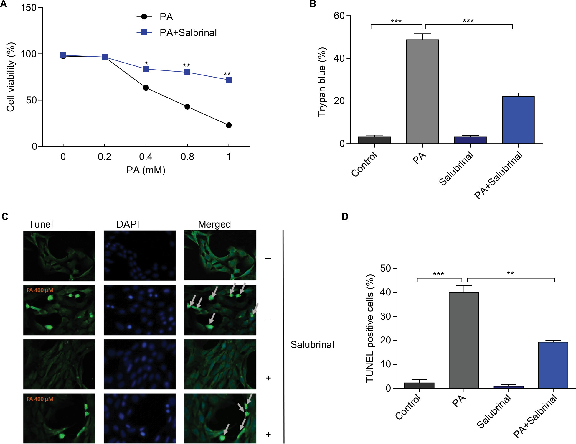 | Figure 1 Salubrinal protects hypothalamic cells against PA-induced cell death. Notes: mHypoE-44 cells were pretreated with salubrinal at 20 µM for 1 hour prior to PA treatment at the indicated concentrations. After 24 hours, cell viability was tested by Cell Counting Kit-8 (A) and TUNEL assay (C, D); cell death was assessed by trypan blue staining assay (B). Data were expressed as mean ± SEM of three independent experiments. *P<0.05, **P<0.01, ***P<0.001 compared with control groups. |
PA induces hypothalamic leptin resistance and ER stress
To study the effects of PA in regulating leptin signaling and ER stress in the hypothalamic cells, mHypoE-44 cells were treated with PA at indicated doses for 24 hours prior to leptin treatment. After 45 minutes, cells were collected and prepared for Western blot analysis. Leptin treatment increased levels of phospho-JAK2 and phospho-STAT3 where this increase was significantly diminished by PA challenge in a dose-dependent manner (Figure 2A–C). These results indicate that PA treatment is able to induce hypothalamic leptin resistance. ER stress is well known as one major cause of leptin resistance. To further assess the role of PA in ER stress, we treated mHypoE-44 cells with PA only and subjected them to Western blot analysis for ER stress markers, phospho-PERK and CHOP (Figure 2D–F). Significant induction of these two markers was found in response to PA treatment, which implicates that PA promotes hypothalamic leptin resistance and ER stress.
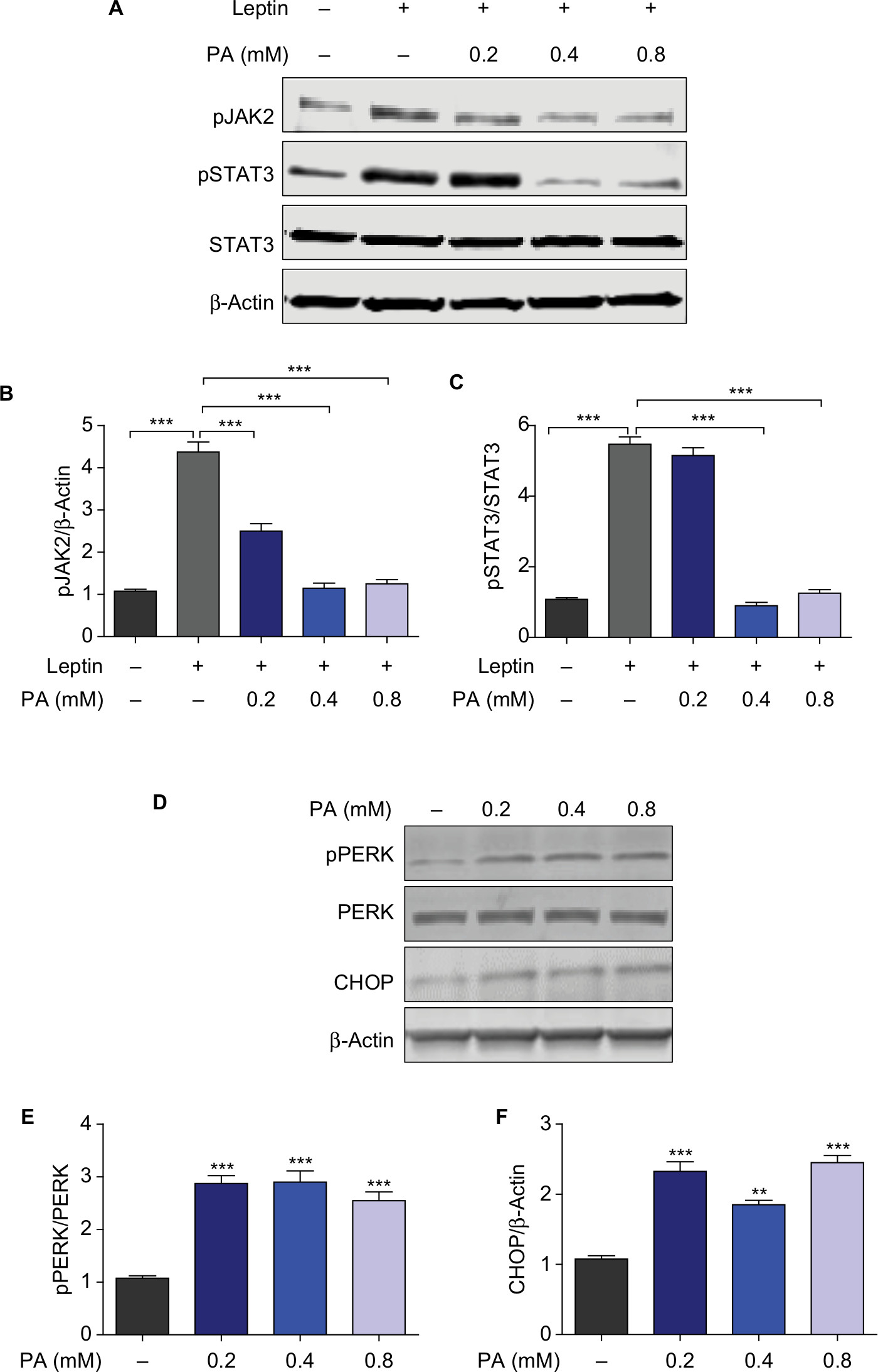 | Figure 2 PA provokes hypothalamic leptin resistance and ER stress. Notes: (A) mHypoE cells were treated with PA at indicated doses for 24 hours prior to leptin treatment. After 45 minutes, cells were prepared for Western blot analysis. Representative Western blots show levels of phospho-JAK2 (pJAK2), phospho-STAT3 (pSTAT3), total-STAT3, and β-Actin levels. (B, C) Quantification of pJAK2 and pSTAT3 normalized to β-Actin and total-STAT3, respectively. (D) mHypoE cells were treated with PA at indicated doses for 24 hours and subjected to Western blot analysis. The results show the levels of phosphor-PERK (pPERK), total PERK, CHOP, and β-Actin. (E,F) Quantification of pPERK and CHOP normalized to total-PERK and β-Actin, respectively. Results were expressed as mean ± SEM of three independent experiments at least. **P<0.01, ***P<0.001 compared with control groups. Abbreviations: ER, endoplasmic reticulum; JAK2, Janus-activated kinase 2; STAT3, signal transducer and activator of transcription 3. |
Salubrinal promotes hypothalamic leptin sensitivity via inhibiting PA-induced ER stress response
To verify the effect of salubrinal on PA-Induced leptin resistance and ER stress, mHypoE cells were co-treated with salubrinal, PA, and leptin. Cells were then subjected to Western blot analysis and probed for the expressions of phosoho-STAT3 and CHOP. Results demonstrated that the elevated expression levels of CHOP induced by PA treatment were significantly attenuated by co-treatment with salubrinal (Figure 3A, C). In addition, PA-induced impairment of leptin signaling was also rescued by salubrinal (Figure 3A, B). These results suggest that salubrinal promotes leptin sensitivity and protects hypothalamic cells against PA-induced ER stress.
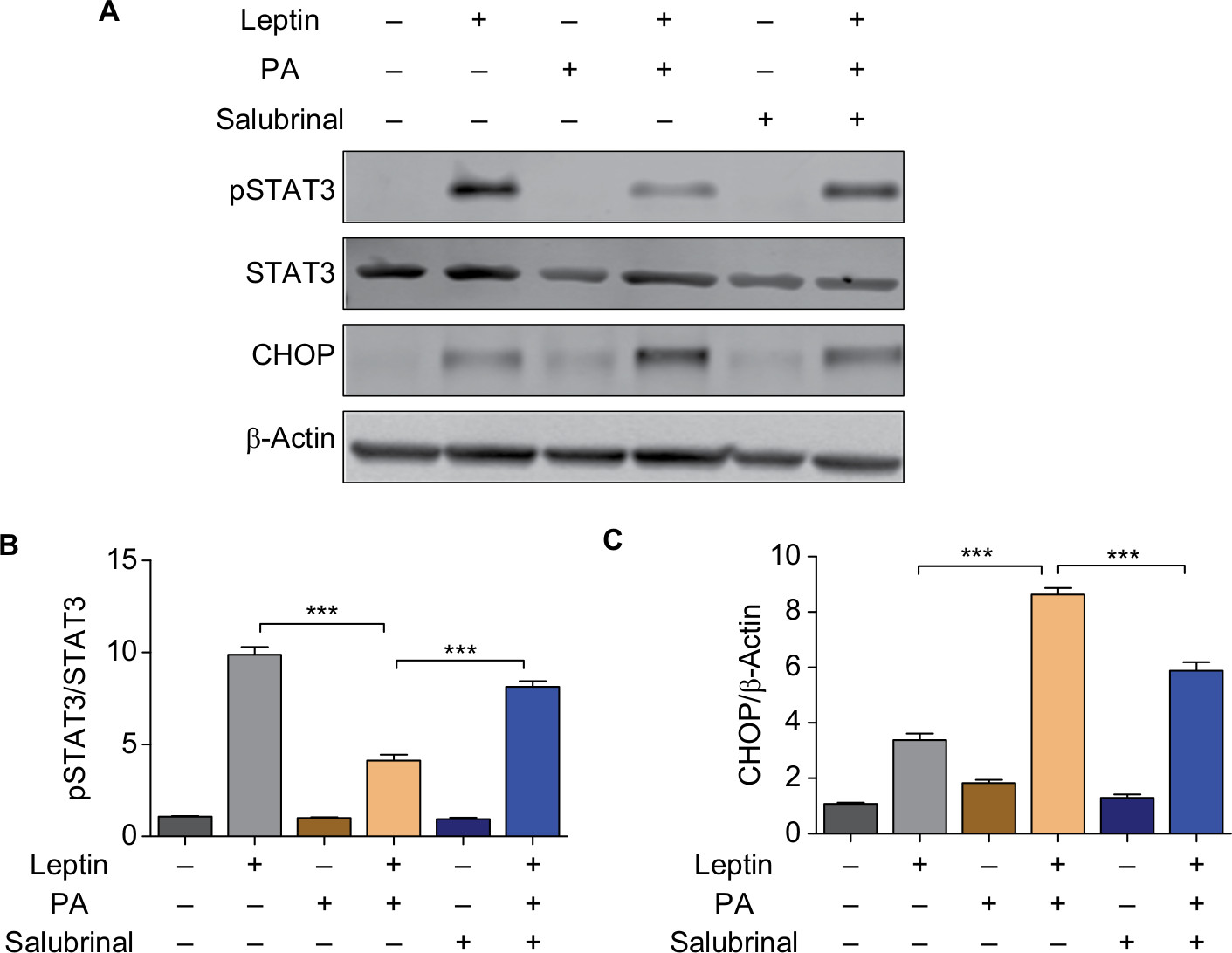 | Figure 3 Salubrinal promotes hypothalamic leptin sensitivity by inhibiting PA-induced ER stress response. Notes: (A) mHypoE cells were pretreated with salubrinal prior to PA treatment and then subjected to leptin challenge. The expressions of pSTAT3, STAT3, CHOP, and β-Actin expression were assessed by Western blot. (B) Protein expressions of pSTAT3 were quantified by Image J software relative to levels of total STAT3. (C) Protein expressions of CHOP were quantified by Image J software relative to levels of β-Actin. Data were expressed as mean ± SEM of three independent experiments. ***P<0.001 compared with control group. Abbreviations: ER, endoplasmic reticulum; JAK2, Janus-activated kinase 2; pSTAT3, phospho-STAT3; STAT3, signal transducer and activator of transcription 3. |
Salubrinal inhibits the activation of hypothalamic nuclear factor kappa-light-chain-enhancer of activated B cells (NF-κB) pathway induced by PA
NF-κB pathway is implicated in many biological progresses in the nervous system, including inflammation, stress response, and cell survival.20,21 To test whether PA treatment can activate NF-κB pathway and salubrinal is able to inhibit the activation, we challenged hypothalamic cells with salubrinal and PA. Western blot analysis probing for the NF-κB pathway demonstrated that salubrinal notably blocked the transportation of NF-κB p65 from cytoplasm to nucleus induced by PA treatment (Figure 4A, B, D, and E). Furthermore, the degradation of IκB induced by PA was also diminished by salubrinal (Figure 4C, F). In sum, these results indicate that salubrinal attenuated the activation of the NF-κB pathway induced by PA treatment in hypothalamic cells.
 | Figure 4 Salubrinal inhibits the activation of hypothalamic NF-κB pathway induced by PA treatment. Notes: (A) mHypo-E44 cells were pretreated with salubrinal prior to PA treatment. Cytoplasmic extracts were isolated and subjected to Western blot analysis probing for NF-κB p65 and β-Actin. (B) Nucleus fraction was extracted and prepared for Western blot for NF-κB p65 and Lamin B1. (C) Whole cell lysates were prepared and subjected to Western blot analysis for Phosphor-IκBα, total-IκBα, and β-Actin. The levels of cytoplasmic NF-κB p65 (D), nucleus NF-κB p65 (E), p-IκBα, and total-IκBα (F) were quantified by Image J software relative to their loading controls. Data shown were representative as mean ± SEM for three independent experiments. **P<0.01, ***P<0.001 compared with control groups. Abbreviation: NF-κB, nuclear factor kappa-light-chain-enhancer of activated B cells, |
Discussion
With an increase in consumption of the Western-style diet, characterized as an HFD, obesity has become a global problem related to the rise of a wide range of diseases. Much effort has been made to explore novel chemicals that may help protect against HFD-induced metabolic syndromes. In the present study, we aim to testify the protective role of salubrinal against PA-induced hypothalamic cell death, leptin resistance, and ER stress. Furthermore, we want to determine the mechanism underlying its protective role.
The hypothalamus is the main site in the central nervous system in controlling energy homeostasis. In the present study, we performed our experiments using the hypothalamic cell line, mHypoE-44. This cell line has a neuronal morphology and is derived and immortalized from mouse embryonic day 17 hypothalamic primary cultures by retroviral transfer of SV40 T-Ag. In addition, mHypoE-44 cells are known to express neuropeptide Y, AgRP, and leptin receptor. As a major component in the HFD, PA is reported to cause hypothalamic inflammation and insulin resistance.22,23 In addition, PA also leads to insulin signaling impairment. However, the correlation between PA and hypothalamic cell death is still elusive. ER stress, which is well known as a major cause of leptin resistance, can also regulate neuron death, neuron inflammation, and neurogenesis.24–26 In our study, we found that exposure to PA results in hypothalamic cell death, leptin resistance, and ER stress. Co-treatment with salubrinal largely reverses these effects of PA and protects hypothalamic cells from apoptosis. These results delineate the specific role of PA in hypothalamic leptin signaling and establish a putative agent, salubrinal, to reverse the adverse effects of over-nutrition.
Finally, we aimed to determine the underlying mechanism by which salubrinal acts in the hypothalamic cell death. The pro-inflammatory NF-κB pathway plays various roles in the nervous system, including regulation of neuroprotection, neuron proliferation, and inflammation.27 It has been reported that HFD induces chronic hypothalamic inflammation and increases the secretion of pro-inflammatory cytokines in the hypothalamus.28 Moreover, leptin resistance is also associated with neuroinflammation.29 This is consistent with our findings in the present study that PA treatment activates the NF-κB pathway. When activated, NF-κB enters into the nucleus and induces the transcription of pro-inflammatory genes. Meanwhile, IκB is phosphorylated then degraded. Currently, we have verified that salubrinal inhibits the activation of the NF-κB pathway and attenuates the degradation of IκB induced by PA treatment. These results suggest that the NF-κB pathway is critical for the protective role of salubrinal against PA-induced hypothalamic cell death and ER stress.
In sum, we have revealed the protective role of salubrinal in PA-induced hypothalamic cell death via inhibiting ER stress and promoting leptin sensitivity. Our results demonstrate that salubrinal can be a potential therapeutic chemical used in preventing HFD-induced obesity.
Acknowledgment
This research was supported by Funds for Major Science and Technology Projects from Changzhou Health and Family Planning Commission (No. ZD201512).
Disclosure
The authors report no conflicts of interest in this work.
References
Friedman JM. Obesity: causes and control of excess body fat. Nature. 2009;459(7245):340–342. | ||
Wang YC, McPherson K, Marsh T, Gortmaker SL, Brown M. Health and economic burden of the projected obesity trends in the USA and the UK. Lancet. 2011;378(9793):815–825. | ||
Hodson L, Skeaff CM, Fielding BA. Fatty acid composition of adipose tissue and blood in humans and its use as a biomarker of dietary intake. Prog Lipid Res. 2008;47(5):348–380. | ||
Hudgins LC, Hellerstein M, Seidman C, Neese R, Diakun J, Hirsch J. Human fatty acid synthesis is stimulated by a eucaloric low fat, high carbohydrate diet. J Clin Invest. 1996;97(9):2081–2091. | ||
Karmi A, Iozzo P, Viljanen A, et al. Increased brain fatty acid uptake in metabolic syndrome. Diabetes. 2010;59(9):2171–2177. | ||
Ubhayasekera SJ, Staaf J, Forslund A, Bergsten P, Bergquist J. Free fatty acid determination in plasma by GC-MS after conversion to Weinreb amides. Anal Bioanal Chem. 2013;405(6):1929–1935. | ||
Penke M, Schuster S, Gorski T, Gebhardt R, Kiess W, Garten A. Oleate ameliorates palmitate-induced reduction of NAMPT activity and NAD levels in primary human hepatocytes and hepatocarcinoma cells. Lipids Health Dis. 2017;16(1):191. | ||
Zhang Y, Proenca R, Maffei M, Barone M, Leopold L, Friedman JM. Positional cloning of the mouse obese gene and its human homologue. Nature. 1994;372(6505):425–432. | ||
Hübschle T, Thom E, Watson A, Roth J, Klaus S, Meyerhof W. Leptin-induced nuclear translocation of STAT3 immunoreactivity in hypothalamic nuclei involved in body weight regulation. J Neurosci. 2001;21(7):2413–2424. | ||
Yang G, Lim CY, Li C, et al. FoxO1 inhibits leptin regulation of pro-opiomelanocortin promoter activity by blocking STAT3 interaction with specificity protein 1. J Biol Chem. 2009;284(6):3719–3727. | ||
Pagliassotti MJ, Kim PY, Estrada AL, Stewart CM, Gentile CL. Endoplasmic reticulum stress in obesity and obesity-related disorders: an expanded view. Metabolism. 2016;65(9):1238–1246. | ||
Ozcan L, Ergin AS, Lu A, et al. Endoplasmic reticulum stress plays a central role in development of leptin resistance. Cell Metab. 2009;9(1):35–51. | ||
Purkayastha S, Zhang H, Zhang G, Ahmed Z, Wang Y, Cai D. Neural dysregulation of peripheral insulin action and blood pressure by brain endoplasmic reticulum stress. Proc Natl Acad Sci U S A. 2011;108(7):2939–2944. | ||
Kim JS, Heo RW, Kim H, et al. Salubrinal, ER stress inhibitor, attenuates kainic acid-induced hippocampal cell death. J Neural Transm (Vienna). 2014;121(10):1233–1243. | ||
Wu L, Luo N, Zhao HR, et al. Salubrinal protects against rotenone-induced SH-SY5Y cell death via ATF4-parkin pathway. Brain Res. 2014;1549:52–62. | ||
Cakir I, Cyr NE, Perello M, et al. Obesity induces hypothalamic endoplasmic reticulum stress and impairs proopiomelanocortin (POMC) post-translational processing. J Biol Chem. 2013;288(24):17675–17688. | ||
Schwartz MW, Woods SC, Porte D, Seeley RJ, Baskin DG. Central nervous system control of food intake. Nature. 2000;404(6778):661–671. | ||
He W, Lam TK, Obici S, Rossetti L. Molecular disruption of hypothalamic nutrient sensing induces obesity. Nat Neurosci. 2006;9(2):227–233. | ||
Lam TK, Schwartz GJ, Rossetti L. Hypothalamic sensing of fatty acids. Nat Neurosci. 2005;8(5):579–584. | ||
Baldwin AS. Series introduction: the transcription factor NF-kappaB and human disease. J Clin Invest. 2001;107(1):3–6. | ||
Hoesel B, Schmid JA. The complexity of NF-κB signaling in inflammation and cancer. Mol Cancer. 2013;12:86. | ||
Posey KA, Clegg DJ, Printz RL, et al. Hypothalamic proinflammatory lipid accumulation, inflammation, and insulin resistance in rats fed a high-fat diet. Am J Physiol Endocrinol Metab. 2009;296(5):E1003–E1012. | ||
Delarue J, Magnan C. Free fatty acids and insulin resistance. Curr Opin Clin Nutr Metab Care. 2007;10(2):142–148. | ||
Brown MK, Naidoo N. The endoplasmic reticulum stress response in aging and age-related diseases. Front Physiol. 2012;3:263. | ||
Anuncibay-Soto B, Pérez-Rodriguez D, Santos-Galdiano M, et al. Salubrinal and robenacoxib treatment after global cerebral ischemia. Exploring the interactions between ER stress and inflammation. Biochem Pharmacol. 2018;151:26–37. | ||
Sprenkle NT, Sims SG, Sánchez CL, Meares GP. Endoplasmic reticulum stress and inflammation in the central nervous system. Mol Neurodegener. 2017;12(1):42. | ||
Wang R, Chen S, Liu Y, et al. All-trans-retinoic acid reduces BACE1 expression under inflammatory conditions via modulation of nuclear factor κB (NFκB) signaling. J Biol Chem. 2015;290(37):22532–22542. | ||
Morselli E, Fuente-Martin E, Finan B, et al. Hypothalamic PGC-1α protects against high-fat diet exposure by regulating ERα. Cell Rep. 2014;9(2):633–645. | ||
de Git KC, Adan RA. Leptin resistance in diet-induced obesity: the role of hypothalamic inflammation. Obes Rev. 2015;16(3):207–224. |
 © 2018 The Author(s). This work is published and licensed by Dove Medical Press Limited. The
full terms of this license are available at https://www.dovepress.com/terms
and incorporate the Creative Commons Attribution
- Non Commercial (unported, 3.0) License.
By accessing the work you hereby accept the Terms. Non-commercial uses of the work are permitted
without any further permission from Dove Medical Press Limited, provided the work is properly
attributed. For permission for commercial use of this work, please see paragraphs 4.2 and 5 of our Terms.
© 2018 The Author(s). This work is published and licensed by Dove Medical Press Limited. The
full terms of this license are available at https://www.dovepress.com/terms
and incorporate the Creative Commons Attribution
- Non Commercial (unported, 3.0) License.
By accessing the work you hereby accept the Terms. Non-commercial uses of the work are permitted
without any further permission from Dove Medical Press Limited, provided the work is properly
attributed. For permission for commercial use of this work, please see paragraphs 4.2 and 5 of our Terms.