Back to Journals » Pediatric Health, Medicine and Therapeutics » Volume 11
Salt Treatment for Umbilical Granuloma – An Effective, Cheap, and Available Alternative Treatment Option: Case Report
Authors Haftu H , Gebremichael TG , Kebedom AG
Received 24 July 2020
Accepted for publication 11 September 2020
Published 30 September 2020 Volume 2020:11 Pages 393—397
DOI https://doi.org/10.2147/PHMT.S269114
Checked for plagiarism Yes
Review by Single anonymous peer review
Peer reviewer comments 3
Editor who approved publication: Professor Roosy Aulakh
Hansa Haftu,1 Teklu Gebrehiwot Gebremichael,2 Abel Gidey Kebedom1
1Mekelle University, College of Health Science, Pediatric, and Child Health, Mekelle, Ethiopia; 2Mekelle University, College of Health Science, Mekelle, Ethiopia
Correspondence: Hansa Haftu
Mekelle University, College of Health Science, Pediatric, and Child Health, Tigray, Mekelle, Ethiopia
Tel +251948487877
Email [email protected]
Introduction: Umbilical granuloma (UG) is the most common cause of umbilical mass and it is formed in the first few weeks of life after the umbilical cord separates. Though there are different options of treatment described in the literature, there is no clear consensus on the best option of treatment. In our case, we will describe the complete resolution of granuloma with salt treatment with no adverse effect.
Case Presentation: An 18-day-old female infant presented to the outpatient department (OPD) with concerns of swelling over the umbilicus with a yellowish discharge of one-day duration noticed after separation of the umbilical cord. The discharge was, initially, odorless, with no fecal or urine content, no pain, and behavioral change in the neonate. The mother was advised on sponge wash and to apply silver nitrate or liquid nitrogen. After five days, the patient presents with purulent discharge from the umbilical swelling of two-day duration but no other complaint. Discharge was noted to be purulent but no erythema in the surrounding skin. The patient had no leukocytosis on labs. A teaspoon of cooking salt was applied to cover the whole granuloma and packed the umbilicus for 30 minutes with gauze. After 30 minutes on the pack, the salt was removed and cleaned with normal saline. Subsequently, after the salt was applied, the granuloma changed from red to blush hue. After three applications of salt pack, the granuloma decreased in size, became dry, and separated. There was no side effect noted and the infant was followed up with no recurrence noted at 3 months of age.
Conclusion: Salt treatment appears to be an effective, available, and less costly treatment option for an umbilical granuloma.
Keywords: salt, umbilicus, granuloma, neonate
Introduction
An umbilical granuloma is the most common umbilical mass in the neonatal period. Though the exact cause is not known, this pink friable mass is thought to be formed due to excessive inflammation in the base of the umbilical cord likely due to infection which may result in delayed cord separation.1,2 The umbilical cord stump usually dries and separates within 1–2 weeks afterbirth.2 An umbilical granuloma looks like a small piece of bright red, moist flesh that remains in the umbilicus after cord separation when normal healing should have occurred.3 Though there are no reports on the spontaneous regression of umbilical granuloma without treatment, some authors recommend observational conservative management without any intervention.2,4 There are other alternative treatment options of granulomas, like the double-ligature technique which is simple to perform and provides good cosmetic and functional results with only minor complications like bleeding.5 The first report of the use of salt for the treatment of umbilical granuloma was reported by Schmitt in 1972 and then detailed by Kesaree in 1983. Worldwide practice and most principal pediatric and Neonatology textbooks on pediatrics and Neonatology recommend silver nitrate as a first-line treatment option. Application of silver nitrate is associated with superficial burns and needs frequent application.1,6,7 Though there are different options of treatment in the literature, there is no clear consensus on the management of UG.6 Although the most commonly used treatment modality is the application of silver nitrate, there are other options of management in the literature, like salt, a topical antiseptic, and steroid. Midwives and neonatologists have shown some reservations on salt treatment because of limited evidence.7 One of the reasons why medical reference books have not yet supported the use of common salt for the treatment of umbilical granuloma is the small number of studies conducted in this area.7 Our case report documents the role of salt treatment in complete resolution of UG and with no adverse effect and non-recurrence in patients with umbilical granuloma.
Case Presentation
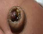
An 18-day-old neonate from Mekelle presented to the OPD with pinkish swelling over the umbilicus and yellowish discharge of one-day duration after separation of the cord. The discharge was, initially, odorless, with no fecal or urine content. According to the mother’s description, there was no pain and behavioral change in the neonate. On physical examination, the infant was alert with stable vital signs. There was no hypothermia or fever. The red-colored umbilical mass measured about 15mm in diameter associated with yellow discharges consisted of umbilical granuloma (Figure 1). There was no edema and color change of the surrounding skin, purulent drainage from the umbilicus. The mother was advised to have granular treated with silver nitrate or liquid nitrogen in the nearby health centers. But she cannot find the drug. After five days, the patient presented with purulent discharge from around the umbilical swelling of two-day duration but no other complaint. Vital signs were normal. There was a purulent discharge from the root of the umbilical granuloma with swelling around the umbilicus, but no erythema of the skin around the umbilicus (Figure 2). On investigation, the patient had no leukocytosis. The patient underwent wound care with topical antibiotics (TTC) for weeks and it became clear with no discharge. The patient was discharged with counseling on hygiene. Again, after 3 days of antibiotics and wound care discontinuation, and the patient starts to have minimal purulent discharge from the swelling but stable vital signs. A cooking salt was applied to cover the entire lesion after the area was cleaned with normal saline. The area was covered with a gauze dressing and held it in place for 30 minutes. This was performed two times per day. After the single application of the salt in the granuloma, there was a color change (Figure 3). Subsequently, after applying three times, the granuloma decreased size with color change and separated (Figure 4). There was no visible side effect of the salt in the skin (no erythema) or no bleeding during the application. There is no recurrence of the granuloma on subsequent follow-ups at the 3 months. Now the patient is a one-year old healthy with normal growth and development.
 |
Figure 1 The Initial appearance of umbilical granuloma with a yellowish discharge at its base after the cord falls off. |
 |
Figure 2 Infected umbilical granuloma with pussy discharge, but no erythema of the surrounding skin. |
 |
Figure 3 The immediate color change of Umbilical granuloma after salt was applied and packed for 30 minutes. |
 |
Figure 4 Umbilical granuloma changes in the subsequent salt application. (A) After the salt was applied for a second time. (B) After the salt was applied for the third time. |
Discussion and Conclusion
An umbilical granuloma is a condition that can develop in a newborn baby’s umbilical stump. Umbilical granulomas develop in about 1 out of 500 births.6 It usually looks like a soft pink or red lump and often is moist with small amounts of clear or yellow fluid. Almost all standard references, recommend treating granulation tissue by cauterization with silver nitrate, repeated at intervals of several days until the base is dry.2 It is most common in the first few weeks of a baby’s life.4 Like our patient, this commonly appears after the cord falls off and with swelling and discharge from the granuloma. There are two well-known important methods of management. The common treatment is the application of a 75% silver nitrate stick, usually repeated two to three times of clinic visits. Burns have been reported following spillage onto the surrounding tissues, another method is using excision and the application of absorbable hemostatic materials, but they have their own limitation recurrence, bleeding.1,3,7 The first report of salt as treatment of umbilical granuloma was initially reported by Schmitt in 1972 and then detailed by Kesaree in 1983.6 The salt will create an osmolality difference resulting in drawing the water from the granuloma leading to drying of the granuloma and separation. This application of salt has shown to be effective for the treatment of pyogenic granuloma.6,7 Five patients with granuloma were treated with salt leading to complete resolution with no recurrence at one-month follow-up and no major adverse effects of salt except mild burning pain with slight bleeding which later resolved during the first application. The time for the complete resolution of the granuloma was 7–14 days depending on the size.6 Similarly, the patient described in our report showed a complete resolution in two days with no recurrence in the subsequent follow-up. In our patient, we describe the process involved in the application of salt for the treatment of UG. The granuloma and the surrounding skin were cleaned before with normal saline. Apply a small pinch of the table or cooking salt onto the umbilical granuloma. Cover the area with a gauze dressing and secure it in place for 30 minutes. This should be repeated twice a day for at least two days. In approximately two or three days, the granuloma is expected to reduce in size, change color and dry off. Over the next few days, the area will gradually heal.4,8 In our patient, we used the cooking salt and this procedure was followed and the color change was noticed even earlier (Figure 3) than expected and the granuloma separated on the second day (Figure 4). The initial evidence related to salt treatment is based on lesser quality studies conducted in developing countries. However, the results of this pragmatic method appear to be consistent and indicate a good clinical outcome.1 Our case report shows the efficacy, availability, and easy application of the salt. Kesaree et al reported a 100% success rate in their study population for the treatment of granuloma with salt.9 One prospective study, Hossain et al reported that table salt application resulted in excellent outcomes in 91.7% of their patients with no side effects. In this study, the silver nitrate treatment group also achieved good results, but with side effects (burns and pain). The authors did not recommend silver nitrate, because of small burns and pain at the umbilicus in some patients.10 Jimish et al report complete resolution of umbilical granuloma after a table salt was carefully applied over the lesion. The granuloma was then occluded with surgical adhesive tape for 24 hours. Cases were followed up the next day to remove the occlusive tape and for assessment of improvement. All seventeen cases responded well to this approach with complete resolution of lesions at 24 hours. A small clot like shrunken tissue was found at the site of granuloma, which was easily scraped off during gentle cleansing. No major complication or recurrence was noted in 3 months of follow-up.11 As previously reported in the literature, we can conclude that complete resolution of umbilical granuloma can be achieved with table salt application to be continued for 2–3 days two times per day. Salt causes shrinkage of granuloma inside the occluded hyperosmolar chamber by the desiccant effect. The advantages of this treatment option are that it is easy to apply with low cost, available everywhere, has low cost, and causes rapid resolution without complications.
Abbreviations
UG, umbilical granuloma; OPD, outpatient department.
Data Sharing Statement
Please contact the corresponding author for data requests.
Ethics Approval and Consent for Publication
Written informed consent was obtained from the patient’s mother for publication of this case with the inclusion of symptoms, signs and images of the patients in the manuscript paper. A copy of the written consent is available for review by the Editor-in-Chief of this journal. There is no need to have Ethical approval of case reports in our institution.
Acknowledgment
I thank pediatric residents, seniors, and other staff in Ayder hospitals who contributed to the hospital management of our patients. I am also thankful for the patient and her mother for their willingness for the publication and Mr. Teklu and Dr. Abel for their support in writing this case report.
Author Contributions
All authors made a significant contribution to the work reported, whether that is in the conception, study design, execution, acquisition of data, analysis and interpretation, or in all these areas; took part in drafting, revising or critically reviewing the article; gave final approval of the version to be published; have agreed on the journal to which the article has been submitted; and agree to be accountable for all aspects of the work.
Funding
No specific funding was received.
Disclosure
The authors declare that they have no competing interests in this work.
References
1. Karaguzel G, Aldemir A. Umbilical Granuloma: modern Understanding of Etiopathogenesis, Diagnosis, and Management. J Pediatr Neonatal Care. 2016;4(3):1–5. doi:10.15406/jpnc.2016.04.00136
2. Amy TN, The Umbilicus, Granuloma; Nelson pediatrics 21 editions. 4175–4176.
3. Ali N, Muslim K, Razzaq J, et al. Management of umbilical granuloma. Thi-Qar Med J. 2010;4(4):82–87.
4. Whiston Hospital Children’s Community Nursing Team. Salt treatment for umbilical granuloma in babies, St Helens, and Knowsley a teaching hospital. 2018.
5. Gad L, Baruch K, Yigal E. Double-ligature: a treatment for pedunculated umbilical granulomas in children. Am Fam Physician. 2002;65(10):2067–2068.
6. Sanober B, Rachita S. A pinch of salt is all it takes! - The novel use of table salt for the effective treatment of pyogenic granuloma. J Pre-Proof. 2019;1–7.
7. Bedfordshire and Luton Joint Prescribing Committee. Community health services bedfordshire, household salt for treatment of umbilical granuloma. 2017;1–9.
8. Child health information, umbilical granuloma in babies, royal united hospitals bath NHS foundation trust. 2015; 1–2.
9. Kesaree N, Babu PS, Banapurmath CR, Krishnamurthy SN. Umbilical granuloma. Indian Pediatr. 1983;20(9):690–692.
10. Hossain AZ, Hasan GZ, Islam KD. Therapeutic effect of common salt (table/cooking salt) on umbilical granuloma in infants. Bangladesh J Child Health. 2010;34(3):99–102. doi:10.3329/bjch.v34i3.10360
11. Jimish B, Saurabh J, Krishna B, et al. Pinch of salt: a modified technique to treat umbilical granuloma. Natl Inst Health. 2019;36(4):561–563.
 © 2020 The Author(s). This work is published and licensed by Dove Medical Press Limited. The full terms of this license are available at https://www.dovepress.com/terms.php and incorporate the Creative Commons Attribution - Non Commercial (unported, v3.0) License.
By accessing the work you hereby accept the Terms. Non-commercial uses of the work are permitted without any further permission from Dove Medical Press Limited, provided the work is properly attributed. For permission for commercial use of this work, please see paragraphs 4.2 and 5 of our Terms.
