Back to Journals » OncoTargets and Therapy » Volume 13
Ribosome Binding Protein 1 Correlates with Prognosis and Cell Proliferation in Bladder Cancer
Authors Lv S, Shi Z, Wang X, Zheng P, Li H, Han Q, Li Z ![]()
Received 1 March 2020
Accepted for publication 13 May 2020
Published 7 July 2020 Volume 2020:13 Pages 6699—6707
DOI https://doi.org/10.2147/OTT.S252043
Checked for plagiarism Yes
Review by Single anonymous peer review
Peer reviewer comments 2
Editor who approved publication: Dr Sanjeev K. Srivastava
Shuang-wu Lv, Zhen-guo Shi, Xiao-hui Wang, Peng-yi Zheng, Hui-bing Li, Qing-jiang Han, Zhi-jun Li
The First Affiliated Hospital and College of Clinical Medicineof Henan University of Science and Technology, Luoyang, Henan 471003, People’s Republic of China
Correspondence: Zhi-jun Li
The First Affiliated Hospital and College of Clinical Medicine of Henan University of Science and Technology, Luoyang 471003, Henan Province, People’s Republic of China
Email [email protected]
Introduction: Ribosome binding protein 1 (RRBP1) is reported to be correlated with tumor formation and progression. However, the role of RRBP1 in bladder cancer is unclear. In this study, we aimed to investigate the expression of RRBP1 and its influence on cell proliferation in bladder cancer.
Methods: Quantification real-time polymerase chain reaction (qRT-PCR) and immunohistochemistry (IHC) were used to detect the expression levels of RRBP1 in 138 bladder cancer and matched adjacent normal bladder tissues. Then, the clinical significance of RRBP1 in bladder cancer was evaluated. The effect of RRBP1 on cell proliferation and its potential mechanism were further explored.
Results: Results show that the mRNA levels of RRBP1 in bladder cancer were significantly higher compared with those in normal tissues (P< 0.001). IHC results show the high-expression rate of RRBP1 in bladder cancer was 68.8%, which was significantly greater than those in normal tissues (40.6%, P< 0.001). RRBP1 high-expression was significantly associated with differentiation, T stage and lymph node metastasis in bladder cancer (P< 0.05). The overall survival time of patients with RRBP1 high-expression was significantly reduced compared to those with RRBP1 low-expression. Moreover, RRBP1 overexpression significantly promoted cell proliferation, which was correlated with Smad1/Smad3/TGF-β 1 signal pathway.
Conclusion: RRBP1 high-expression correlates with prognosis and promotes cell proliferation in bladder cancer, which could be a potential biomarker.
Keywords: RRBP1, prognosis, bladder cancer, survival, biomarker
Introduction
Bladder cancer is one of the most common urological carcinomas in the world.1–3 It is reported that the incidence of bladder cancer is still increasing.4,5 Furthermore, bladder cancer presents a high recurrence rate and mortality rate because of the absence of typical symptoms at the early stage.6,7 No reliable biomarkers are helpful for clinical therapy and survival estimation.8–10 Therefore, identifying effective biomarkers for early diagnosis and prognosis assessment is needed for the clinical therapy of bladder cancer.
Ribosome binding protein 1 (RRBP1) is an endoplasmic reticulum membrane protein which plays a crucial role in the secretion and transportation of nascent proteins in mammalian cells.11–15 In addition, RRBP1 is involved in the endoplasmic reticulum stress and unfolded protein response.16,17 Nowadays, RRBP1 high-expression is reported in lung cancer,18 breast cancer,19,20 colorectal cancer,21,22 esophageal carcinoma,23 prostate cancer,24 endometrial endometrioid adenocarcinoma25 and ovarian cancer.26 Moreover, RRBP1 correlates with patients’ survival in breast cancer,19 colorectal cancer,22 esophageal carcinoma,23 prostate cancer,24 endometrial endometrioid adenocarcinoma25 and ovarian cancer.26 However, the role of RRBP1 in bladder cancer remains unknown.
In the present study, the expression of RRBP1 and its clinical significance in bladder cancer were investigated. Moreover, the effect of RRBP1 expression on cell proliferation and its potential mechanism were explored.
Patients and Methods
Patients and Samples
One hundred thirty-eight bladder cancer and matched adjacent normal tissues were obtained from the First Affiliated Hospital of Henan University of Science and Technology. All patients received surgical resection during 2010 to 2017. None of the patients were treated with chemotherapy or radiotherapy before surgery. Clinical-pathological characters including age, sex, history of smoking, lymph node metastasis, T stage and differentiation were obtained from hospital records. The follow-up period for patients was recorded from the day of surgery. The follow-up time was from 7 to 65 months. All patients agreed to participate in this study and provided written informed consent. All experiments were approved by the ethics committee of Henan University of Science and Technology, and performed in line with the Declaration of Helsinki and relevant laws and regulations. All animal experiments were performed in accordance with policies of the NIH Guide for the Care and Use of Laboratory Animals and the Institutional Animal Care and Use Committee (IACUC) of the First Affiliated Hospital of Henan University of Science and Technology.
Cell Culture and Transfection
Human EJ, UM-UC-3 and T24 bladder cancer cell lines were obtained from the Cell Bank of Type Culture Collection of Chinese Academy of Sciences (Shanghai, China). Cells were cultured with RPMI-1640 (Gibco) with 10% heat-inactivated fetal bovine serum (Hyclone) at 37 °C in a humidified incubator with 5% CO2. Based on the manufacturer’s instructions, cell transfection was successfully completed by LipofectamineTM 2000 (Invitrogen, Thermo Fisher Scientific, Inc., USA). RRBP1 downregulation was performed by transferring shRNA lentivirus vectors (Genepharma, Suzhou, China) and RRBP1 overexpression was performed by transferring recombinant lentivirus vectors (Genepharma, Suzhou, China). Non-target shRNA (Genepharma, Suzhou, China) lentivirus vectors were treated as the negative control.
Quantification Real-Time Polymerase Chain Reaction (qRT-PCR)
After surgery, all fresh tissues were collected and immediately stored in liquid nitrogen until RNA extraction. Total RNAs were obtained from fresh tissues and cells by using the manufacture’s protocol. The RNA was reversed into cDNA using a PrimeScript RT kit (Takara, Dalian, China). Real-time quantitative PCR was done by SYBR1 Premix Ex Taq. Primers for RRBP1 were 5′-AACCTAATGGGAAGATACCTGA-3′ (F) and 5′-CATGGCTGGAACTGTGGC-3′ (R). The primer sequence for GAPDH were 5′-CTGAACGGGAAGCTCACTGG-3′ (F) and 5′-TGAGGTCCACCACCCTGTTG-3′ (R). GAPDH was regarded as the internal control and each experiment was repeated three times. The relative expression level of RRBP1 was compared using the 2–ΔΔCt method.
Western Blotting
Proteins were obtained by radioimmunoprecipitation assay lysis buffer (Abcam Corp, USA) and quantified by bicinchoninic acid. Protein, 50 μg per sample, was separated onto 10% SDS–PAGE gels and transferred onto a polyvinyl difluoride (PVDF) filter membrane. Then, PVDF membranes were incubated with 5% non-fat dried milk, anti-RRBP1 (Abcam Corp., USA), Smad-1 (Abcam Corp., USA), p-Smad-1 (Abcam Corp., USA), Smad-3 (Abcam Corp., USA), p-Smad-3 (Abcam Corp., USA), TGF-β1 (Abcam Corp., USA) and anti-GAPDH (Abcam Corp., USA), respectively. Finally, membranes were washed with phosphate-buffered saline (PBS) and incubated with secondary antibodies. Signals were developed using an enhanced chemiluminescence reaction kit (Applygen Technologies, Beijing, China).
Immunohistochemistry (IHC)
All tissue sections were fixed with 0.4% formulation, rehydrated and embedded into paraffin. Tissue sections (3 μm thick) were deparaffinized with xylene and rehydrated with alcohol. Antigen retrieval was completed by sodium citrate buffer (pH 6.0) for 5 minutes at 100 °C. Then, sections were blocked into 0.3% hydrogen peroxide solution for 20 min and incubated with rabbit polyclonal RRBP1 antibody (Abcam Corp., USA) overnight at 4 °C. Washing with PBS, sections were incubated with biotin-labeled secondary antibodies. Signals were developed by diaminobenzidine tetrahydrochloride. PBS substituted with primary antibody was used as negative controls. Prostate cancer tissues with positive expression of RRBP1 were used as positive controls.24 The scores were scored according to the number of positive cells and staining intensity. Percentage of cells was scored as follows: 1 (<25%), 2 (25%–50%), 3 (>51%). Staining intensity was divided into blank (0), weak (1), moderate (2) and strong (3). Total score ranged from 0 to 9. Using semiquantitative analysis, RRBP1 expression was scored as low-expression (<4) and high-expression (≥4).24
Cell Counting Kit-8 (CCK-8) Analysis
Cell proliferation was assessed by CCK-8 assay. Cells were transferred and seeded in 96-well plates (1.5 × 10³ cells/well) for 5 days. According to the manufacturer’s instructions, cells were cultured with 10 µL of CCK-8 for 3 h, and the optical density of each well was determined by a microplate reader at 450 nm. Each experiment was repeated in triplicate at the same condition.
Colony Formation Analysis
Transfected cells were seeded into 6-well plates at a density of 1 × 10³ cells/well. After 2 weeks, cells were obtained and fixed with 100% methanol. Then, cells were incubated with 0.1% crystal violet and stained with hematoxylin for counting under an optical microscope. Each experiment was repeated in triplicate at the same condition.
Tumor Xenograft in vivo
Balb/c nude mice (4–5 weeks old) were obtained from the Animal Center and kept on specific pathogen-free conditions. To evaluate the tumor growth between the shRNA group and negative control group, 5 × 10⁶ bladder cancer cells were subcutaneously injected into the flank regions of legs (4 mice per group). Tumor size was recorded every 3–5 days. Finally, tumor weight was measured and tumor volumes were calculated by the formula: volume = (length × width2)/2.
Statistical Analysis
Data were characterized as mean ± standard deviation (SD), and performed by SPSS software (version 19.0; SPSS, Chicago, IL, USA). The expression levels of RRBP1 between bladder cancer and matched adjacent normal tissues were compared by paired t-test. The association between RRBP1 expression and clinical-pathological characters was analyzed by Chi-square test (χ2). Survival analysis was compared by using the Kaplan–Meier method with Log rank test. Hazard ratio was estimated using Cox’s proportional hazards model. Statistical significance was P < 0.05.
Results
RRBP1 Expression and its Clinical Significance in Bladder Cancer
To evaluate the expression of RRBP1, the mRNA levels of RRBP1 were detected in 138 cases of bladder cancer and normal bladder tissues by qRT-PCR. As shown in Figure 1A, the mRNA levels of RRBP1 in bladder cancer was significantly higher compared with adjacent normal bladder tissues (P< 0.001). Then, the protein expression levels of RRBP1 were further investigated by IHC analysis. Results show that positive staining of RRBP1 was mainly distributed in the cell cytoplasm and stained as brown and yellow (Figure 1B–E). Semiquantitative analysis of IHC score show the high-expression rate of RRBP1 in bladder cancer was 68.8%, which was significantly greater than those in normal tissues (40.6%, P< 0.001, Table 1).
 |
Table 1 The Expression of RRBP1 Was Examined in Bladder Cancer by IHC |
 |
Figure 1 The expression levels of RRBP1 were examined in bladder cancer. (A) The mRNA levels of RRBP1 were tested by qRT-PCR (N: normal tissues; T: tumor tissues). (B) RRBP1 low-expression in normal bladder tissues by IHC (N= 82). (C) RRBP1 high-expression in normal bladder tissues by IHC (N= 56). (D) RRBP1 low-expression in bladder cancer tissues by IHC (N= 43). (E) RRBP1 high-expression in bladder cancer tissues by IHC (N= 95). |
Subsequently, the association between RRBP1 expression and clinical-pathological characters was investigated based on the IHC scores. Results show RRBP1 high-expression was significantly associated with differentiation, T stage and lymph node metastasis in bladder cancer (P< 0.05, Table 2), while not correlated with age, sex and smoking history (P> 0.05, Table 2). In addition, the prognostic value of RRBP1 was further investigated in bladder cancer. Kaplan–Meier analysis revealed that the overall survival time of patients with RRBP1 high-expression was significantly reduced compared to those with RRBP1 low-expression (Figure 2, P= 0.001). Furthermore, overall survival time was correlated with differentiation, T stage and lymph node metastasis (P< 0.05, Table 3). Multivariate Cox regression analysis showed that RRBP1 expression, differentiation, T stage and lymph node metastasis were independent prognostic factors for overall survival in bladder cancer (Table 4, P< 0.05). However, age, sex and smoking history were not significantly correlated with prognosis.
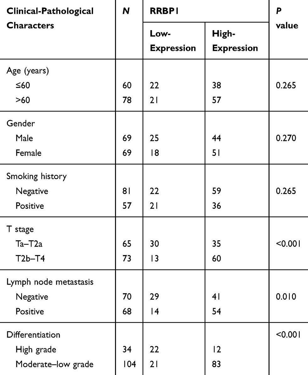 |
Table 2 RRBP1 Expression Correlated with Clinical-Ppathological Characters in Bladder Cancer |
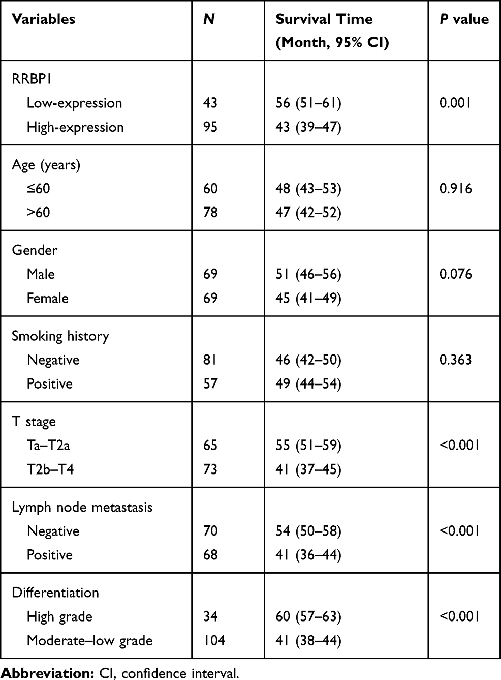 |
Table 3 Survival Analysis Performed by the Kaplan–Meier Method |
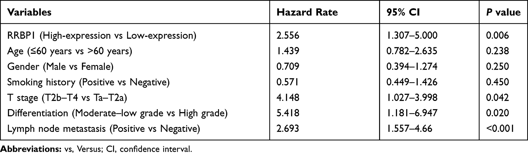 |
Table 4 Prognostic Factors Evaluated by Multivariate Cox Regression Analysis |
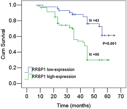 |
Figure 2 Kaplan–Meier survival analysis shows patients with RRBP1 overexpression had unfavorable survival. |
RRBP1 Promotes Cell Proliferation in Bladder Cancer
To investigate the biological role of RRBP1 in bladder cancer, the effect of RRBP1 expression on cell proliferation was investigated. As shown in Figure 3A, RRBP1 was highly expressed in EJ cells, but was relatively low in UM-UC-3 and T24 cells. Therefore, RRBP1 expression was downregulated in EJ cells and upregulated in T24 cells. As shown in Figure 3B, Western blot analysis shows RRBP1 downregulation and upregulation were successfully performed. Then, the proliferation and clone formation of bladder cancer cells were evaluated in vitro. Results showed that RRBP1 knockdown significantly inhibited proliferation in EJ cells (Figure 3C), while RRBP1 overexpression significantly promoted proliferation in T24 cells. Meanwhile, the colony number in cells with RRBP1 downregulation was significantly decreased, while it was significantly increased in cells with RRBP1 overexpression (Figure 4A). To further investigate the effects of RRBP1 expression on cell proliferation, xenografts in vivo were performed. The results showed that RRBP1 knockdown significantly suppressed the growth of bladder cancer xenografts (Figure 4B).
 |
Figure 3 RRBP1 overexpression promoted cell proliferation in vitro. (A) The protein expression levels of RRBP1 were detected in bladder cancer cell lines by Western blot assay. (B) Western blot analysis revealed that RRBP1 downregulation and upregulation were successfully performed. (C) CCK-8 assay shows RRBP1 downregulation inhibited cell proliferation, while RRBP1 overexpression promoted cell proliferation. |
 |
Figure 4 RRBP1 overexpression promoted cell proliferation in vivo. (A) RRBP1 overexpression promoted colony formation. (B) RRBP1 knockdown inhibited the growth of bladder cancer xenografts in vivo. (C) RRBP1 overexpression activated the TGF-β/Smad pathway. |
To further explore the potential mechanism of RRBP1 in bladder, the TGF-β1/Smad pathway was investigated. As shown in Figure 4C, the expression of TGF-β1 was significantly decreased with the knockdown of RRBP1. The protein expression levels of Smad1 and Smad3 were unchanged regardless of the RRBP1 expression interference, while RRBP1 downregulation resulted in the decrease of p-Smad1/3.
Discussion
Recently, RRBP1 overexpression has been reported in several types of tumor,16–26 and could be regarded as a potential prognostic marker.19–24 However, the role of RRBP1 in bladder cancer remains unknown. In this study, the expression and clinical significance of RRBP1 was investigated in bladder cancer. Results show that the mRNA levels of RRBP1 in bladder cancer were significantly greater than those in normal bladder tissues. Meanwhile, IHC results show that RRBP1 was positively expressed in bladder cancer and located in the cell cytoplasm. The high-expression rate of RRBP1 in bladder cancer was 68.8%, which was significantly higher than those in normal tissues (40.6%, P <0.001). These observations indicated that RRBP1 overexpression was associated with the formation of bladder cancer, which was consistent with current reports.16–25 Abnormal high-expression of RRBP1 in bladder cancer might be helpful for the diagnosis of bladder cancer. In addition, RRBP1 overexpression was significantly associated with differentiation, T stage and lymph node metastasis, which was in line with the reports in breast cancer,19 colorectal cancer,22 esophageal carcinoma23 and prostate cancer.24 Moreover, survival analysis revealed that patients with RRBP1 high-expression had unfavorable survival. RRBP1 as well as differentiation, T stage and lymph node metastasis were independent prognostic factors in bladder cancer. These data indicated that RRBP1 expression correlated with the progression of bladder and prognosis, which was consistent with the reports in esophageal carcinoma23 and prostate cancer.24
Subsequently, the influence of RRBP1 expression on cell proliferation and its potential mechanism were explored. Results show RRBP1 downregulation significantly inhibited cell proliferation, while RRBP1 overexpression significantly promoted cell proliferation in vitro. Moreover, RRBP1 knockdown significantly suppressed the growth of bladder cancer xenografts in vivo. To investigate the potential mechanism of RRBP1, the TGF-β/Smad pathway was evaluated. TGF-β, as an oncogene, is widely involved in cell proliferation, migration and invasion.27–29 TGF-β/Smad signaling plays a key role in the epithelial–mesenchymal transition pathway and carcinogenesis in many cancer types.30–32 Results show TGF-β/Smad signaling was activated by RRBP1 overexpression. Therefore, these data indicated that RRBP1 overexpression promoted cell proliferation, which was connected with the TGF-β/Smad pathway.
In conclusion, these data indicate that RRBP1 is highly expressed in bladder cancer and correlates with clinical-pathological characters and prognosis. Moreover, RRBP1 overexpression promotes cell proliferation, which is connected with the TGF-β/Smad pathway.
Acknowledgment
This study did not receive supports from any foundation.
Author Contributions
All authors contributed to data analysis, drafting or revising the article, gave final approval of the version to be published, and agree to be accountable for all aspects of the work.
Disclosure
All authors declare they have no financial disclosure and conflicts of interest.
References
1. Li Z, Wei D, Zhu C, Zhang Q. Effect of a patient education and rehabilitation program on anxiety, depression and quality of life in muscle invasive bladder cancer patients treated with adjuvant chemotherapy. Medicine. 2019;98(44):e17437. doi:10.1097/MD.0000000000017437
2. Wu ZS, Ding W, Cai J, Bashir G, Li YQ, Wu S. Communication of cancer cells and lymphatic vessels in cancer: focus on bladder cancer. Onco Targets Ther. 2019;12:8161–8177. doi:10.2147/OTT.S219111
3. Li WM, Wu WJ. Transgelin in bladder cancer: a potential biomarker and therapeutic target. EBioMedicine. 2019;48:16–17. doi:10.1016/j.ebiom.2019.09.011
4. Fukuokaya W, Kimura T, Miki J, et al. Effectiveness of intravesical doxorubicin immediately following resection of primary non-muscle-invasive bladder cancer: a propensity score-matched analysis. Clin Genitourin Cancer. 2019;19:30274–30275.
5. Loftfield E, Freedman ND, Inoue-Choi M, Graubard BI, Sinha R. A prospective investigation of coffee drinking and bladder cancer incidence in the United States. Epidemiology. 2017;28(5):685–693. doi:10.1097/EDE.0000000000000676
6. Thiel T, Ryk C, Renström-Koskela L, et al. Intravesical BCG treatment causes a long-lasting reduction of recurrence and progression in patients with high-risk non-muscle-invasive bladder cancer. World J Urol. 2019;37(1):155–163. doi:10.1007/s00345-018-2375-7
7. Schinkel JK, Shao S, Zahm SH, McGlynn KA, Shriver CD, Zhu K. Overall and recurrence-free survival among black and white bladder cancer patients in an equal-access health system. Cancer Epidemiol. 2016;42:154–158. doi:10.1016/j.canep.2016.04.012
8. Ericson KJ, Murthy PB, Bryk DJ, et al. Bladder-sparing treatment of nonmetastatic muscle-invasive bladder cancer. Clin Adv Hematol Oncol. 2019;17(12):697–707.
9. Yao Z, Zheng Z, Ke W, et al. Prognostic nomogram for bladder cancer with brain metastases: a national cancer database analysis. J Transl Med. 2019;17(1):411. doi:10.1186/s12967-019-2109-7
10. Chen S, Zhang N, Shao J, Wang T, Wang X. A novel gene signature combination improves the prediction of overall survival in urinary bladder cancer. J Cancer. 2019;10(23):5744–5753. doi:10.7150/jca.30307
11. Chen W, Su M. Characterization of the role of Rrbp1 gene in regulating cardiovascular function. Basic Clin Pharmacol Toxicol. 2014;115:13.
12. Cui XA, Zhang H, Ilan L, Liu AX, Kharchuk I, Palazzo AF. mRNA encoding Sec61 beta, a tail-anchored protein, is localized on the endoplasmic reticulum. J Cell Sci. 2015;128(18):3398–3410. doi:10.1242/jcs.168583
13. Hung V, Lam SS, Udeshi ND, et al. Proteomic mapping of cytosol-facing outer mitochondrial and ER membranes in living human cells by proximity biotinylation. Elife. 2017;6:e24463. doi:10.7554/eLife.24463
14. Ortega A, Rosello-Lleti E, Tarazon E, et al. Endoplasmic reticulum stress induces different molecular structural alterations in human dilated and ischemic cardiomyopathy. PLoS One. 2014;9(9):e107635. doi:10.1371/journal.pone.0107635
15. Gao WQ, Li Q, Zhu RY, Jin J. La autoantigen induces ribosome binding protein 1 (RRBP1) Expression through internal ribosome entry site (IRES)-mediated translation during cellular stress condition. Int J Mol Sci. 2016;17(7):1174. doi:10.3390/ijms17071174
16. Becker F1, Block-Alper L, Nakamura G, Harada J, Wittrup KD, Meyer DI. Expression of the 180-kD ribosome receptor induces membrane proliferation and increased secretory activity in yeast. J Cell Biol. 1999;146(2):273–284. doi:10.1083/jcb.146.2.273
17. Spear E, Ng DT. The unfolded protein response: no longer just a special teams player. Traffic. 2001;2(8):515–523. doi:10.1034/j.1600-0854.2001.20801.x
18. Tsai HY, Yang YF, Wu AT, et al. Endoplasmic reticulum ribosome-binding protein 1 (RRBP1) overexpression is frequently found in lung cancer patients and alleviates intracellular stress-induced apoptosis through the enhancement of GRP78. Oncogene. 2013;32(41):4921–4931. doi:10.1038/onc.2012.514
19. Liang XS, Sun SS, Zhang XY, et al. Expression of ribosome-binding protein 1 correlates with shorter survival in Her-2 positive breast cancer. Cancer Sci. 2015;106(6):740–746. doi:10.1111/cas.12666
20. Telikicherla D, Marimuthu A, Kashyap MK, et al. Overexpression of ribosome binding protein 1 (RRBP1) in breast cancer. Clin Proteomics. 2012;9(1):7. doi:10.1186/1559-0275-9-7
21. Krasnov GS, Oparina N, Khankin SL, et al. Colorectal cancer 2D-proteomics: identification of altered protein expression. Mol Biol. 2009;43:348–356. doi:10.1134/s0026893309020186
22. Pan Y, Cao F, Guo A, et al. Endoplasmic reticulum ribosome-binding protein 1, RRBP1, promotes progression of colorectal cancer and predicts an unfavourable prognosis. Br J Cancer. 2015;113:763–77210. doi:10.1038/bjc.2015.260
23. Wang L, Wang M, Zhang MY, Li XD, Zhu ZC, Wang HY. Expression and significance of RRBP1 in esophageal carcinoma. Cancer Manag Res. 2018;10:1243–1249. doi:10.2147/CMAR.S158013
24. Li TQ, Wang QQ, Hong XQ, et al. RRBPI is highly expressed in prostate cancer and correlates with prognosis. Cancer Manag Res. 2019;11:3021–3027. doi:10.2147/CMAR.S186632
25. Liu S, Lin M, Ji HY, et al. RRBP1 overexpression is associated with progression and prognosis in endometrial endometrioid adenocarcinoma. Diagn Pathol. 2019;14(1):7. doi:10.1186/s13000-019-0784-6
26. Ma J, Ren SJ, Ding J, et al. Expression of RRBP1 in epithelial ovarian cancer and its clinical significance. Biosci Rep. 2019;39(7):
27. Gao S, Yin H, Tong H, et al. Nucleolar and spindle associated protein 1 (NUSAP1) promotes bladder cancer progression through the TGF-β signaling pathway. Onco Targets Ther. 2020;28(13):813–825. doi:10.2147/OTT.S237127
28. Shi Z, Li X, Wu D, et al. Silencing of HMGA2 suppresses cellular proliferation, migration, invasion, and epithelial-mesenchymal transition in bladder cancer. Tumour Biol. 2016;37(6):7515–7523. doi:10.1007/s13277-015-4625-2
29. Zhou HQ, Liu MS, Deng TB, et al. The TGF-β/Smad pathway inhibitor SB431542 enhances the antitumor effect of radiofrequency ablation on bladder cancer cells. Onco Targets Ther. 2019;12:7809–7821. doi:10.2147/OTT.S212596
30. Huang Y, Li G, Wang K, et al. Collagen type VI alpha 3 chain promotes epithelial-mesenchymal transition in bladder cancer cells via transforming growth factor β (TGF-β)/Smad pathway. Med Sci Monit. 2018;24:5346–5354. doi:10.12659/MSM.909811
31. Barros TMB, Lima APB, Almeida TC, da Silva GN. Inhibition of urinary bladder cancer cell proliferation by silibinin. Environ Mol Mutagen. 2020;61(4):445–455. doi:10.1002/em.22363
32. Tong H, Yin H, Hossain MA, et al. Starvation-induced autophagy promotes the invasion and migration of human bladder cancer cells via TGF-β1/Smad3-mediated epithelial-mesenchymal transition activation. J Cell Biochem. 2019;120(4):5118–5127. doi:10.1002/jcb.27788
 © 2020 The Author(s). This work is published and licensed by Dove Medical Press Limited. The
full terms of this license are available at https://www.dovepress.com/terms
and incorporate the Creative Commons Attribution
- Non Commercial (unported, 3.0) License.
By accessing the work you hereby accept the Terms. Non-commercial uses of the work are permitted
without any further permission from Dove Medical Press Limited, provided the work is properly
attributed. For permission for commercial use of this work, please see paragraphs 4.2 and 5 of our Terms.
© 2020 The Author(s). This work is published and licensed by Dove Medical Press Limited. The
full terms of this license are available at https://www.dovepress.com/terms
and incorporate the Creative Commons Attribution
- Non Commercial (unported, 3.0) License.
By accessing the work you hereby accept the Terms. Non-commercial uses of the work are permitted
without any further permission from Dove Medical Press Limited, provided the work is properly
attributed. For permission for commercial use of this work, please see paragraphs 4.2 and 5 of our Terms.