Back to Journals » International Journal of Women's Health » Volume 15
Revolutionizing Fallopian Tube Evaluation in Infertility: Transvaginal Sonography Case Study
Authors Sun X, Cai J, Yu H, Zhang T, Zhang L
Received 8 October 2023
Accepted for publication 16 November 2023
Published 29 November 2023 Volume 2023:15 Pages 1895—1899
DOI https://doi.org/10.2147/IJWH.S435879
Checked for plagiarism Yes
Review by Single anonymous peer review
Peer reviewer comments 2
Editor who approved publication: Dr Everett Magann
Xiaofeng Sun,1 Junhong Cai,1 Hongkui Yu,1 Ting Zhang,1 Lanlang Zhang2
1Department of Ultrasound, Shenzhen Baoan Women’s and Children’s Hospital, Shenzhen, Guangdong Province, 518102, People’s Republic of China; 2Department of Hemodialysis, Fuyong People’s Hospital of Baoan District, Shenzhen, Guangdong Province, 518103, People’s Republic of China
Correspondence: Lanlang Zhang, Department of Hemodialysis, Fuyong People’s Hospital of Baoan District, Shenzhen, Guangdong Province, 518103, People’s Republic of China, Tel +86-13603098960, Email [email protected]
Background: Transvaginal four-dimensional hysterosalpingo-contrast sonography (TVS 4D-HyCoSy) is a pivotal diagnostic tool in the assessment and management of infertility. Conventionally, a 20mL syringe is employed for contrast agent injection, either at a constant or pulsatile pressure. However, in cases of bilateral fallopian tube obstruction, continued injection can lead to discomfort and excessive pressure within the uterine cavity, necessitating discontinuation of the examination.
Case Presentation: In this illuminating case study, a patient underwent TVS 4D-HyCoSy due to infertility concerns. Initial contrast agent injection failed to visualize both fallopian tubes, accompanied by acute pain. Bilateral tubal obstruction was diagnosed, prompting an innovative approach. A 2.5mL syringe was chosen for pulsed injection, leading to successful visualization of patency in one fallopian tube. Remarkably, the patient achieved natural pregnancy within three months of the examination.
Conclusion: Pulsed injection using a small-volume syringe emerges as a promising technique in cases of fallopian tube obstruction during TVS 4D-HyCoSy. This method not only enhances patient comfort but also improves the likelihood of visualizing fallopian tube patency, contributing to accurate infertility assessments. As a supplementary technique, it addresses limitations associated with constant pressure injection and offers a novel approach to enhance diagnostic success.
Keywords: hysterosalpingography, tubal obstruction, contrast agent, pulsed injection, small-volume syringe
Introduction
Infertility, a complex medical concern with multifaceted causes encompassing the uterus, fallopian tubes, ovaries, and pelvic region, affects a significant proportion of individuals seeking to conceive. Among these contributing factors, fallopian tube obstruction is prevalent, accounting for approximately 30–40% of infertility cases.1 Historically, the assessment of fallopian tube patency has revolved around methodologies like laparoscopic chromotubation and X-ray hysterosalpingography (HSG).2 However, the former approach is associated with substantial financial burdens, invasiveness, and the potential for complications. In contrast, HSG has emerged as a less invasive alternative for gauging fallopian tube patency.3 Despite this, concerns persist surrounding HSG, such as the exposure of women to ionizing radiation and the potential for allergic reactions induced by contrast agents. Furthermore, discomfort and pain often accompany HSG procedures, potentially deterring patients from undergoing the evaluation.4 Throughout, ultrasound technology has played an important role in reproductive medicine due to its simplicity and speed.5 In response to these challenges, modern ultrasound-based techniques, specifically the transvaginal four-dimensional hysterosalpingo contrast sonography (TVS 4D-HyCoSy), have gained prominence. This technology offers dynamic visualization of contrast agent flow within the uterine cavity, fallopian tubes, and the pelvic region, allowing for three-dimensional depictions of fallopian tube morphology, course, and patency.6 This marks a significant advancement in infertility assessment. Moreover, contemporary medical practices strive to bridge the gap between conventional methods and advanced imaging technologies, aiming for more precise diagnostics and patient-centric care. Recognizing the limitations of existing techniques, particularly the discomfort associated with HSG, this paper explores an innovative approach utilizing pulsed injection with a small-volume syringe. The goal is to enhance patient comfort and improve fallopian tube visualization during TVS 4D-HyCoSy procedures. This case study demonstrates the application of this technique, aiming to optimize fallopian tube assessment and address initial non-visualization issues. Furthermore, it opens avenues for investigating the long-term implications of this method, its impact on fallopian tube patency, and its correlation with pregnancy outcomes. By bridging technological advancements with innovative methodologies, this study seeks to contribute to enhanced infertility diagnostics and patient care, providing a holistic approach that combines clinical effectiveness and patient well-being.
Case Presentation
The case revolves around a 33-year-old woman who presented with primary infertility concerns to our fertility clinic on June 3, 2022. She had been trying to conceive for over a year without success. Her medical history was unremarkable, with regular menstrual cycles lasting 28–30 days and moderate menstrual flow. The patient has no history of ectopic pregnancy, or abortion, and no medical history of hyperthyroidism, hypertension, nephritis, or diabetes. Her obstetric history is G0P0A0 (no previous pregnancies, no deliveries, no abortions). She reported no history of dysmenorrhea or pelvic pain. Both the patient and her husband underwent a thorough evaluation, which including microbiological laboratory tests for chlamydia and mycoplasma, as well as a comprehensive semen analysis, which all yielded normal results. She was treated with azithromycin and doxycycline, and on September 10, 2022, a follow-up test confirmed the cure of the Chlamydia infection. Despite these efforts, ovulation monitoring did not result in pregnancy. Given the absence of evident causes for her infertility, we decided to conduct a more comprehensive assessment of the patient’s reproductive system. Recognizing the potential significance of fallopian tube obstruction in infertility cases, we opted for transvaginal hysterosalpingo contrast sonography (TVS 4D-HyCoSy) to evaluate the patency of her fallopian tubes was performed on March 22, 2023.
Methods
The TVS 4D-HyCoSy procedure was conducted using a Mindray ResonaR9S ultrasound diagnostic device and the SonoVue microbubble contrast agent from Italy’s Bracco company.7 Prior to the procedure, the sulfur hexafluoride microbubbles powdered contrast agent was reconstituted with 5mL of saline and then further diluted in a 1:10 ratio with additional saline as recommended previously.8 The imaging procedure began with a transvaginal ultrasound scan to assess the patient’s uterine and ovarian anatomy. After obtaining a clear visualization of these structures, a Foley catheter (size 12) was gently inserted into the uterine cavity. To prevent contrast agent leakage through the cervix, the balloon at the catheter’s distal end was inflated with sterile water, effectively sealing the cervical os.
Imaging Procedure
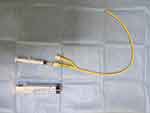
Initially, a regular 20mL syringe was employed for the injection, revealing a blockage in both fallopian tubes. However, due to the severe pain experienced during pulsed pressure injection, a switch was made to a 2.5mL syringe. If the status of tubal patency had been unknown at the outset and a 2.5mL syringe was used right away, it might not have been the most efficient approach due to the slower progress (Figure 1). The pulsed injection approach was chosen to optimize patient comfort while ensuring a controlled and gradual buildup of pressure.9 This method involved alternating between gentle and slightly stronger pulses of contrast agent injection, allowing the fallopian tubes to be visualized without causing excessive discomfort. Although the patient reported some initial discomfort during the injection process, her tolerance improved as the proce dure continued. The gradual pressure increase allowed for successful visualization of the fallopian tubes, which revealed a partial obstruction in the right fallopian tube. The left fallopian tube, on the other hand, displayed complete patency with the contrast agent.
 |
Figure 1 Contrast Agent Injection Using 2.5mL Syringe with Pulsed Technique. |
Outcome and Follow-Up
Three months following the TVS 4D-HyCoSy procedure, the patient returned for a follow-up visit on June 10, 2023. To our delight, the patient revealed that she had achieved a natural pregnancy because we found her Human Chorionic Gonadotropin (HCG) level was found to be 147.82 mIU/mL. Subsequent confirmation through ultrasound imaging demonstrated a gestational sac consistent with early pregnancy (Figure 2). This positive outcome underscores the potential clinical utility of the innovative pulsed injection technique utilizing a small-volume syringe during TVS 4D-HyCoSy.
 |
Figure 2 Ultrasound Confirmation of Gestational Sac in Early Pregnancy. |
Discussion
The presented case report introduces a novel approach that addresses the challenges of fallopian tube obstruction during transvaginal four-dimensional hysterosalpingo-contrast sonography (TVS 4D-HyCoSy), a promising technique for infertility assessment. The utilization of pulsed injection employing a small-volume syringe demonstrates several noteworthy aspects that warrant further discussion.
Advantages of Pulsed Injection Technique: The adoption of a pulsed injection technique using a small-volume syringe offers distinct advantages. Unlike traditional continuous injection methods, pulsed injection minimizes discomfort by allowing for controlled and incremental pressure buildup. This is particularly crucial in cases of fallopian tube obstruction, where the patient’s comfort can significantly impact the procedure’s success. The ability to tailor injection pressure within the patient’s tolerance range is a significant step towards achieving more patient-centric diagnostics.10
Enhanced Visualization and Diagnostic Precision: The pulsed injection technique contributes to improved visualization of the fallopian tubes by mitigating patient discomfort.11 This enhanced visualization is essential, especially in cases of partial obstruction. The case study demonstrates that even in instances where initial attempts result in non-visualization, the pulsed injection method allows for successful fallopian tube imaging. The precise control over injection pressure and the resulting image quality has implications for accurate diagnostic conclusions.
Potential Clinical Implications: The successful visualization of partial fallopian tube obstruction is of clinical significance. Such observations may guide clinicians in personalized treatment strategies, enabling targeted interventions to improve fertility outcomes. Additionally, the positive correlation between the pulsed injection technique and the patient’s subsequent natural pregnancy further underscores the clinical potential of this approach.
Patient-Centric Care and Comfort: The patient’s experience and comfort are paramount in any medical procedure. By incorporating the pulsed injection technique, clinicians can offer a more patient-centric approach to infertility assessments12. Reducing discomfort not only contributes to a more positive patient experience but also enhances patient compliance and cooperation during the procedure. While the presented case highlights the potential of the pulsed injection technique, it is essential to acknowledge its limitations. The technique’s applicability to various patient populations and the reproducibility of outcomes warrants further investigation. Additionally, the study’s scope is limited to a single patient case, necessitating larger-scale studies to establish the broader impact and effectiveness of this approach.
Conclusion
The fusion of innovative methodologies within established imaging techniques offers significant promise in the field of infertility assessment. By introducing the pulsed injection technique using a small-volume syringe to address challenges associated with fallopian tube obstruction during TVS 4D-HyCoSy, this case report emphasizes the potential for improved diagnostic precision, patient comfort, and positive clinical outcomes. As the medical community continues to explore the intersection of technology and patient-centric care, approaches like the one presented here contribute to advancing infertility diagnostics and treatment strategies. Further research and collaborative efforts are essential to validate the efficacy, reproducibility, and broader applicability of this technique, ultimately enriching the landscape of reproductive medicine.
Ethics Approval and Consent to Participate
This study was conducted in accordance with the ethical principles outlined in the Declaration of Helsinki and was approved by Shenzhen Baoan Women’s and Children’s Hospital, Shenzhen, Guangdong Province, 518000, China by LLSCHY-2023-09-06.
Consent for Publication
Written informed consent was obtained from the patient for publication of this case report and any accompanying images.
Author Contributions
All authors made a significant contribution to the work reported, whether that is in the conception, study design, execution, acquisition of data, analysis and interpretation, or all these areas; took part in drafting, revising or critically reviewing the article; gave final approval of the version to be published; have agreed on the journal to which the article has been submitted; and agree to be accountable for all aspects of the work.
Funding
There is no funding to report.
Disclosure
The authors declare no conflicts of interest in this work.
References
1. Ambildhuke K, Pajai S, Chimegave A, Mundhada R, Kabra P. A review of tubal factors affecting fertility and its management. Cureus. 2022;14(11):e30990. doi:10.7759/cureus.30990
2. Christianson MS, Legro RS, Jin S, et al. Comparison of sonohysterography to hysterosalpingogram for tubal patency assessment in a multicenter fertility treatment trial among women with polycystic ovary syndrome. J Assist Reprod Genet. 2018;35(12):2173–2180. doi:10.1007/s10815-018-1306-2
3. Sharaf MF, Fawzy I, Elkhateb IT, et al. Diagnostic accuracy of hysterosalpingo-lidocaine-foam sonography combined with power Doppler (HyLiFoSy-PD) compared to laparoscopy and dye testing in tubal patency assessment in cases of infertility. Middle East Fertil Soc J. 2022;7:34. doi:10.1186/s43043-022-00125-3
4. Jimah BB, Appiah AB, Sarkodie BD, Anim D. Ketamine use in hysterosalpingography (the Jimah Procedure): a follow-up of bilateral tubal evaluation of 27 infertile women at a Teaching Hospital, Ghana. Radiol Res Pract. 2021;2021:6657137. doi:10.1155/2021/6657137
5. Cucinella G, Gullo G, Etrusco A, et al. Early diagnosis and surgical management of heterotopic pregnancy allows us to save the intrauterine pregnancy. Menopause Rev. 2021;20(4):222–225. doi:10.5114/pm.2021.111277
6. Gu P, Yang X, Zhao X, Xu D. The value of transvaginal 4-dimensional hysterosalpingo-contrast sonography in predicting the necessity of assisted reproductive technology for women with tubal factor infertility. Quant Imaging Med Surg. 2021;11(8):3698–3714. doi:10.21037/qims-20-1193
7. Greis C. Technology overview: sonoVue (Bracco, Milan). Eur Radiol. 2004;14(Suppl 8):P11–P15.
8. Cheng KT. Stabilized sulfur hexafluoride microbubbles SF6 Microbubbles. Molecular Imaging and Contrast Agent Database (MICAD); 2008.
9. Gomez J, Koozekanani DD, Feng AZ, et al. Strategies for improving patient comfort during intravitreal injections: results from a survey-based study. Ophthalmol Ther. 2016;5(2):183–190. doi:10.1007/s40123-016-0058-2
10. Zhang Y, Wang Q, Gao CY, et al. Evaluation of the safety and effectiveness of tubal inflammatory drugs in patients with incomplete tubal obstruction after four-dimensional hysterosalpingo-contrast-sonography examination. BMC Pregnancy Childbirth. 2022;22(1):395. doi:10.1186/s12884-022-04722-y
11. Panchal S, Nagori C. Imaging techniques for assessment of tubal status. J Hum Reprod Sci. 2014;7(1):2–12. doi:10.4103/0974-1208.130797
12. Ayinde O, Hayward RS, Ross JDC, Peña Fernández MÁ. The effect of intramuscular injection technique on injection associated pain; a systematic review and meta-analysis. PLoS One. 2021;16(5):e0250883. doi:10.1371/journal.pone.0250883
 © 2023 The Author(s). This work is published and licensed by Dove Medical Press Limited. The full terms of this license are available at https://www.dovepress.com/terms.php and incorporate the Creative Commons Attribution - Non Commercial (unported, v3.0) License.
By accessing the work you hereby accept the Terms. Non-commercial uses of the work are permitted without any further permission from Dove Medical Press Limited, provided the work is properly attributed. For permission for commercial use of this work, please see paragraphs 4.2 and 5 of our Terms.
