Back to Journals » Journal of Multidisciplinary Healthcare » Volume 15
Review on Nanomaterials and Nano-Scaled Systems for Topical and Systemic Delivery of Antifungal Drugs
Received 3 February 2022
Accepted for publication 15 August 2022
Published 27 August 2022 Volume 2022:15 Pages 1819—1840
DOI https://doi.org/10.2147/JMDH.S359282
Checked for plagiarism Yes
Review by Single anonymous peer review
Peer reviewer comments 3
Editor who approved publication: Dr Scott Fraser
Gamachu Diba Nagasa,1,* Anteneh Belete2,*
1Department of Pharmacy, Ambo University, Ambo, Ethiopia; 2Department of Pharmaceutics and Social Pharmacy, School of Pharmacy, Addis Ababa University, Addis Ababa, Ethiopia
*These authors contributed equally to this work
Correspondence: Gamachu Diba Nagasa, Email [email protected]
Abstract: Fungal infections are human infections that topically affect the skin, mucous membranes, or more serious, invasive, and systemic diseases of the internal organs. The design and advancement of the formulation and approach of administration for therapeutic agents depend on many variables. The correlation between the formulations, mode of administration, pharmacokinetics, toxicity and clinical indication must be thoroughly studied for the successful evolution of suitable drug delivery systems. There are several NP formulations that serve as good delivery approaches for antifungal drugs. This paper covers various groups of nanoparticles utilized in antifungal drug delivery, such as phospholipid-based vesicles (nanovesicles), non-phospholipid vesicles, polymeric nanoparticles, inorganic nanoparticles and dendrimers, whereby their advantages and drawbacks are emphasized. Many in vitro or cell culture studies with NP formulations achieve an adequate high drug-loading capacity; they do not reach the clinically significant concentrations anticipated for in vivo studies. Because of this, the transfer of these nano-formulations from the laboratory to the clinic could be aided by focusing studies on overcoming problems related to nanoparticle stability, drug loading, and high production and standardization costs.
Keywords: nano-scaled, anti-fungal drugs, nano-vesicles, polymeric nanoparticles, inorganic nanoparticles
Introduction
Fungal infections are one of the major contributors to skin diseases worldwide. Fungi can affect human life and cause various diseases, from superficial infections of the skin and mucosal surfaces to invasive infections of internal organs, or from superficial infections to systemic infections. They are human diseases that affect topical skin and mucous membranes, or more severe, invasive and systemic infections of the internal organs.1 Over the past two decades, the incidence of invasive fungal disease has increased in parallel with the number of patients at risk for opportunistic fungal infections (eg patients with AIDS or neoplastic diseases, recipients of organ transplants or patients who have undergone aggressive surgery).2 Superficial fungal diseases are a main cause for patient visits to dermatologists, and superficial mycoses suffer from about 2025% of the human population.3 It is estimated that more than 10% of the German population are affected by fungal infections. The prevalence of invasive fungal infections varies from country to countryfor instance, in France the total frequency of this invasive fungal infection is around 5.9 cases per 100,000 per year, with a mortality rate of 27.6%.4
Approximately 1.5 million people die each year from an invasive fungal infection, making it one of the deadliest communicable diseases along with lower respiratory infections, tuberculosis, and diarrhea. They are increasingly becoming a major cause of morbidity and mortality, especially in patients with acquired immunodeficiency syndrome (AIDS), hematologic malignancies, severe aplastic anemia, myelodysplasia, immunocompromised patients, organ transplant recipients, premature babies, and the elderly. The study reports that the total deaths from invasive fungal infections are expected to be 1.5 million people per year.5
Globally, fungal infection is very diverse with three million to six million fungal species. From those fungal species, approximately around 150–300 species are most common cause of disease in human being. There are two mechanisms that are required for pathogenesis (Figure 1). The first process is survival and growth of the infecting microorganism, and the second process is damage to the host, a disruption of homeostasis manifested as disease symptoms.6
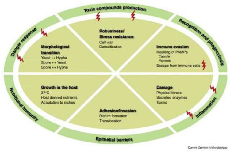 |
Figure 1 Virulence attributes of a prototypic human pathogenic fungus in interaction with the host. |
Acquisition of essential micro- and macronutrients is a well-known pathogenic for fungi virulence. Phosphorus, copper and iron are some of the essential macronutrient for all organisms as it is contained in nucleic acids, sugars, proteins and phospholipids. There is much known about the relationship between phosphate acquisition strategies that promote the virulence of many bacterial pathogens. However, there is less known about the relationship between phosphate acquisition and virulence in pathogenic fungi. Currently, study shows that there is occurrence of perturbation of phosphate acquisition in two common species of human fungal pathogens (Candida albicans and Cryptococcus neoformans).7 The strategy to acquire Fe ions employed by the host is similar, which is indicative of the typical response of the host to fungal pathogens. C. albicans, C. neoformans and A. fumigatus typically face low Fe levels in the host. The pathogenicity of fungi uses different processes in their iron acquisition, including Fe uptake from ferritin, hemoglobin or through siderophores (Figure 2).8
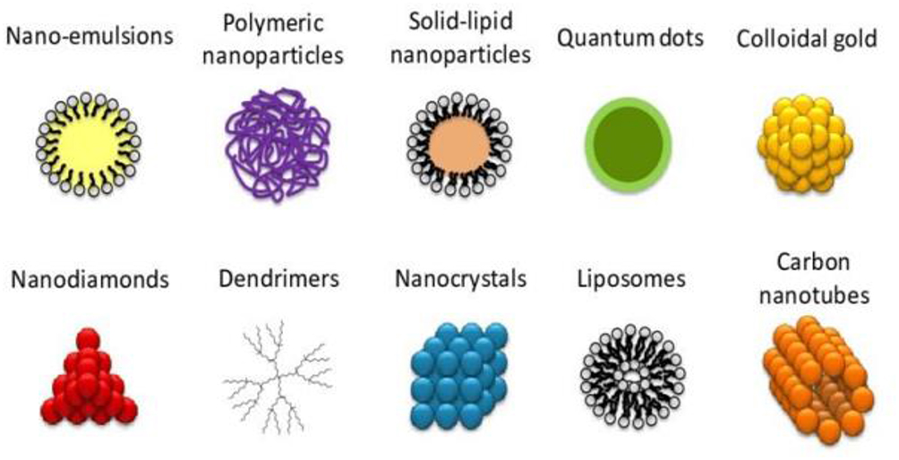 |
Figure 2 Iron homeostasis in Saccharomyces cerevisiae and major human pathogenic fungi. |
To ensure its direct consequence and attain a higher retention rate at the target, topical administration of fungal medications is the suitable method to combat superficial fungal infections. In addition to this topical antifungal administration, they assist in reducing systemic toxicity and circumventing presystemic metabolism. Great progresses in the growth of new antifungal agents have resulted in the accessibility of recent generations with improved therapeutic results in these vulnerable patient populations. Various drugs such as ketoconazole, itraconazole, clotrimazole are adopted as topical administration to the skin by spreading or rubbing.9 Although there is advancement in the efficacy and safety profile of the drugs, some invasive fungal infections such as Aspergillus spp., Zygomycetes, Fusarium spp., Scedosporium spp. and non-Albicans Candida spp. pose a major [main] risk to vulnerable patients. In addition, numerous of these recently established antifungal agents have important limitations in terms of their spectrum of activity, their physicochemical and biopharmaceutical properties, their pharmacokinetics, drug interactions, and pharmacodynamic properties.5,10
Objective
This review aimed to provide an in-depth presentation of the types, benefits, and drawbacks of various NPs used for topical and systemic antifungal drug delivery. It also highlights the potential of various antifungal NPs against superficial, systemic fungal infections, and also discusses the challenges this area is facing that are hampering the clinical implementation of some promising nano-formulations.
Method
The current work summarizes the approaches of novel drug delivery systems for antifungal agents from secondary sources and discusses published information with the ultimate aim of bringing the reader up-to-date with the current literature considering into account the articles published since 2008. This has been achieved by using large electronic databases, such as PubMed, Medline, Google Scholar, Scientific Journals, Cochrane Library (library genesis) and Science Direct, which provide access to scientific and medical research.
Need to Develop Novel Drug Delivery Systems for Antifungal Agents and Strategies for Treating Fungal Infection
The design, formulation development, and drug delivery system for therapeutic agents depend on numerous variables. The researcher should cautiously consider the association between formulation, route of administration, toxicity, pharmacokinetics, and clinical indication in order to successfully develop a relevant drug delivery system.11 Several antifungal agents are hydrophobic, resulting in limited water solubility, poor oral bioavailability, and limited formulation approaches. The most hydrophobic and poorly watersoluble antifungals encompass clotrimazole, miconazole, econazole, oxiconazole, tioconazole and sertaconazole, while amphotericin-B (AmB) is one of the most notorious toxic antifungal drugs (infusion-related reactions) and enhances the toxicity of several different drugs, such as nephricin and cyclosporine, and aminoglycosides.12
Despite the accessibility of numerous conventional dosage forms for antifungal drugs including tablets, creams, IV infusions, etc., they appeared to be ineffective in overcoming these limitations. Hence, there is a strong desire to develop innovative drug delivery approaches to address these problems. In order to improve drug performance and overcome the above limitation, the production of a rationally designed drug is very important. Indeed, lipid-based formulations of amphotericin-B (AmB), such as the AmB-lipid complex (ABLC), the AmB colloidal dispersion (ABCD) and the liposomal AmB (L-AmB), showed a substantial reduction in AmB nephrotoxicity,while having their broad spectrum antifungal activity. For treating validated invasive fungal infections, liposomal amphotericin B was more effective than amphotericin B deoxycholate in patients with disseminated histoplasmosis and AIDS, while it was in patients with acute cryptococcal meningitis and AIDS.13
In order to improve the safety profile of antifungal drugs while maintaining or optimizing their effectiveness, the production of nanoparticles is a promising result of the application of various recent drug delivery systems. These nanoparticles (NPs) have developed into an innovative and promising platform that is able to minimize undesirable drug side effects, maintaining and optimizing therapeutic efficacy.
Many of the unfavorable features of active ingredients can be overwhelmed by nanoparticles due to their versatility, multi-functionality and wide range properties or their ability to kill different species of fungi. The other advantage of this preparation is its capability to improve the penetration of the drug through the skin and consequently help eliminate deep fungal infections, sustained drug release, improved drug stability, targeted infected tissue, reduction of side effects outside of the target area, increase in residence time in the blood and improved effectiveness of drugs.14,15
The antifungal drugs act on different targets. Drugs that act on the cell membrane encompass polyene antibiotics such as amphotericin B lipid formulations, nystatin (topical), and azole antifungal drugs, such as ketoconazole, itraconazole, fluconazole, voriconazole, miconazole, and clotrimazole. DNA synthesis is another target for antifungal therapy, and this therapy encompasses drugs such as pyrimidine analogs, eg Flucytosin; the antifungal agents that act on the cell wall are echinocandins, and caspofungin acetate.16 Novel topical formulation approaches, such as solid lipid nanoparticles, liposomes, niosomes, microemulsions, nanoemulsions, etc., are appearing to be superior for overcoming the skin permeation barrier. The main objective of this review is to provide a detail of the types, benefits, and limitations of various NPs used for topical and systemic antifungal drug delivery. It also highlights the capability of these nanoparticles in the fight against superficial and invasive fungal diseases.
Nanomaterial’s and Nano-Scaled Systems Used in Antifungal Drug Delivery
Fungal infections are one of the biggest health issues in the world. The administration of antifungal agents, which are utilized to treat superficial and systemic infections, has great consequences in terms of both therapeutic aspects and safety. Although there are multiple conventional dosage forms, they have some disadvantages. In order to overcome the drawbacks of this conventional DF, nanoscale active ingredient carriers play a crucial role in improving the efficacy and guaranteeing the safety of the treatment. According to their composition, NPs used in drug delivery applications can be generally classified into phospholipid vesicles (liposomes, malleable liposomes, ethosomes, transfersomes, transethosomes, etc.), non-phospholipid vesicles (niosomes and chip plastics), polymeric NPs, polymeric micelles, solid lipid nanoparticles, nanostructured lipid carriers, nanoemulsions and dendrimers. Nanoscale active ingredient carriers dominate the superiority of enhancing the water solubility of lipophilic active ingredients, prolonging the release of active ingredients, minimizing side effects of active ingredients such as irritation, keeping active ingredients away from environmental determinants and targeting active ingredients in certain tissues. Schematic representation of the most commonly used nanomedicine types composed of different kinds of materials is shown in Figure 3.
 |
Figure 3 Schematic representation of the most commonly used nanomedicine types composed of different kinds of materials. |
Phospholipid-Based Vesicles (Nanovesicles) Antifungal Drug Delivery
Liposomes
Liposomes are tiny spherical vesicles containing one or more phospholipid bilayer vesicles with an aqueous core and one or more concentric phospholipid membranes. Depending on their size, liposomes are divided into unilamellar vesicles, multilamellar vesicles and multi-vesicular vesicles. It consists of natural or synthetic phospholipids, which primarily contain phosphatidylethanolamine and phosphatidylcholine. The incorporation of cholesterol to saturated phospholipids increases the fluidity of the bilayer, whereas the incorporation of cholesterol to unsaturated phospholipids improves the packing and provides controlled release of drugs. In addition, liposomes have both lipophilic and lipophobic properties and can consequently all kinds of drugs.2,17 Other interesting features of liposomes as drug delivery systems encompass biocompatibility, minimal toxicity, high drug loading capacity, improved drug bioavailability and stability, sustained drug release, and minimization of drug side effects.18
The study on nystatin by Saadat et al, 2016 illustrated that liposomal nystatin was as active or more active as the free drug or more active than the free drug 0.5 g/mL compared to the free drug with an MIC of 2 g/mL (p < 0.05). In addition, it facilitated intravenous administration of nystatin, heightened the maximum tolerated dose in mice from 4 to 16 mg/kg body weight, dramatically improved the survival rate of Candida albicans infected mice, and offered a therapeutic benefit in systemic urinary tract infections and were at high level encapsulation stable.19
Liposomal amphotericin B is another antifungal agent that has been successfully included into liposomal formulations for the treatment of systemic fungal infections. It is the first and highly successful commercial nano-formulation of antifungal drugs. Today, most patients with disseminated histoplasmosis and AIDS, candidal meningitis, or endophthalmitis are treated with liposomal AmB as first-line therapy. Liposomal AmB achieves dose-dependent higher drug concentrations in the liver, spleen, lung tissue and brain, while renal drug levels are similar to those of conventional AmB.20,21
Since (Zoubek et al, 2009) AmBisome improves the tolerability of AmB treatment, significantly improve the antifungal activity in infected mice with no obvious toxicity compared to the free drug, reduces toxicity in recipients of bone marrow and organ transplants, the dosage became good tolerated and consequently the dose have consistently increased with conventional AMB to a daily dose of 4.0 mg/kg/day to 6.0 mg/kg/day. AmBisome seems to be able to attain higher tissue concentrations of AMB than with conventional AMB. This observation corroborates the low toxicity of AmBisome compared to conventional AMB. After infusions of liposomal AMB, there were neither chills nor high fever, and no kidney failure happened despite too high doses.21,22
The desirability of topical drug delivery depends on achieving a high drug concentration in a local area, resulting in a lower drug dose, fewer side effects and decreased therapy costs. In addition to this topical antifungal agent, it offers improved patient compliance with simple application and removal. Liposomal topical antifungal agents have an advantage over traditional ones in their localized active ingredient for extended time, sustained release, biocompatibility, good skin penetration, improved therapeutic efficiency and decreased side effects. For example, prepared miconazole nitrate-loaded liposomes illustrated better penetration and retention into the skin layers, fighting fungi in deep layers of the skin, and overcoming the inadequacies of conventional miconazole creams.23,24
As mentioned above, liposomal nanoscale formulations have the capability to overcome the limitations of traditional eye drops, such as through the subconjunctiva. Encapsulating drugs in liposomes could defend drugs from degradation by metabolic enzymes on conjunctival and corneal surfaces and in tear fluids. As Salem, Ahmed and Omar, 2016, flucytosine encapsulated in liposomes improved its intraocular penetration and illustrated a cure of 86.4% after 3 weeks, compared to a 50% cure for fluconazole solution after the same time.25 The liposomes significantly slowed the clearance of the drug, resulting in a higher concentration in the vitreous humor. This led to an approximately 7-fold increment in the fluconazole half-life.26
Transfersomes
Transfersomes are referred to as highly deformable or elastic liposomes or edge activators, which lead to the so-called deformable liposomes or transfersomes, which gives the liposome structure flexibility. The edge activator is responsible for weakening the lipid bilayers of the vesicles and increasing their deformability. Transfersomes have been successfully assessed as topical and transdermal carriers for drugs and have also been demonstrated to be effective carriers for genetic material and vaccines. Voriconazole transethosomes showed significantly enhanced skin permeation of drugs compared to regulatory and other vesicles, such as deformable and classical liposomes.27 The study illustrates deformable membrane vesicles (DMVs) for topical administration of griseofulvin as a potential treatment for dermatophytosis and achieved substantially higher drug permeation and skin retention compared to classical liposomes.28
Ethosomes
Ethosomes are classic liposomes with a high alcohol content that demonstrate penetration into deeper layers of the skin and the systemic circulation. The high ethanol content of ethosomes can enhance the solubility of more lipophilic drugs and make it easier to enhance permeation. Ethanol disrupts the intercellular lipid structure of the stratum corneum owing to the phospholipids in their content.29 Compared to liposomes, ethosomes attain improved penetration through the skin owing to the ability of ethanol to fluidize ethosomal lipids and intercellular lipids of the stratum corneum.30 As topical delivery approaches for antifungal drugs, ethosomes showed great potential and had better drug entrapment effectiveness, improved transdermal flow, and deeper skin penetration. Ethosomes had the highest zone of inhibition against Candida albicans compared to liposomes and drug formulations and showed noticeably higher drug permeation and flux through the skin of rats, as well as better antifungal activity compared to liposomal gel and hydroethanolic gel.
The ethosomes attain deeper penetration into the skin, with confocal laser scanning microscopy showing that the ethosomes reached the final layer of the epidermis (stratum basale).31 A recent study showed that ethosomal voriconazole was 2 times more efficient against Aspergillus flavus compared to the drug solution in dimethyl sulfoxide, but less active than the hydroalcoholic drug solution. Voriconazole ethosomes had a 6-fold higher permeability through the abdominal skin of rats compared to the hydroalcoholic solution of the drug. The drawbacks of the ethosomes encompass higher release rates of the trapped active ingredients at an ethanol concentration >30%, skin irritation and contact dermatitis due to repeated topical utilization of high ethanol concentrations, which can be overwhelmed by prepared Cavamax W7 composite ethosomes in order to reduce the amount of ethanol in the vesicles.3
Ufasomes
Ufasomes (unsaturated fatty acid vesicles) are suspensions of closed lipid bilayers, which consist of fatty acids and their ionized species (soap) and are limited to a narrow pH range of 7 to 9. In Ufasomes, the fatty acid molecules are aligned so that their hydrocarbon tails are directed towards the inside of the membrane and the carboxyl groups come into interact with water. A stable Ufasome formulation crucially relies on the correct selection of fatty acid, the amount of cholesterol, the buffer, the pH range, and the amount of lipoxygenase and the existence of divalent cations.32 The study indicates that fatty acid vesicles (ufasomes) for topical administration of clotrimazole have high drug entrapment, sustained release of the drug from the vesicular dispersion, and accumulation of the drug in the epidermal part of the skin. The in vivo investigation confirmed the prolonged release of drugs from oleic acid vesicles for up to five days, indicating their efficacy for long-term therapy.
Non-Phospholipid Vesicles Antifungal Drug Delivery
Niosomes
Niosomes are analogous to liposomes, but their bilayers are generated up to nonionic surfactants with a single alkyl chain instead of phospholipids. These are types of spherical lipid vesicles manufactured by nonionic surfactants. Its skin permeation relies on the nature of surfactants, the characteristics of the drug adopted and the morphological characteristics of the niosome preparations. It has been observed that the therapeutic activity of ketoconazole is increased in niosomal preparations. Itraconazole and miconazole niosomes have also been proven to be effective and proven to be effective carrier approaches for antifungal drugs. Fluconazole-loaded niosomes made using multiple surfactants (Span 40, Span 60) showed prolonged localized and sustained implications of fluconazole.2,33 The study illustrated that niosomal gel was approximately 6.5 times higher drug localization in the skin compared to pure carbopol gel, which signifies better target accumulation of niosomal gel. The outcome of niosome gel loaded with clotrimazole illustrates a sustained and controlled-release pattern with good tolerance at the tissue level in rats for suitable local vaginal therapy.34
The advantages of niosomes over liposomes include improved chemical stability, cheaper price, and the ability to store them under normal conditions.35 Niosomes have a high drug loading capacity, and their properties can be tailored by carefully selecting their composition and manufacturing method. The niosomal formulations showed a gradual increase in the zones of inhibition due to the controlled release of the active ingredient.
Purified niosomes containing only the trapped drug and niosomes containing all of the drug (trapped and free drug) were incorporated into the gel, and their antifungal properties were compared to the marketed drug formulation.36 An improvement in the antifungal effect of ketoconazole against Aspergillus niger was observed for the drug incorporated in niosome gel.37 The nystatin release from niosome gel was greatly prolonged compared to conventional gel. In addition, the niosome gels showed a two-fold improvement in nystatin deposition in pig skin compared to conventional gels. The in vitro antifungal activity against Candida albicans was higher with the niosome dispersion than with the niosome gel, probably due to the faster release of nystatin from the dispersion.38
Solid Lipid Nanoparticles
Solid lipid nanoparticles (SLNs) are colloidal, nanostructured active ingredient carriers that consist of physiologically compatible lipids that are dispersed in aqueous surfactant solutions. SLNs offer numerous drug delivery benefits, including improved permeability through biological barriers, lipid chemical stability, and the ability to modify their surface, the ability to deliver multiple drugs together, and protect the built-in drug from degradation. However, SLNs have several disadvantages including poor drug loading capacity and drug expulsion during storage. Amphotericin NP stabilizes amphotericin B against degradation at different pH values (1.2, 4, 6.8) and increases its relative BA compared to a drug alone. In a recent study, AmbiOnp was found to contain a non-toxic super-aggregated form of AmB resulting from the probe sonication-assisted nano-precipitation technique used in SLN preparation.39
The relative bioavailability of AmbiOnp was 1.05-fold with a Cmax of 1109.31–104.79 ng/mL at the end of 24 hours, which was comparable to a Cmax of 1417.49_85.52 ng/mL obtained with IV Fungizone1 was achieved which confirms the potential of this new oral formulation. For AmbiOnp, compared to Fungizone1, very low AmB levels and lower kidney toxicity were found.39
Solid lipid nanoparticles (SLN) designed for topical administration of econazole nitrate (ECN) show encapsulation efficiency values of 100%. According to Sanna et al, 2007, SLNs show in ex vivo tests that they were able to control drug release by the SC; the release rate depended on the lipid content of the nanoparticles, while an in-vitro study shows that SLN promoted rapid penetration of ECN through the SC after 1 hour and improved the diffusion of the drug in deeper skin layers after 3 hours of use compared to the reference.40 In another study, fluconazole-loaded solid lipid nanoparticles (FLZ-SLNs) were prepared and the effectiveness of the preparation was investigated for optimal formulation on fluconazole (FLZ)-resistant strains of several Candida species. In this study, the author creates FLZ SLN using probe ultrasound techniques and identifies the morphology by field emission scanning electron microscopy. In addition, the minimum inhibitory concentrations of the new formulations against fluconazole-resistant Candida strains were investigated. The result of the study shows that FLZ-SLNs have a spherical shape with a mean diameter, zeta potential and inclusion efficiency of 84.8 nm, 25 mV and 89.6%, respectively.
The drug release from fluconazole (FLZ) showed a burst release in the initial stage (1.30 minutes), followed by a sustained release over 24 hours. FLZ-resistant yeast strains behaved as susceptible strains after treatment with FLZ-SLNs (8 g/mL). The MIC50 drug concentrations were 2 g/mL, 1 g/mL and 2 g/mL for FLZ-resistant strains of C. albicans, C. parapsilosis and C. glabrata, respectively. A novel drug delivery system thus shows that it combats the Candida strain better than the conventional formulation, which shows a lower susceptibility as a first-line treatment.41
Nanostructured Lipid Carriers
To overcome the limitations of SLNs, a second-generation SLN, called Nanostructured Lipid Carriers (NLCs), was introduced. NLC has a solid lipid matrix at room and body temperature, which consists of a mixture of a solid lipid and oil in certain proportions, which leads to better drug loading, modulation of the drug release and long-term stability.42,43 For pulmonary use, an itraconazole-loaded formulation with nanostructured lipid carriers (NLC) was developed. NLC loaded with itraconazole had a narrow particle size distribution with inclusion efficiency of 99.98%; the formulation was found to have a good storage stability over 6 months, which indicates good tolerability after inhalation.
When the formulation was nebulized with a new nano-nebulizer in a therapeutic chamber for the treatment of falcons, no physical instabilities of the itraconazole-loaded NLC could be detected. It could be shown that the particle size of the aerosol, which was generated when nebulizing itraconazole-loaded NLC with the nebuliser, was in the nanometer range, which offers the possibility of penetrating into the respiratory tract of hawks. The deposition of itraconazole-loaded NLC in the lungs and alveoli of a patient could be demonstrated by means of scintigraphy, as a prerequisite for the pulmonary treatment of aspergillosis in falcons.44
NLC dispersion loaded with voriconazole (VRC), consisting of Precirol ATO 5, Labrafil 1944 CS and Tween 80, was produced by high-pressure homogenization and embedded in Carbopol 940 hydrogel. The lipid nanoparticles in the hydrogel were approximately 210 nm in size, had a spherical shape and a zeta potential of 30 mV. In this study, the skin permeation of the NLC gel was superior to the conventional gel formulation based on cream and microemulsion and showed 2.8 and 1.7 times higher flow values and a significantly higher accumulation of VRC in deeper skin layers compared to the reference formulations, and finally the author suggests that there is an alternative treatment for skin infections like candidiasis, with less potential for systemic side effects than oral therapy45 (Song et al, 2014). A (Singh et al, 2016) study evaluated the potential of ultra-small nanostructured lipid carriers (USNLC)-based topical gels made from miconazole nitrate for the treatment of athlete’s foot. It shows that the drug release from usNLC showed an initial rapid release followed by a sustained release with 91.99% of the drug release in 24 hours. The antifungal activity against trichophyton mentagrophytes showed a wider zone of inhibition, 6.6 1.5 mm for the optimized usNLC3 gel, while the gel formulation marketed was 3.71.2 mm. An improved dermal delivery of miconazole by usNLC gel was achieved for the treatment of athlete’s foot.46
Nanoemulsions Antifungal Drug Delivery
The nanoemulsions have similar droplet dimensions as the microemulsions in the range of <200 nm and in some cases <100 nm. Nanoemulsions are thermodynamically metastable because phase separation occurs over time. However, nanoemulsions are given kinetic stability because there is no gravitational separation and droplet aggregation due to the reduced force of attraction between the small droplets.47 NE has interesting properties for antifungal drug delivery, such as small size, good kinetic stability, high drug solubility capacity and improved penetration through the skin. In addition, NE can be used as an alternative to the less stable lipidic nanocarriers (eg liposomes).
NE also achieved better antifungal activity of the drug compared to free drugs due to the presence of the drug in solubilized nanospheres form. The large surface area due to the reduced particle size and the lipophilic nature of the NE formulation improved the drug permeation through biological membranes, which led to better drug effectiveness.48,49 NE formulations could also enable targeted topical antifungal drug delivery to maximize local drug effects and avoid systemic toxicity such as nephrotoxicity. The AmB-NE gel (18.09 0.6 mg/cm2/h) and NE (15.74 0.4 mg/cm2/h) showed the highest percutaneous permeation flow rate of the skin compared to the drug solution (4, 59 0.01 mg/cm2/h), which suggests a better alternative to painful and nephrotoxic intravenous administration. Hemolytic and histopathological results indicated the safe administration of the drug. Based on the combined results, NE and AmB-NE gel could be viewed as an efficient, stable and safe carrier for improved and sustained topical delivery of AmB for local skin fungal infections. AmB-laden NE achieved a 3.3- or 2.2-fold higher drug permeation through the skin of albino rats compared to AmB solution or commercial cream Fungisome1.48
Better drug permeability of NE has been attributed to its ability to alter both the lipophilic and polar pathways through synergistic interaction of its component with the stratum corneum. In other studies, excipients with innate antifungal activity were used to formulate AmB-laden NE in order to exploit a possible synergistic effect between AmB and NE excipients.50 Nystatin NE was manufactured and tested as a potential treatment for oral and skin candidiasis. NE-loaded nystatin had a minimal inhibitory concentration against Candida albicans and Saccharomyces cerevisiae that was two-fold lower than that of the free drug, confirming the drug’s improved antifungal efficacy. Systemic administration of nystatin leads to serious toxicities. Therefore, ex vivo porcine oral permeability studies were performed to confirm the safety of nystatin NE. Negligible drug permeation through the mucosa was observed, confirming the safety of this delivery system. Studies on drug retention on oral pig mucosa showed that about 50% of the applied drug dose remained in the tissue after 6 hours. This was about 310 times higher than the minimum inhibitory concentration value of the drug against Candida albicans, confirming the effectiveness of this treatment.51
NE has also been used to improve ocular delivery of antifungal drugs. Thus, terbinafine hydrochloride was loaded into NE and dispersed in an in situ gel. The NEs were small in size (<30 nm) and had good thermodynamic stability. In-situ NE gel showed the lowest eye irritation potential, a 1.7-fold higher mean residence time of the drug in the aqueous humor and a 2.7-fold higher bioavailability of the drug in the eye compared to the oily drug solution.52 NE has also been a proven useful vaginal antifungal drug delivery system. Therefore, ITZ was encapsulated in NE with tea tree oil (TTO) as a potential treatment for recurrent vaginal Candidiasis.53 NE mucoadhesive gel had a significantly higher ITZ permeability compared to conventional gel. In vivo studies on oophorectomized rats treated with estradiol and infected with Candida albicans showed synergistic and accelerated fungal clearance for an optimized NE gel with ITZ and TTO compared to a gel with an individual ITZ or TTO. In addition to the above-mentioned studies of improved topical antifungal drug delivery, NE formulations have also been found useful in improving the oral bioavailability of drugs. Thus, ITZ NE was tested for its ability to improve the intestinal permeability and oral bioavailability of the drug ex vivo. Antifungal studies showed a much higher zone of inhibition against Aspergillus niger for ITZ NE (42 mm) compared to drug suspension (12 mm) or pure NE (11 mm). In addition, ITZ NE had better permeation through both the stomach and the intestines compared to the drug suspension and the marketed preparation (Sporanox1). In vivo studies on healthy male Wistar rats showed an approximately 2-fold higher bioavailability for ITZ NE compared to the drug suspension.54
Spanlastics
Spanlastics are compositional edge activators that form a new class of malleable nanovesicular carriers to improve niosome permeability across biological membranes. They mainly consist of spans as well as edge activators like tweens or others. They are analogues of transfersomes and transethosomes in the sense that they all contain edge activators. The first reported Spanlast ik-based delivery system was designed for ocular delivery of ketoconazole using Span1 60 and Tween1 80 as edge activators.55 The spanlastics had a 2-fold better ketoconazole corneal permeation compared to conventional niosomes. They showed good stability and were safe from genotoxicity, cytotoxicity, acute skin/eye irritation/corrosion, and chronic eye irritation/corrosion tests. Fluconazole-loaded spanlastics were 3 times smaller than their corresponding niosomes and had three times higher drug permeability through the cornea of the pig compared to the commercially available drug eye drops.56 Spanlastics loaded with itraconazole had a drug entrapment efficiency of greater than 88%, slower drug release, and better permeability through excisions. The bovine cornea is compared to conventional niosomes.57 Terbinafine hydrochloride was encapsulated in Span1 60 or 65 Spanlastics, which contained Tween1 80 or sodium deoxycholate as edge activators for the treatment of onychomycosis.58 Spanlastics loaded with terbinafine showed better ex vivo permeability through the nails than the commercial cream. With the exception of this later study, spanlastics were used solely for ocular administration of antifungal agents. Therefore, their potential for systemic and topical administration of antifungal agents has yet to be recognized.
Polymeric Nanoparticles Antifungal Drug Delivery
Polymer nanoparticles can be defined as solid colloidal particles in the size range of 1100 nm and preferably consist of polymers that are obtained from natural, synthetic or semi-synthetic sources and can be either biodegradable or non-biodegradable. The functionalized polymeric nanoparticles have a variety of uses ranging from drug delivery to the vagina, the brain, cancer treatment, gene therapy, and much more.59 NPS has shown an excellent ability to improve the therapeutic properties of drugs while minimizing its side effects/toxicity. Nanoparticles are promising candidates for ocular drug delivery because of their small size, which results in little irritation, and a sustained-release property that avoids frequent administration offers advantages. Enormous types of nanocarriers have been studied by researchers to i) improve the permeation of drugs through the horny layer of the skin, ii) achieve controlled and prolonged drug release, and iii) improve the effectiveness of topical treatment.17 Many polymeric NP-based cytotoxic drug formulations have already made it into the clinic, while many others are in the development phase. According to their manufacturing and architectural methods, polymeric nanoparticles are divided into nanospheres, nanocapsules and polymeric micelles.5
Nanospheres and Nanocapsules
Drug-loaded nanoparticles can be nanocapsules or nanospheres. In nanocapsules, the drug is enclosed in the polymer shell, while it is in nanospheres; the drug is evenly distributed in the polymer matrix.60 Nanospheres are solid NPs of the colloidal matrix type in which the active ingredient is dissolved or enclosed in the matrix or adsorbed on the surface, while nanocapsules are colloidal particles with a core that is liquid or semi-solid at room temperature, surrounded by a solid polymer shell. The core is a lipophilic solvent, usually an oil, and is used as a reservoir for the encapsulation of hydrophobic drugs.61,62 Several studies have shown that incorporating antifungal drugs into nanospheres and nanocapsules prolongs their release, increases their antifungal potency, and decreases their toxicity. The following sections summarize some of these studies based on the antifungal drug tested.
Campos et al, (2016) show that the fungicides tebuconazole and carbendazim were synthesized with phenyleneethynylene polymer and characterized by UV-Vis spectroscopy, fluorescence spectroscopy and TEM and were used to color polymeric nanocapsules. The nanocapsules showed good stability over time without changes in shape or fluorescence and were suitable for use in drug delivery systems with an average particle size of about 430 nm, a polydispersity index of less than 0.2 and a zeta potential of about −13 mV. It was observed that nanocapsules containing the fluorescent polymer retained the ability to modulate the release of the fungicides tebuconazole and carbendazim (used as a model drug) even after 4 days. Preliminary results indicated that staining with the fluorescent phenyleneethynylene polymer could be used as a valuable tool to follow the behavior of polymer systems in the environment.63
Nanocapsules were loaded with amphotericin B (AMB) with encapsulation efficiencies (EE%) depending on the active ingredient and the concentrations and degrees of substitution (DS) of the acetylated Sterculia striata polysaccharide (ASSP). A maximum EE% of 99.2% was achieved and the loaded AMB was found to be in a monomeric form, even at a concentration hundreds of times higher than that commonly used for commercial aqueous AMB solutions. Loaded nanocapsules show an in vitro controlled release of AMB. Because the AMB monomeric state reduces drug toxicity, AMB loaded ASSP nanocapsules have the potential for use as a drug delivery system. NC loaded with AMB retains its activity against 5 tested strains of Candida albicans.64 In another study, a nanocapsule with a degree of substitution (DS) of 2.53 (NCP 2.53) was selected for encapsulation, biocompatibility and antifungal evaluation against Candida albicans strains. A maximum of 98.3% of the AMB encapsulation was achieved. Encapsulated AMB was in its monomeric form and showed good biocompatibility and antifungal activity against four C. albicans strains. The data suggest that propionate Sterculia striata polysaccharide (PSSP) has potential as a nanocarrier system for AMB.65
Another study also shows that PLGA-NPs with AmB drug improve oral bioavailability (8 times higher than just AmB) and minimize side effects. The release of AmB NPS was biphasic, with an initial rapid-release phase followed by a sustained-release phase. It also lowers hemolysis and nephrotoxicity in rats compared to bare AmB.66 The NPS showed good biocompatibility, which was demonstrated by a non-significant decrease in hemoglobin concentration and hematocrit and a non-significant increase in blood urea nitrogen and serum creatinine level when tested on adult male Swiss albino mice. Candida albicans antibody (CDA) AmB NPs had significantly lower hemolytic activity against red blood cells and lower in vitro toxicity compared to the immortalized human renal tubular epithelial cell line (HKC) and lower in vivo nephrotoxicity. In addition, in vivo studies showed that CDA-AmB-NPS effectively eliminates fungi in the kidney, liver and spleen compared to free AmB.67,68
Incorporation of PLGA nanospheres into itraconazole with the optimal nanospheres had an average diameter of about 200 nm with a unimodal size distribution, a negative surface charge, and an encapsulation efficiency of about 72%. It shows a sustained-release formulation for intravenous administration and in vivo studies in rats showed that the nanosphere formulation had similar systemic bioavailability as the commercial drug formulation and sustained plasma levels (>100 ng/mL) for up to 24 hours exhibited.69 Another study shows that itraconazole-PLGA nanospheres completely inhibited the growth of Aspergillus flavus for 11 days at a drug concentration of 30 mg/mL, improved the antifungal efficacy of ITZ, no cytotoxicity on HeLa cells and fibroblasts at a high concentration of 25 mg/mL had a high survival rate and duration in mice after loading with Da-tocopheryl PEG 1000 succinate-b-poly (e-caprolactone-ran-glycolide). Pectin NPS improved ITZ dispersibility in water, the rate of dissolution and changed its crystallinity. The highest resolution was observed for NPs made from HMP. The improved dissolution properties were retained after 6 months of storage. In vivo studies on fasting rats showed that ITZ/pectin-NP had a 1.3-fold higher absorption rate than the commercial drug formulation.60,70
The reuse of itraconazole-loaded PLGA nanoparticles was more effective than ITR solution in inducing pro-apoptotic Bax and p53 while reducing anti-apoptotic Bcl2 protein expression. ITRNPs were more effective than ITR solution in arresting cells in both the G0/G1 and G2/M phases of the cell cycle. Therefore, reusing itraconazole by encapsulating it in chitosan-coated PLGA NPs is a potentially promising approach in the treatment of lung cancer.71
The nanocapsules were stable for up to 2 months and improved the clotrimazole stability to UV radiation. In addition, clotrimazole loaded with nanocapsules was more effective than the free drug against Candida albicans and Candida glabrata strains, which are sensitive and resistant to fluconazole. Clotrimazole and econazole were loaded into PLGA NPs and alginate NPs stabilized with chitosan to improve the bioavailability of the two active ingredients. The encapsulation efficiency of alginate NPs was almost twice that of PLGA NPs. Biodistribution/pharmacokinetic data following oral administration of the NPS to mice indicated controlled drug release for 56 days. In contrast, the unencapsulated drugs were excreted within 34 hours of oral and intravenous administration. NP-loaded drugs were detected in the tissues (lungs, liver, and spleen) for up to 68 days, while the free drugs were eliminated within 12 hours. In general, the alginate formulation was better than PLGA for drug encapsulation effectiveness, biodistribution, and pharmacokinetic profile.72
According to (Endo et al, 2020) Ketoconazole-PLA, the characterization and improvement of the antifungal activity in vitro against Candida and dermatophytes shows that nanoparticles containing ketoconazole exhibited superior antifungal activity against all tested fungal strains than free ketoconazole. Inhibition of yeast biofilm formation was also achieved. Ketoconazole PLA nanoparticles resulted in better antifungal activity of ketoconazole nanoparticles as a free agent against dermatophytes and Candida species, indicating a promising tool for developing therapeutic strategies.73
Polymeric Micelles
Micellar delivery systems are self-organizing nanoscale (100 nm) and most commonly used carrier systems to formulate therapeutic agents in clear aqueous solutions. In general, these nano-micelles are made with amphiphilic molecules. These molecules can be surface-active or polymeric. Because of its high drug encapsulation ability, ease of manufacture, small size, and hydrophilic nanomicellar corona-generating aqueous solution, nanomicell-based drug delivery technology has grown dramatically in recent times.60 The hydrophilic corona maintains the water solubility and colloidal stability of the micelles, reduces their uptake by the cells of the immune system and extends their circulation time in the blood. Due to its hydrophilicity and biocompatibility, poly(ethylene glycol) (PEG) is the most frequently used polymer as a hydrophilic micellar corona and forms a steric barrier that reduces the adsorption of opsonin proteins on the micelle surface, which leads to a prolongation of the micelle circulation time in the blood.1 As (Oerlemans et al, 2010), due to their lower critical association concentration, polymeric micelles show better stability towards dilution, a property that is of the greatest importance due to the high degree of dilution after intravenous injection compared to surfactant micelles.74
Polymer micelles have become one of the most promising platforms for drug delivery due to their improvement in water solubility, biodegradability, and/or biocompatibility, in that they can target drugs to specific tissues in the body and achieve high drug concentrations in tumor and inflammatory tissues and to increase the effectiveness of antifungal drugs. Polymer micelles are one of them with their specific ability to encapsulate hydrophilic active ingredients. These carriers can enhance the therapeutic effectiveness and minimize the systemic side effects of the drugs. In normal and dermatological diseases such as psoriasis and acne, polymer micelles could improve the deposition of active ingredients on specific areas of the skin. Nevertheless, there is still a need to investigate the mechanism of action of these carriers and the fate of polymeric micelles in the skin.75–77
Ketoconazole loaded with methoxy-poly (ethylene glycol) -b-poly (-valerolactone) copolymer micelles improved skin penetration and distribution. The micelles loaded with active ingredient were obtained with an encapsulation efficiency of 86.39% and a particle diameter of about 12 nm. It increases the water solubility of ketoconazole by 86 times over the raw one. Micelles loaded with ketoconazole increased skin deposition and exhibited an antifungal effect similar to that of ketoconazole cream available on the market. Fluorescein-loaded micelles showed higher skin deposition than fluorescein-water solution. These results indicate that the MPEG-PVL micelle is a potential delivery system for ketoconazole in the area of skin delivery.78 Bachhav et al (2011) show that clotrimazole (CLZ), econazole nitrate (ECZ) and fluconazole (FLZ) identify micelles with the help of novel amphiphilic methoxy-poly (ethylene glycol)-hexyl-substituted polylactide (MPEG-hexPLA) block copolymers which preparation is used can improve the encapsulation efficiency.79
ECZ was incorporated into MPEG-dihexPLA micelles with an efficiency of 98.3%, while others have lower encapsulation efficiency. The ECZ micelle formulation showed significantly higher penetration than the commercial liposomal gel in both pig and human skin. (Bachhav et al, 2011) concluded that better skin release is due to the smaller size of the formulation, while the commercial formulation contains numerous penetration enhancers.79
According to (Rodriguez et al, 2020), AmB encapsulated in polymer micelles showed improved antifungal activity against Candida albicans and Candida auris strains compared to AmB alone (Fungizon), which was derived from the low minimal inhibitory concentration.80
In another study, polymeric micelles from AmB: lecithin: d-alpha-tocopheryl-polyethylene glycol succinate and 1,2-stearoyl-sn-glycerol-3-phosphoethanolamine-methoxy (poly (ethylene glycol) −2000 (DSPE-PEG2K) (Ambicelles) shows that the effectiveness of the active ingredient encapsulation was 90.14% and the active ingredient loading (7.51%) and the solubility was increased from 0.001 to 5 mg/mL and orally increased 2.18 and 1.50 times, respectively. In terms of in vitro cytotoxicity, Ambicelles had higher cell viability than AmB free solution or Fungizone.67
Another attempt to increase the concentration of AmB in the brain for the treatment of candidal meningitis was made using antitransferrin receptor-modified AmB-loaded PLA-PEG micelles. Micelle-laden AmB had a significant reduction in fungal exposure in the brain and increased mouse survival. Polymeric micelles showed improved water solubility, antifungal efficacy, and controlled release of several other antifungal drugs.81
Dendrimers Antifungal Drug Delivery
Dendrimers are macromolecular compounds that consist of a series of branches around an inner core and are characterized as nanoscale, highly branched, star-shaped polymer systems. The terminal functional group plays an important role in dendrimers. These branched polymer systems are available in various molecular weights with terminal amine, hydroxyl or carboxyl groups.82 They are novel synthetic polymer systems with improved physical and chemical properties due to their unique three-dimensional architecture. They have a well-defined size, shape, molecular weight and monodispersity. These are compatible with drug groups as well as bioactive molecules such as DNA, heparin, and other polyanions, and can include hydrophobic and hydrophilic drugs in their dendritic structures by modifying the end groups for targeting purposes and providing miscibility, reactivity, and solubility. Toxicity problems associated with cationic dendrimers are avoided by using PEGylation, which neutralizes their charge.83
To improve the water solubility and potency of clotrimazole, clotrimazole has been loaded into generation 2 (G2) and generation 3 (G3) PAMAM dendrimers with amine (PAMAM-NH2) or hydroxy surface groups (PAMAM-OH). PAMAM-NH2 dendrimers showed better drug solubilization than that of PAMAM-OH. Drug-loaded PAMAM-NH2 had a 4–32-fold increase in antifungal activity compared to drug-neat and the most potent dendrimer was G2 PAMAM-NH2. A similar improvement in the water solubility and antifungal activity of drugs (up to 16-fold against Candida strains) was achieved for ketoconazole-loaded PAMAM-NH2-G2 dendrimers.84
A novel formulation of ketoconazole called poly(amidoamine) PAMAM dendrimers was developed and characterized and it was found that the surface charge had a strong influence on its solubility and antifungal activity. The MIC and MFC values obtained by the broth dilution method indicated that PAMAM-NH2 dendrimers increased the antifungal activity of ketoconazole against Candida strains by up to 16-fold and the antifungal activity produced by the Plate diffusion method was higher than pure drug hydrogel and the commercially available product, proving that it is a significant drug delivery system.85 Similar studies showed an increase in solubility and antifungal activity (4–32 fold) of clotrimazole only in PAMAM-NH2 dendrimers compared to the pure drug.86 Various drugs such as nystatin and terbinafine showed antifungal activity against Candida albicans, Aspergillus niger and Saccharomyces cerevisiae when incorporated into PAMAM and poly(propyleneimine) (PPI) dendrimers, compared to pure nystatin and terbinafine dissolved in DMSO (Dimethyl sulfoxide). These observations indicate that dendrimers could be considered as potential carriers for antimycotics and provide further impetus to evaluate these polymers for use in basic drug delivery studies and to develop semi-solid dosage forms based on dendrimers with antimicrobial agents.87
Nanofibers
Polymeric nanofibers have been extensively studied for their potential application in the delivery of biopharmaceuticals due to their interesting properties such as fine diameters from submicrometers to several nanometers, large surface area per unit mass, high porosity, high gas permeability, and small pore size.88 In a study conducted by (Imren Esentrk, 2019), voriconazole-incorporated polyvinyl alcohol/sodium alginate electrospun nanofibers were made and then crosslinked with glutaraldehyde for topical antifungal treatment. The study shows that the release rate of voriconazole from nanofibers crosslinked with glutaraldehyde was slower than that of non-crosslinked nanofibers, their deposition in deeper skin layers from nanofiber formulations was higher than in the control formulation (voriconazole solution in propylene glycol (1% (w/v)), antifungal activity against Candida albicans, with no confirmed cytotoxicity on mouse fibroblast cells.3
The antifungal activity of eugenol on polyacrylonitrile (PAN) nanofibers against Candida albicans was determined by.89 The average diameter of nanofibers in pure PAN nanofibers was 127 nm. The results showed that the average diameter of the nanofibers was increased after increasing the eugenol ratio (from 127 to 179–218 nm). The drug release profile of the samples was gradual and was completed after 150 hours. According to the results, these nanofiber mats loaded with eugenol can be used as a coating on a tissue substrate or as a temporary wound dressing for the treatment of cutaneous mucocutaneous candidiasis in high-risk patients.89
Another study aims to investigate the localized delivery of fluconazole using mucoadhesive polymeric nanofibers. Active ingredient-loaded polymeric nanofibers were produced by the electrospinning process using polyvinyl alcohol (PVA) as the polymeric component. The result shows that the nanofibers produced are uniform, not bead-shaped and not woven, with a fiber diameter of 150 to 180 nm, which indicates a sustained release of fluconazole over 6 hours.
Activity against Candida albicans compared to the pure drug.88
Studies show that poly(d, l-lactide-co-glycolide) poly(-caprolactone) nanofibers encapsulated in quercetin were produced using electrospinning technology, which showed an effect against prodimorphic sessile fungal growth. The subsequent characterization showed the formation and encapsulation of active ingredients in the nanofibers, which proved to be effective against Candida albicans. The cytocompatibility of quercetin-encapsulated nanofibers was similar to that of control and pure polymeric nanofibers based on the 3-(4,5-dimethylthiazol-2-yl)-2,5- diphenyltetrazolium bromide test against human embryonic kidney (HEK-293) Cell lines. These manufactured nanofibers showed the potential as a coating on biomedical devices to inhibit microbial contamination. Other modifications involved embedding silver nanoparticles in poly(vinyl alcohol) (PVA) and poly(vinylpyrrolidone) (PVP) nanofibers with a particle size of 518 nm. The biological activity of yeast cells was enhanced by these PVP nanofibers with embedded Ag-effectively inhibited nanoparticles.90,91
Nanosponges
NS is a class of polymer-based colloidal structures with nanoscale cavities. A wide variety of topical agents can be safely incorporated into NS to take advantage of these systems. This is a tiny, net-like, nano-porous structure in which a wide variety of substances can be encapsulated or suspended and then incorporated into a dosage form. Nanosponges are more of a three-dimensional network or scaffold. The predictable release is one of the main advantages of this system compared to other nanoparticle delivery systems under development.92 These are used for passive targeting of cosmetic active ingredients on the skin and thereby achieve great advantages such as reducing the total dose, avoiding systemic absorption and keeping the dosage form on the skin.
Nanosponges (NS) loaded with fluconazole (an antifungal agent) were produced by an emulsion- solvent diffusion process using ethyl cellulose as the polymer and tested for various physicochemical parameters and in vitro drug release. Fluconazole NS were discrete, free-flowing nanoparticles with perforated orange peel-like morphology as shown by SEM analysis.
A topical hydrogel formulation based on the drug-loaded NS showed a prolonged-release profile of the drug. Kinetic modeling of the released data indicated that the best-fit model was the Higuchi model and the release mechanism was through Fickian diffusion. The lack of any drug–polymer interaction and stability of the drug in the delivery system is confirmed by FTIR and PXRD.93 Another study shows that the sustained-release pattern of the drug-loaded nano-sponges of fluconazole was 43.9 3.2% of the drug release after 6 h.94
According to,92 Clotrimazole nanosponge loaded, hydrogel-based gel formulation was made using Carbopol 934 and estimated for pH, viscosity, spreadability and in vitro drug release. The author concludes that the formulations have been optimized to be safe and effective for topical application and show a controlled-release effect with reduced side effects. A feasibility study of the same using econazole nitrate containing polyvinyl alcohol: ethyl cellulose (3: 2) has been formulated. A hydrogel with Carbopol 934 NF as the polymer with different concentrations of permeation enhancers such as propylene glycol and N-methyl-2-pyrrolidone was used to load these nanosponges. The optimization studies showed that econazole nitrate was stable in the nano-sponge delivery system and that there was no drug–polymer interaction in nano-sponges.95
Inorganic Nanoparticles Antifungal Drug Delivery
Silver Nanoparticles
About 12% of all metallic nanoparticles are used in cosmetics. It is used to impart improved sensory properties, stability to cosmetics, to impart better aesthetic properties and spreadability to the cosmetic formulation, to impart better sun protection and to have a unique property of an improved antimicrobial spectrum. A study of the clinical activity of synthesized silver nanoparticles against isolates and strains of Trichophyton mentagrophytes and Candida species shows a strong activity inhibition concentration (IC80, 1–7 g/mL). This activity of Nano-Ag was comparable to that of amphotericin B and superior to that of fluconazole (amphotericin B IC80, 1–5 g/mL; fluconazole IC80, 10–30 g/mL)96,97 The research used a green approach to synthesizing the nanoparticles with yeast that involved extracellular biosynthesis of silver nanoparticles (Ag-NPs) with Kluyveromyces marxianus, C. Utilis. The author assessed antifungal activity against various species and particle sizes ranging from 3 to 12 nm and 6 to 20 nm. This is considered to be a promising environmentally friendly alternative to chemical methods that show maximum absorption in the visible range at 430–450.98
A novel process called the Tollens process has been used to produce silver nanoparticles that are further sensitive to pathogenic Candida spp. were examined by determining the minimum inhibitory concentration (MIC), the minimum fungicidal concentration (MFC) and the time dependence of the inhibition of yeast growth. It showed an inhibitory effect against the yeasts tested at a concentration of only 0.21 mg/L silver. The inhibitory effect of silver NPs was increased by their stabilization and the lowest MIC of 0.05 mg/L was determined for silver NPs which were stabilized against C. albicans by sodium dodecyl sulfate. The MICs obtained for the silver NPs and in particular the stabilized silver NPS were comparable and in some cases even better than the MICs of conventional antimycotics determined in the E-test. The silver NPs effectively inhibited the growth of the tested yeasts at concentrations below their cytotoxic limit against the tested human fibroblasts, determined at a concentration of 30 mg/l Ag. In contrast, ionic silver inhibited the growth of the tested yeasts at concentrations comparable to the cytotoxic level (approx. 1 mg/L) of ionic silver against the tested human fibroblasts.99
Gold Nanoparticles
Gold nanoparticles are among those nanoparticles that have received a great deal of attention due to their unique physical and chemical properties. Their surfaces can be easily modified chemically and can be modified by many bioactive molecules. These properties allow for widespread use in a variety of biological applications including dermal drug delivery. However, the cost remains worrying. Novel Approaches for Antifungal Therapy Summery is shown in Table 1.
Gold nanoparticles have been successfully synthesized by the solvothermal method using tin chloride (SnCl2) and NaBH4 as reducing agents. Studies have shown the highly crystalline and monophasic nature of gold nanoparticles with a face-centered cubic structure by X-ray diffraction. Transmission electron microscopic examinations showed the formation of nearly spherical gold nanoparticles with an average size of 15 nm using SnCl2, however NaBH4 produced very uniform, monodisperse and spherical gold nanoparticles with an average grain size of 7 nm. A high surface area of 329 m2/g was found for 7 nm and 269 m2/g were observed for 15 nm gold nanoparticles. The gold nanoparticles showed excellent size-dependent antifungal activity and a greater biocidal activity against Candida isolates for 7 nm gold nanoparticles, which restricted the transmembrane H + outflow of the Candida species than 15 nm gold nanoparticles.100 Gold nanoparticles improved the photodynamic therapy of methylene blue against recalcitrant pathogenic Candida albicans biofilm. The characterization of these conjugate systems showed a significant reduction in the biofilm and undesirable effects against Candida cells in the presence of conjugate. A fluorescence spectroscopic study confirmed the phototoxicity of type I against biofilm. A photodynamic therapy mediated by gold nanoparticle conjugate can be used against nosocomially acquired refractory Candida albicans biofilms.101
Triangular gold nanoparticles are new inhibitors of the sap enzyme that may be useful in the treatment of candidiasis. The synthesis of this enzyme requires the incubation of hydrogen tetrachloroaurate and hexadecyltrimethylammonium bromide in tin meso-tetra (N-methyl-4- pyridyl) porphintetratosylate chloride. In addition, clinical isolates of C. albicans were obtained and the required treatment was carried out, resulting in nanoparticles with a particle size of 70 nm. The evaluation of the juice activity showed that this system can inhibit the enzyme and was inversely proportional to the incubation time and concentration.102
Challenges Facing Clinical Translation of NPS
There are several NP formulations that serve as efficient delivery systems for antifungal drugs.
Although there have been various types of research in the field and many published articles on NP, the only commercially available antifungal in the world is AmB. This large gap can be due to several reasons, the main ones being industry-related issues, limitations of the NPS itself, and issues related to preclinical and clinical trials. Most of the research in the field of NP is done in the academic field with no real support from the pharmaceutical industry.15
In addition, complex nano-architectures with surface coatings or multiple components require complex production and evaluation steps that could significantly increase production costs and make the scale-up process inaccessible. Poor physical stability, insufficient uptake capacity for drugs, drug leakage during storage and problems with pharmacokinetics/biodistribution, premature drug release when administered in vivo, NP aggregation or precipitation when mixing with biological fluids, or accumulation in off-target sites of NPs are boundaries in vivo areas that prevent the clinical translation of NPs. Quality assurance and high production costs are major barriers to NP production in the pharmaceutical industry. Even if many in vitro or cell culture studies with NP formulations achieve a sufficiently high drug loading capacity, they have not reached the clinically relevant concentrations required for in vivo studies.103–105 Regulatory problems, the lack of standardized methods for NP synthesis, preparation and in-vitro/in-vivo characterization also delay the implementation of NP formulations from the laboratory to the market.106 In addition to the above limitation, many promising NP formulations exhibit some degree of cytotoxicity and/or immunogenicity, and the effects of their chronic use on the human body are not certain.107 A summary of the challenges in translation of nanoparticles in stages of development and different steps in the clinical trial is shown in Table 2.
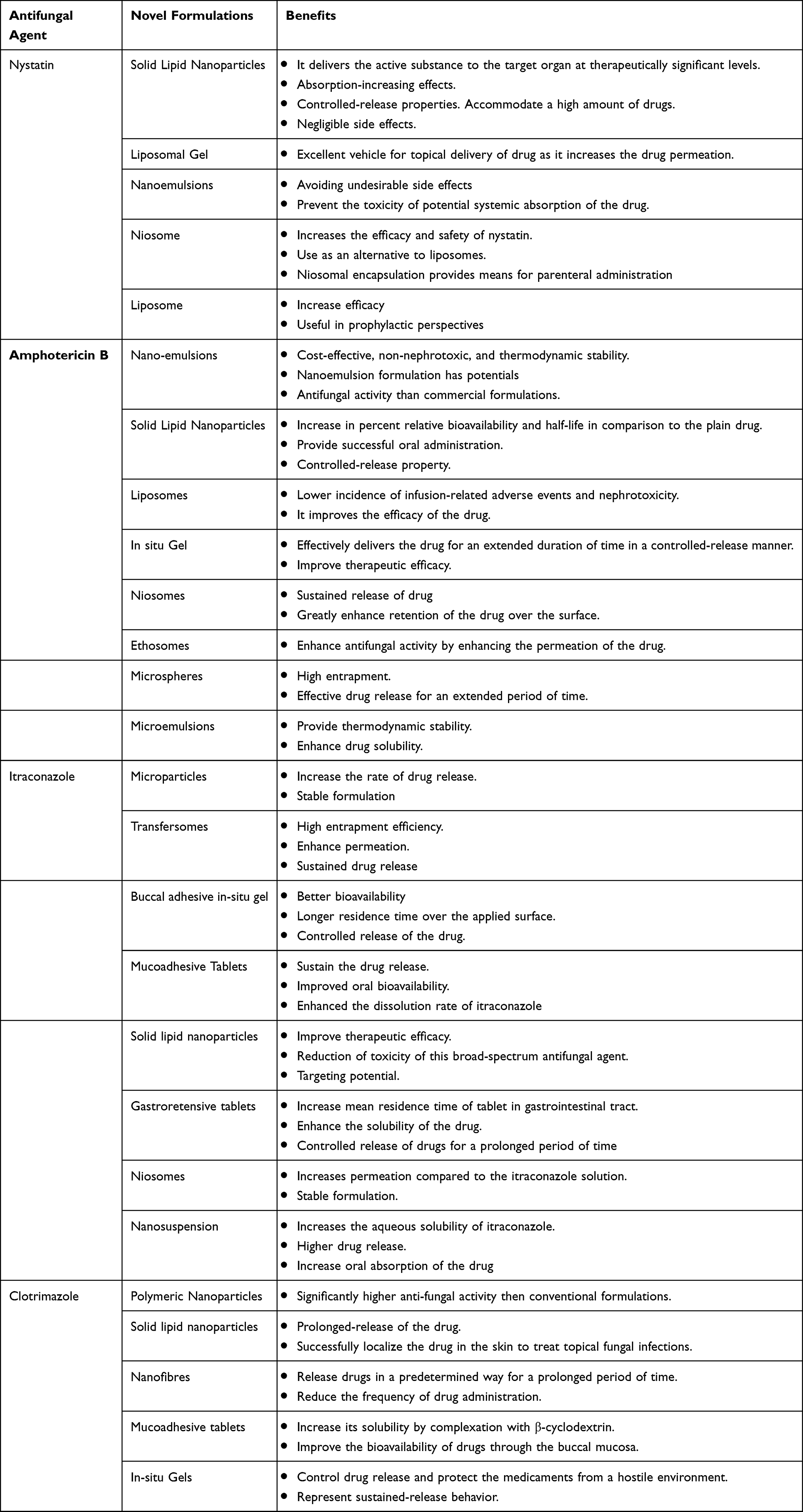 |
Table 1 Novel Approaches for Antifungal Therapy Summery |
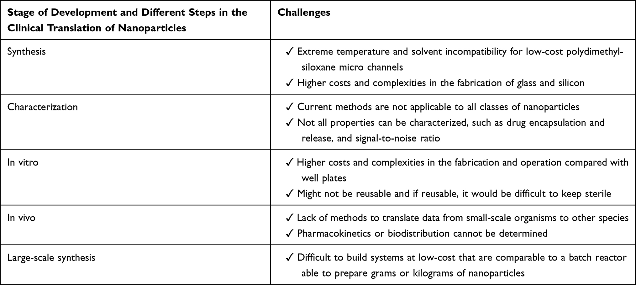 |
Table 2 Summary of Challenges in Translation of Nanoparticles in Stage of Development and Different Steps in the Clinical Trial |
Future Prospective
In the case of antifungal agents currently in clinical use, new formulations and delivery systems may, in principle, enable the development of targeted therapies that should lead to improved efficacy and reduced toxicity, leading to overall improved patient outcomes. The same applies to the application of antifungal pharmacokinetic/pharmacodynamic (PK/PD) principles, better in vitro and in vivo PK/PD models, more accurate clinical antifungal PK/PD predictions, and therapeutic monitoring can result in optimized antifungal dosing lead and increase the likelihood of a successful outcome in patients with fungal infections. Finally, we would like to highlight the use of nanotechnology using nanoscale materials as an alternative strategy for the development of antifungal drugs, which has grown in importance in recent years. These so-called nanomaterials or nano-antibiotics can be described as individual structures whose size in at least one of their three dimensions is less than 100 nm. The increasing interest in nanomaterials is due to their new or improved physicochemical properties such as durability, chemical reactivity, biocompatibility, conductivity and reduced toxicity. Although a wide variety of nanomaterials have been evaluated, the majority of research on the topic has used metal nanoparticles synthesized by various methods and assessed their direct antifungal activity.108
Conclusion
Fungal infections are a major contributor to skin diseases worldwide, causing significant morbidity and mortality. Although there are several effective antifungal agents, their therapeutic utility is limited by high toxicity or poor physicochemical properties. Due to their favorable properties, such as small size, multifunctionality and biocompatibility, nanoparticles, have the potential to overcome many of these limitations. Lipid-based nanocarriers (liposomes, solid lipid nanoparticles, and nanostructured lipid carriers) have been studied best for antifungal drug delivery compared to other nanocarriers. Many of these nanocarriers (eg liposomes) have undergone multiple clinical trials for the treatment of invasive mycoses. Indeed, the commercialization of liposomal amphotericin B has been a major advance, allowing this potent antifungal agent to be used in clinical practice with minimal or no toxicity. Many other nanoparticle formulations have shown promising results in improving the water solubility of antifungal drugs, improving their antifungal efficacy, improving stability, and targeting infected tissue. However, amphotericin B is almost the only antifungal agent that has made it to market in clinical trials and in nano formulations. Therefore, research in the area of antifungal drug delivery should focus on overcoming the challenges that hinder the clinical translation of nanoparticle-based formulations.
Abbreviations
ABLC, AmB lipid complex; ABCD, AmB colloidal dispersion; L-AmB, liposomal AmB; MIC, minimum inhibitory concentration; DMVs, deformable membrane vesicles; PLGA, poly(lactic-co-glycolic acid); CDA, Candida albicans antibody; SLNs, solid lipid nanoparticles; NLCs, nanostructured lipid carriers; NS, nanosponges; MFC, minimum fungicidal concentration.
Disclosure
The authors report no conflicts of interest in this work.
References
1. Suk JS, Xu Q, Kim N, Hanes J, Ensign LM. PEGylation as a strategy for improving nanoparticle-based drug and gene delivery. Adv Drug Deliv Rev. 2016;99:28–51. doi:10.1016/j.addr.2015.09.012
2. Garg A, Sharma GS, Goyal AK, Ghosh G, Si SC, Rath G. Recent advances in topical carriers of anti-fungal agents. Heliyon. 2020;6(8):e04663. doi:10.1016/j.heliyon.2020.e04663
3. Faisal W, Soliman GM, Hamdan AM, Faisal W, Soliman GM, Hamdan AM. Enhanced skin deposition and delivery of voriconazole using ethosomal preparations. J Liposome Res. 2018;28(1):14–21. doi:10.1080/08982104.2016.1239636
4. Bitar D, Lortholary O, Strat Le Y, et al. Deaths attributable to carbapenem-resistant Enterobacteriaceae infections. Emerg Infect Dis J. 2014;20(7):1149–1155.
5. Soliman GM. Nanoparticles as safe and effective delivery systems of antifungal agents: achievements and challenges. Int J Pharm. 2017;523(1):15–32. doi:10.1016/j.ijpharm.2017.03.019
6. Brunke S, Mogavero S, Kasper L, Hube B. Virulence factors in fungal pathogens of man. Curr Opin Microbiol. 2016;32:89–95. doi:10.1016/j.mib.2016.05.010
7. Ikeh M, Ahmed Y, Quinn J. Phosphate acquisition and virulence in human fungal pathogens. Microorganism. 2017;5(3):1–17. doi:10.3390/microorganisms5030048
8. Ding C, Festa RA, Sun TS. Iron and copper as virulence modulators in human fungal pathogens. Mol Microbiol. 2014;93(1):10–23. doi:10.1111/mmi.12653
9. Nett JE, Andes DR. Antifungal agents: spectrum of activity, pharmacology, and clinical indications. Infect Dis Clin North Am. 2016;30(1):51–83. doi:10.1016/j.idc.2015.10.012
10. Walsh TJ, Groll A, Hiemenz J, Fleming R, Roilides E, Anaissie E. Infections due to emerging and uncommon medically important fungal pathogens. Clin Microbiol Infect. 2004;10(SUPPL. 1):48–66. doi:10.1111/j.1470-9465.2004.00839.x
11. Kumar JR, Muralidharan S, Parasuraman S. Antifungal agents: new approach for novel delivery systems. J Pharm Sci Res. 2014;6(5):229–235.
12. Lewis RE. Current concepts in antifungal pharmacology. Mayo Clin Proc. 2011;86(8):805–817. doi:10.4065/mcp.2011.0247
13. Macesic N, Stone NRH, Wingard JR. Liposomal amphotericin B. In: Kucers’ the Use of Antibiotics a Clinical Review of Antibacterial, Antifungal, Antiparasitic, and Antiviral Drugs.
14. Furst T, Piette M, Lechanteur A, Evrard B, Piel G. Mucoadhesive cellulosic derivative sponges as drug delivery system for vaginal application. Eur J Pharm Biopharm. 2015;95(February):128–135. doi:10.1016/j.ejpb.2015.01.019
15. Zazo H, Colino CI, Lanao JM. Current applications of nanoparticles in infectious diseases. J Control Release. 2016;224:86–102. doi:10.1016/j.jconrel.2016.01.008
16. Khatry S, Sirish Shastri N, Sadanandam M. Novel drug delivery systems for antifungal therapy. Int J Pharm Pharm Sci. 2010;2(4):6–9.
17. Waghule T, Sankar S, Rapalli VK, et al. Emerging role of nanocarriers based topical delivery of anti-fungal agents in combating growing fungal infections. Dermatol Ther. 2020;33(6). doi:10.1111/dth.13905.
18. Bozzuto G, Molinari A. Liposomes as nanomedical devices. Int J Nanomedicine. 2015;10:975–999. doi:10.2147/IJN.S68861
19. Saadat E, Dinarvand R, Ebrahimnejad P. Encapsulation of nystatin in nanoliposomal formulation: characterization, stability study and antifungal activity against Candida albicans. Pharm Biomed Res. 2016;2(1):44–54. doi:10.18869/acadpub.pbr.2.1.44
20. Adler-Moore JP, Gangneux JP, Pappas PG. Comparison between liposomal formulations of amphotericin B. Med Mycol. 2016;54(3):223–231. doi:10.1093/mmy/myv111
21. Stone NRH, Bicanic T, Salim R, Hope W. Liposomal Amphotericin B (AmBisome®): a review of the pharmacokinetics, pharmacodynamics, clinical experience and future directions. Drugs. 2016;76(4):485–500. doi:10.1007/s40265-016-0538-7
22. Zoubek A, Emminger W, Schmidmeier WE, et al. Conventional vs. liposomal amphotericin B in immunosuppressed children. Pediatr Hematol Oncol. 2009;3:18.
23. Akhtar N. Vesicles: a recently developed novel carrier for enhanced topical drug delivery. Curr Drug Deliv. 2014;11(1):87–97. doi:10.2174/15672018113106660064
24. Pandit J, Garg M, Jain NK. Miconazole nitrate bearing ultraflexible liposomes for the treatment of fungal infection. J Liposome Res. 2014;24(2):163–169. doi:10.3109/08982104.2013.871025
25. Salem HF, Ahmed SM, Omar MM. Liposomal flucytosine capped with gold nanoparticle formulations for improved ocular delivery. Drug Des Devel Ther. 2016;10:277–295. doi:10.2147/DDDT.S91730
26. Gupta SK, Velpandian T, Dhingra N, Jaiswal J. Intravitreal pharmacokinetics of plain and liposome-entrapped fluconazole in rabbit eyes. J Ocul Pharmacol Ther. 2000;16(6):511–518. doi:10.1089/jop.2000.16.511
27. Song CK, Balakrishnan P, Shim CK, Chung SJ, Chong S, Kim DD. A novel vesicular carrier, transethosome, for enhanced skin delivery of voriconazole: characterization and in vitro/in vivo evaluation. Colloids Surf B Biointerfaces. 2012;92:299–304. doi:10.1016/j.colsurfb.2011.12.004
28. Aggarwal N, Goindi S. Preparation and evaluation of antifungal efficacy of griseofulvin loaded deformable membrane vesicles in optimized Guinea pig model of microsporum canis - Dermatophytosis. Int J Pharm. 2012;437(1–2):277–287. doi:10.1016/j.ijpharm.2012.08.015
29. Mahima M, Devi VK. Potential of novel drug delivery systems in the management of topical candidiasis. J Drug Target. 2017. doi:10.1080/1061186X.2017.1331352
30. Campani V, Biondi M, Mayol L, et al. Nanocarriers to enhance the accumulation of vitamin K1 into the skin. Pharm Res. 2016;33(4):893–908. doi:10.1007/s11095-015-1836-6
31. Verma P, Pathak K. Nanosized ethanolic vesicles loaded with econazole nitrate for the treatment of deep fungal infections through topical gel formulation. Nanomed Nanotechnol Biol Med. 2012;8(4):489–496. doi:10.1016/j.nano.2011.07.004
32. Patel DM, Jani RH, Patel CN. Ufasomes: a vesicular drug delivery. Syst Rev Pharm. 2011;2(2):72–78. doi:10.4103/0975-8453.86290
33. Gupta M, Vaidya B, Mishra N, Vyas SP. Effect of surfactants on the characteristics of fluconazole niosomes for enhanced cutaneous delivery. Artif Cells Blood Substitutes Biotechnol. 2011;39(6):376–384. doi:10.3109/10731199.2011.611476
34. Barakat HS, Darwish IA, El-Khordagui LK, Khalafallah NM. Development of naftifine hydrochloride alcohol-free niosome gel. Drug Dev Ind Pharm. 2009;35(5):631–637. doi:10.1080/03639040802498864
35. Moghassemi S, Hadjizadeh A. Nano-niosomes as nanoscale drug delivery systems: an illustrated review. J Control Release. 2014;185(1):22–36. doi:10.1016/j.jconrel.2014.04.015
36. Abdul Hasan Sathali A, Rajalakshmi G. Evaluation of transdermal targeted niosomal drug delivery of terbinafine hydrochloride. Int J PharmTech Res. 2010;2(3):2081–2089.
37. Shirsand S, Kanani K, Keerthy D, Nagendrakumar D, Para M. Formulation and evaluation of Ketoconazole niosomal gel drug delivery system. Int J Pharm Investig. 2012;2(4):201. doi:10.4103/2230-973X.107002
38. Alam M, Dwivedi V, Khan AA, Mohammad O. Efficacy of niosomal formulation of diallyl sulfide against experimental candidiasis in Swiss albino mice. Nanomedicine. 2009;4(7):713–724. doi:10.2217/nnm.09.60
39. Chaudhari MB, Desai PP, Patel PA, Patravale VB. Solid lipid nanoparticles of amphotericin B (AmbiOnp): in vitro and in vivo assessment towards safe and effective oral treatment module. Drug Deliv Transl Res. 2016;6(4):354–364. doi:10.1007/s13346-015-0267-6
40. Sanna V, Gavini E, Cossu M, Rassu G, Giunchedi P. Solid lipid nanoparticles (SLN) as carriers for the topical delivery of econazole nitrate: in-vitro characterization, ex-vivo and in-vivo studies. J Pharm Pharmacol. 2007;59(8):1057–1064. doi:10.1211/jpp.59.8.0002
41. Moazeni M, Kelidari HR, Saeedi M, et al. Time to overcome fluconazole resistant Candida isolates: solid lipid nanoparticles as a novel antifungal drug delivery system. Colloids Surf B Biointerfaces. 2016;142:400–407. doi:10.1016/j.colsurfb.2016.03.013
42. Geszke-Moritz M, Moritz M. Solid lipid nanoparticles as attractive drug vehicles: composition, properties and therapeutic strategies. Mater Sci Eng C. 2016;68:982–994. doi:10.1016/j.msec.2016.05.119
43. Khan AB, Saha C. A review on vaginal drug delivery system. Rajiv Gandhi Univ Heal Sci J Pharm Sci. 2015;4(4):142–147.
44. Pardeike J, Weber S, Zarfl HP, Pagitz M, Zimmer A. Itraconazole-loaded nanostructured lipid carriers (NLC) for pulmonary treatment of aspergillosis in falcons. Eur J Pharm Biopharm. 2016;108:269–276. doi:10.1016/j.ejpb.2016.07.018
45. Song SH, Lee KM, Kang JB, Lee SG, Kang MJ, Choi YW. Improved skin delivery of voriconazole with a nanostructured lipid carrier-based hydrogel formulation. Chem Pharm Bull. 2014;62(8):793–798. doi:10.1248/cpb.c14-00202
46. Hussain A, Singh VK, Singh OP, Shafaat K, Kumar S, Ahmad FJ. Formulation and optimization of nanoemulsion using antifungal lipid and surfactant for accentuated topical delivery of Amphotericin B. Drug Deliv. 2016;23(8):3101–3110. doi:10.3109/10717544.2016.1153747
47. Aswathanarayan JB, Vittal RR. Nanoemulsions and their potential applications in food industry. Front Sustain Food Syst. 2019;3:
48. Hussain A, Samad A, Singh SK, et al. Nanoemulsion gel-based topical delivery of an antifungal drug: in vitro activity and in vivo evaluation. Drug Deliv. 2016;23(2):652–667. doi:10.3109/10717544.2014.933284
49. Mahtab A, Anwar M, Mallick N, Naz Z, Jain GK, Ahmad FJ. Transungual delivery of Ketoconazole Nanoemulgel for the effective management of onychomycosis. AAPS PharmSciTech. 2016;17(6):1477–1490. doi:10.1208/s12249-016-0488-0
50. Hussain A, Singh S, Webster TJ, Ahmad FJ. New perspectives in the topical delivery of optimized amphotericin B loaded nanoemulsions using excipients with innate anti-fungal activities: a mechanistic and histopathological investigation. Nanomed Nanotechnol Biol Med. 2017;13(3):1117–1126. doi:10.1016/j.nano.2016.12.002
51. Psimadas D, Georgoulias P, Valotassiou V, Loudos G. Molecular nanomedicine towards cancer . J Pharm Sci. 2012;101(7):2271–2280. doi:10.1002/jps.23146
52. Tayel SA, El-Nabarawi MA, Tadros MI, Abd-Elsalam WH. Promising ion-sensitive in situ ocular nanoemulsion gels of terbinafine hydrochloride: design, in vitro characterization and in vivo estimation of the ocular irritation and drug pharmacokinetics in the aqueous humor of rabbits. Int J Pharm. 2013;443(1–2):293–305. doi:10.1016/j.ijpharm.2012.12.049
53. Mirza MA, Ahmad S, Mallick MN, Manzoor N, Talegaonkar S, Iqbal Z. Development of a novel synergistic thermosensitive gel for vaginal candidiasis: an in vitro, in vivo evaluation. Colloids Surf B Biointerfaces. 2013;103:275–282. doi:10.1016/j.colsurfb.2012.10.038
54. Thakkar HP, Khunt A, Dhande RD, Patel AA. Formulation and evaluation of Itraconazole nanoemulsion for enhanced oral bioavailability. J Microencapsul. 2015;32(6):559–569. doi:10.3109/02652048.2015.1065917
55. Kakkar S, Kaur IP. Spanlastics-A novel nanovesicular carrier system for ocular delivery. Int J Pharm. 2011;413(1–2):202–210. doi:10.1016/j.ijpharm.2011.04.027
56. Kaur IP, Rana C, Singh M, Bhushan S, Singh H, Kakkar S. Development and evaluation of novel surfactant-based elastic vesicular system for ocular delivery of fluconazole. J Ocul Pharmacol Ther. 2012;28(5):484–496. doi:10.1089/jop.2011.0176
57. ElMeshad AN, Mohsen AM. Enhanced corneal permeation and antimycotic activity of itraconazole against Candida albicans via a novel nanosystem vesicle. Drug Deliv. 2016;23(7):2115–2123. doi:10.3109/10717544.2014.942811
58. ElSherif MS, Brown C, Mackinnon-Cameron D, et al. Assessing the safety and immunogenicity of recombinant vesicular stomatitis virus Ebola vaccine in healthy adults: a randomized clinical trial. CMAJ. 2017;189(24):E819–E827. doi:10.1503/cmaj.170074
59. Pugazhendhi A, Prabakar D, Jacob JM, Karuppusamy I, Saratale RG. Synthesis and characterization of silver nanoparticles using Gelidium amansii and its antimicrobial property against various pathogenic bacteria. Microb Pathog. 2018;114:41–45. doi:10.1016/j.micpath.2017.11.013
60. Patel NR, Leonardi C, Sabliov CM. Itraconazole-loaded poly (lactic-co-glycolic) acid nanoparticles for improved antifungal activity. Nanomedicine. 2010;5(7):1037–1050. doi:10.2217/nnm.10.68
61. Mora-Huertas CE, Fessi H, Elaissari A. Polymer-based nanocapsules for drug delivery. Int J Pharm. 2010;385(1–2):113–142. doi:10.1016/j.ijpharm.2009.10.018
62. Pourgholi F, Hajivalili M, Farhad JN, Kafil HS, Yousefi M. Nanoparticles: novel vehicles in treatment of Glioblastoma. Biomed Pharmacother. 2016;77:98–107. doi:10.1016/j.biopha.2015.12.014
63. Campos EVR, Oliveira JL, Zavala-Betancourt SA, et al. Development of stained polymeric nanocapsules loaded with model drugs: use of a fluorescent poly(phenyleneethynylene). Colloids Surf B Biointerfaces. 2016;147:442–449. doi:10.1016/j.colsurfb.2016.08.031
64. Matoso Sombra F, Richter AR, de Araújo AR, et al. Nanocapsules of Sterculia striata acetylated polysaccharide as a potential monomeric amphotericin B delivery matrix. Int J Biol Macromol. 2019;130:655–663. doi:10.1016/j.ijbiomac.2019.02.076
65. Sombra FM, Richter AR, Araújo De AR, et al. Development of Amphotericin B-loaded Propionate Sterculia striata. Int J Biol Macromol. 2019;146:1133–1141.
66. Italia JL, Yahya MM, Singh D, Ravi Kumar MNV. Biodegradable nanoparticles improve oral bioavailability of amphotericin B and show reduced nephrotoxicity compared to intravenous fungizone®. Pharm Res. 2009;26(6):1324–1331. doi:10.1007/s11095-009-9841-2
67. Tang X, Dai J, Xie J, et al. Enhanced antifungal activity by Ab-modified amphotericin B-loaded nanoparticles using a pH-responsive block copolymer. Nanoscale Res Lett. 2015;10(1):1–11. doi:10.1186/s11671-015-0969-1
68. Van De Ven H, Paulussen C, Feijens PB, et al. PLGA nanoparticles and nanosuspensions with amphotericin B: potent in vitro and in vivo alternatives to Fungizone and AmBisome. J Control Release. 2012;161(3):795–803. doi:10.1016/j.jconrel.2012.05.037
69. Bian X, Liang S, John J, et al. Development of PLGA-based itraconazole injectable nanospheres for sustained release. Int J Nanomedicine. 2013;8:4521–4531. doi:10.2147/IJN.S54040
70. Burapapadh K, Takeuchi H, Sriamornsak P. Development of pectin nanoparticles through mechanical homogenization for dissolution enhancement of itraconazole. Asian J Pharm Sci. 2016;11(3):365–375. doi:10.1016/j.ajps.2015.07.003
71. Alhakamy NA, Shadab M. Repurposing itraconazole loaded PLGA nanoparticles for improved antitumor efficacy in non-small cell lung cancers. Pharmaceutics. 2019;11(12):12. doi:10.3390/pharmaceutics11120685
72. Pandey R, Ahmad Z, Sharma S, Khuller GK. Nano-encapsulation of azole antifungals: potential applications to improve oral drug delivery. Int J Pharm. 2005;301(1–2):268–276. doi:10.1016/j.ijpharm.2005.05.027
73. Endo EH, Makimori RY, Companhoni MVP, Ueda-Nakamura T, Nakamura CV, Dias Filho BP. Ketoconazole-loaded poly-(lactic acid) nanoparticles: characterization and improvement of antifungal efficacy in vitro against Candida and dermatophytes. J Mycol Med. 2020;30(3):101003. doi:10.1016/j.mycmed.2020.101003
74. Oerlemans C, Bult W, Bos M, Storm G, Nijsen JFW, Hennink WE. Polymeric micelles in anticancer therapy: targeting, imaging and triggered release. Pharm Res. 2010;27(12):2569–2589. doi:10.1007/s11095-010-0233-4
75. Maeda M, Moriuchi S, Sano A, Yoshimine T. New drug delivery system for water-soluble drugs using silicone and its usefulness for local treatment: application of GCV-silicone to GCV/HSV-tk gene therapy for brain tumor. J Control Release. 2002;84(1–2):15–25. doi:10.1016/S0168-3659(02)00236-5
76. Reddy BPK, Yadav HKS, Nagesha DK, Raizaday A, Karim A. Polymeric micelles as novel carriers for poorly soluble drugs-A review. J Nanosci Nanotechnol. 2015;15(6):4009–4018. doi:10.1166/jnn.2015.9713
77. Bobo D, Robinson KJ, Islam J, Thurecht KJ, Corrie SR. Nanoparticle-based medicines: a review of FDA-approved materials and clinical trials to date. Pharm Res. 2016;33(10):2373–2387. doi:10.1007/s11095-016-1958-5
78. Deng P, Teng F, Zhou F, Song Z, Meng N, Feng R. Methoxy poly (ethylene glycol)-b-poly (δ-valerolactone) copolymeric micelles for improved skin delivery of ketoconazole. J Biomater Sci Polym Ed. 2017;28(1):63–78. doi:10.1080/09205063.2016.1244371
79. Bachhav YG, Mondon K, Kalia YN, Gurny R, Möller M. Novel micelle formulations to increase cutaneous bioavailability of azole antifungals. J Control Release. 2011;153(2):126–132. doi:10.1016/j.jconrel.2011.03.003
80. Rodriguez YJ, Quejada LF, Villamil JC, Baena Y, Parra-Giraldo CM, Perez LD. Development of amphotericin b micellar formulations based on copolymers of poly(Ethylene glycol) and poly(ε-caprolactone) conjugated with retinol. Pharmaceutics. 2020;12(3):3. doi:10.3390/pharmaceutics12030196
81. Tang X, Liang Y, Zhu Y, et al. Anti-transferrin receptor-modified amphotericin B-loaded PLA-PEG nanoparticles cure Candidal meningitis and reduce drug toxicity. Int J Nanomedicine. 2015;10:6227–6241. doi:10.2147/IJN.S84656
82. Chauhan AS. Dendrimers for Drug Delivery. Molecules. 2018;23(4):4. doi:10.3390/molecules23040938
83. Tripathy S, Das MK. Dendrimers and their applications as novel drug delivery carriers. J Appl Pharm Sci. 2013;3(9):142–149.
84. Rao BN, Fathima SR, Viswanath V, Prakash KG, Padmini DS, Reddy PS. Novel citric acid dendritic hydrogels for the delivery of econazole nitrate and its antifungal activity. J Appl Pharm Sci. 2016;6(12):094–102. doi:10.7324/JAPS.2016.601213
85. Winnicka K, Wroblewska M, Wieczorek P, Sacha PT, Tryniszewska E. Hydrogel of Ketoconazole and PAMAM dendrimers: formulation and antifungal activity. Molecules. 2012;17(4):4612–4624. doi:10.3390/molecules17044612
86. Sosnowska K. PAMAM dendrimers affect the in vitro release of clotrimazole from hydrogels irrespective of its molecular state. African J Pharm Pharmacol. 2013;7(10):560–573. doi:10.5897/AJPP2013.3470
87. Khairnar GA, Chavan-Patil AB, Palve PR, Bhise SB, Mourya VK, Kulkarni CG. Dendrimers: potential tool for enhancement of antifungal activity. Int J PharmTech Res. 2010;2(1):736–739.
88. Sharma R, Garg T, Goyal AK, Rath G. Development, optimization and evaluation of polymeric electrospun nanofiber : a tool for local delivery of fluconazole for management of vaginal candidiasis. Artif Cells, Nanomed Biotechnol. 2014;44(2):524–531. doi:10.3109/21691401.2014.966194
89. Albicans C. Antifungal activity of eugenol loaded electrospun PAN nanofiber mats against Candida albicans. Curr Drug Deliv. 2018;15(6):860–866. doi:10.2174/1567201815666180226120436
90. Pourdeyhimi B, Khan SA. Preservation of cell viability and protein conformation on immobilization within nano fibers via electrospinning functionalized yeast. ACS Appl Mater Interfaces. 2013;5(19):9349–9354. doi:10.1021/am4022768
91. Huang S, Zhou L, Li M, Wu Q, Kojima Y. Preparation and properties of electrospun poly (Vinyl Pyrrolidone)/ cellulose nanocrystal/silver nanoparticle composite fibers. Materials. 2016;9(7):523. doi:10.3390/ma9070523
92. Clotrimazole K. Formulation and evaluation of antifungal nanosponge loaded hydrogel for topical delivery. Int J Pharm Pharm Res. 2018;13:362–379.
93. Access O, Press H. Formulation and evaluation of fluconazole loaded nanospongies for improved topical drug delivery. Br J Pharm. 2018;2(2):41–42.
94. Abbas N, Parveen K, Hussain A, Latif S, Shah PA, Ahsan M. Nanosponge-based hydrogel preparation of fluconazole for improved topical delivery. Trop J Pharm Re. 2019;18:215–222. doi:10.4314/tjpr.v18i2.1
95. Sharma R, Pathak K. Polymeric nanosponges as an alternative carrier for improved retention of econazole nitrate onto the skin through topical hydrogel formulation. Pharm Dev Technol. 2011;16(4):367–376. doi:10.3109/10837451003739289
96. Gajbhiye S, Sakharwade S. Silver nanoparticles in cosmetics. J Cosmet Dermatol Sci Appl. 2016;06(01):48–53. doi:10.4236/jcdsa.2016.61007
97. Lee J, Kim K, Sung WS, Kim JG, Lee DG. The silver nanoparticle (Nano-Ag): a new model for antifungal agents the silver nanoparticle (Nano-Ag): a new model for antifungal agents. Silver Nanoparticles. 2014;1:295–308.
98. Ashour SM. Article Silver nanoparticles as antimicrobial agent from Kluyveromyces marxianus and Candida utilis. Int J Curr Microbiol Appl Sci. 2014;3(8):384–396.
99. Kola M, Vec R, Prucek R, Soukupova J, Hamal P, Zbor R. Biomaterials Antifungal activity of silver nanoparticles against Candida sppˇova. Biomaterials. 2009;30:6333–6340. doi:10.1016/j.biomaterials.2009.07.065
100. Tharwat NA, Al-bedak OA, Hamouda RE, Shreif El RH. Antifungal effect of gold nanoparticles on fungi isolated from onychomycosis patients. Al-Azhar J Pharm sci. 2019;60(2):26–42. doi:10.21608/ajps.2019.70234
101. Alam F, Azam A, Khan AU. Gold nanoparticles enhance methylene blue – induced photodynamic therapy : a novel therapeutic approach to inhibit Candida albicans biofilm. Int J Nanomedicine. 2012;7:3245–3257. doi:10.2147/IJN.S31219
102. Jebali A, Haji F, Hajjar E, et al. Triangular gold nanoparticles conjugated with peptide ligands : a new class of inhibitor for Candida albicans secreted aspartyl proteinase. Biochem Pharmacol. 2014;90(4):349–355. doi:10.1016/j.bcp.2014.05.020
103. Allen TM, Cullis PR. Liposomal drug delivery systems: from concept to clinical applications. Adv Drug Deliv Rev. 2013;65(1):36–48. doi:10.1016/j.addr.2012.09.037
104. Couvreur P. Nanoparticles in drug delivery: past, present and future. Adv Drug Deliv Rev. 2013;65(1):21–23. doi:10.1016/j.addr.2012.04.010
105. Sercombe L, Veerati T, Moheimani F, Wu SY, Sood AK, Hua S. Advances and challenges of liposome assisted drug delivery. Front Pharmacol. 2015;6:1–13. doi:10.3389/fphar.2015.00286
106. Sainz V, Conniot J, Matos AI, et al. Regulatory aspects on nanomedicines. Biochem Biophys Res Commun. 2015;468(3):504–510. doi:10.1016/j.bbrc.2015.08.023
107. Lewinski N, Colvin V, Drezek R. Cytotoxicity of nanoparticles. Small. 2008;4(1):26–49. doi:10.1002/smll.200700595
108. Wall G, Lopez-Ribot JL. Current antimycotics, new prospects, and future approaches to antifungal therapy. Antibiotics. 2020;9(8):1–10. doi:10.3390/antibiotics9080445
 © 2022 The Author(s). This work is published and licensed by Dove Medical Press Limited. The
full terms of this license are available at https://www.dovepress.com/terms
and incorporate the Creative Commons Attribution
- Non Commercial (unported, 3.0) License.
By accessing the work you hereby accept the Terms. Non-commercial uses of the work are permitted
without any further permission from Dove Medical Press Limited, provided the work is properly
attributed. For permission for commercial use of this work, please see paragraphs 4.2 and 5 of our Terms.
© 2022 The Author(s). This work is published and licensed by Dove Medical Press Limited. The
full terms of this license are available at https://www.dovepress.com/terms
and incorporate the Creative Commons Attribution
- Non Commercial (unported, 3.0) License.
By accessing the work you hereby accept the Terms. Non-commercial uses of the work are permitted
without any further permission from Dove Medical Press Limited, provided the work is properly
attributed. For permission for commercial use of this work, please see paragraphs 4.2 and 5 of our Terms.