Back to Journals » Clinical, Cosmetic and Investigational Dermatology » Volume 16
Retrospective Analysis of 397 Dermatoses Inpatients Associated with Blood Eosinophilia
Authors Zhao Y, Tian J, Gao C ![]() , Liu L
, Liu L ![]() , Pan L, Song Z
, Pan L, Song Z
Received 5 July 2023
Accepted for publication 7 October 2023
Published 2 December 2023 Volume 2023:16 Pages 3455—3463
DOI https://doi.org/10.2147/CCID.S429183
Checked for plagiarism Yes
Review by Single anonymous peer review
Peer reviewer comments 3
Editor who approved publication: Dr Jeffrey Weinberg
Ying Zhao, Jing Tian, Cuie Gao, Lu Liu, Linxin Pan, Zhiqiang Song
Department of Dermatology, Southwest Hospital, Army Medical University, Chongqing, People’s Republic of China
Correspondence: Zhiqiang Song, Department of Dermatology, Southwest Hospital, Army Medical University, No. 30, Gaotanyan Street, Shapingba District, Chongqing, 400038, People’s Republic of China, Email [email protected]
Background: Blood eosinophilia is often associated with various dermatoses, such as atopic eczema, urticaria, drug eruption, bullous pemphigoid, and hypereosinophilic syndrome (HES). Differential diagnosis is very challenging due to the similarities of clinical and pathological characteristics.
Purpose: To investigate and analyze the clinical characteristics of dermatoses associated with blood eosinophilia (DABE) to further optimize disease management.
Patients and Methods: We conducted a retrospective analysis on 397 DABE patients with blood absolute eosinophil count (AEC) greater than or equal to 0.5× 10⁹/L. Clinical characteristics, laboratory values, treatment course, and associated diagnoses were evaluated. All DABE patients were grouped based on the severity of eosinophilia as mild group (0.5 ≤ AEC× 109/L < 1.5), moderate group (1.5 ≤ AEC× 109/L < 3), and severe group (AEC× 109/L ≥ 3).
Results: Our study revealed three distinct patterns: (1) Mild eosinophilia associated with localized skin lesions, atopic history, mildly elevated total serum IgE level, diagnosed with eczema/dermatitis, and frequent antihistamines use. (2) Moderate eosinophilia has the characteristics of both mild group and severe group. (3) The severe eosinophilia group had a high proportion of elderly people without atopic history, but with acute onset, generalized skin lesions, and high level of lactate dehydrogenase, and the majority of them were diagnosed with systemic diseases (HES or tumor).
Conclusion: We summarize the clinical rules of dermatoses associated with blood eosinophilia, hoping to facilitate the diagnosis and treatment for patients.
Keywords: eosinophilia, diagnosis, eczema, psoriasis, drug eruption
Introduction
Eosinophils have a wide range of biological functions and play an important role in anti-infection, inflammatory response, anti-tumor, and tissue damage and repair.1,2 Eosinophils enter the blood circulation after maturation in the bone marrow. There are no eosinophils in normal skin tissue, and eosinophils are recruited from peripheral blood to skin tissue only when inflammation occurs.3 Most dermatoses associated with blood eosinophilia (DABE) belong to allergy-related skin diseases, such as atopic eczema, contact dermatitis, urticaria, prurigo, and drug eruption; Second, blood eosinophilia can also be seen in parasitic infections and autoimmune bullous diseases. The skin is also the first and most commonly affected organ of hypereosinophilic syndrome (HES).4,5
It is estimated that for every additional eosinophil in the blood, there is a corresponding increase of 100 eosinophils in the tissue.6 Potential mechanisms leading to eosinophilia are divided into primary intrinsic mechanisms and secondary reactive mechanisms.7,8 The clonal expansion of eosinophils mediated by FIP1L1-PDGFRA (F/P) fusion gene belongs to the primary disease, while the secondary eosinophilia is mainly caused by eosinophilopoietic cytokines (IL-3, IL-5 and GM-CSF).7,9 The continuous increase of eosinophils can secrete a series of cytotoxic mediators, such as eosinophil cationic protein (ECP), eosinophil-derived neurotoxin (EDN), and eosinophil peroxidase (EPO), leading to multiple organ damage and possibly life-threatening.10 However, the evaluation and treatment of dermatoses with blood eosinophilia is challenging because of the significant clinical and histopathological overlap between different DABE diseases.
In this study, we divided DABE patients into three groups according to blood absolute eosinophil count (AEC) levels: mild eosinophilia group, moderate eosinophilia group, and severe eosinophilia group, and summarized the demographics, clinical characteristics, laboratory results, related diagnoses, and treatments, hope to help the differential diagnoses of DABE patients to further optimize disease management.
Methods
This is a retrospective cross-sectional study, including all inpatients with AEC greater than or equal to 0.5×10⁹/L who visited the Department of Dermatology, Southwest Hospital of Army Military Medical University from January 2018 to January 2023. Since eosinophilia associated with dermatoses is usually not very high, the categorization used for HES does not make sense and important information in the mild to moderate group might have been overlooked.11 According to the degree of elevated blood AEC, patients were divided into the mild eosinophilia group (0.5 ≤ AEC×109/L < 1.5), moderate eosinophilia group (1.5 ≤ AEC×109/L < 3), and severe eosinophilia group (AEC×109/L ≥ 3). Electronic medical records were reviewed for all cases, and data collected included demographics, patient history, clinical manifestations, laboratory results, diagnoses, and treatment. The Ethics Committee of Southwest Hospital of Army Medical University approved this study (KY2023100). Patient consent is not required for this retrospective study. The study conformed to the ethical guidelines of the Declaration of Helsinki.
Statistical analyses were performed using SPSS Statistics (V22; IBM SPSS Corp., Armonk, NY, USA). All tests were considered significant at P < 0.05. The Kruskal–Wallis test was used to analyze variations in the age, duration of eosinophilia, serum total Immunoglobulin.E (IgE) values, and lactate dehydrogenase (LDH) values in different groups. Categorical data were analyzed using chi-square test or Fisher’s exact test, including sex ratios, medical history ratios, clinical manifestations (prevalence of pruritus, distribution of lesions, ratio of lesion types), diagnostic rates, and drug use rates.
Results
Demographics and History
A total of 397 DABE patients (267 males, 67.3%; median 59 years, range:45–70 years) were included and grouped according to blood AEC: mild eosinophilia, 0.5 ≤ AEC < 1.5 (n = 292, 73.6%); moderate eosinophilia, 1.5 ≤ AEC < 3 (n = 70, 17.6%); severe eosinophilia, AEC ≥ 3 (n = 35, 8.8%, Table 1).
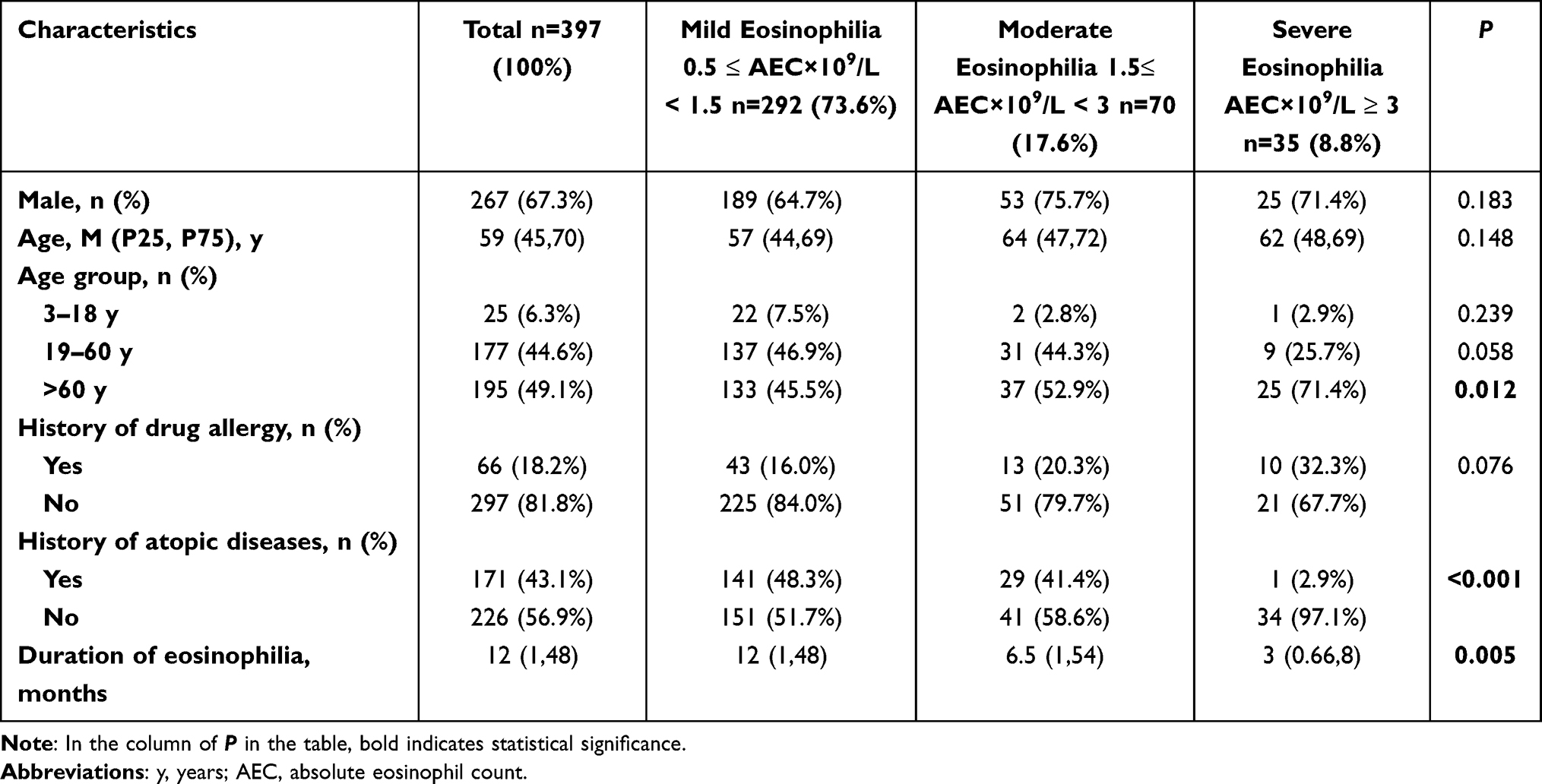 |
Table 1 Demographic and Historical Characteristics in the Study Groups |
There were statistically significant differences in the age distribution (P = 0.012) and the proportion of atopic history (P < 0.001) among the three groups. The severe eosinophilia group had a higher proportion (23/35, 71.5%) of old patients and a lower proportion (1/35, 2.9%) of atopic history. 18.2% (66/397) of the patients had lesions associated with elevated blood eosinophils due to drug exposure. The proportion of drug sensitization in the severe eosinophilia group (10/35, 32.3%) was higher than that in the other two groups, but the difference was not statistically significant (P = 0.076). The severe group had the shortest duration of eosinophilia compared with the mild and moderate groups (P = 0.005, Table 1).
Clinical Manifestations
Almost all DABE patients (383/397, 96.5%) exhibited pruritus symptoms, which were independent of blood eosinophil levels (P = 0.549). Localized skin lesions were more common in the mild eosinophilia group, while generalized skin lesions were observed in the moderate and severe eosinophilia groups (P < 0.001). The morphological spectrum of skin lesions in DABE patients was wide, and the most common lesions were erythema (348/397, 87.7%) and papules (208/397, 52.4%). The incidence of skin vesicles was significantly higher in the moderate eosinophilia group than in the mild and severe groups (P = 0.03, Table 2). The incidence of other lesion types was independent of blood eosinophil levels.
 |
Table 2 Clinical Manifestations in the Study Groups |
Laboratory Results
We selected two blood parameters, serum total IgE and LDH, to analyze their association with blood eosinophilia. Serum total IgE was elevated in 68.4% (132/193) of DABE patients, and LDH levels were elevated in 27.7% (67/242) of DABE patients. In the mild eosinophilia group, the serum total IgE median was significantly lower than those in the other two groups (P < 0.001). In contrast to mild and moderate groups, elevated LDH was more common in the severe group, and their LDH levels were also higher (P < 0.001, Table 3).
 |
Table 3 The Laboratory Results in the Study Groups |
Then, we were interested in whether increased blood eosinophilia corresponded to eosinophilic infiltration in the skin and bone marrow. Histopathological examination of skin biopsies showed cutaneous eosinophilic infiltration in 71.9% (105/155) patients. There was no significant difference in skin eosinophil infiltration among the three groups (P = 0.629). The most common histopathologic characteristics are spongiosis and hyperplasia. Bone marrow biopsy histopathology showed that 93.2% (41/44) of DABE patients were accompanied by bone marrow eosinophil infiltration, and most of them were from the moderate or severe eosinophilia groups (Table 3). Screening for the F/P fusion gene, which has been associated with HES, was negative in 2 patients. For the immunophenotype analysis of peripheral blood lymphocytes, no abnormal T and B lymphocytes were found in all 3 patients.
Diagnosis
The most common diagnosis in DABE patients was eczema/dermatitis (207/397, 52.1%), followed by drug eruption (66/397, 16.6%), systemic disease (50/397, 12.6%) including HES or tumor, autoimmune bullous diseases (34/397, 8.6%), psoriasis (23/397, 5.8%), and other diseases (17/397, 4.3%). The diagnosis of eczema/dermatitis was dominant in the mild eosinophilia group (P < 0.001), while the diagnosis of systemic disease (HES or tumor) was more common in the severe eosinophilia group (P < 0.001, Table 4).
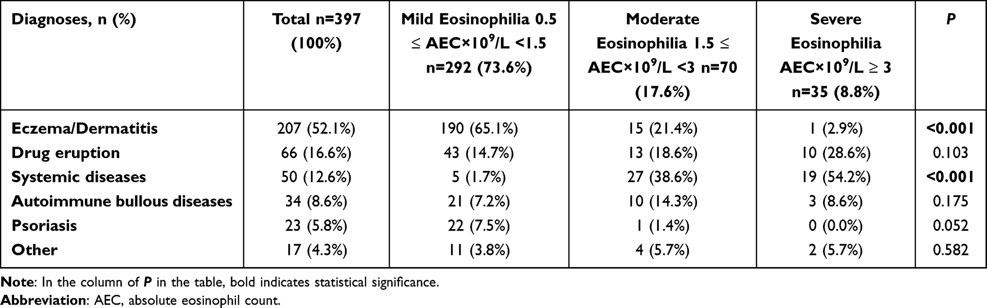 |
Table 4 Final Diagnoses in the Study Groups |
Treatment
In this study, we also evaluated the response to therapeutic drugs in three groups with elevated blood eosinophils. Glucocorticoids (370/392, 93.2%) were the most commonly used drug to treat DABE, followed by antihistamines (322/397, 81.1%), immunosuppressants (129/392, 32.5%), antibiotics (14/392, 3.5%), retinoids (14/392, 3.5%), biologics (5/392, 1.3%), other drugs (7/392, 1.8%). In the mild eosinophilia group, the usage rate of antihistamines was significantly higher than that in the moderate and severe groups (P = 0.032), while the usage rate of antibiotics was opposite (P < 0.001, Table 5). There was no statistically significant difference in the usage rate of other drugs among the three groups.
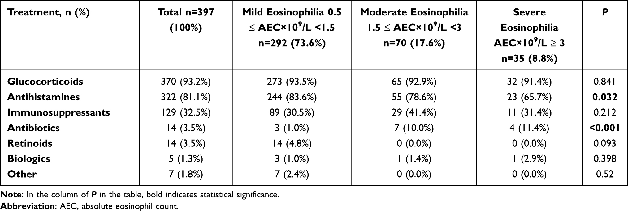 |
Table 5 Treatment in the Study Groups |
Discussion
An elevated level of blood eosinophils may be the first important clue in a laboratory result. We analyzed in detail the demographics, clinical characteristics, laboratory results, related diagnoses and treatments in DABE patients. Our results demonstrated that blood eosinophil level is associated with different clinical types in DABE patients. Blood eosinophil level plus other information such as age, history of atopy, history of drug sensitization, disease duration, distribution of skin lesions, and abnormal blood parameters may be helpful for further diagnosis.
Our study revealed three distinct patterns: (1) Mild eosinophilia associated with localized skin lesions, atopic history, mildly elevated total serum IgE level, diagnosed with eczema/dermatitis, and frequent antihistamines use. (2) Moderate eosinophilia has the characteristics of both mild group and severe group. (3) The severe eosinophilia group had a high proportion of elderly people without atopic history, but with acute onset, generalized skin lesions, and high blood LDH levels, and the majority of them were diagnosed with systemic diseases (HES or tumor) (Figure 1).
 |
Figure 1 The characteristics of demographics, history, lesion manifestation, examination, and diagnoses in DABE patients with mild, moderate, and severe blood eosinophilia. (-) The laboratory result was negtive. ↑The laboratory result was mildly elevated. ↑↑ The laboratory result was dramatically elevated. Abbreviations: DABE, dermatoses associated with blood eosinophilia; IgE, immunoglobulin E; LDH, lactate dehydrogenase. |
The predominance of eosinophils in eczema is not surprising since T helper 2 (Th2) lymphocytes always induce the recruitment of eosinophils in inflamed areas.12 Dermatological results showed that eosinophil activation and toxic granule protein deposition were involved in the acute and chronic lesions of atopic eczema.13 Cetinkaya et al suggested that transient, mild eosinophilia in children is associated with atopic eczema, whereas persistent, severe eosinophilia may be associated with congenital immune deficiency.14 This is similar to our data, where patients under the age of 18 were mostly clustered in the mild eosinophilia group and were diagnosed with eczema. Our results also show that atopic eczema is often related to mild blood eosinophilia, and this relation is more pronounced when accompanied by atopic history and mildly elevated serum total IgE.15
The skin is one of the organs most commonly affected by adverse drug reactions (ADRs). Eosinophils play a key role in drug-induced lesions.16 The incubation period for drug exposure can vary from days to years. Correspondingly, the duration of drug eruption includes acute exacerbation and chronic relapse. Our study showed that 16.6% (66/397) of the patients had lesions and different degrees of elevated blood eosinophils due to drug exposure. The incidence of drug sensitization was 14.7%, 18.6%, and 28.6% in the mild, moderate, and severe eosinophilia groups, but there was no statistical difference among the three groups. Severe eosinophilia with organ damage of the heart, liver, and kidney is associated with more serious systemic adverse drug reactions, such as drug eruption with eosinophilia and systemic symptoms (DRESS).17 Similar to the study of Yang et al, we suggested that the circulating eosinophil count was positively correlated with the severity of drug eruption, and the circulating eosinophil count could also be a prognostic indicator of drug eruption.18 Therefore, any patient with unexplained eosinophilia must obtain a detailed medication history.
Although eosinophilia is generally considered to be insignificant in psoriasis, our results showed 22 cases of psoriasis with mild eosinophilia, including 14 cases of vulgaris, 5 cases of pustulosa, 3 cases of erythrodermic. Retinoids are the first-line drugs for the treatment of psoriasis. Correspondingly, the use of retinoids is clustered in the mild eosinophilia group. There are no published reports investigating the overall incidence of eosinophilia associated with psoriasis. In psoriasis patients, the number of eosinophils labeled with ECP polyclonal antibody was significantly higher than that in healthy controls.19 Sueki et al reported a case of psoriasis vulgaris in which peripheral blood eosinophilia paralleled with the Psoriasis Area and Severity Index (PASI) score, and improvement in psoriasis was directly correlated with decline in eosinophilia.20 Another study showed that peripheral blood eosinophilia appears to be associated with severe forms of psoriasis, such as generalized pustulosa and erythrodermic forms.6 In conclusion, the combination of mild blood eosinophilia and psoriasis appears to be a relatively common condition. It would be significant to further investigate this association in a larger series of cohorts.
Although parasitic infection is one of the most important causes of eosinophilia,21 only 1% (4/397) of DABE in our study were caused by parasitic infection (including 1 case of hookworm infection, 1 case of insect bite dermatitis, 2 cases of scabies) and both were from the mild eosinophilia group. It was reported that 1.0% of children with eosinophilia had parasitic infections, compared with 4.8% of non-parasitic infections.14 In another study, the frequency of parasitic infections in hypereosinophilia was 5.7%.22 Differences between these studies may be strongly related to various socioeconomic levels. Based on the epidemiological importance of parasitic infection, we suggest that for DABE patients with a history of travel to endemic areas and persistent eosinophilia, it is necessary to develop further stool and dermatoscopy to detect eggs and parasites.
Peripheral blood eosinophilia has been reported in 61% of bullous pemphigoid (BP) cases and 46% of pemphigus cases.23,24 In our study, 8.6% (34/397) of DABE patients were diagnosed with autoimmune bullous diseases. Research has shown the strong relation between circulating eosinophil counts and the classic phenotype of BP (vesicles and erosions).25 There was a positive correlation between the severity of BP and peripheral blood eosinophils in the study of Gore Karaali et al.26 Diagnosis of autoimmune bullous disease was mostly in the moderate eosinophilia group through semantic connectivity map analysis.27 Unfortunately, we did not detect these patterns, which may be due to the lack of enough patients with autoimmune bullous disease in our cohort.
In the severe eosinophilia group, DABE patients were more diagnosed with systemic diseases (HES and tumor), and they had a lower proportion with atopic history and a higher proportion of older age. Severe blood eosinophilia was associated with higher levels of IgE and LDH. HES is a diagnosis of exclusion, excluding allergies, infections, rheumatism, and other diseases.28 In various studies, more than 50% of patients with HES develop pleomorphic skin lesions, often delaying diagnosis and treatment.29,30 Khallaayoune et al reported a case diagnosed with BP who showed resistance to conventional treatment and persistent eosinophilia, and finally this patient was considered as BP-associated HES.31 Patients with HES should be carefully examined, especially bone marrow biopsy, F/P fusion gene, and immunophenotyping of peripheral blood lymphocytes. Because HES with the fusion gene is at risk of developing to the malignant end, eventually progressing to eosinophilic leukemia. It is worth noting that some HES patients have allergies, rhinitis, asthma, and other comorbidities at the same time, which is difficult to distinguish from atopic eczema.30
Conclusion
This study could help to better understand the relationship between dermatoses and blood eosinophilia, potentially improving diagnoses and treatments for patients. The level of blood eosinophilia corresponds to different dermatoses, and careful history and targeted examination are crucial for differential diagnosis.
Ethics Approval
This study was approved by the Ethics Committee of Southwest Hospital of Army Medical University (KY2023100). This retrospective study conformed to the ethical guidelines of the Declaration of Helsinki, and patients’ privacy and personal identity information are protected. Exemption from informed consent will not have any adverse impact on patients’ health and rights. Therefore, the patient consent is not required for this retrospective study.
Acknowledgments
The authors thank all participants in this study for their enthusiastic cooperation.
Funding
This research was funded by Natural Science Foundation of China (82073442).
Disclosure
The authors report no conflicts of interest in this work.
References
1. Wechsler ME, Munitz A, Ackerman SJ, et al. Eosinophils in health and disease: a state-of-the-art review. Mayo Clin Proc. 2021;96(10):2694–2707. doi:10.1016/j.mayocp.2021.04.025
2. Rothenberg ME, Hogan SP. The eosinophil. Annu Rev Immunol. 2006;24:147–174. doi:10.1146/annurev.immunol.24.021605.090720
3. Long H, Zhang G, Wang L, Lu Q. Eosinophilic skin diseases: a comprehensive review. Clin Rev Allergy Immunol. 2016;50(2):189–213. doi:10.1007/s12016-015-8485-8
4. Leiferman KM, Peters MS. Eosinophil-related disease and the skin. J Allergy Clin Immunol Pract. 2018;6(5):1462–1482.e1466. doi:10.1016/j.jaip.2018.06.002
5. Radonjic-Hoesli S, Brüggen MC, Feldmeyer L, Simon HU, Simon D. Eosinophils in skin diseases. Semin Immunopathol. 2021;43(3):393–409. doi:10.1007/s00281-021-00868-7
6. Mansur AT, Göktay F, Yaşar SP. Peripheral blood eosinophilia in association with generalized pustular and erythrodermic psoriasis. J Eur Acad Dermatol Venereol. 2008;22(4):451–455. doi:10.1111/j.1468-3083.2007.02489.x
7. Shomali W, Gotlib J. World Health Organization-defined eosinophilic disorders: 2022 update on diagnosis, risk stratification, and management. Am J Hematol. 2022;97(1):129–148. doi:10.1002/ajh.26352
8. Leru PM. Eosinophilic disorders: evaluation of current classification and diagnostic criteria, proposal of a practical diagnostic algorithm. Clin Transl Allergy. 2019;9(1):36. doi:10.1186/s13601-019-0277-4
9. Hougaard M, Thomsen GN, Kristensen TK, et al. A retrospective cohort study of patients with eosinophilia referred to a tertiary centre. Dan Med J. 2022;69(4): A07210558
10. Acharya KR, Ackerman SJ. Eosinophil granule proteins: form and function. J Biol Chem. 2014;289(25):17406–17415. doi:10.1074/jbc.R113.546218
11. Valent P, Klion AD, Horny HP, et al. Contemporary consensus proposal on criteria and classification of eosinophilic disorders and related syndromes. J Allergy Clin Immunol. 2012;130(3):607–612.e609. doi:10.1016/j.jaci.2012.02.019
12. Akdis CA, Arkwright PD, Brüggen MC, et al. Type 2 immunity in the skin and lungs. Allergy. 2020;75(7):1582–1605. doi:10.1111/all.14318
13. Kiehl P, Falkenberg K, Vogelbruch M, Kapp A. Tissue eosinophilia in acute and chronic atopic dermatitis: a morphometric approach using quantitative image analysis of immunostaining. Br J Dermatol. 2001;145(5):720–729. doi:10.1046/j.1365-2133.2001.04456.x
14. Cetinkaya PG, Aytekin ES, Esenboga S, et al. Eosinophilia in children: characteristics, etiology and diagnostic algorithm. Eur J Pediatr. 2023;182(6):2833–2842. doi:10.1007/s00431-023-04961-x
15. Crnković HT, Bendelja K, Šimić Klarić A, Tomić Rajić M, Drkulec V, Aberle N. Family history and cord blood eosinophil count as predictors for atopic manifestations. Cent Eur J Public Health. 2019;27(4):267–271. doi:10.21101/cejph.a5601
16. Hoetzenecker W, Nägeli M, Mehra ET, et al. Adverse cutaneous drug eruptions: current understanding. Semin Immunopathol. 2016;38(1):75–86. doi:10.1007/s00281-015-0540-2
17. Duong TA, Valeyrie-Allanore L, Wolkenstein P, Chosidow O. Severe cutaneous adverse reactions to drugs. Lancet. 2017;390(10106):1996–2011. doi:10.1016/S0140-6736(16)30378-6
18. Yang J, Yang X, Li M. Peripheral blood eosinophil counts predict the prognosis of drug eruptions. J Investig Allergol Clin Immunol. 2013;23(4):248–255.
19. Kim TY, Park HJ, Kim CW. Eosinophil cationic protein (ECP) level and its correlation with eosinophil number or IgE level of peripheral blood in patients with various skin diseases. J Dermatol Sci. 1997;15(2):89–94. doi:10.1016/S0923-1811(97)00614-2
20. Sueki H, Nakada T, Iijima M. A case of psoriasis vulgaris with peripheral blood eosinophilia, parallelling the psoriasis area and severity index (PASI) score. Clin Exp Dermatol. 2004;29(5):549–550. doi:10.1111/j.1365-2230.2004.01566.x
21. Rothenberg ME. Eosinophilia. N Engl J Med. 1998;338(22):1592–1600. doi:10.1056/NEJM199805283382206
22. Rosenberg HF, Dyer KD, Foster PS. Eosinophils: changing perspectives in health and disease. Nat Rev Immunol. 2013;13(1):9–22. doi:10.1038/nri3341
23. Crotty C, Pittelkow M, Muller SA. Eosinophilic spongiosis: a clinicopathologic review of seventy-one cases. J Am Acad Dermatol. 1983;8(3):337–343. doi:10.1016/S0190-9622(83)70036-8
24. Morais KL, Miyamoto D, Maruta CW, Aoki V. Diagnostic approach of eosinophilic spongiosis. An Bras Dermatol. 2019;94(6):724–728. doi:10.1016/j.abd.2019.02.002
25. Garrido PM, Aguado-Lobo M, Espinosa-Lara P, Soares-Almeida L, Filipe P. Association of peripheral blood and cutaneous eosinophils with bullous pemphigoid disease severity and treatment outcomes. Actas Dermosifiliogr. 2022;113(9):881–887. doi:10.1016/j.ad.2022.05.021
26. Gore Karaali M, Koku Aksu AE, Cin M, Leblebici C, Kara Polat A, Gurel MS. Tissue eosinophil levels as a marker of disease severity in bullous pemphigoid. Australas J Dermatol. 2021;62(2):e236–e241. doi:10.1111/ajd.13547
27. Radonjic-Hoesli S, Martignoni Z, Cazzaniga S, et al. Characteristics of dermatological patients with blood eosinophilia: a retrospective analysis of 453 patients. J Allergy Clin Immunol Pract. 2022;10(5):1229–1237.e1228. doi:10.1016/j.jaip.2022.02.018
28. Salomon G, Severino M, Casassa E, et al. Skin manifestations of hypereosinophilic syndrome are polymorphous and difficult to treat: a retrospective cohort study. Ann Dermatol Venereol. 2022;149(2):139–141. doi:10.1016/j.annder.2021.12.002
29. Ogbogu PU, Bochner BS, Butterfield JH, et al. Hypereosinophilic syndrome: a multicenter, retrospective analysis of clinical characteristics and response to therapy. J Allergy Clin Immunol. 2009;124(6):1319–1325.e1313. doi:10.1016/j.jaci.2009.09.022
30. Neve S, Beukers S, Kirtschig G. Hypereosinophilic syndrome in an atopic patient. Clin Exp Dermatol. 2009;34(8):e643–646. doi:10.1111/j.1365-2230.2009.03356.x
31. Khallaayoune M, Sialiti S, Meziane M, Senouci K. Bullous pemphigoid-like rash revealing hypereosinophilic syndrome. BMJ Case Rep. 2021;14(6). doi:10.1136/bcr-2021-242695
 © 2023 The Author(s). This work is published and licensed by Dove Medical Press Limited. The
full terms of this license are available at https://www.dovepress.com/terms
and incorporate the Creative Commons Attribution
- Non Commercial (unported, 3.0) License.
By accessing the work you hereby accept the Terms. Non-commercial uses of the work are permitted
without any further permission from Dove Medical Press Limited, provided the work is properly
attributed. For permission for commercial use of this work, please see paragraphs 4.2 and 5 of our Terms.
© 2023 The Author(s). This work is published and licensed by Dove Medical Press Limited. The
full terms of this license are available at https://www.dovepress.com/terms
and incorporate the Creative Commons Attribution
- Non Commercial (unported, 3.0) License.
By accessing the work you hereby accept the Terms. Non-commercial uses of the work are permitted
without any further permission from Dove Medical Press Limited, provided the work is properly
attributed. For permission for commercial use of this work, please see paragraphs 4.2 and 5 of our Terms.
Recommended articles
Therapeutic Inertia in the Management of Psoriasis: A Quantitative Survey Among Indian Dermatologists and Patients
Rajagopalan M, Dogra S, Godse K, Kar BR, Kotla SK, Neema S, Saraswat A, Shah SD, Madnani N, Sardesai V, Sekhri R, Varma S, Arora S, Kawatra P
Psoriasis: Targets and Therapy 2022, 12:221-230
Published Date: 25 August 2022
Anxiety and Depression in People with Eczema or Psoriasis: A Comparison of Associations in UK Biobank and Linked Primary Care Data
Matthewman J, Mansfield KE, Hayes JF, Adesanya EI, Smith CH, Roberts A, Langan SM, Henderson AD
Clinical Epidemiology 2023, 15:891-899
Published Date: 7 August 2023