Back to Journals » Drug Design, Development and Therapy » Volume 18
Research Progress of Triptolide Against Fibrosis
Authors Jiang M, Xie Y, Wang P, Du M ![]() , Wang Y
, Wang Y ![]() , Yan S
, Yan S ![]()
Received 8 March 2024
Accepted for publication 1 July 2024
Published 25 July 2024 Volume 2024:18 Pages 3255—3266
DOI https://doi.org/10.2147/DDDT.S467929
Checked for plagiarism Yes
Review by Single anonymous peer review
Peer reviewer comments 2
Editor who approved publication: Dr Qiongyu Guo
Minmin Jiang,1,* Yongxia Xie,2,* Ping Wang,1 Mengyu Du,3 Ying Wang,4 Shuxun Yan1
1Department of Endocrinology, The First Affiliated Hospital of Henan University of Chinese Medicine, Zhengzhou, People’s Republic of China; 2Department of Respiratory Diseases, The First Affiliated Hospital of Henan University of Chinese Medicine, Zhengzhou, People’s Republic of China; 3The First Clinical Medical College, Henan University of Chinese Medicine, Zhengzhou, People’s Republic of China; 4Department of International Medicine, The First Affiliated Hospital of Zhengzhou University, Zhengzhou, People’s Republic of China
*These authors contributed equally to this work
Correspondence: Shuxun Yan; Ying Wang, Email [email protected]; [email protected]
Abstract: Fibrosis leads to organ failure and death, which is the final stage of many chronic diseases. Triptolide (TPL) is a terpenoid extracted from the traditional Chinese medicine Tripterygium wilfordii Hook. F (TwHF). Triptolide and its derivatives (Omtriptolide, Minnelide, (5R)-5-hydroxytriptolide) have been proven to have a variety of pharmacological effects. This study comprehensively reviewed the antifibrotic mechanism of TPL and its derivatives, and discussed the application of advanced nanoparticles (NPs) drug delivery system in the treatment of fibrotic diseases by TPL. The results show that TPL can inhibit immune inflammatory response, relieve oxidative stress and endoplasmic reticulum stress (ERS), regulate collagen deposition and inhibit myofibroblast production to play an anti-fibrosis effect and reduce organ injury. A low dose of TPL has no obvious toxicity. Under pathological conditions, a toxic dose of TPL has a protective effect on organs. The emergence of TPL derivatives (especially Minnelide) and NPs drug delivery systems promotes the anti-fibrosis effect of TPL and reduces its toxicity, which may be the main direction of anti-fibrosis research in the future.
Keywords: triptolide, derivatives, anti-fibrosis, nanoparticles
Introduction
Fibrosis is a pathological deviation from injury repair caused by various causes, like autoimmunity, chronic viral infection, and toxicity (such as drugs, radiation, chronic ischemia, or alcohol), which is usually accompanied by over-deposition of extracellular matrix (ECM) (primarily collagen) and organ function damage.1,2 Almost every organ system is affected by fibrosis, which can include the heart, liver, kidneys, pulmonary system, and skin.3 However, the results of clinical treatment of fibrosis are not satisfactory. Tissue fibroproliferative disorders are responsible for nearly 45% of annual deaths from all diseases.4
TwHF (Figure 1) was first documented in the Compendium of Materia Medica and is an essential Chinese medicine.5 It has a long-term clinical application and functions in dispelling wind and dehumidification, promoting blood circulation and dredging collaterals, killing insects and detoxifying, and reducing swelling and pain. Many clinical researches have confirmed the therapeutic effects of TwHF on various diseases including interstitial pneumonia, systemic lupus erythematosus, multiple sclerosis, oral lichen planus, and rheumatoid arthritis.6–8 Triptolide (TPL) (Figure 2), as the most abundant terpenoid constituent of TwHF, is regarded as the active component with the greatest potential for translation from traditional to modern medicine.9 Previous investigations have shown that TPL has beneficial roles such as anticancer, anti-inflammatory, and regulation of immunity.10–12 In recent years, it has also been found to have therapeutic effects on fibrotic diseases, such as renal fibrosis (RF) caused by diabetic nephropathy.13 However, TPL has limited water solubility, oral bioavailability, and high doses cause serious toxicity, so a variety of TPL derivatives (Figure 2) have been exploited, such as Minnelide, Omtriptolide (PG490-88), (5R)-5-hydroxytriptolide (LLDT-8).14,15 In addition, the development of advanced technologies such as NPs has completely changed the drug delivery of TPL.16 In this review, we not only systematically review the pharmacological mechanism of TPL and its derivatives (Table 1, Figure 3), but also introduce the application of the cutting-edge NPs delivery system in TPL anti-fibrosis (Table 2), hoping to provide help for the future research and application of TPL.
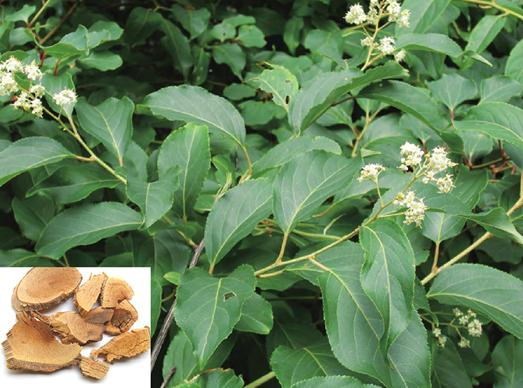 |
Figure 1 Tripterygium wilfordii Hook. F and its pieces of traditional Chinese medicine. |
 |
Figure 2 The structures of triptolide and its derivatives. |
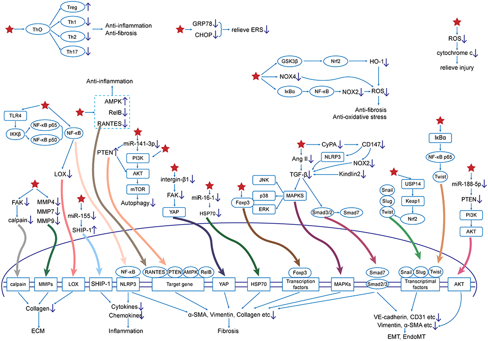 |
Figure 3 Mechanism diagram of triptolide and its derivatives against fibrosis. Star indicates TPL and its derivatives. |
 |
Table 1 Pharmacological Mechanism of TPL and Its Derivatives |
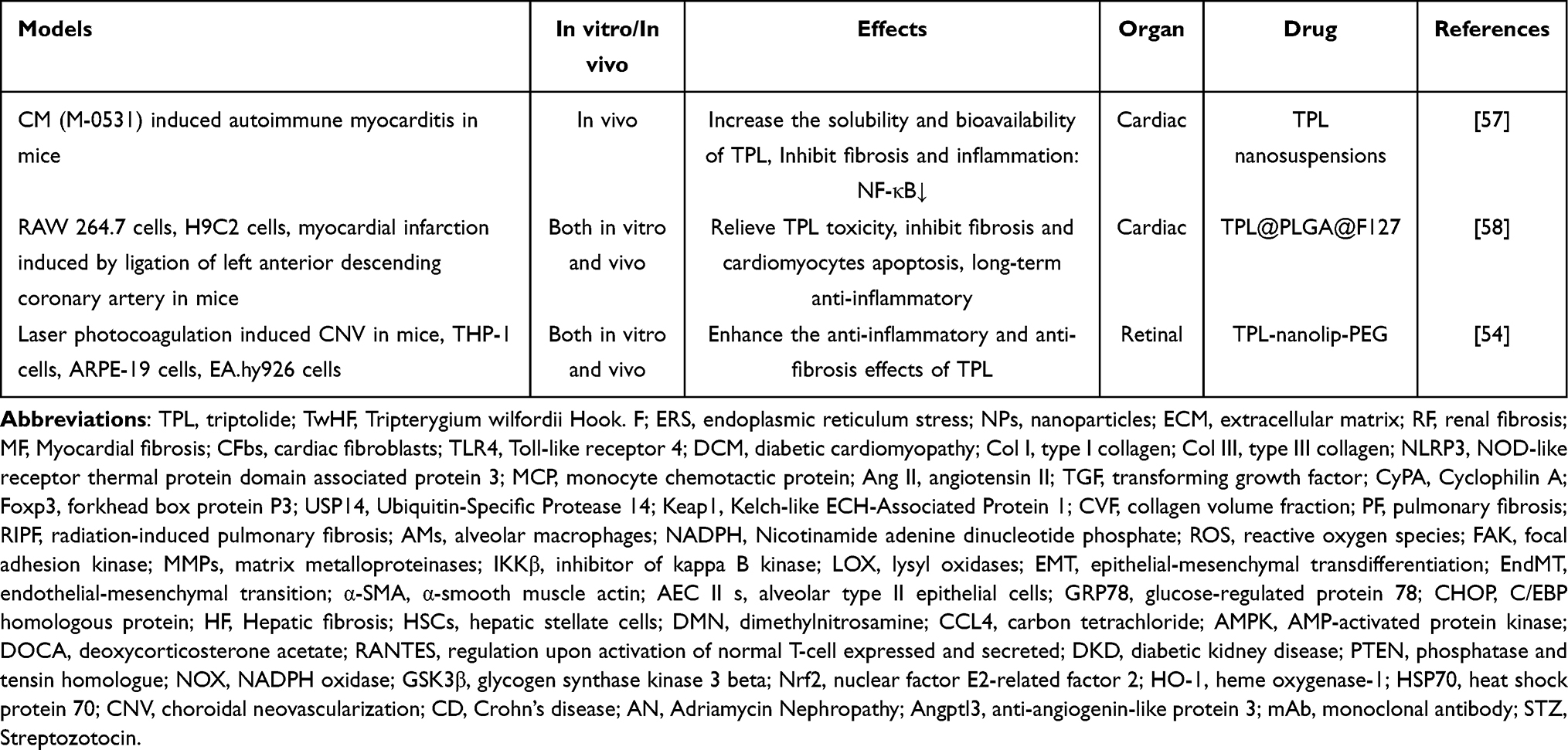 |
Table 2 Application of NPs Delivery System in TPL Anti-Fibrosis |
Anti-Fibrotic Effect of TPL
Heart
Myocardial fibrosis (MF) exists in almost all chronic heart diseases, and damages heart function, playing a crucial role in the progression and consequences of heart failure. Abnormal proliferation and activation of cardiac fibroblasts (CFbs), deposition of ECM, and formation of scar tissue are its major pathological features.59,60
Inflammatory mediators are implicated in the pathogenesis of MF. Long-term chronic inflammation may lead to cardiomyocyte necrosis, and necrotic cardiomyocytes are replaced by collagen scars to cause reparative fibrosis.61 NF-κB pathway is a classic inflammatory pathway. It has been shown that TPL can inhibit Toll-like receptor 4 (TLR4) /NF-κB and NF-κB pathways to improve cardiac immune inflammatory response and alleviate myocardial fibrosis in rats with diabetic cardiomyopathy (DCM).17,18 TPL can improve the expression of fibrosis-associated factors, such as transforming growth factor (TGF)-β1, type I collagen (Col I), and type III collagen (Col III) mRNA, in rats by inhibiting the activation of NOD-like receptor thermal protein domain associated protein 3(NLRP3) inflammasome mediators downstream of the NF-κB pathway.19 TPL also depresses pro-fibrotic TGF-β1 pathways and inflammatory mediators downstream of NLRP3 inflammasome, such as IL-1β, IL-18, monocyte chemotactic protein (MCP)-1 and vascular cell-adhesion molecule −1, and inhibits infiltration of macrophages in a dose-dependent manner.20
Via direct and TGF-β-mediated actions, the local release of angiotensin II (Ang II) plays an effective role in activating and stimulating cardiac fibrosis.62,63 In vitro, TPL inhibited Ang II-induced CFbs proliferation and decreased Ang II-induced fibrosis signaling TGF-β1 /Smad3 expression.21 In vivo, TPL treatment could inhibit the generation of pro-fibrosis factors such as Ang II and TGF-β induced by pressure overload, and significantly suppress left ventricular end-diastolic pressure, myocardial collagen volume fraction (CVF), and Col I/III deposition.22 The down-regulation of pro-inflammatory cytokines (IL-1β and IL-6) in serum and NF-κB in myocardial tissue was also observed in the TPL group.22 Further studies have shown that TPL can alleviate MF by attenuating Ang II-induced TGF- β signal by inhibiting intracellular and extracellular Cyclophilin A (CyPA) / CD147.23 Transcriptional factors participate in the modulation of MF. TPL can improve myocardial injury and myocardial fibrosis score and inhibit cardiac hypertrophy by up-regulating the expression of forkhead box protein P3 (Foxp3).24 TPL can also significantly improve endothelial-mesenchymal transition (EndMT) and alleviate MF by regulating Ubiquitin-Specific Protease 14 (USP14)/ Kelch-like ECH-Associated Protein 1 (Keap1)/ Nuclear factor erythroid 2-related factor 2 (Nrf2) pathway and transcription factors such as Snail, Slug and Twist.25 In addition, the effect of TPL on reducing myocardial collagen content, perivascular collagen area, and myocardial CVF was related to the up-regulation of phosphatase and tensin homologue (PTEN).26
Lung
A group of chronic lung conditions known as pulmonary fibrosis (PF) causes progressive damage to the pulmonary interstitium, impairing gas exchange, producing dyspnea, lowering quality of life, and ultimately leading to respiratory failure and death.64
Studies have confirmed that inflammation and oxidative stress are closely associated with the occurrence and development of PF.65 Yang et al66 found that TPL can inhibit the production of pro-fibrotic cytokines (IL-1β, TGF- β1, and IL-13) in the radiation-induced pulmonary fibrosis (RIPF) model, and improve the 5-month survival rate, lung density, and function. Subsequent studies have identified alveolar macrophages (AMs) as a major source of reactive oxygen species (ROS) in RIPF, and TPL exerts its antifibrotic effects by inhibiting myofibroblast activation and collagen accumulation via the nicotinamide adenine dinucleotide phosphate (NADPH) oxidase-ROS axis in AMs.27 TPL also represses migration and invasion of lung fibroblasts and inhibits the expression of fibrosis factors Col I and Col III and the production of inflammatory factors IL-6 through the focal adhesion kinase (FAK)/troponin axis.28
The increase in myofibroblast production and ECM protein deposition are some pathological features associated with the development of PF.67 It has been shown that TPL can not only regulate matrix metalloproteinases (MMPs), which are involved in ECM deposition but also mediate TGF-β1/Smad fibrosis signal pathway to inhibit the transformation of lung fibroblasts into myofibroblasts.29,68 It was also found that TPL could block integrin-β1/FAK/YAP signal transduction in the biomechanical stress transduction pathway and attenuate the pro-fibrotic effect of fibrogenic ECM on fibroblasts via integrin suppression.29 In addition, TPL can inhibit PF through inhibitor of kappa B kinase (IKKβ)/NFκB pathway and reduce the production of lysyl oxidases (LOX) which catalyzes matrix protein cross-linking.29,30 Zhang et al31 found that TPL can inhibit TGF-β1/ERK/Smad3 pathway and reduce myofibroblast activation both in vivo and in vitro. In addition, in PF, myofibroblasts can also originate from cell sources other than fibroblasts, such as epithelial and endothelial cells, which gain a mesenchymal phenotype through epithelial-mesenchymal transdifferentiation (EMT) or EndMT, and show classic markers of myofibroblast differentiation, like vimentin, α-smooth muscle actin (α-SMA) expression and others.69,70 Many studies demonstrated that TPL inhibits the migration and invasion of human lung epithelial cells and inhibits the expression of EMT-related factors through TGF-β1 signal.32,33 TPL can reverse the EMT of alveolar type II epithelial cells (AEC II s) and relieve PF by regulating NF-κB/Twist1 signal.34 TPL can also inhibit the expression of glucose-regulated protein 78 (GRP78) and C/EBP homologous protein (CHOP) in AEC II s, relieve ERS, reduce collagen deposition in lung tissue, and significantly improve lung function in mice.35
Liver
Hepatic fibrosis (HF) is mainly induced by chronic hepatotoxic injury (such as chronic hepatitis and non-alcoholic steatohepatitis) and cholestatic injury (such as primary and secondary cholangitis, primary sclerosing cholangitis and biliary atresia).71
The main mesenchymal cells in the liver are hepatic stellate cells (HSCs), and activated HSCs is a key factor in the pathological progression of HF.72 Chong et al37 have shown that TPL can inhibit the expression of fibrosis factor α-SMA through anti-NF-κB activation pathway. In addition, TPL treatment significantly reduced the increase of inflammatory cytokines (TNF-α and IL-6) induced by dimethylnitrosamine (DMN) in rats. Immune regulation is a key factor in the pathological process of fibrosis. Jiang et al38 reported for the first time that TPL attenuates carbon tetrachloride (CCL4)-induced HF by modulating the differentiation of CD4+T (Th2, Th1, Th17, and Treg) cells. RelB is related to liver fibrosis. By lowering the expression of RelB in bile duct cells, TPL can suppress the bile duct response brought on by common bile duct ligation, hence lowering liver damage, fibrosis, and inflammation.39 TPL can also improve liver lipogenesis, HF, and fatty acid oxidation in nonalcoholic fatty liver disease mice by activating AMP-activated protein kinase (AMPK).40
Kidney
RF, particularly tubulointerstitial fibrosis, is the primary indicator and dependable prognostic index of renal insufficiency, including glomerulosclerosis, renal tubule atrophy, and renal interstitial fibrosis.73,74 It is also a common marker and pathway of several progressive chronic kidney diseases.
TPL has multiple protective effects on RF, including reducing inflammatory cell infiltration and fibrosis, reducing the expression of many chemokines and cytokines, and reducing renal injury. It was shown that TPL significantly decreased macrophage and myofibroblast infiltration and collagen deposition in the renal interstitial fibrosis model.75 Similarly, in the model of renal injury induced by deoxycorticosterone acetate (DOCA)-salt hypertension, TPL can inhibit the NF-κB pathway to protect cells from inflammatory damage and reduce renal collagen levels.41 Zhu et al42 found that TPL can inhibit ECM synthesis in NRK-49F cells by regulating the activities of Smad2, p38, and ERK1/2. It was shown that TPL ameliorated glomerulosclerosis and interstitial fibrosis in diabetic rats in association with inhibition of regulation upon activation of normal T-cell expressed and secreted (RANTES) overexpression in renal tissues.43 MicroRNAs are small non-coding RNAs that play an influential role in the fight against RF. Li et al44 found that TPL can relieve RF caused by diabetic kidney disease (DKD) by restoring autophagy through miR-141-3p/ PTEN/Akt/mTOR pathway. Subsequent studies confirmed that TPL can improve renal tubulointerstitial fibrosis by targeting miR-188-5p/PTEN/PI3K/AKT signal pathway to reverse tubulointerstitial fibrosis induced by high glucose.45 More importantly, TPL can delay the development of nephropathy in diabetic rats, especially in the stage of massive albuminuria, which may be related to the inhibition of monocyte-macrophage aggregation and the reduction of inflammatory factor expression by TPL.76 Fan et al46 have shown that TPL can not only improve the proteinuria of DKD mice, but also regulate the expression of superoxide dismutase (SOD), ROS, and prototype NADPH oxidase (NOX) 4 in kidney tissue to improve oxidative stress and alleviate RF in DKD by regulating glycogen synthase kinase 3 beta (GSK3β)/ Nrf2/heme oxygenase-1 (HO-1) signal transduction pathway. In addition, TPL was protective against structural damage and loss of function of podocytes in DKD mice.47 TPL alleviates diabetes-induced podocyte EMT by inhibiting TGF- β1/Smad3 signal pathway and kindlin-2.47
Intestine
Intestinal fibrosis is a common complication of chronic inflammatory bowel diseases, such as ulcerative colitis and Crohn’s disease (CD).77 Previous studies have shown that TPL inhibits IL-1β-induced expression of IL-8, MCP-1, and MMP-3 in human colonic subepithelial myofibroblasts by suppressing NF-κB activity.78 In the colon fibrosis model of chronic colitis, TPL can reduce the deposition of ECM and the production of total collagen in the colon, and inhibit the expression of Col I protein and collagen Iα1 mRNA in myofibroblasts.50 Ileocecal anastomotic fibrosis and stricture are common complications after ileocecal resection of CD. Hou et al51 reported that TPL can improve the inflammation and fibrosis of CD anastomotic fibrosis mice through miR-16-1/heat shock protein 70 (HSP70) signal. A subsequent study showed that TPL could also inhibit the migration, proliferation, and fibrosis of fibroblasts derived from ileocolon anastomosis in CD patients by regulating miR-16-1/HSP70 pathway in vitro.52 Similarly, TPL can also target miR-155/SHIP-1 signal pathway to reduce the expression of pro-inflammatory cytokines in CD anastomotic fibrosis mice.53
Others
Retinal fibrosis is one of the end-stage complications of neovascular age-associated macular degeneration, leading to severe, permanent, and high risk of irreversible visual impairment.79 The studies of Lai et al54 showed that TPL inhibited the production of vascular endothelial growth factor and neovascularization in retinal fibrosis mice related to choroidal neovascularization (CNV), promoted M2 macrophage polarization, and mediated TGF-β1/Smad signal pathway to improve EMT/EndoMT. In vivo studies have shown that TPL inhibits the expression of IL-6 and α-SMA mediated by the NF-κB/p65 pathway, thereby alleviating pancreatic fibrosis in mice with chronic pancreatitis.55 Ileocolic anastomotic fibrosis and stenosis are common complications after CD ileocolic resection. TPL can improve epidural fibrosis by inhibiting PI3K/AKT/mTOR signal, inhibiting fibroblast proliferation, and stimulating apoptosis and autophagy.56
Antifibrotic Effect of Triptolide Derivatives
When a phosphate group is added to TPL, a water-soluble derivative called Minnelide is produced, which is widely used in the treatment of cancer.10 Li et al48 have shown that Minnelide alone can significantly alleviate proteinuria and renal injury in Adriamycin Nephropathy (AN) mice, which is related to the ROS-mediated mitochondrial pathway. However, Minnelide combined with anti-angiogenin-like protein 3 (Angptl3)-FLD monoclonal antibody (mAb) almost completely improved the proteinuria and restored the ultrastructure of podocytes in AN mice, which was associated with the promotion of podocyte autophagy and suppression of apoptosis.80 Subsequent studies have demonstrated that Minnelide can reduce the expression of pro-inflammatory cytokines (TNF-α, IL-6, and IL-1β) in Angptl3 knockout AN mice, and improve apoptosis and fibrosis through TGF-β1/Smad2 and p53 signal.49
Compared with TPL, LLDT-8 substituted hydrogen with a hydroxyl group at C-5 position, and PG490-88 introduced fatty acid structure at C-14 position. They play an active role in the therapy of PF. In the bleomycin (BLM)-induced PF mouse model, LLDT-8 could alleviate weight loss and increase lung index, reduce the production of inflammatory cells (neutrophils and lymphocytes) and cytokines (IL-4, TNF-α, and TGF-β), promote the activity of antioxidant factor SOD, inhibit the level of hydroxyproline and improve lung histological injury.81 In the same model, PG490-88 significantly decreased the number of myofibroblasts and blocked the increase of TGF-β gene expression induced by BLM in human lung fibroblasts.36 The latest studies have shown that LLDT-8 can suppress the generation of inflammatory and fibrogenic factors by macrophages to improve proteinuria and structural renal damage and delay fibrosis in DKD mice.82
Application of Nanoparticle Drug Delivery System in Anti-Fibrosis of TPL
Although studies have shown that low-dose TPL has no obvious toxicity, a toxic dose of TPL has a protective effect on organs under pathological conditions.15,83 But the toxicity of TPL is still a concern. The use of NPs can not only improve the dissolution, transport and cellular uptake of bioactive components, but also reduce the toxicity of TPL.16,84 At present, the NPs carrier technology used in TPL anti-fibrosis includes nano-suspension, nano-gel, and nano-liposome. Nanosuspensions are a versatile formulation method for improving drug delivery of hydrophobic drugs and one of the most prosperous ways to improve the performance of poorly water-soluble drugs.85 Li et al57 used TPL nano-suspension to treat autoimmune myocarditis rats, delayed left ventricular remodeling, and significantly improved fibrosis indexes such as myocardial collagen proliferation, CVF, and perivascular collagen area, I/ III collagen ratio. NF-κB pathway may partially mediate the decrease of peripheral blood inflammatory factors, as IL-1, TNF- α, IL-6, and MCP-1. Wang et al58 combined with FDA-approved polylactic acid-glycolic acid copolymer (PLGA) NPs and F127 hydrogel, prepared TPL-loaded nano-hydrogel platform (TPL@PLGA@F127), which can reduce the hepatotoxicity by releasing TPL more slowly and stably. The study has shown that TPL@PLGA@F127 can promote the polarization of macrophages to M2 (anti-inflammatory cells) on the 3rd day after myocardial infarction to play a long-term anti-inflammatory effect, inhibit myocardial fibrosis, and protect cardiomyocytes, and improve cardiac function58 Liposome is one of the most mature nano-delivery carriers, and it is one of the few nano-preparations used in clinical treatment. Its particle size ranges from 50 to 1000nm.86 Lai et al54 showed that polyethylene glycol nanoliposomes (TPL-nanolip-PEG) loaded with TPL could enhance the inhibitory effect of TPL on the infiltration of retinal fibrosis and M2 macrophages, and had no toxic effect on the morphology and function of the retina.
Discussion and Prospect
TPL and its derivatives have an obvious anti-fibrosis effect and can play a role in a variety of tissues and organs. The anti-fibrosis mechanisms of TPL and its derivatives can be summarized as follows: (1) relieving oxidative stress and ERS; (2) inhibiting immune inflammatory response; (3) regulating collagen deposition (4) inhibiting myofibroblast production; (5) regulating cell migration, proliferation, apoptosis, and autophagy.
However, at present, the anti-fibrosis studies of TPL and its derivatives are based on the level of cellular and animal research, and lack of clinical data verification, so further strict large-scale randomized controlled trials and further scientific research should be carried out to fully evaluate its clinical efficacy and safety. In addition, the application of TPL derivatives, especially Minnelide, and NPs delivery systems, to overcome the water solubility and toxicity of TPL is a promising treatment and should be focused on.
Acknowledgments
This work has been supported by Henan Province Traditional Chinese Medicine Inheritance and Innovation Talent Project (Zhongjing Plan) (CZ0237-02), Henan Province Traditional Chinese Medicine Research Special Project (20-21ZY1016), Zhengzhou Collaborative Innovation Project (2023XTCX048) and Henan University of Traditional Chinese Medicine 2022 graduate research and Innovation Ability Improvement plan (2022KYCX043).
Disclosure
The authors report no conflicts of interest in this work.
References
1. Lurje I, Gaisa NT, Weiskirchen R, et al. Mechanisms of organ fibrosis: emerging concepts and implications for novel treatment strategies. Mol Aspects Med. 2023;92:101191. doi:10.1016/j.mam.2023.101191
2. Zhao Y, Qu Y, Hao C, et al. PD-1/PD-L1 axis in organ fibrosis. Front Immunol. 2023;14:1145682. doi:10.3389/fimmu.2023.1145682
3. Wei L, Liu L, Bai M, et al. CircRNAs: versatile players and new targets in organ fibrosis. Cell Commun Signal. 2023;21(1):90. doi:10.1186/s12964-023-01051-1
4. Henderson NC, Rieder F, Wynn TA. Fibrosis: from mechanisms to medicines. Nature. 2020;587(7835):555–566. doi:10.1038/s41586-020-2938-9
5. Song CY, Xu YG, Lu YQ. Use of Tripterygium wilfordii Hook F for immune-mediated inflammatory diseases: progress and future prospects. J Zhejiang Univ Sci B. 2020;21(4):280–290. doi:10.1631/jzus.B1900607
6. Shan Y, Zhao J, Wei K, et al. A comprehensive review of Tripterygium wilfordii hook. f. in the treatment of rheumatic and autoimmune diseases: bioactive compounds, mechanisms of action, and future directions. Front Pharmacol. 2023;14:1282610. doi:10.3389/fphar.2023.1282610
7. Luo Y, Kuai L, Chen J, et al. Efficacy and safety of Tripterygium wilfordii Hook. f. for oral lichen planus: evidence from 18 randomized controlled trials. Phytother Res. 2020;34(9):2180–2191. doi:10.1002/ptr.6672
8. Li Y, Zhu W, He H, et al. Efficacy and Safety of Tripterygium Wilfordii Hook. F for Connective Tissue Disease-Associated Interstitial Lung Disease: A Systematic Review and Meta-Analysis. Front Pharmacol. 2021;12:691031. doi:10.3389/fphar.2021.691031
9. Corson TW, Crews CM. Molecular understanding and modern application of traditional medicines: triumphs and trials. Cell. 2007;130(5):769–774. doi:10.1016/j.cell.2007.08.021
10. Noel P, Von Hoff DD, Saluja AK, et al. Triptolide and Its Derivatives as Cancer Therapies. Trends Pharmacol Sci. 2019;40(5):327–341. doi:10.1016/j.tips.2019.03.002
11. Yu GM, Zhou LF, Zeng BX, et al. The antioxidant effect of triptolide contributes to the therapy in a collagen-induced arthritis rat model. Redox Rep. 2021;26(1):197–202. doi:10.1080/13510002.2021.2004047
12. Huang Y, Ba X, Wang H, et al. Triptolide alleviates collagen-induced arthritis in mice by modulating Treg/Th17 imbalance through the JAK/PTEN-STAT3 pathway. Basic Clin Pharmacol Toxicol. 2023;133(1):43–58. doi:10.1111/bcpt.13880
13. Liu P, Zhang J, Wang Y, et al. The Active Compounds and Therapeutic Target of Tripterygium wilfordii Hook. f. in Attenuating Proteinuria in Diabetic Nephropathy: a Review. Front Med. 2021;8:747922. doi:10.3389/fmed.2021.747922
14. Gao J, Zhang Y, Liu X, et al. Triptolide: pharmacological spectrum, biosynthesis, chemical synthesis and derivatives. Theranostics. 2021;11(15):7199–7221. doi:10.7150/thno.57745
15. Hou Z, Yan M, Li H, et al. Variable p53/Nrf2 crosstalk contributes to triptolide-induced hepatotoxic process. Toxicol Lett. 2023;379:67–75. doi:10.1016/j.toxlet.2023.03.011
16. Sun R, Dai J, Ling M, et al. Delivery of triptolide: a combination of traditional Chinese medicine and nanomedicine. J Nanobiotechnology. 2022;20(1):194. doi:10.1186/s12951-022-01389-7
17. Wen HL, Liang ZS, Zhang R, et al. Anti-inflammatory effects of triptolide improve left ventricular function in a rat model of diabetic cardiomyopathy. Cardiovasc Diabetol. 2013;12(1):50. doi:10.1186/1475-2840-12-50
18. Guo X, Xue M, Li CJ, et al. Protective effects of triptolide on TLR4 mediated autoimmune and inflammatory response induced myocardial fibrosis in diabetic cardiomyopathy. J Ethnopharmacol. 2016;193:333–344. doi:10.1016/j.jep.2016.08.029
19. Shen J, Ma H, Wang C. Triptolide improves myocardial fibrosis in rats through inhibition of nuclear factor kappa B and NLR family pyrin domain containing 3 inflammasome pathway. Korean J Physiol Pharmacol. 2021;25(6):533–543. doi:10.4196/kjpp.2021.25.6.533
20. Li R, Lu K, Wang Y, et al. Triptolide attenuates pressure overload-induced myocardial remodeling in mice via the inhibition of NLRP3 inflammasome expression. Biochem Biophys Res Commun. 2017;485(1):69–75. doi:10.1016/j.bbrc.2017.02.021
21. Liu M, Yeh J, Huang Y, et al. EFFECT OF TRIPTOLIDE ON PROLIFERATION AND APOPTOSIS OF ANGIOTENSIN II-INDUCED CARDIAC FIBROBLASTS IN VITRO: a PRELIMINARY STUDY. Afr J Tradit Complement Altern Med. 2016;14(1):145–154. doi:10.21010/ajtcam.v14i1.16
22. Zhang Z, Qu X, Ni Y, et al. Triptolide protects rat heart against pressure overload-induced cardiac fibrosis. Int J Cardiol. 2013;168(3):2498–2505. doi:10.1016/j.ijcard.2013.03.001
23. Cao M, Zhao Q, Xia H, et al. Intracellular and extracellular Cyclophilin a promote cardiac fibrosis through TGF-β signaling in response to angiotensin II. Biochem Pharmacol. 2024;225:116271. doi:10.1016/j.bcp.2024.116271
24. Ding YY, Li JM, Guo FJ, et al. Triptolide Upregulates Myocardial Forkhead Helix Transcription Factor p3 Expression and Attenuates Cardiac Hypertrophy. Front Pharmacol. 2016;7:471. doi:10.3389/fphar.2016.00471
25. Ba L, Mingyao E, Wang R, et al. Triptolide attenuates cardiac remodeling by inhibiting pyroptosis and EndMT via modulating USP14/Keap1/Nrf2 pathway. Heliyon. 2024;10(2):e24010. doi:10.1016/j.heliyon.2024.e24010
26. Liu M, Chen J, Yao JR, et al. Triptolide Attenuates Cardiac Remodeling in Isoprenaline-induced Chronic Heart Failure Rats via Upregulating PTEN Pathway. J Sun Yat-Sen Univer. 2017;38(01):29–35.
27. Chen C, Yang S, Zhang M, et al. Triptolide mitigates radiation-induced pulmonary fibrosis via inhibition of axis of alveolar macrophages-NOXes-ROS-myofibroblasts. Cancer Biol Ther. 2016;17(4):381–389. doi:10.1080/15384047.2016.1139229
28. Zhang P, Liu J, Zong R. Triptolide protects against TGF-β1-induced pulmonary fibrosis by regulating FAK/calpain signaling. Exp Ther Med. 2019;18(6):4781–4789. doi:10.3892/etm.2019.8127
29. Lin W, Song Y, Li T, et al. Triptolide attenuates pulmonary fibrosis by inhibiting fibrotic extracellular matrix remodeling mediated by MMPs/LOX/integrin. Biomed Pharmacother. 2023;166:115394. doi:10.1016/j.biopha.2023.115394
30. Guo K, Chen J, Chen Z, et al. Triptolide alleviates radiation-induced pulmonary fibrosis via inhibiting IKKβ stimulated LOX production. Biochem Biophys Res Commun. 2020;527(1):283–288. doi:10.1016/j.bbrc.2020.04.023
31. Zhang YW, Zhang ZQ, Wu LX, et al. TPL’s suppression on activation of myofibroblasts in radiation induced lung fibrosis related to its inhibition on TGF-β1/ERK/Smad3 pathway. Chin Pharmacol Bulletin. 2017;33(05):630–636.
32. Li Q, Chen Q, Chen H. Mechanism of triptolide inhibiting pulmonary fibrosis induced by paraquat through the transforming growth factor β1 pathway. China J Tradition Chin Med Pharm. 2023;38(02):815–818.
33. Chen H, Chen Q, Jiang CM, et al. Triptolide suppresses paraquat induced idiopathic pulmonary fibrosis by inhibiting TGFB1-dependent epithelial mesenchymal transition. Toxicol Lett. 2018;284:1–9. doi:10.1016/j.toxlet.2017.11.030
34. Chen H, Chen Q, Li Q. Analysis on the inhibitory effect of triptolide on NF-κB/Twist 1 signaling pathway on interstitial differentiation of alveolar epithelial cells. China J Tradition Chin Med Pharm. 2022;37(03):1384–1388.
35. Song QN, Li M, Li GX, et al. Triptolide improves pulmonary function and mechanism of endoplasmic reticulum stress in mice with pulmonary fibrosis induced by bleomycin. Lishizhen Medicine and Materia Medica Res. 2022;33(12):2971–2973.
36. Krishna G, Liu K, Shigemitsu H, et al. PG490-88, a derivative of triptolide, blocks bleomycin-induced lung fibrosis. Am J Pathol. 2001;158(3):997–1004. doi:10.1016/S0002-9440(10)64046-1
37. Chong LW, Hsu YC, Chiu YT, et al. Antifibrotic effects of triptolide on hepatic stellate cells and dimethylnitrosamine-intoxicated rats. Phytother Res. 2011;25(7):990–999. doi:10.1002/ptr.3381
38. Jiang S, Feng J, Jiang Y, et al. Triptolide attenuates CCL4-induced liver fibrosis by regulating the differentiation of CD4+ T cells in mice. Int Immunopharmacol. 2023;125(Pt B):111206. doi:10.1016/j.intimp.2023.111206
39. Yuan Z, Wang J, Zhang H, et al. Triptolide increases resistance to bile duct ligation-induced liver injury and fibrosis in mice by inhibiting RELB. Front Nutr. 2022;9:1032722. doi:10.3389/fnut.2022.1032722
40. Huang R, Guo F, Li Y, et al. Activation of AMPK by triptolide alleviates nonalcoholic fatty liver disease by improving hepatic lipid metabolism, inflammation and fibrosis. Phytomedicine. 2021;92:153739. doi:10.1016/j.phymed.2021.153739
41. Zhang J, Zhu M, Zhang S, Xie S, Gao Y, Wang Y. Triptolide attenuates renal damage by limiting inflammatory responses in DOCA-salt hypertension. Int Immunopharmacol. 2020;89(Pt A):107035. doi:10.1016/j.intimp.2020.107035
42. Zhu B, Wang YJ, Zhu CF, et al. Triptolide inhibits extracellular matrix protein synthesis by suppressing the Smad2 but not the MAPK pathway in TGF-beta1-stimulated NRK-49F cells. Nephrol Dial Transplant. 2010;25(10):3180–3191. doi:10.1093/ndt/gfq239
43. Zhu JJ, Wang BF, Hong YZ, et al. Effect of Triptolide on the Expression of RANTES in the Renal Tissue of Diabetic Nephropathy Rats. Chin J Integr Tradit West Med. 2014;34(10):1231–1237.
44. Li XY, Wang SS, Han Z, et al. Triptolide Restores Autophagy to Alleviate Diabetic Renal Fibrosis through the miR-141-3p/PTEN/Akt/mTOR Pathway. Mol Ther Nucleic Acids. 2017;9:48–56. doi:10.1016/j.omtn.2017.08.011
45. Xue M, Cheng Y, Han F, et al. Triptolide Attenuates Renal Tubular Epithelial-mesenchymal Transition Via the MiR-188-5p-mediated PI3K/AKT Pathway in Diabetic Kidney Disease. Int J Biol Sci. 2018;14(11):1545–1557. doi:10.7150/ijbs.24032
46. Fan D, Ying Z, Yang Y, et al. Deciphering the anti-renal fibrosis mechanism of triptolide in diabetic nephropathy by the integrative approach of network pharmacology and experimental verification. J Ethnopharmacol. 2023;316:116774. doi:10.1016/j.jep.2023.116774
47. Ren L, Wan R, Chen Z, et al. Triptolide Alleviates Podocyte Epithelial-Mesenchymal Transition via Kindlin-2 and EMT-Related TGF-β/Smad Signaling Pathway in Diabetic Kidney Disease. Appl Biochem Biotechnol. 2022;194(2):1000–1012. doi:10.1007/s12010-021-03661-2
48. Ji B, Liu J, Ma Y, et al. Minnelide Markedly Reduces Proteinuria in Mice with Adriamycin Nephropathy by Protecting Against Podocyte Injury. Appl Biochem Biotechnol. 2023;195(12):7379–7396. doi:10.1007/s12010-023-04333-z
49. Ji B, Liu J, Ma Y, et al. Minnelide combined with Angptl3 knockout completely protects mice with Adriamycin nephropathy via suppression of TGF-β1-Smad2 and p53 pathways. Int Immunopharmacol. 2023;115:109656. doi:10.1016/j.intimp.2022.109656
50. Tao Q, Wang B, Zheng Y, Li G, Ren J. Triptolide ameliorates colonic fibrosis in an experimental rat model. Mol Med Rep. 2015;12(2):1891–1897. doi:10.3892/mmr.2015.3582
51. Hou HW, Wang JM, Wang D, et al. Triptolide exerts protective effects against fibrosis following ileocolonic anastomosis by mechanisms involving the miR-16-1/HSP70 pathway in IL-10-deficient mice. Int J Mol Med. 2017;40(2):337–346. doi:10.3892/ijmm.2017.3016
52. Chen M, Wang JM, Wang D, et al. Triptolide inhibits migration and proliferation of fibroblasts from ileocolonic anastomosis of patients with Crohn’s disease via regulating the miR 161/HSP70 pathway. Mol Med Rep. 2019;19(6):4841–4851. doi:10.3892/mmr.2019.10117
53. Wu R, Li Y, Guo Z, et al. Triptolide ameliorates ileocolonic anastomosis inflammation in IL-10 deficient mice by mechanism involving suppression of miR-155/SHIP-1 signaling pathway. Mol Immunol. 2013;56(4):340–346. doi:10.1016/j.molimm.2013.05.006
54. Lai K, Li Y, Li L, et al. Intravitreal injection of triptolide attenuates subretinal fibrosis in laser-induced murine model. Phytomedicine. 2021;93:153747. doi:10.1016/j.phymed.2021.153747
55. Chen H, Tan P, Qian B, et al. Hic-5 deficiency protects cerulein-induced chronic pancreatitis via down-regulation of the NF-κB (p65)/IL-6 signalling pathway. J Cell Mol Med. 2020;24(2):1488–1503. doi:10.1111/jcmm.14833
56. Dai J, Sun Y, Chen D, et al. Negative regulation of PI3K/AKT/mTOR axis regulates fibroblast proliferation, apoptosis and autophagy play a vital role in triptolide-induced epidural fibrosis reduction. Eur J Pharmacol. 2019;864:172724. doi:10.1016/j.ejphar.2019.172724
57. Li W, Gong K, Ding Y, et al. Effects of triptolide and methotrexate nanosuspensions on left ventricular remodeling in autoimmune myocarditis rats. Int J Nanomed. 2019;14:851–863. doi:10.2147/IJN.S191267
58. Wang K, Zhu K, Zhu Z, et al. Triptolide with hepatotoxicity and nephrotoxicity used in local delivery treatment of myocardial infarction by thermosensitive hydrogel. J Nanobiotechnology. 2023;21(1):227. doi:10.1186/s12951-023-01980-6
59. Frangogiannis NG. Cardiac fibrosis: cell biological mechanisms, molecular pathways and therapeutic opportunities. Mol Aspects Med. 2019;65:70–99. doi:10.1016/j.mam.2018.07.001
60. López B, Ravassa S, Moreno MU, et al. Diffuse myocardial fibrosis: mechanisms, diagnosis and therapeutic approaches. Nat Rev Cardiol. 2021;18(7):479–498. doi:10.1038/s41569-020-00504-1
61. Frangogiannis NG. Cardiac fibrosis. Cardiovasc Res. 2021;117(6):1450–1488. doi:10.1093/cvr/cvaa324
62. Kagami S, Border WA, Miller DE, et al. Angiotensin II stimulates extracellular matrix protein synthesis through induction of transforming growth factor-beta expression in rat glomerular mesangial cells. J Clin Invest. 1994;93(6):2431–2437. doi:10.1172/JCI117251
63. Li X, Li L, Lei W, et al. Traditional Chinese medicine as a therapeutic option for cardiac fibrosis: pharmacology and mechanisms. Biomed Pharmacother. 2021;142:111979. doi:10.1016/j.biopha.2021.111979
64. Koudstaal T, Funke-Chambour M, Kreuter M, et al. Pulmonary fibrosis: from pathogenesis to clinical decision-making. Trends Mol Med. 2023;29(12):1076–1087. doi:10.1016/j.molmed.2023.08.010
65. Otoupalova E, Smith S, Cheng G, et al. Oxidative Stress in Pulmonary Fibrosis. Compr Physiol. 2020;10(2):509–547.
66. Yang S, Zhang M, Chen C, et al. Triptolide Mitigates Radiation-Induced Pulmonary Fibrosis. Radiat Res. 2015;184(5):509–517. doi:10.1667/RR13831.1
67. Gupta D, Kumar A, Mandloi A, et al. Renin angiotensin aldosterone system in pulmonary fibrosis: pathogenesis to therapeutic possibilities. Pharmacol Res. 2021;174:105924. doi:10.1016/j.phrs.2021.105924
68. Roque W, Boni A, Martinez-Manzano J, et al. A Tale of Two Proteolytic Machines: matrix Metalloproteinases and the Ubiquitin-Proteasome System in Pulmonary Fibrosis. Int J Mol Sci. 2020;21(11):3878. doi:10.3390/ijms21113878
69. Zhang Q, Gan C, Liu H, et al. Cryptotanshinone reverses the epithelial-mesenchymal transformation process and attenuates bleomycin-induced pulmonary fibrosis. Phytother Res. 2020;34(10):2685–2696. doi:10.1002/ptr.6699
70. Phan THG, Paliogiannis P, Nasrallah GK, et al. Emerging cellular and molecular determinants of idiopathic pulmonary fibrosis. Cell Mol Life Sci. 2021;78(5):2031–2057. doi:10.1007/s00018-020-03693-7
71. Kisseleva T, Brenner D. Molecular and cellular mechanisms of liver fibrosis and its regression. Nat Rev Gastroenterol Hepatol. 2021;18(3):151–166. doi:10.1038/s41575-020-00372-7
72. Yao C, Dai S, Wang C, et al. Luteolin as a potential hepatoprotective drug: molecular mechanisms and treatment strategies. Biomed Pharmacother. 2023;167:115464. doi:10.1016/j.biopha.2023.115464
73. Webster AC, Nagler EV, Morton RL, et al. Chronic Kidney Disease. Lancet. 2017;389(10075):1238–1252. doi:10.1016/S0140-6736(16)32064-5
74. Nastase MV, Zeng-Brouwers J, Wygrecka M, et al. Targeting renal fibrosis: mechanisms and drug delivery systems. Adv Drug Deliv Rev. 2018;129:295–307. doi:10.1016/j.addr.2017.12.019
75. Yuan XP, He XS, Wang CX, et al. Triptolide attenuates renal interstitial fibrosis in rats with unilateral ureteral obstruction. Nephrology. 2011;16(2):200–210. doi:10.1111/j.1440-1797.2010.01359.x
76. Ma RX, Liu LQ, Xu Y, et al. Protective effect of triptolide on kidney of type 2 diabetic rats. Chin J Hypertens. 2008;16(12):1120–1124.
77. Wang J, Lin S, Brown JM, et al. Novel mechanisms and clinical trial endpoints in intestinal fibrosis. Immunol Rev. 2021;302(1):211–227. doi:10.1111/imr.12974
78. Tao QS, Ren JA, Li JS. Triptolide suppresses IL-1beta-induced chemokine and stromelysin-1 gene expression in human colonic subepithelial myofibroblasts. Acta Pharmacol Sin. 2007;28(1):81–88. doi:10.1111/j.1745-7254.2007.00482.x
79. Shu DY, Butcher E, Saint-Geniez M. EMT and EndMT: emerging Roles in Age-Related Macular Degeneration. Int J Mol Sci. 2020;21(12):4271. doi:10.3390/ijms21124271
80. Ji B, Liu J, Yin Y, Xu H, Shen Q, Yu J. Minnelide combined with anti-ANGPTL3-FLD monoclonal antibody completely protects mice with Adriamycin nephropathy by promoting autophagy and inhibiting apoptosis. Cell Death Dis. 2023;14(9):601. doi:10.1038/s41419-023-06124-0
81. Ren YX, Zhou R, Tang W, et al. (5R)-5-hydroxytriptolide (LLDT-8) protects against bleomycin-induced lung fibrosis in mice. Acta Pharmacol Sin. 2007;28(4):518–525. doi:10.1111/j.1745-7254.2007.00524.x
82. Jianbin X, Peng D, Jing Z, et al. (5R)-5-hydroxytriptolide ameliorates diabetic kidney damage by inhibiting macrophage infiltration and its cross-talk with renal resident cells. Int Immunopharmacol. 2024;126:111253. doi:10.1016/j.intimp.2023.111253
83. Jiang S, Wan F, Lian H, et al. Friend or foe? The dual role of triptolide in the liver, kidney, and heart. Biomed Pharmacother. 2023;161:114470. doi:10.1016/j.biopha.2023.114470
84. Alfei S, Schito AM, Zuccari G. Nanotechnological Manipulation of Nutraceuticals and Phytochemicals for Healthy Purposes: established Advantages vs. Still Undefined Risks. Polymers. 2021;13(14):2262. doi:10.3390/polym13142262
85. Ma Y, Cong Z, Gao P, et al. Nanosuspensions technology as a master key for nature products drug delivery and In vivo fate. Eur J Pharm Sci. 2023;185:106425. doi:10.1016/j.ejps.2023.106425
86. Torchilin V. Antibody-modified liposomes for cancer chemotherapy. Expert Opin Drug Deliv. 2008;5(9):1003–1025. doi:10.1517/17425247.5.9.1003
 © 2024 The Author(s). This work is published and licensed by Dove Medical Press Limited. The
full terms of this license are available at https://www.dovepress.com/terms
and incorporate the Creative Commons Attribution
- Non Commercial (unported, 3.0) License.
By accessing the work you hereby accept the Terms. Non-commercial uses of the work are permitted
without any further permission from Dove Medical Press Limited, provided the work is properly
attributed. For permission for commercial use of this work, please see paragraphs 4.2 and 5 of our Terms.
© 2024 The Author(s). This work is published and licensed by Dove Medical Press Limited. The
full terms of this license are available at https://www.dovepress.com/terms
and incorporate the Creative Commons Attribution
- Non Commercial (unported, 3.0) License.
By accessing the work you hereby accept the Terms. Non-commercial uses of the work are permitted
without any further permission from Dove Medical Press Limited, provided the work is properly
attributed. For permission for commercial use of this work, please see paragraphs 4.2 and 5 of our Terms.