
Back to Journals » International Journal of Nanomedicine » Volume 21
Research Progress of Injectable Hydrogels Targeting Specific Pathogenesis for Osteonecrosis of the Femoral Head
Authors Chen X, Salama AM, Wang Z, Kong C, Duan Y, Sun G, Wang Y, Wang H, Zhang Q, Chen J
Received 14 April 2026
Accepted for publication 17 June 2026
Published 17 July 2026 Volume 2026:21 616821
DOI https://doi.org/10.2147/IJN.S616821
Checked for plagiarism Yes
Review by Single anonymous peer review
Peer reviewer comments 2
Editor who approved publication: Professor Eng San Thian
Xiangyu Chen,1 Ahmed M Salama,1 Zesheng Wang,1 Can Kong,1 Yaxing Duan,2 Guoyuan Sun,3 Yake Wang,4 Haoning Wang,1 Qidong Zhang,2 Jianbin Chen1
1School of Chemistry and Chemical Engineering, Qilu University of Technology (Shandong Academy of Sciences), Jinan, 250353, People’s Republic of China; 2Department of Orthopedics, China-Japan Friendship Hospital, Beijing, 100029, People’s Republic of China; 3Graduate School of Chinese Academy of Medical Sciences & Peking Union Medical College, Beijing, 100029, People’s Republic of China; 4Beijing University of Chinese Medicine, Beijing, 100029, People’s Republic of China
Correspondence: Qidong Zhang, Email [email protected] Jianbin Chen, Email [email protected]
Abstract: Osteonecrosis of the femoral head (ONFH) remains a significant clinical challenge in orthopedics, characterized by the poor self-regenerative ability of necrotic bone tissue. Without timely and effective intervention, over 70% of affected patients will eventually suffer from femoral head collapse and require total hip arthroplasty(THA). ONFH arises from different pathways and causes, including traumatic vascular disruption and nontraumatic factors such as steroid-induced ONFH(SONFH) and alcohol-induced ONFH(AIONFH) . Based on these complex pathways, injectable hydrogels have emerged as a minimally invasive therapeutic option for targeted treatment. Herein, this review systematically assesses the strategically designed hydrogels targeting specific pathogenic pathways of ONFH to prevent structural collapse. This review mainly discusses the advantages of injectable hydrogel in stimulating angiogenesis and neovascularization to restore blood supply; facilitating anti-inflammatory modulation to foster a pro-regenerative microenvironment; enhancing bone mineralization through osteoconduction-osteoinduction mechanism; and regulating bone resorption. Additionally, it analyzes various injectabl hydrogel formulations for targeting distinct etiologies, along with their associated clinical studies, and offers recommendations for the future. This review establishes a targeted design for hydrogels that mimic ONFH pathological characteristics, providing a framework for developing effective, minimally invasive strategies that prevent or delay joint replacement.
Keywords: osteonecrosis, hydrogel, angiogenesis, pathogenic pathways
The Clinical Challenge of Femoral Head Osteonecrosis
Osteonecrosis of the femoral head (ONFH) represents a serious disorder mostly impacting young, active adults aged 20 to 40, with an estimated 20,000 to 30,000 incident cases documented annually within the United States.1–3 Initially, ONFH presents as asymptomatic hip pain accompanied by a noticeable limitation in hip mobility.4,5 Early diagnosis remains crucial for the effective treatment of ONFH, with intervention initiated at an early stage to mitigate the risk of femoral head collapse.6–8 It poses a diagnostic challenge, as the initial ischemic phase does not affect bone mineral density, making it undetectable on routine radiography.9,10 Conversely, bone scintigraphy is more sensitive for detecting avascular necrosis (AVN) by showing diminished radiotracer uptake, suggesting reduced ischemia of the femoral head. Also, classification of ONFH poses significant challenges for clinicians in assessing collapse risk and selecting the optimal treatment protocol. ONFH can be classified using international standard systems such as the Ficat and Arlet system, the Steinberg classification, the ARCO system, and the Chinese Medical Association (Figure 1).11,12 This different classification is mainly based on accurate diagnosis via X-ray, computed tomography (CT), and magnetic resonance imaging (MRI), which have been used to diagnose ONFH.4,13,14
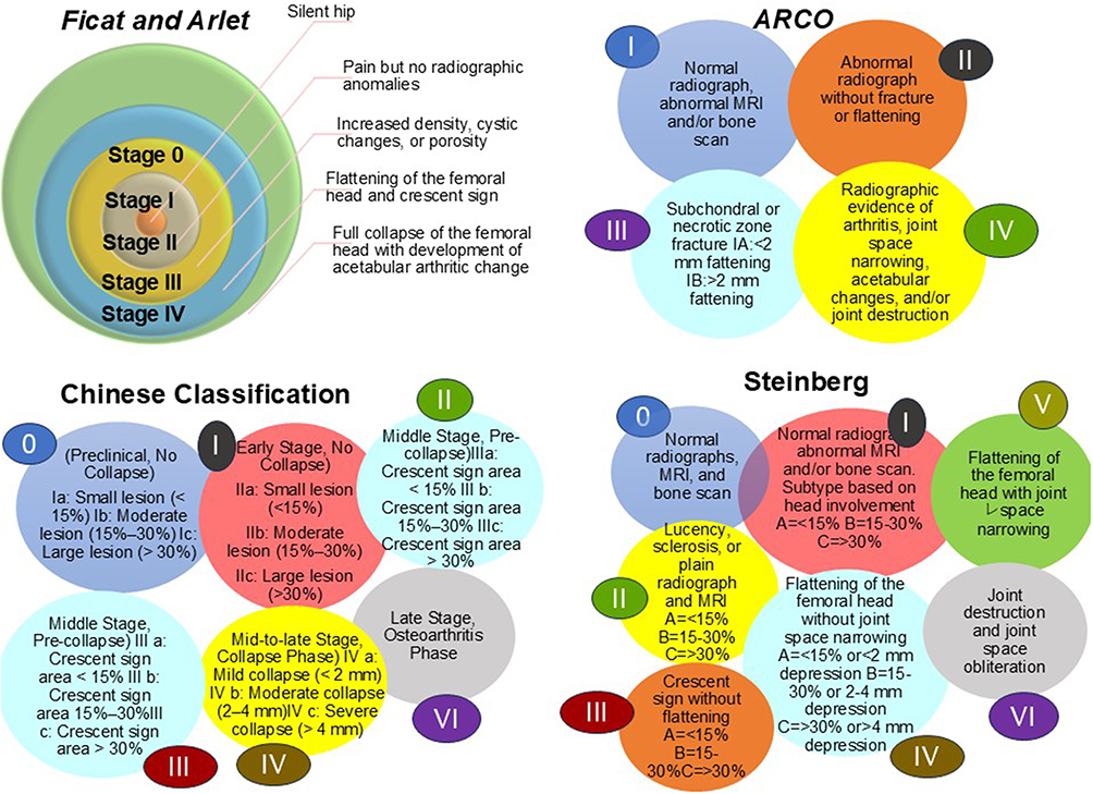 |
Figure 1 Classification systems of ONFH based on Ficat and Arlet, ARCO, Chinese, and Steinberg.10 |
ONFH presents two mechanisms: traumatic and non-traumatic, with distinct etiologies.15,16 Specifically, the etiologies related to non-traumatic mechanisms include steroid-induced osteonecrosis (SONFH), alcohol-induced osteonecrosis (AIONFH), COVID-19-associated ONFH, and pediatric Legg-Calvé-Perthes Disease (LCPD). These causes still suffer from unclarified and poorly understood pathogenic pathways.17,18 In contrast, the traumatic mechanism is well understood and linked to fractures and dislocations.19,20 This infarction leads to the formation of a central necrotic zone bordered by an ischemic rim. Within this transitional zone, the inner portion suffers severe ischemic damage, whereas the outer region exhibits reactive hyperemia, reflecting an attempt at compensatory perfusion.21,22
Limitations of Current Conventional Treatment
Current conventional therapeutic strategies for ONFH, including core decompression (CD) and THA, are the most commonly performed in hospitals. Specifically, THA represents the common treatment; it is associated with complications, including postoperative thrombosis, infection, intraoperative fractures, joint dislocation, leg length discrepancy, implant loosening, and rare nerve injury.23 Moreover, elevated costs and a prolonged postsurgical rehabilitation period impose considerable burdens on patients. Recently, preclinical studies for introducing different biomaterials, such as injectable hydrogels, have been extensively reported for ONFH treatment as effective localized drug-delivery systems and to address the limitations of conventional treatment mentioned above.24
Compared with traditional drug delivery, ONFH treatment via injectable hydrogel offers an effective platform due to its ability to retain water, its tunable physicochemical properties, and its capacity to mimic the natural extracellular matrix (ECM).25 Based on previous studies, injectable hydrogels serve as matrices for encapsulating various active species, such as nanoparticles, growth factors, and peptides. Injectable hydrogels have emerged as versatile platforms for ONFH treatment, including natural polymer-based materials (gelatin, hyaluronic acid, alginate, chitosan), synthetically derived polymers (PEG, Pluronic), and their composites.26 It serves as a drug-delivery system, promoting osteogenesis, angiogenesis, and vascularization for the treatment of ONFH.27
Targeting well-established pathophysiological mechanisms is an effective approach to enhance the efficacy of hydrogel for ONFH treatment. In addition to the physicochemical characteristics of injectable hydrogels, enhancing therapeutic effects requires a pathogenesis-targeted material design that inhibits pathological cascades and halts disease progression by encapsulation of bioactive substances or functional nanomaterials, thereby delaying the need for THA. Furthermore, these targeted therapeutic agents are essential for inhibiting excessive inflammation, suppressing abnormal osteoclast activation, alleviating oxidative stress, and reversing vascular endothelial damage, thereby preventing the progressive deterioration of the femoral head at the early pathological stage. Taken together, the pathogenesis-driven hydrogel design not only accelerates bone regeneration but also fundamentally prevents pathological progression, a significant development that addresses the limitations of conventional ONFH treatment.
In our previous review in ACS Applied Bio Materials, we discussed the application of injectable hydrogels for bone diseases in a general context, rather than focusing intensely on ONFH-specific pathological features.26 Because of the unique clinical challenges of ONFH, this review presents a core concept for a pathogenesis-targeted hydrogel platform. Specifically, we assess the potential of advanced hydrogels for precise targeting and inhibition of key pathological biomolecules.
This comparative review systematically provides the first mechanistic framework for hydrogel-based therapeutic strategies targeting specific ONFH pathogenic pathways, along with a clinical trial. A systematic literature search across PubMed and Google Scholar utilizing key terms including “hydrogel + osteonecrosis of the femoral head,” “hydrogel + Non-Traumatic + osteonecrosis of the femoral head,” and “hydrogel + Non-Traumatic + femoral head necrosis,” encompassing all publications up to 2026 (Table 1). A significant portion of the existing literature addresses non-specific etiologies of osteonecrosis. Herein, we will first examine these studies independently. Subsequently, we will investigate injectable hydrogel systems developed for specific etiologies, including steroid-induced osteonecrosis (SONFH), alcohol-induced osteonecrosis (AIONFH), and other distinct pathological mechanisms.
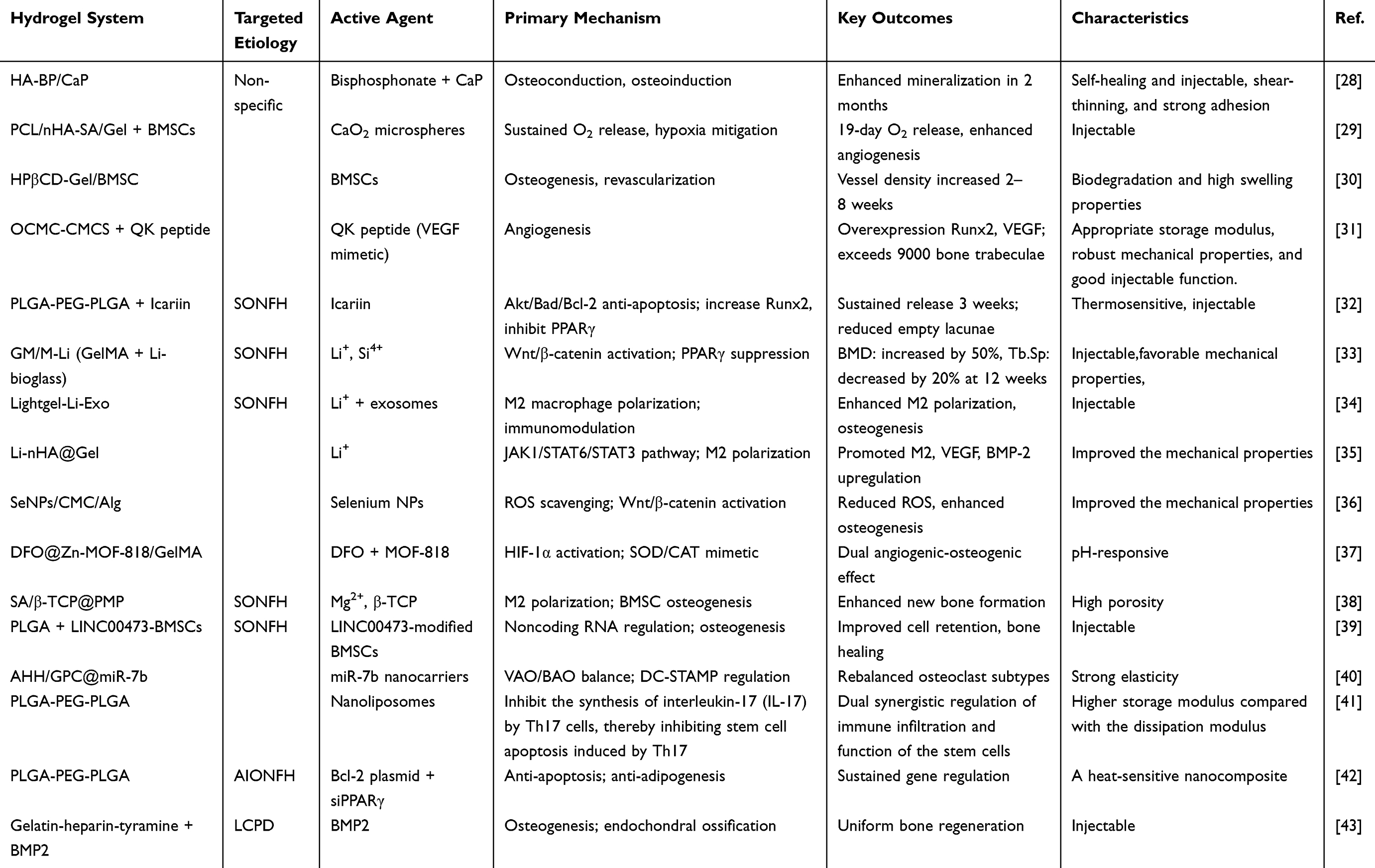 |
Table 1 Previous Studies Related to Injectable Hydrogels for Non-Traumatic Osteonecrosis of the Femoral Head |
Hydrogel for Non-Specific Etiologies of Osteonecrosis
Promoting Osteogenic Differentiation
Based on the unique characteristics of Calcium phosphate (CaP), such as osteoinductivity and osteoconductivity, Wang et al evaluated the effectiveness of injectable hydrogel HA modified by bisphosphonate (BP) (HA-BP) encapsulated with CaP for ONFH treatment. HA-BP/CaP presents a promising platform for clinical use due to its simpler operation, smaller skin and surrounding soft-tissue wounds, shorter operating time, and a shape of the injected hydrogel that matches the bone defect region. It exhibits enhanced proliferation and osteogenic differentiation, and the mineralized content within newly formed bone was increased at 1 and 2 months (Figure 2).28
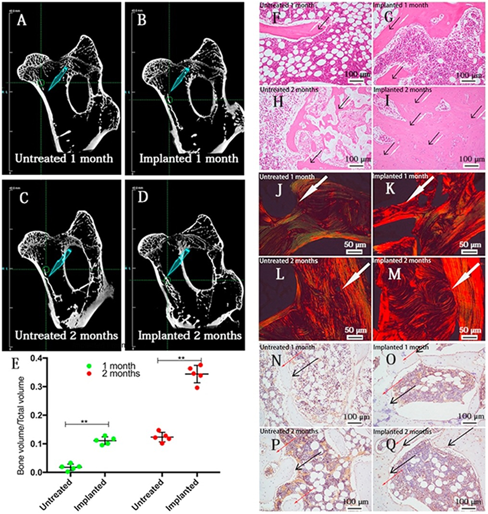 |
Figure 2 (A–D) 3D reconstruction views, (A) corresponds to 1 month after surgery without hydrogel placement; (B) to 1 month after hydrogel implantation; (C) corresponds to 2 months after surgery without hydrogel placement; (D) to 2 months after hydrogel implantation. (E) Quantification of the newly formed bone volume per osteonecrosis area. H and E after hydrogel implantation for 1 month, **p < 0.01. (G) and 2 months (I), without hydrogel for 1 month (F) and 2 months (H). Black arrows: collagen, white arrows: osteoblast cells. The scale bar is 100 after hydrogel implantation for 1 month (was perioK) and 2 (M) months, without hydrogel for 1 month (J) and 2 months (L). More swirling bone units were easily found in the implanted group (K and M). The scale bar is 50 implanted group r is 100 after hydrogel implantation. (N) untreated one month. (O) implant one month. (P) untreated two months. (Q) Implant two months. Black arrows: periostin, red arrows: collagen, White arrows: collagen I. Reproduced with permission from Wang et al,28 Elsevier 2019. |
Promoting Osteogenic and Angiogenic Effects
To promote osteogenic and angiogenic effects by targeting the hypoxia pathway, introducing oxygen-releasing biomaterials, such as liquid and solid peroxides, presents a promising, innovative strategy for ONFH treatment.44 However, the volume of oxygen released by solid peroxides, such as CaO2, poses a significant challenge due to their severe cytotoxicity, including high levels of H2O2 and ROS. So, Wang et al have explored an effective approach for mitigating the limitations of CaO2. A composite of CaO2/gelatin microspheres/three-dimensionally printed polycaprolactone/nano-hydroxyapatite (PCL/nHA)/sodium alginate/gelatin hydrogel grafted with bone marrow-derived mesenchymal stromal cells (BMSCs) (OMGs-PCL/nHA-SA/gel) has been synthesized. The oxygen released can consistently provide oxygen for 19 days. The results indicated that these scaffolds containing oxygen-generating microspheres markedly improved osteogenic and angiogenic effects, reduced apoptosis in the local microenvironment, and increased the viability of transplanted cells within the surrounding tissue (Figure 3).29
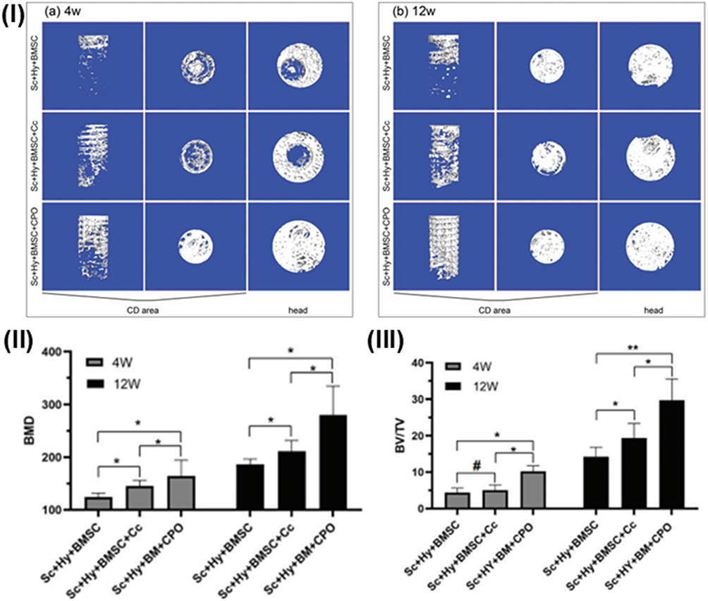 |
Figure 3 (I) Micro-CT evaluation of femoral head bone regeneration at (a) 4 weeks and (b) 12 weeks. Quantitative analysis of (II) bone mineral density and (III) bone volume fraction (BV/TV) within the core decompression area at both point of time. Data presented as mean ± SEM (n = 3); #p > 0.05, *p < 0.05, **p < 0.01. Figures reproduced with permission from C. Wang et al,29 Royal Society of Chemistry, 2021. Abbreviations: Sc, PCL/nHA scaffold; Hy, hydrogel; Cc, calcium carbonate; CPO, calcium peroxide. |
Growth factors are attracting interest in bone tissue engineering due to their enhanced osteoinductive and vascular properties. They include recombinant human bone morphogenetic protein-9 (rhBMP9), recombinant human bone morphogenetic protein-2 (rhBMP2), Bone Morphogenetic Protein 9 (BMP9), and recombinant human bone morphogenetic protein-7 (rhBMP7). Their mechanism is based on activating new blood vessel formation by binding to specific vascular endothelial growth factor (VEGF) receptors. Under physiological conditions, sustained hydrogel release is particularly advantageous for prolonging healing in osteonecrosis, as demonstrated by Lv et al, who assessed the mechanism of rhBMP9 encapsulation in a chitosan-Pluronic F-127 (PF)- tripolyphosphate hydrogel formulation. The CS/PF/TPP hydrogel exhibited remarkable pH sensitivity, thermal responsiveness, and regulated release of rhBMP9 in the acidic environment. The average pore diameter reached 45 μm, within the 40–100 μm range considered most effective for SONFH therapy, while exceeding the approximately 5 μm ideal size for vascular ingrowth, thereby facilitating nutrient delivery, metabolite removal, and bone/vessel regeneration. The optimal CS1.5-PF20-TPP0.5 exhibited minimal swelling, owing to its elevated crosslink density and compact structure. This hydrogel exhibited more rapid biodegradation at pH 6.0 (66% in 14 days) than at pH 7.4 (42% in 14 days). It demonstrated an initial rapid release of rhBMP9, succeeded by a prolonged delivery, with 75% of the drug released at pH 6.0 and 52% at pH 7.4 over 14 days, while sustained release persisted for over 25 days at pH 6.0 and more than 36 days at pH 7.4. This hydrogel enhances the proliferation and motility of bone marrow-derived mesenchymal stem cells (BMSCs) and human umbilical vein endothelial cells(HUVECs), while promoting bone formation and vascularization (Figure 4).45
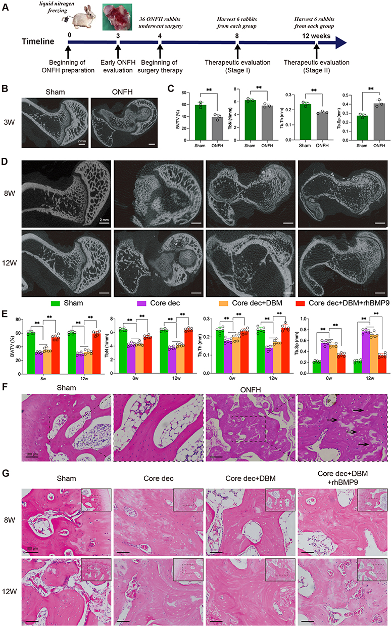 |
Figure 4 Micro-CT analysis and histological examination of bone regeneration in the ONFH rabbits treated by hydrogels with or without rhBMP9. (A) Experimental schema for the preparation of rabbit ONFH model and treatment of different groups. (B) Sham group and ONFH rabbits detected by Micro-CT at week 3, bar = 2 mm. (C) BV/TV, Tb.Th, Tb.N and Tb.Sp were calculated based on reconstructed CT images of the Sham and ONFH rabbits at week 3. Tb.Th: trabecular thickness; BV/TV: bone volume/tissue volume; Tb.N: trabecular number; Tb.Sp: trabecular separation. (D) Micro-CT images of the femoral heads in different treatment groups at week 8 and 12, bar = 2 mm. (E) BV/TV, Tb.N, Tb.Th and Tb.Sp were calculated based on reconstructed CT images in different treatment groups at week 8 and 12. (F) Sham group and early osteonecrosis detected by HE staining at week 3. The black arrows indicate multiple diffuse empty lacunas, bar = 100 μm. (G) H and E staining of the femoral head in different treatment groups at week 8 and 12, bar = 100 μm. Data were depicted as mean± SD of 6 rabbits each group, **p < 0.01. Figures reproduced with permission from Lv et al45 Elsevier, 2025. |
Researchers assessed the limitations of growth factors, notably short half-life of VEGF, which leads to rapid clearance upon introduction into the host environment. Nevertheless, a substantial amount of VEGF can be incorporated into the scaffold and administered to the ischemic region to elicit the desired angiogenic response. So, Peyravian et al assessed the impact of the injectable, shear-thinning hydrogel OCMC-CMCS, encapsulated with an angiogenesis-stimulating peptide (QK), on ONFH repair, emphasizing its sustained, localized drug-delivery system to enhance bone regeneration. OCMC-CMCS-QK hydrogel possessed an interconnected porous structure with an average pore size of approximately 101 μm. The hydrogel undergoes gradual degradation over 21 days in pH 7.4 phosphate-buffered saline (PBS), a condition suitable for bone tissue engineering, with significant swelling rates. These findings confirm a significant overexpression of the Runx2, Osteocalcin, Collagen I, VEGF, and CD34 genes (**p < 0.01 and ***p < 0.001, respectively). Furthermore, femoral head necrosis was effectively prevented, and an increased number of blood vessels was observed in the defect area treated with OCMC-CMCS hydrogel infused with QK peptide (bone trabeculae >9000, ***p < 0.001) (Figure 5).31
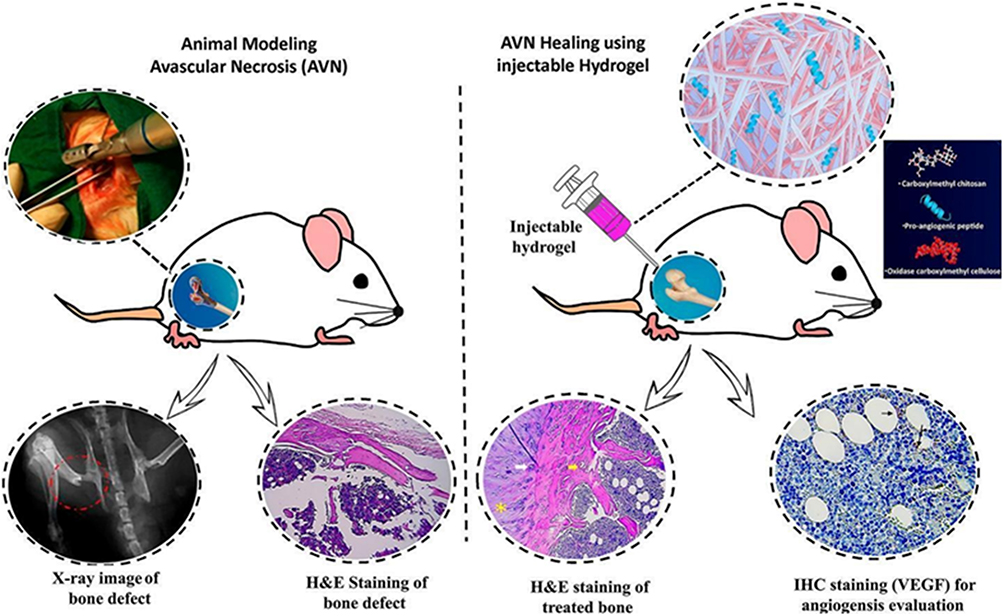 |
Figure 5 Schematic representation and biological evaluation of QK peptide-functionalized injectable hydrogel. (Top) Graphical illustration of the OCMC-CMCS hydrogel platform incorporating QK peptide for enhanced angiogenesis and osteogenesis. (Bottom) Histological analysis of bone defects via H and E staining, alongside immunohistochemical assessment of VEGF expression, demonstrating the hydrogel’s pro-angiogenic effects in vivo. Figures reproduced with permission from N. Peyravian et al,31 Copyright 2024 Elsevier. |
Promoting Osteogenic Differentiation and Revascularization
To promote osteogenic differentiation and revascularization, Shuai Yuan et al evaluated the impact of an injectable hydrogel encapsulated within BMSCs on osteogenic differentiation and revascularization. The HPβCD-Gel/BMSCs hydrogel system represented a promising approach for ONFH treatment by increasing stem cell viability, osteogenic differentiation, and revascularization. It serves as an injectable that gels within 6 minutes at ambient temperature. It forms an interconnected three-dimensional porous network through the crosslinking of HPβCD with gelatin. Increasing HPβCD concentration increases crosslink density and decreases pore size.
Furthermore, it possesses significant water absorption capacity, and pronounced swelling accelerates in vitro breakdown. Hydrogel exhibits significant water absorption capacity, and increased swelling accelerates in vitro breakdown. Moreover, it facilitates BMSC differentiation into osteocytes within 14 days more effectively than a standard osteogenic medium, and vascularization was observed within the first 2 weeks. Thereafter, vessel density and mean vessel diameter significantly increased from 2 to 8 weeks, subsequently leading to augmented new bone development (Figure 6).30
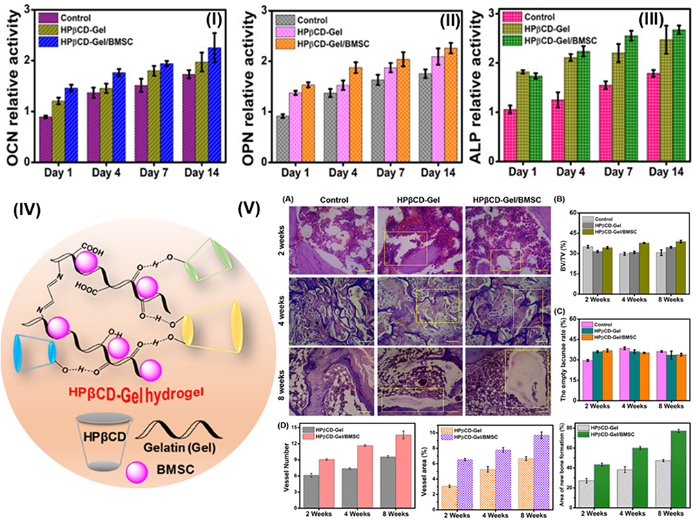 |
Figure 6 HPβCD-Gel hydrogel system for BMSC-mediated ONFH regeneration. (I–III) Osteogenic marker expression (OCN, OPN, ALP) in stem cells cultured with control, HPβCD-Gel, or HPβCD-Gel/BMSC at days 1, 4, 7, and 14 (P < 0.05). (IV) Schematic of interpenetrating polymer network synthesis via carbonyldiimidazole crosslinking. (V) In vivo rat ONFH model: (A) H and E staining at 2, 4, 8 weeks post-treatment; quantitative analysis of (B) bone volume, (C) empty lacunae, and (D) vascular parameters (vessel count, vessel area, new bone area). Scale bar = 100 μm. Data presented as mean±SD (P < 0.05). Figures reproduced with permission from Yuan et al30 Elsevier 2022. |
Hydrogel for Specific Etiologies of Osteonecrosis
SONFH
Steroid-induced osteonecrosis of the femoral head (SONFH) is the leading cause of non-traumatic ONFH approximately 40% to 60% of all non-traumatic cases. It demonstrated a rise of 75% in non-traumatic ONFH related to glucocorticoid therapy based on excessive use of steroidssuch as anti-inflammatory, anti-allergic, anti-toxic, immunosuppressive metabolic hormones with anti-shock effects.46
Various pathophysiological mechanisms contributing to SONFH have been proposed, including (1) the Sclerostin/Wnt/β-catenin Axis: Elevated serum sclerostin concentrations in necrotic regions impede the Wnt/β-catenin signaling pathway. Its pathological mechanisms involve disruption of bone remodeling by impeding osteoblast differentiation and blocking the formation of type-H vessels, thereby decreasing angiogenesis and accelerating the progression of osteonecrosis.47–49
(2) Vascular Dysfunction and Hemodynamic Compromise: Metabolic anomalies, such as increased lipids, result in the development of fat emboli and hypertrophy of marrow adipocytes, hence elevating intraosseous pressure. Together with a hypercoagulable state, this mechanical compression disturbs microcirculation and promotes vasoconstriction, ultimately resulting in ischemic necrosis.50 A distinct mechanism of adiposity entails altering the differentiation equilibrium of bone marrow stromal cells through the suppression of the BMP/Runx2 osteogenic pathway and the activation of the transcription factor Peroxisome Proliferator-Activated Receptor Gamma (PPARγ) adipogenic pathway, resulting in elevated Ca2⁺ and phosphate concentrations, diminished osteoblastic activity, and lowered bone matrix protein levels.
(3) Hypoxic Signaling and Inhibition Impaired HIF-1α Signaling Diminishes VEGF-Mediated Angiogenesis. Simultaneously, elevated concentrations of Nitric Oxide (NO) reduce CXCR4 receptor expression on endothelial cells, hindering their motility and the repair of vascular structures. The impairment of angiogenesis, along with oxidative stress and the formation of reactive oxygen species, disturbs chondrocyte homeostasis and leads to cell death.51
(4) Inflammatory and Immune Pathways: Hyperactivation of the TLR4/NF-κB pathway, influenced by genetic factors (CR2, cytokines),results in the overproduction of inflammatory cytokines such as Interleukin-4(IL-4),Interleukin-6(IL-6),and Tumor Necrosis Factor-alpha(TNF-α).These cytokines enhanced a pro-inflammatory milieu that hastens bone resorption and impedes recovery.52
(5) Apoptosis and Programmed Cell Death Pathways: SONFH activates multiple programmed cell death pathways in osteoblasts and endothelial cells, encompassing apoptosis through mitochondrial pathways (Akt/Bad/Bcl-2), TNF-α signaling, and p53 activation; necroptosis via the RIPK1/RIPK3/MLKL pathway, which exacerbates inflammation; and ferroptosis through the downregulation of SLC7A11 and GPX4, leading to ROS accumulation and the dysregulation of HIF-1α and FOXO1 signaling.53
(6) Glucocorticoid Receptor (GR) Activation: The inhibition of various pro-inflammatory response genes occurs through the enhancement of anti-inflammatory protein synthesis and the repression of pro-inflammatory transcription factors, such as nuclear factor-kappaB (NF-kappaB) and activator protein-1 (AP-1).54
We assess their treatment technique through a particular process. Various options for targeting the distinct pathways of SONFH processes are shown through the encapsulation of functional substances, including pharmaceuticals, natural products, metals, nanoparticles, exosomes, stem cells, RNA, and peptides.
Target the Phosphorylation of the Akt/Bad/Bcl-2 Signaling Pathway
Icariin (ICA) is a bioactive molecule extracted from epimedium-derived flavonoids that regulates the osteogenesis and adipogenesis of bone mesenchymal stem cells.55 Xu et al enveloped icariin into injectable thermosensitive PLGA-PEG-PLGA hydrogels to improve its limited bioavailability. Sustained release was observed for up to 3 weeks in the 200 μM group, with a plateau reached. The release rate of ICA was postponed as the copolymer concentration rose from 15 wt% to 25 wt%. After four weeks, the cumulative release rates were 89.85% (15 wt%), 82.2% (20 wt%), and 78.7% (25 wt%), indicating that denser copolymer matrices facilitate prolonged, more regulated release. The hydrogels exhibited accelerated degradation at higher ICA concentrations; the 2000 μM ICA and 25 wt% PLGA-PEG-PLGA hydrogels degraded within 42 days. This hydrogel was meticulously designed to target Akt/Bad/Bcl-2 signaling phosphorylation to suppress apoptosis and enhance the survival of vascular and bone cells. This approach encompasses a substantial decrease in bone marrow edema, improvement of trabecular bone, reduction of empty lacunae, and diminished adipocyte accumulation, alongside the promotion of Runx-2 expression and suppression of PPARγ expression in the femoral head (Figure 7).32
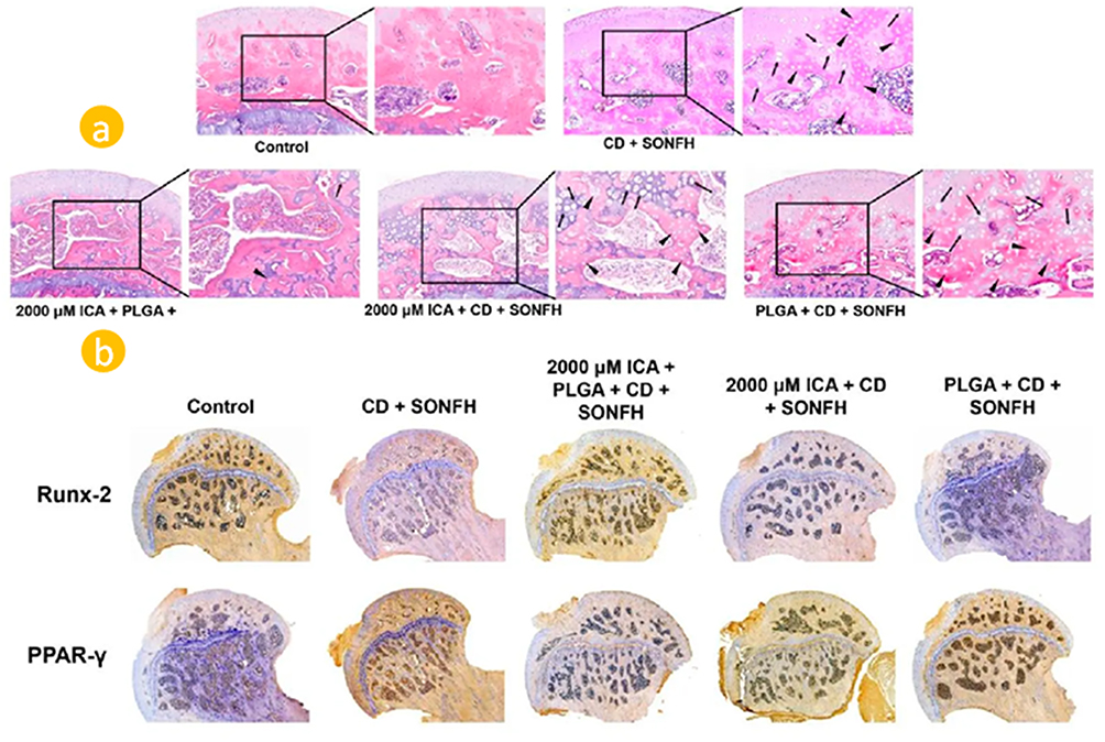 |
Figure 7 Histological analysis of SONFH rat models after treatment with ICA/CD-loaded thermosensitive PLGA-PEG-PLGA hydrogels. (a) H and E staining showing bone morphology. (b) Immunohistochemical analysis of protein expression. Black arrows indicate pathological changes associated with SONFH, including: (1) empty osteocyte lacunae within trabecular bone, (2) adipocyte hypertrophy/accumulation in the bone marrow cavity, and (3) disrupted/necrotic trabecular bone architecture. Figures reproduced with permission from Xu et al32 Frontiers, 2025. |
Target Activating the Wnt Pathway
Metal ions, increasingly recognized as additives in biomaterials such as hydrogels, have been shown to influence cellular metabolism, signaling pathways, and ion channels by functioning as cofactors for various enzymes in vivo. Lithium (Li), used as a metal-ion addition in bioactive scaffolds, has been demonstrated in numerous studies to enhance bone repair. Several hypotheses have been proposed to clarify the mechanisms of lithium’s action; however, none has achieved conclusive status, and in some cases, the results even contradict each other. A prior study indicated that lithium-mediated suppression of GSK-3β activity leads to the stabilization and nuclear accumulation of β-catenin, thereby initiating transcription of Wnt target genes and activating the Wnt pathway. Moreover, stimulation of the Wnt pathway is essential for osteoblast differentiation and osteogenesis. Tang et al found that LiCl can inhibit GSK-3β, thereby modulating osteogenesis in BMSCs by stimulating Wnt signaling. The interaction between LiCl and Wnt pathway activation has been previously established; however, the mechanisms by which LiCl activates the Wnt pathway remain unresolved. Research indicates that Li+ influences many downstream signaling pathways by activating the canonical Wnt/β-catenin signaling pathway. It also facilitated the migration and proliferation of adipose-derived stem cells (hASCs) and the expression of genes associated with osteogenic development. LiCl is reported to facilitate interfacial bone fusion by reducing osteoclast differentiation and inflammation, while promoting osteogenesis. Chen et al examined the function of Li in promoting osteogenic differentiation in BMSCs by activating β-catenin signaling via autocrine signaling facilitated by exosomal Wnt10a. They identified a novel regulatory route involving MARK2-mediated trafficking of Rab11a and Rab11FIP1 complexes, linked to the secretion of Wnt10a from BMSCs.56
Z. Wu et al recently developed a lithium-loaded bioglass/GelMA hydrogel (GM/M-Li) designed to modulate osteogenesis, angiogenesis, and adipogenesis. This hydrogel exhibits precise swelling behavior and an appropriate compressive modulus of 4 kPa, resulting from the encapsulation of lithium bioglass. Also, it shows sustained release of lithium ions (Li⁺) exerts a dual mechanism of action: activation of the canonical Wnt/β-catenin pathway to promote bone formation, coupled with inhibition of the adipogenic regulator PPARγ to suppress fat accumulation. Additionally, Li⁺ combined with ions released from mesoporous bioglass (notably Si4⁺) was proposed to enhance bone-vascular network formation synergistically. This multifunctional platform addresses the shortcomings of previous single-function materials, offering a comprehensive approach to bone regeneration in SONFH. GM/M-Li demonstrates continuous Li⁺ and Si4⁺ release persisted for over 35 days, significantly augmenting the proliferation and migration of bone marrow-derived mesenchymal stem cells and human umbilical vein endothelial cells, while concurrently stimulating osteogenic and angiogenic differentiation in vitro and in vivo. It exhibits a 50% increase in bone mineral density (BMD) and a 20% decrease in Trabecular spacing (Tb.Sp) compared to model controls at 12 weeks, further confirming markedly enhanced osteogenesis and angiogenesis following treatment (Figure 8).33
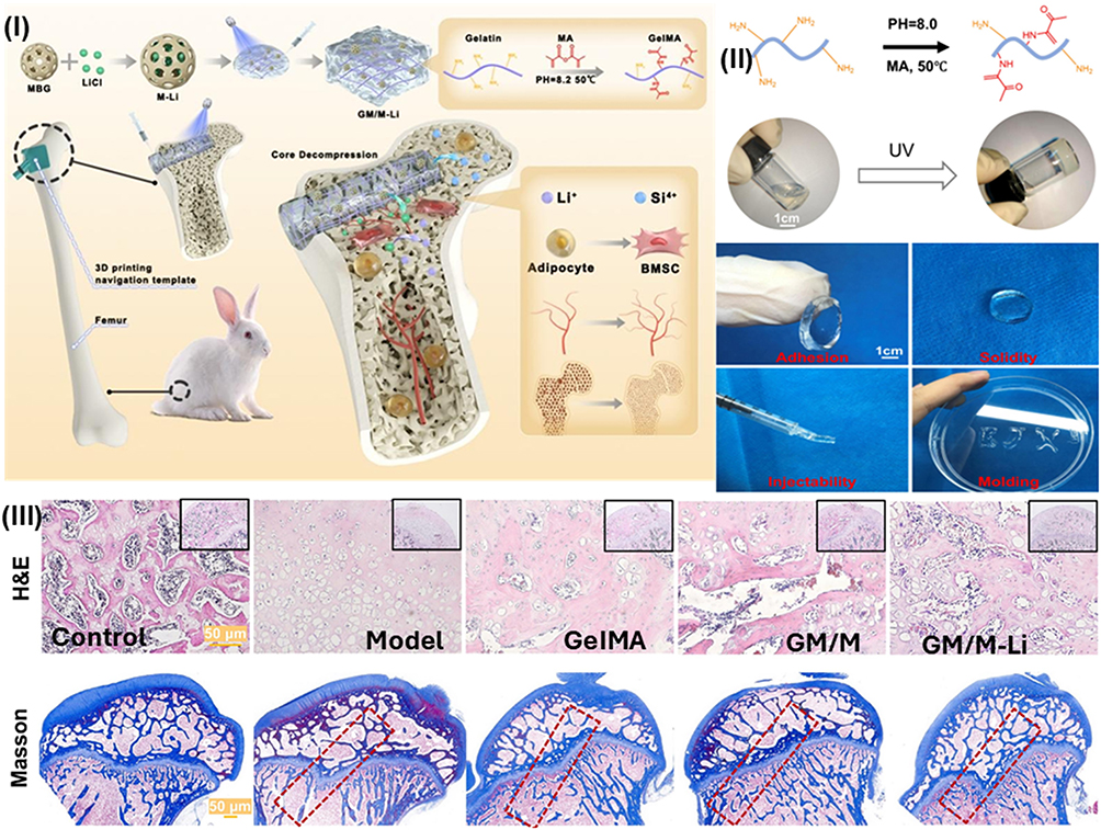 |
Figure 8 GM/M-Li hydrogel system for ONFH treatment: (I) Therapeutic strategy schematic: core decompression combined with Li⁺-releasing hydrogel inhibits adipogenesis, stimulates angiogenesis, and promotes osteoblast differentiation from MSCs. (II) UV-induced morphological transformation of hydrogel. (III) Histological evaluation at 12 weeks showing (A) H and E and (B) Masson staining of femoral head tissue. Figures reproduced with permission from Wu et al,33 Elsevier 2026. |
Selenium nanoparticles (SeNPs) have demonstrated therapeutic efficacy in SONFH treatment by mitigating oxidative stress and activating the Wnt/β-catenin signaling pathway. Although the importance of this route in bone homeostasis is well established, its regulation in the setting of SONFH remains poorly described. C. Liu et al examined the encapsulation of SeNPs within a carboxymethyl cellulose (CMC)/alginate hydrogel matrix to facilitate controlled and sustained release, subsequently implanting the SeNPs/CMC/Alg composite into the femoral head post-CD surgery for SONFH intervention. SeNPs/CMC/Alg hydrogel serves as a localized delivery system for SONFH by enhancing ROS clearance and upregulating Wnt/β-catenin signaling. It has an interconnected porous structure with a porosity of around 74.41%. The swelling capacity is slightly reduced following the addition of SeNPs. However, it remains superior to that of pure alginate hydrogel, and the incorporation of CMC improves crosslink density and thermal stability of the network. It exhibits greater mechanical stiffness than pure alginate; however, SeNPs have a minimal effect on the hydrogel’s mechanical properties. It also suppresses noticeable burst release and promotes sustained selenium nanoparticle delivery for at least 7 days, achieving over 70% drug entrapment efficiency. 10 μg/mL SeNPs is the optimal loading dose, balancing substantial antioxidant efficacy with moderate cytotoxicity (Figure 9).36
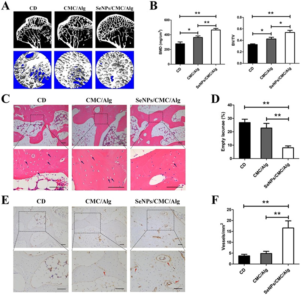 |
Figure 9 Repair of SONFH in each group by radiographic and histological evaluation. (A) Micro-CT images of the subchondral region of the femoral head in each group. (B) Quantitative analysis of the trabecular bone parameters (BMD and BV/TV) based on reconstructed 3D images. (C) H and E staining of the empty lacunae in the necrotic region of the femoral head. The blue arrows indicate the empty lacunae in the necrotic region. (D) Percentage of empty lacunae in each group. (E) Immunohistochemical staining for CD31 in the femoral head in each group. The red arrows indicate the vessels. (F) Vessel density in the necrotic region of the femoral head in each group. Scale bars = 100 μm. *p < 0.05, **p < 0.01. Figures reproduced with permission from Liu et al,36 Elsevier, 2024. |
A heparin-lithium hydrogel (Li-hep-gel) was synthesized by Li et al as a delivery system for both lithium ions and microRNA-functionalized tetrahedral DNA nanostructures (MiR@TDNs). MiR@TDNs/Li-hep-gel exhibits an interconnected porous architecture with pore dimensions between 200 and 400 μm, and the pore characteristics remain consistent following the incorporation of lithium and MiR@TDNs nanoparticles. The hydrogels demonstrated medium-dependent degradation, with approximately 40% weight loss in PBS and around 60% in cell supernatants, while the three experimental groups exhibited nearly identical degradation kinetics during the incubation period. All hydrogel formulations exhibited compressive moduli of 11.8–12.3 kPa, and the addition of lithium and MiR@TDNs did not significantly affect the overall mechanical properties of the heparin-based network. In vitro findings demonstrate increased alkaline phosphatase activity and calcium deposition, reduced lipid droplet accumulation in BMSCs, and enhanced secretion of vascular endothelial growth factor, along with the formation of vascular-like structures, with MiR@TDNs therapy. Upon implantation into the SONFH model, the MiR@TDNs/Li-hep-gel composite facilitated robust bone defect repair, evidenced by extensive new bone formation, neovascularization, and diminished empty lacunae. This dual-delivery system enables synergistic activation of the Wnt signaling pathway through combined lithium and MiR@TDNs release, thereby enhancing bone regeneration in complex defects and positioning it as a promising therapeutic strategy for SONFH management.57
Cell-Based Therapy
Cell-based therapeutic strategies employing mesenchymal stromal cells as an adjunct to core decompression have emerged as a viable intervention for early-stage osteonecrosis of the femoral head. Recent studies have elucidated the therapeutic potential of immunomodulatory osteogenesis mediated by BMSCs preconditioned with pro-inflammatory cytokines (pMSCs), as well as the capacity of mesenchymal stem cells genetically engineered to overexpress anti-inflammatory cytokines to accelerate inflammatory resolution. In this context, Maruyama et al investigated the efficacy of adjunctive cell-based therapy for SONFH treatment in a rabbit model, utilizing either perinatal mesenchymal stem cells (pMSCs) or interleukin-4-modified mesenchymal stem cells (IL4-MSCs) delivered via an injectable hydrogel (HG). The authors hypothesized that pMSCs would facilitate accelerated new bone formation, whereas IL4-MSCs would reduce the population of necrotic cells within the lesioned area. So, they encapsulated pMSCs within injectable hydrogels and delivered them into the bone tunnel, and studied their impact on both angiogenesis and osteogenesis in the femoral head using a SONFH rabbit model. These findings suggest that an adjunctive cell-based therapy combining CD with pMSCs and IL4-MSCs may represent a viable therapeutic approach for restoring osteonecrotic lesions during the initial stages of ONFH. It is imperative, however, that such interventions be executed in a temporally coordinated manner to avoid disruption of the critical acute inflammatory phase essential for optimal bone repair (Figure 10).58
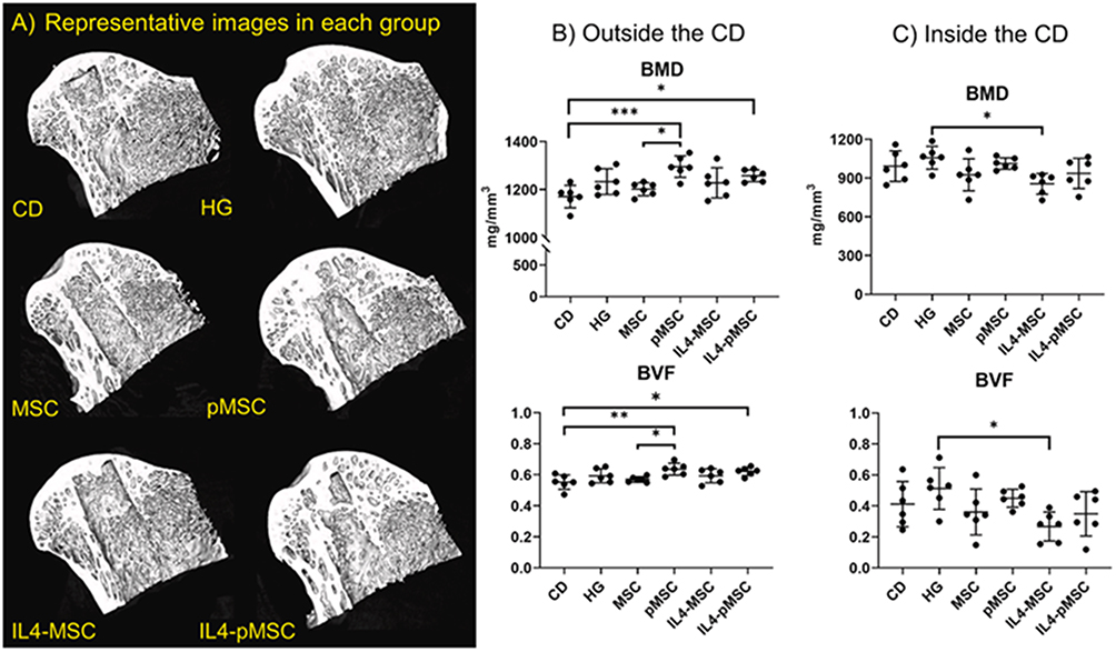 |
Figure 10 Quantitative MicroCT evaluation of bone regeneration in the femoral head following treatment. (A) Representative images and corresponding data analysis demonstrated differential effects based on anatomical region and treatment group. (B and C) bone mineral density (BMD) and bone volume fraction (BVF)outside and inside CD. For the area outside the CD, bone marrow density (BMD) and bone volume fraction (BVF) in the pMSC group were significantly higher than in the CD and MSC groups (p < 0.05). Furthermore, BMD and BVF in the IL4-pMSC group were significantly higher than in the CD group (p < 0.05). Interestingly, for the area inside the CD, BMD and BVF in the IL4-MSC group were significantly lower than in the HG group (p < 0.05). *p < 0.05, **p < 0.01, ***p < 0.005. Figures reproduced with permission from M. Maruyama et al,58 Copyright 2021, Elsevier International Publisher. |
Numerous genes, particularly noncoding RNAs, play pivotal roles in regulating BMSC function. Previous studies have confirmed the essential roles of LINC00473 and miR-23a-3p in mediating osteogenic and adipogenic differentiation, as well as apoptotic pathways, in BMSCs. Xu et al subsequently explored a therapeutic approach employing LINC00473-modified rat-derived bone marrow mesenchymal stem cells (rBMSCs) embedded within an injectable thermosensitive poly(lactic-co-glycolic acid) (PLGA) hydrogel for SONFH treatment. The results revealed that the PLGA hydrogel created a favorable milieu for rBMSC retention, substantially improving bone regeneration and structural restoration of the necrotic femoral head in a SONFH rat model. Notably, unlike unencapsulated BMSCs transplantation, rBMSCs delivered via the PLGA hydrogel successfully migrated from the medullary cavity into the femoral head, highlighting the capacity of the hydrogel scaffold to enhance targeted cellular delivery (Figure 11).39
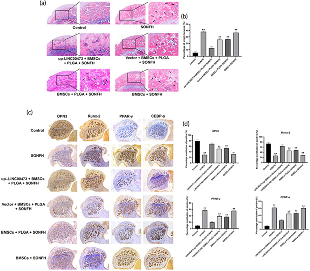 |
Figure 11 Histological evaluation of PLGA hydrogel-mediated delivery of LINC00473-overexpressing rBMSCs for SONFH treatment. (a) Representative hematoxylin and eosin (H and E)-stained sections of the femoral head subchondral bone across experimental groups at 12 weeks post-treatment. (b) Quantitative analysis of empty lacunae percentage within bone trabeculae of the femoral head following 12 weeks of treatment. (c) Immunohistochemical (IHC) staining showing expression patterns of adipogenic markers PPARγ and CEBP-α in femoral head sections at 12 weeks. (d) Semiquantitative analysis of IHC staining intensity. Data expressed as mean ± standard deviation. **p < 0.01 versus Control group and up-LINC00473 + BMSCs + PLGA + SONFH group. Figures reproduced with permission from Y. Xu et al.39 Copyright 2022, Wiley-VCH International Publisher. Abbreviations: CEBP-α, CCAAT/enhancer-binding protein-α; H and E, hematoxylin and eosin; IHC, immunohistochemistry; LINC00473, long intergenic non-protein coding RNA 00473; PLGA, poly(lactic-co-glycolic acid); PPAR-γ, peroxisome proliferator-activated receptor-γ; rBMSCs, rat bone marrow-derived mesenchymal stem cells; SONFH, steroid-induced osteonecrosis of the femoral head. |
Subsequent investigations into the functional roles of noncoding RNAs have identified a distinct osteoclast subtype termed vessel-associated osteoclasts (VAOs). These investigations have opened new therapeutic avenues for the treatment of ONFH. Building upon this paradigm, Quan et al developed an injectable alginate/hydroxyapatite hydrogel (AHH) functionalized with graphene oxide-based miR-7b nanocarriers (designated GPC@miR) for ONFH intervention through targeted modulation of osteoclast subtypes, providing valuable insights into VAO-mediated angiogenesis and osteogenesis. The AHH/GPC@miR composite hydrogel exhibited modifiable gelation duration, swelling characteristics, and compressive modulus (3–14 kPa) contingent upon the quantities of alginate and GDL, while maintaining water content between 92% and 96% across all formulations. Controlled release of GPC@miR from the AHH/GPC@miR composite suppressed bone-associated osteoclasts (BAOs) formation through downregulation of dendritic cell-specific transmembrane protein (DC-STAMP), thereby attenuating pathological bone resorption. Concurrently, mono- and bi-nucleated VAOs were preserved and expanded, promoting type H vessel angiogenesis and osteogenesis via paracrine secretion of platelet-derived growth factor-BB (PDGF-BB) and vascular endothelial growth factor-A (VEGF-A). Intraosseous administration of AHH/GPC@miR restored the equilibrium between VAOs and BAOs, reconstituted the femoral head microenvironment, and enhanced both vascularization and bone regeneration in rat models of ONFH (Figure 12).40
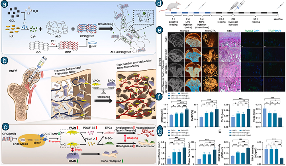 |
Figure 12 Schematic illustration of the fabrication and function of AHH/GPC@miR. (a) AHH was fabricated via ionic cross-linking to deliver GPC@miR. The homogeneous Ca-ALG gel was formed by in situ release of calcium cations from HA through lowering the pH with gradual hydrolysis of GDL. (b) AHH/GPC@miR facilitated vascularization and bone formation in ONFH by restoring the balance between VAOs and BAOs. (c) AHH/GPC@miR inhibited the formation of BAOs by targeting DC-STAMP, thereby blocking osteoclastic bone resorption. (d) Schematic representation of steroid-induced ONFH rat modeling and treatments. (e) Representative images of microCT in section and superficial views, microCTA in superficial view, H and E staining, and immunofluorescence staining of RUNX2 or TRAP in the femoral head (bar = 100 μm). (f) Quantitative analysis of microCT by measuring BMD, BV/TV, Tb.Th, and Tb.Sp (n = 6). (g) Quantitative analysis of microCTA by measuring vessel volume and surface (n = 6). (E) Quantitative analysis of RUNX2 intensity and TRAP intensity (n = 6). Data are shown as means ± SD (C, D, and E). Statistical significance was determined by one-way ANOVA with Tukey’s multiple comparisons test (C, D, and E). Asterisks indicate statistically significant differences (ns: P > 0.05, *P < 0.05, **P < 0.01, and ***P < 0.001). Figures reproduced with permission from H. Quan et al40 Copyright 2025, Elsevier International Publisher. |
Targeting Modulation of Immune Infiltration
β-tricalcium phosphate (β-TCP) is frequently used to facilitate bone regeneration, whereas magnesium ions (Mg2+) enhance the proliferation, migration, and osteogenic differentiation of BMSCs. Other studies demonstrated that magnesium-based biomaterials can repolarize inflammatory M1 macrophages to a regenerative M2 phenotype, thereby accelerating inflammation resolution and enhancing BMSC mineralization, thereby accelerating bone regeneration. Consequently, the regulated release of Mg2+ is preferable to ensure prolonged effects on bone mineralization while mitigating potential negative consequences associated with rapid release at elevated levels. Zhou et al designed an injectable hydrogel system comprising sodium alginate, β-TCP, and graded P34HB/MgO+PEG (PMP) coaxial microfibers (0/1/2/4 wt%), crosslinked via progressive Ca2⁺ release from β-TCP/GDL hydrolysis. All prepared hydrogels pass through a 22G needle; the 2% PMP group is the optimal formulation and reaches swelling equilibrium within 90 min, with maximal swelling exceeding 6.8-fold. All hydrogels exhibit an interconnected porous structure, and pore size increases with increasing PMP addition. SA/β-TCP@2%PMP exhibits an advantageous pore size (100–200 μm) suitable for cell infiltration. It demonstrates normal shear-thinning behavior for smooth injection; 2% PMP holds the highest storage modulus among all groups. SA/β-TCP@2%PMP composite hydrogel possesses increased angiogenic and osteogenic properties, facilitating considerable new bone synthesis in a rabbit model of SONFH (Figure 13).38
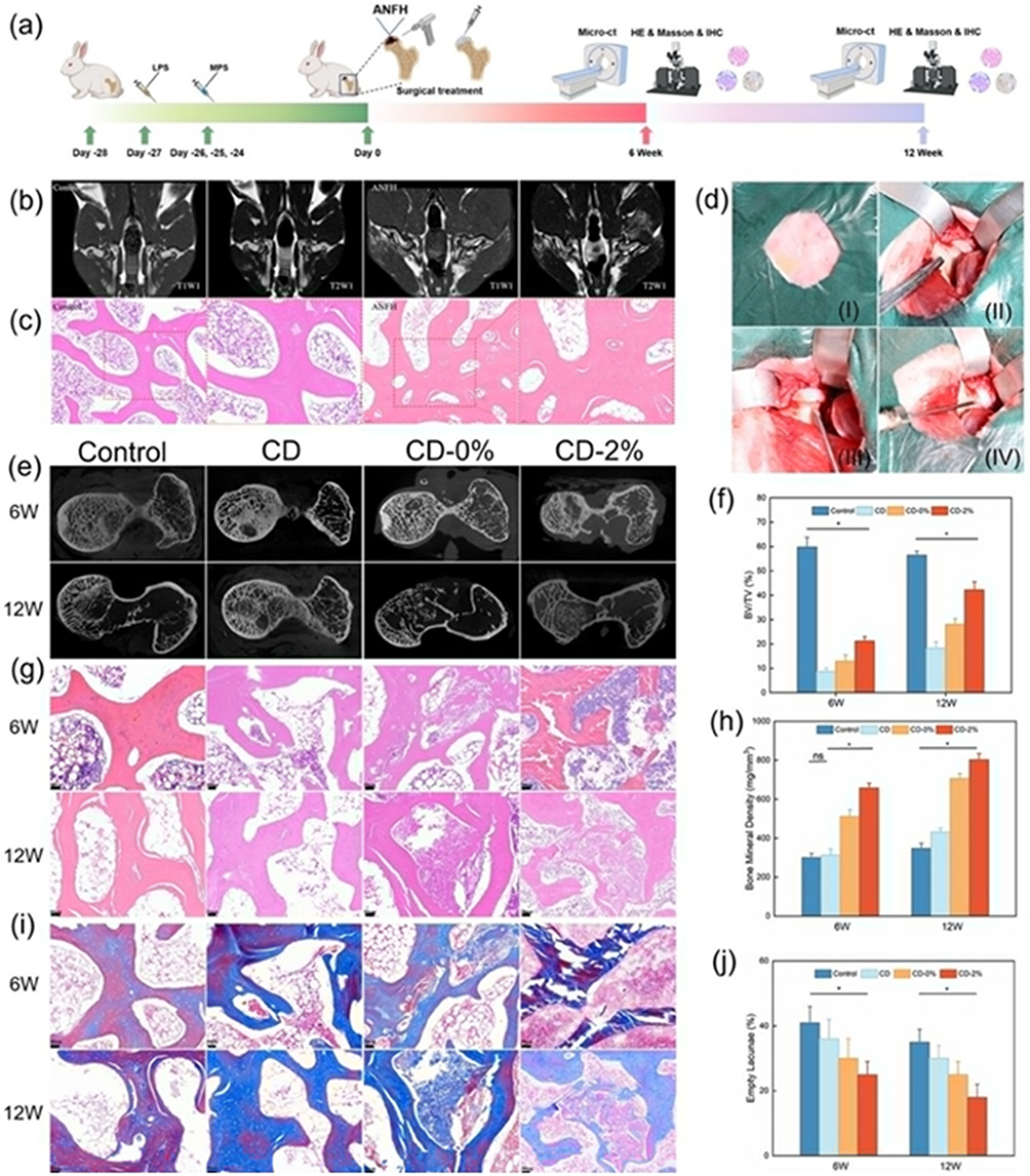 |
Figure 13 Rabbit model of SONFH and evaluation of core decompression combined with functionalized SA/β-TCP hydrogel scaffolds. (a) Schematic illustration of the experimental timeline showing ANFH induction via sequential lipopolysaccharide (LPS) and methylprednisolone (MPS) injections over 4 weeks, followed by randomization into four groups: untreated Control, core decompression alone (CD), CD with SA/β-TCP hydrogel (CD+0%), and CD with SA/β-TCP hydrogel containing 2% PMP (CD+2%), with animals euthanized at designated time points for multimodal analysis. (b) MRI scanning at 4 weeks post-induction revealed uneven signal intensity within the femoral heads, confirming osteonecrosis development. (c) H and E staining at 4 weeks demonstrates characteristic ANFH pathology, including thinner bone trabeculae and numerous empty lacunae. (d) Sequential surgical procedure images showing: ((i) exposure of the greater trochanter tip, (ii) core decompression drilling, (iii) creation of the CD tunnel, and (iv) scaffold implantation via hydrogel injection). (e) Representative micro-CT images of femoral heads at 6 and 12 weeks post-treatment.(f) quantitative analysis of BV/TV. (g) H and E staining. (h) BMD within the bone defect area. (i) Masson’s trichrome (MT) staining of femoral head sections at 6 and 12 weeks post-surgery, with (j) quantitative analysis of empty lacunae percentage. Scale bar = 50 µm for histological images. Data presented as mean ± SD (n = 3 per group); statistical significance indicated as *P < 0.05. Figures reproduced with permission from Y. H. Zhou et al38 Copyright 2025, Wiley-VCH International Publisher. |
In a related context regarding the function of LiCl in SONFH, Chen et al examined the mechanism of co-culturing BMSCs with LiCl incorporated within a Lightgel precursor solution amenable to UV-mediated in situ crosslinking, as well as the impact of this hydrogel on immunomodulation, osteogenesis, and angiogenesis. All produced hydrogels reach swelling equilibrium within 90 min; no precise swelling ratios are provided. Around 20% weight loss after 24 h in collagenase solution (80% residual mass remaining). Exosomes are continuously released over 14 days, with roughly 50% of the total cumulative release. By leveraging the proposed mechanism that promotes polarization toward the M2 phenotype while concurrently inhibiting M1 polarization, the hydrogel serves as an effective delivery vehicle for exosomes, enabling sustained release and enhancing therapeutic efficacy while supporting translational utility. An ECM-mimetic Lightgel hydrogel, formulated from methacryloylated type I collagen, served as the carrier for Li-Exo and Con-Exo incorporation, yielding Lightgel-Li-Exo and Lightgel-Con-Exo formulations. Moreover, the Li-Exo-loaded Lightgel formulation exhibited superior pro-osteogenic and pro-angiogenic effects, with the most potent effects on promoting M2 polarization, bone formation, and vascularization to facilitate bone healing in SONFH (Figure 14).34
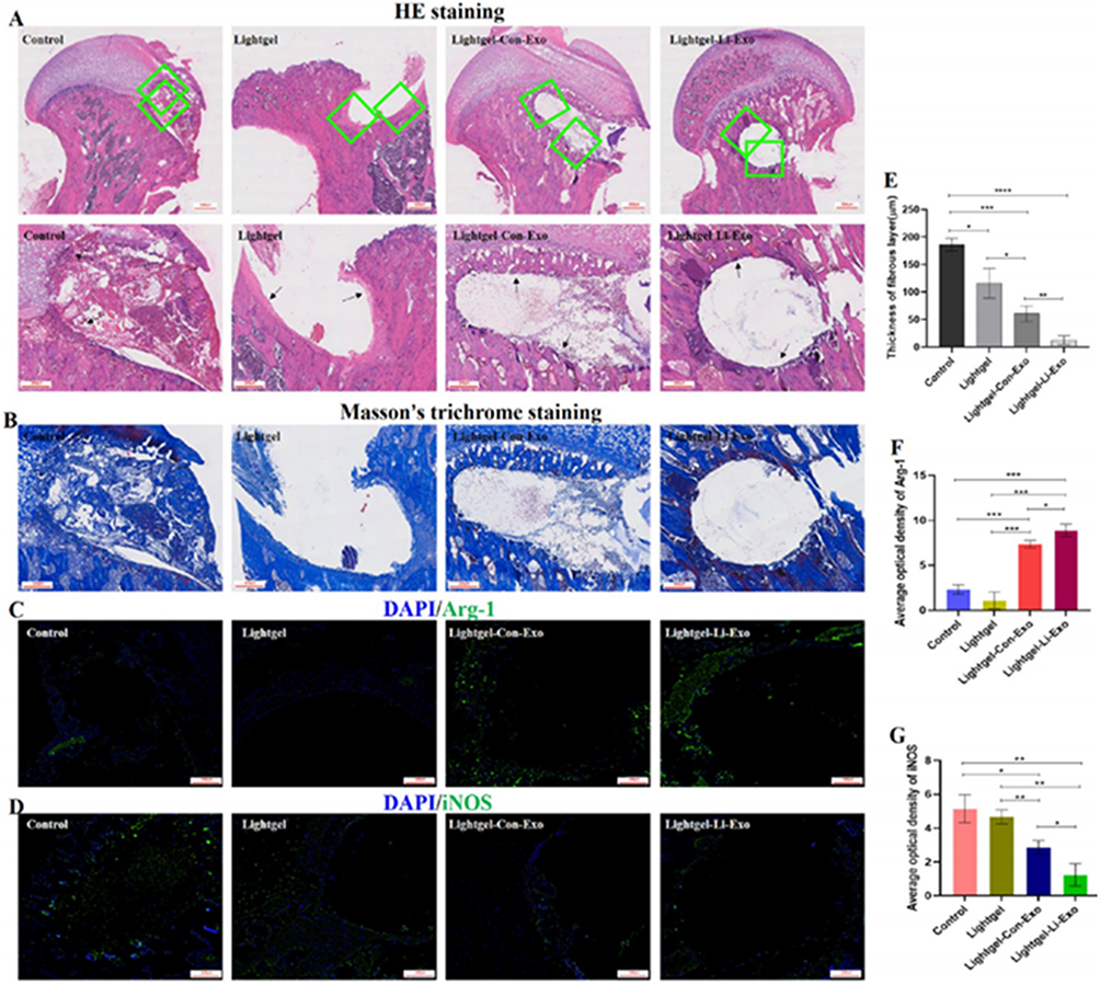 |
Figure 14 Evaluation of the early immune response in rat femoral head surgical channels at 7 days post-surgery. (A) H and E staining showing the fibrous layer (black arrows) within surgical channels; scale bars = 500/250 μm. (B) Masson’s trichrome staining of the surgical channel region; scale bar = 250 μm. Immunofluorescence staining for (C) Arg-1 and (D) iNOS in drilling compression channels; scale bars = 100 μm. Quantitative analyses of (E) fibrous layer thickness, (F) Arg-1 expression, and (G) iNOS expression (n = 3 per group). Data represent mean ± SD; *p < 0.05, **p < 0.01, ***p < 0.001, ****p < 0.0001. Figures reproduced with permission from C. Chen et al34 Copyright 2023, American Chemical Society International Publisher. |
Luo et al proposed a mechanism-based model for SONFH therapy using doped Li, based on the activation of the JAK1/STAT6/STAT3 signaling cascade, which subsequently creates a pro-repair immune milieu and enhances osteogenic and angiogenic differentiation. Thiolated hyaluronic acid (HA-SH) crosslinked with PEGDA via Michael addition; gelation time = 8.6 ±1.6 min, appropriate for in-vivo injection. Composite loaded with Li-doped nano-hydroxyapatite (Li-nHA, rod-shaped, 20–45 nm diameter). Interconnected porous structure with pore size 100–300 μm. It demonstrates a considerably higher compressive modulus (~99 kPa) after Li-nHA inclusion, coupled with lower equilibrium swelling than the hydrogel. Li-nHA@Gel facilitates sustained and prolonged release of Li ions, promoting M2 macrophage polarization via stimulation of the JAK1/STAT6/STAT3 signaling axis, which, in turn, elevates the expression of pro-tissue repair factors VEGF and BMP-2, thereby enhancing osteogenic and angiogenic differentiation (Figures 15 and 16).35
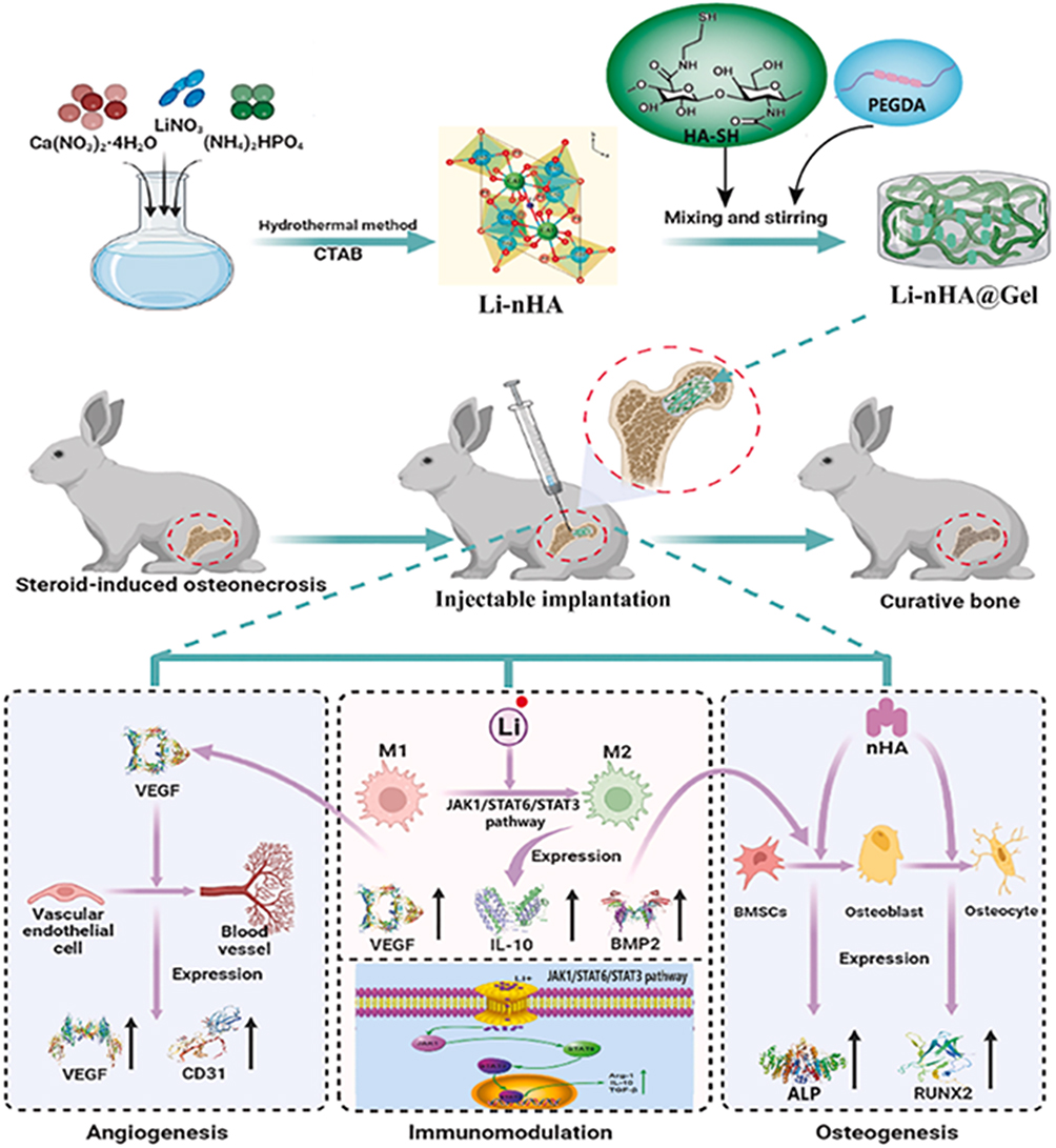 |
Figure 15 Overview of Li-nHA@Gel hydrogel fabrication and its trifunctional biological activities. The schematic outlines the preparation of lithium-doped nano-hydroxyapatite within gelatin hydrogel, followed by assessment of immunomodulatory, osteogenic, and angiogenic properties. Figures reproduced with permission from Y. Luo et al35 Copyright 2024, Elsevier International Publisher. |
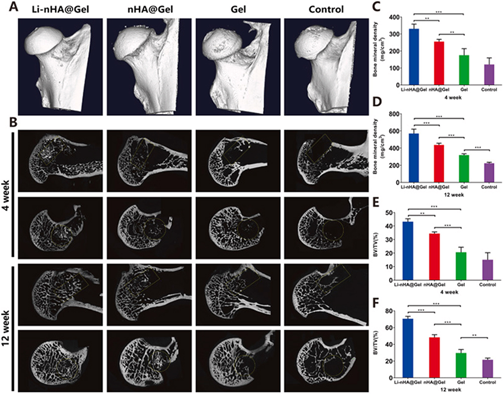 |
Figure 16 Assessment of bone regeneration in drilling channels using Micro-CT analysis at 4 and 12 weeks post-treatment. (A) Three-dimensional reconstructed images of femoral heads. (B) Representative two-dimensional views showing bone defect repair within the drilling channel (highlighted by yellow circles and boxes) across groups. (C–F) Quantitative analysis of BV/TV and BMD. Statistical significance: **P < 0.01, ***P < 0.001. Figures reproduced with permission from Y. Luo et al35 Copyright 2024, Elsevier International Publisher. |
Fu et al have examined the impact of T helper 17 (Th17) cell infiltration on the physiological function of focal stem cells in SONFH treatment, targeting the detrimental effects of immune infiltration by electrical stimulation of an injectable thermosensitive PLGA-PEG-PLGA hydrogel encapsulating IL-17@nanoliposomes to augment stem cell activity. It converts into a solid at physiological temperature and incorporates piezoelectric PLLA nanofibers alongside IL-17 inhibitor-encapsulated liposomes for dual physical–chemical regulation. Upon external ultrasound stimulation, embedded nanofibers generate microcurrents to enhance mitochondrial activity and osteogenic differentiation of mesenchymal stem cells, while reducing chemokine secretion to block Th1 cell recruitment; liposomes deliver sustained IL-17 inhibition over 2 weeks to restrain IL-17 production and stem cell apoptosis. Combined dual effects substantially attenuate the inflammatory microenvironment in steroid-induced femoral head necrosis and promote in situ bone regeneration in animal models. This study has drawbacks, notably the inadequate examination of the regulatory systems governing immune infiltration. The infiltration of various immune cells (eg, NK cells, CD8 T cells) in SONFH lesions has markedly risen, and the impact of these immune cells on SONFH progression requires additional investigation (Figure 17).41
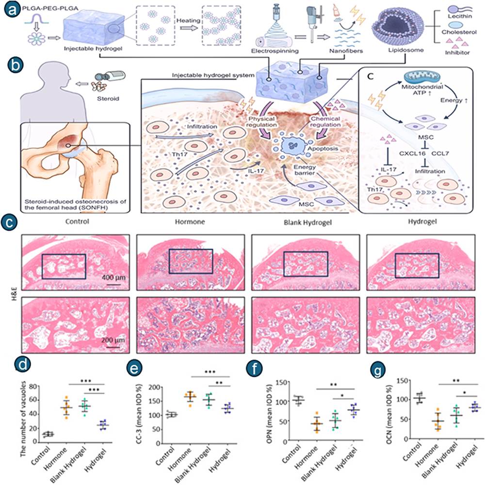 |
Figure 17 (a) presents a schematic of the injectable hydrogel system construction incorporating physical (electrical stimulation) and chemical (IL-17 inhibitor) dual regulation methods. (b) illustrates the disease mechanism whereby Th17 cell infiltration and stem cell physiological dysfunction lead to SONFH progression. (c) displays representative hematoxylin and eosin staining images of rat femoral head sections, while Panel (d–g) quantitative analysis: (d) vacuole numbers within the femoral head across experimental groups, (e) CC-3, (f) OPN, (g) OCN. Statistical significance is indicated as *P < 0.05, **P < 0.01, ***P < 0.001. Figures reproduced with permission from Fu et al.41 Copyright 2025, Elsevier International Publisher. |
Targeting Hypoxia-Inducible Factor (HIF-1α)
Metal–organic frameworks (MOFs) exhibit enzyme-like characteristics owing to their structural variety, elevated porosity, and capacity for delivery. Specifically, MOF-818 emulates superoxide dismutase (SOD) and catalase (CAT), mitigating excess ROS and facilitating wound healing. Cu/Zn-SOD, a crucial antioxidative enzyme in living organisms, demonstrates significant antioxidative activity due to its histidine-bridged bimetallic Cu−Zn site. Deferoxamine (DFO), sanctioned by the US Food and Drug Administration (FDA), functions as an iron chelating agent for the treatment of transfusion-related iron excess. Multiple studies have demonstrated that DFO modulates the signaling pathways of HIF-1α and VEGF, thus promoting angiogenesis and osteogenesis. Motivated by this mechanism, Liu et al integrated DFO-loaded MOF-818 (DFO@Zn-MOF-818) into GelMA as an innovative model for SONFH treatment, enhancing SOD-like activity.
DFO@Zn-MOF@GelMA hydrogel exhibits pH-responsive release, accelerating significantly under acidic conditions while providing sustained long-term distribution from the composite scaffold. Zn2⁺ is consistently released in conjunction with DFO. All prepared samples showed a swelling plateau at 24 hours, with a final swelling ratio of around 40%; no significant variation in swelling was observed following MOF loading. Complete breakdown occurs rapidly within 6 hours after collagenase incubation. The pore size of DFO@Zn-MOF@GelMA increased to 107.9 ± 20.1 μm compared to the GelMA hydrogel, which measured 96.5 ± 19.4 μm. It exhibited a dual mechanism, facilitating angiogenesis and osteogenesis via the HIF-1α pathway and enhanced SOD-like activity (Figure 18).37
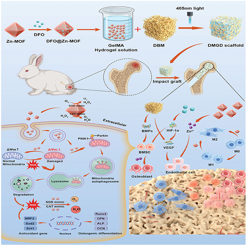 |
Figure 18 Schematic illustration of the biomimetic multifunctional scaffold design for SONFH treatment. Figures reproduced with permission from L. Bai et al.37 Copyright 2025, Wiley-VCH International Publisher. |
AlONFH
The pathophysiology of AlONFH is complex, involving synergistic interactions among direct cellular toxicity, metabolic instability, and hereditary predisposition, culminating in bone necrosis.59 Fundamental research on AlONFH has predominantly concentrated on various pathways, encompassing bone and lipid metabolism, angiogenesis, and genetic predisposition. Alcohol, as a notable external factor, disturbs the equilibrium between osteogenesis and adipogenesis by suppressing Wnt/β-catenin and mTOR signaling pathways while enhancing PPARγ activation. Furthermore, alcohol impairs angiogenesis regulated by VEGF and bFGF, worsening ischemic necrosis of the femoral head. Furthermore, polymorphisms in genes such as ApoB and ApoA1 elucidate the genetic basis of lipid-metabolism diseases. The regulatory functions of non-coding RNAs, including miRNAs, circRNAs, and lncRNAs, particularly in the proliferation, differentiation, and death of osteoclasts and osteoblasts, offer novel avenues for early diagnosis and targeted intervention.
A primary mechanism is the adverse impact of alcohol on bone remodeling. Alcohol disturbs the equilibrium between osteoblast-driven bone growth and osteoclast-driven bone resorption. It inhibits osteoblast differentiation from mesenchymal stem cells, diminishes bone mineralization indicators (alkaline phosphatase, osteocalcin), and triggers apoptosis in osteoblasts and osteocytes. Simultaneously, it facilitates bone resorption. This imbalance, frequently intensified by concurrent malnutrition, vitamin deficiencies (notably D, B1, and B2), and endocrine disorders (affecting PTH, vitamin D, and IGF-1), leads to diminished BMD, osteopenia, and osteoporosis. The damaged bone structure is susceptible to microfractures that cannot be adequately repaired, making it prone to structural failure and collapse.
Furthermore, alcohol consumption may exacerbate the course of ONFH by altering gut microbiota composition, elevating Lactobacillus and Roseburia, and modifying fecal metabolites. An extensive study is underway to clarify the mechanisms underlying AlONFH and to enhance its management. Researchers discovered that betaine exerted a protective effect, inhibiting the advancement of AlONFH through the mammalian target of rapamycin (mTOR) pathway. Moreover, the inhibition of Dickkopf-1 (DKK1) stimulates Wnt/β-catenin signaling and facilitates the nuclear translocation of β-catenin, thereby enhancing osteogenesis and suppressing adipogenesis in bone marrow-derived mesenchymal stem cells (BMSCs).
Preclinical investigations identified multiple potential regulatory mechanisms. For instance, disrupted osteogenic-adipogenic differentiation of stromal cells and altered regulons, including myocyte enhancer factor 2C (MEF2C) and JunD, are linked to AONFH. A subpopulation of endothelial cells expressing atypical chemokine receptor 1 (ACKR1) demonstrated increased chemotaxis but diminished angiogenic capacity. Endothelial cells may modulate stromal cell differentiation via pathways involving nicotinamide phosphoribosyltransferase (NAMPT) and E-selectin.
Unlike SONFH, in which adipogenesis is predominantly stimulated by glucocorticoids in the bone marrow, lipid dysregulation in AlONFH is more closely associated with alcohol’s systemic effects on hepatic lipid metabolism and overall systemic dysfunction. The intricate, multifactorial nature of AlONFH pathogenesis complicates accurate predictions of disease progression, underscoring the need for ongoing research incorporating multi-omics data and the development of targeted molecular interventions to enhance early diagnosis and patient outcomes.
Inhibiting apoptosis and adipogenic differentiation of stem cells using gene therapy may provide a useful technique for treating SONFH. Fu et al developed a heat-sensitive nanocomposite hydrogel to modulate gene expression and sustain gene regulation in lesion cells in AlONFH. Upon injection into the body, the hydrogel can solidify due to body heat, allowing for prolonged retention. As the hydrogel degrades in vivo, its intracellular secondary nanostructures are continuously released. These nanoparticles deliver plasmids and siRNA into lesion stem cells to enhance expression of B-cell lymphoma 2 (which inhibits stem cell death) and suppress peroxisome proliferator-activated receptor γ (PPARγ) expression (which inhibits adipogenic differentiation of stem cells). The physiological activity of stem cells in the ONFH region was ultimately reinstated, facilitating the repair of ONFH. In vivo investigations revealed that this nanocomposite hydrogel can be implanted for prolonged durations, thereby offering sustained therapeutic benefits. Consequently, bone regeneration transpires in the ONFH region, thereby facilitating the management of AlONFH. This nanocomposite hydrogel offers an innovative treatment approach for alcohol-related disorders and may be beneficial as a biomaterial for other gene therapy applications (Figure 19).42
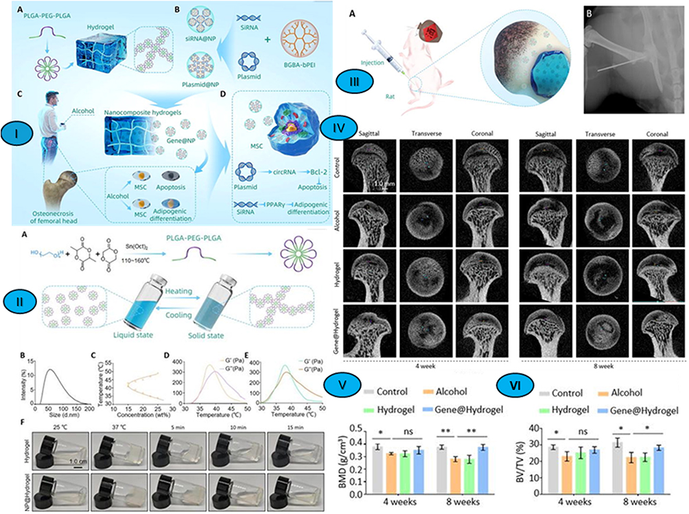 |
Figure 19 Design, characterization, and therapeutic efficacy of nanocomposite hydrogels for alcohol-induced ONFH. (I) Schematic diagram of the nanocomposite hydrogel for the treatment of alcohol-induced ONFH: (a) Schematic diagram of heat-sensitive hydrogel synthesis. (b) Schematic diagram of the gene-loaded nanoparticle synthesis. (c) Pathophysiological mechanism of chronic alcohol consumption that leads to ONFH. (d) Nanocomposite hydrogels inhibit the apoptosis and adipogenic differentiation of MSCs, thus treating ONFH. (II) Characterization of heat-sensitive hydrogels: (a) Schematic diagram of the synthesis and mechanism of action of the heat-sensitive hydrogel. (b) Particle size distribution of nanomicelles. (c) Phase transitions in the water systems of triblock polymers. (d) Change in the modulus of the water system of triblock polymer varies with temperature. (e) Change in the modulus of the nanocomposite hydrogels varies with temperature. (f) Photographs of polymer hydrogels and nanocomposite hydrogels achieving solid and liquid transitions at different temperatures. (III): (a) Schematic illustration of the generation and treatment of the rat model of alcohol-induced ONFH. (b) The liquid nanocomposite hydrogel was injected into the Hip joint using a syringe). (IV) Micro-CT was performed to detect femoral head necrosis in the rats. (V) Statistical analysis of BMD. (VI) Statistical analysis of BV/TV. (h) Statistical analysis of Tb.Th. *P < 0.05, **P < 0.01. Figures reproduced with permission from Z. Fu et al42 Copyright 2023, AIP International Publisher. Abbreviation: ns, nonsignificant. |
COVID-Related
COVID-19-associated osteonecrosis of the femoral head (ONFH) has recently been identified as a new clinical entity, probably linked to severe SARS-CoV-2 infections or intensive corticosteroid therapies administered during therapy.60 The pathophysiology of avascular necrosis can be understood through many theories, as detailed below. The systemic inflammatory response induced by COVID-19 releases cytokines, including Interleukin-17 and Tumor Necrosis Factor-alpha, which subsequently impede osteoblast proliferation and maturation. Secondly, the SARS-CoV-2 virus diminishes the function of angiotensin-converting enzyme 2 (ACE2), hence worsening bone loss. Third, systemic inflammation, along with direct endothelial injury induced by COVID-19, leads to overexpression of tissue factors and dysregulation of the coagulation cascade, frequently culminating in a hypercoagulable state that heightens the risk of bone necrosis. This discovery was validated by specific cadaveric experiments, demonstrating coagulopathy and intravascular thrombosis in individuals post-COVID-19 infection. Furthermore, findings from studies on osteonecrosis of the femoral head following SARS-CoV-2 infection suggest that osteonecrotic lesions generally reduce in size and stabilize over time, unlike ischemic necrosis that arises from steroid usage in other circumstances.61
A prior study posited that significant morphological biomarkers initiating the primary molecular and cellular pathways in the progression of post-COVID-19 ONFH encompass clusters of mast cells, severe fibrosis, numerous arterial and venous thrombi, and giant cell granulomas. The primary contributors are likely mast cells, which, upon interaction with the COVID-19 virus, undergo excessive hyperplasia, activation, and degranulation. The mediators secreted by mast cells subsequently exacerbate inflammation, influence bone and cartilage metabolism, induce vascular dysfunction and thrombosis, and promote fibrosis, thereby accelerating the progression of femoral osteonecrosis.62 While preclinical data on injectable hydrogels for ONFH therapy are promising, research on ONFH related to COVID-19 remains unexamined. The mechanism of action of hydrogels in treating this particular etiological variety has not been elucidated in any published publications. This significant information gap remains unexamined and warrants investigation in future studies.
LCPD-ONFH
LCPD is a pediatric ischemic ONFH that impacts 1 in 1200 children. This illness is among the most severe afflictions of the pediatric hip joint, particularly in adolescents, with over 50% of patients ultimately developing incapacitating osteoarthritis despite therapeutic interventions. ONFH leads to localized osteocyte and bone marrow cell death due to compromised blood supply to the bone, and the ensuing healing process results in necrotic bone resorption and structural abnormalities. The advancement of LCPD results in hip pain, restricted mobility, and physical impairment requiring THA. Nonetheless, total hip replacement is not an ideal therapeutic option for younger patients because of their elevated physical demands and lifespan.43
Consequently, prompt identification and intervention are crucial to alleviating the detrimental effects described above and maintaining hip functionality, underscoring the need for a robust, effective strategy for identifying and treating the complex problem of LCPD. The exact etiology of LCPD remains unclear; nevertheless, it is widely acknowledged that genetic predisposition, vascular anomalies, and mechanical stress on the hip joint significantly contribute to its progression. These variables result in a transient interruption of blood flow to the femoral head, causing the distinctive symptoms of the condition.
Bone morphogenetic protein 2 (BMP2), a member of the transforming growth factor beta family, exhibits significant osteogenic potential. Exogenous recombinant human BMP2 has received approval for the treatment of tibial nonunion fractures and spinal fusion. Local injections of BMP2 is reported to substantially enhance new bone formation and reduce the likelihood of femoral head deformation. Saline has been utilized as a vehicle for localized BMP2 injection for ONFH treatment. Nonetheless, using saline as the carrier was associated with a significant incidence of heterotopic ossification (HO) within the hip capsule, due to its limited capacity to prevent BMP2 leakage after injection. The leakage reduces the localized dosage and distribution at the target site, leading to inadequate bone regeneration and uneven bone repair. To mitigate these limitations, Ma et al devised a BMP2-hydrogel therapy comprising transphyseal bone lavage followed by injection of BMP2-encapsulated hydrogel. In vitro investigations have shown that a gelatin-heparin-tyramine hydrogel preserved BMP2 for a duration of four weeks. The hydrogel injection can effectively avert leakage. The injected hydrogel exhibited extensive diffusion inside the cranial region following the bone wash. In vivo investigations in swine demonstrated that BMP2-hydrogel administration led to uniform bone regeneration without heterotopic ossification. It maintained the subchondral contour and reinstated subchondral endochondral ossification, despite increasing growth plate fusions. The study revealed a promising BMP2-hydrogel for ONFH treatment, particularly in adolescents (Figure 20).43
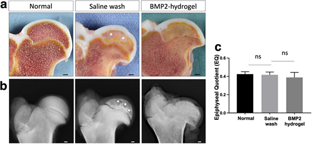 |
Figure 20 Macroscopic and radiographic analysis of femoral heads from normal, saline wash, and BMP2-hydrogel treatment groups. (a) Representative photographs of bisected femoral heads with white asterisks indicating large bone void areas. (b) Representative X-ray images of femoral heads. (c) Quantitative comparison of mean EQ values across groups. Scale bar = 2 mm (a and b). Data expressed as mean ± SD (n = 6); ns = not significant. Figures reproduced with permission from C. Ma et al43 Copyright 2023, Nature International Publisher. |
Comparative Study Related to Key Physicochemical Parameters of Hydrogel Performance for ONFH Therapy
Because there are many different hydrogel systems for treating ONFH, we need to focus on their physicochemical properties to enhance their therapeutic mechanisms. Table 1 summarizes the main features of the hydrogel systems discussed in this review. Swelling, biodegradation, and mechanical characteristics constitute three indispensable core design indices for SONFH-target injectable hydrogels.
Porous Structure
Overall, a rationally tailored porous hydrogel architecture offers significant potential for ONFH treatment while simultaneously enabling the controlled release of loaded drugs and metal ions from the matrix. The optimum pore size (100–200 μm) suited for cell infiltration that matches those of DFO@Zn-MOF@GelMA, SA/β-TCP@2%PMP, and OCMC-CMCS-QK.
Targeting pH-Responsive Release
Due to the acidic microenvironment induced by inflammation and hypoxia in necrotic femoral head lesions, pH-sensitive hydrogels have emerged as promising agents for targeted, expedited release at the lesion site.63 Among the evaluated formulations, two pH-responsive systems are notable: the chitosan-based CS/PF/TPP hydrogel enhances rhBMP9 release in localized acidic pathological conditions, whereas DFO@Zn-MOF/GelMA facilitates pH-dependent deferoxamine delivery, exhibiting significantly increased DFO release in low-pH necrotic environments while minimizing premature leakage in neutral healthy tissue settings. This pH-triggered release facilitates localized high therapeutic concentrations at damaged regions, minimizes systemic side effects, and enhances the efficiency of antioxidative, osteogenic, and angiogenic medicines for the treatment of ONFH.
Swelling
Tunable swelling balances inflammatory fluid absorption and nutrient exchange while governing the diffusion kinetics of loaded therapeutic ions, exosomes, or small-molecule payloads; over-swelling induces peri-lesion tissue compression and exacerbates local femoral ischemia, whereas insufficient hydration restricts cellular nutrient transport. For SONFH hydrogel, moderate, controlled swelling (300–1000% equilibrium) is optimal: ultra-high swelling ratios cause mechanical collapse and tissue compression, whereas too low swelling restricts nutrient exchange and payload release efficiency.64
Degradation
SONFH bone repair requires 6–12 weeks for substantial trabecular reconstruction in rabbit preclinical models; hydrogel degradation rate must match the pace of new bone growth, with dual pitfalls of too-fast/too-slow breakdown.65 SONFH preferred residual mass: 35–50% remaining after 4 weeks in vitro, balancing sustained release delivery and progressive matrix replacement by neo-bone.66,67 Over-fast degradation leads to abrupt burst release of therapeutic cargo (Li, DFO, Mg, exosomes), generating toxic local high concentrations, premature loss of the physical niche for seeded BMSCs, early void formation in the bone defect, and fibrous tissue ingrowth blocking osteogenesis.68 Over-slow degradation leads to undegraded residual polymer acting as a foreign body, triggers chronic M1 macrophage infiltration and fibrous encapsulation around the implant, hindering native bone penetration into the hydrogel space. Degradation rate also dictates long-term bioactive supply: A slowly degrading composite maintains sustained ion/drug release over the entire bone remodeling cycle (4+ weeks, critical for SONFH anti-inflammation and angiogenesis).32
Mechanical Properties
Mechanically, shear-thinning rheology ensures minimally invasive injectability via clinical gauge needles, while a compressive modulus calibrated to 20–100 kPa mimics native cancellous bone mechanics to maintain defect space and mechanically drive BMSC osteogenic differentiation against GC-triggered adipogenic drift.69
Clinical Studies
As previously noted, recombinant human fibroblast growth factor-2 (rhFGF-2) directly promotes the migration, proliferation, and differentiation of vascular endothelial cells, establishing itself as a potent pro-angiogenic mediator. Accordingly, while the aforementioned clinical follow-up studies report favorable outcomes for ONFH management, we contend that vascular density modifications should also have been examined. In a 2016 one-year clinical follow-up study, Kuroda et al administered a single localized injection of 800 μg rhFGF-2 incorporated into a gelatin hydrogel to patients with pre-collapse ONFH (n = 10) classified as Stage 2 or lower. Femoral head collapse occurred in only one subject, whereas the remaining nine individuals exhibited no evidence of collapse. Moreover, increased bone mass was observed within the affected regions of these patients.70 Subsequently, a multicenter Phase II clinical trial was conducted across four Japanese institutions, including The University of Tokyo, Gifu University, Osaka University, and Kyoto University, enrolling 64 patients to evaluate the two-year outcomes following a single administration of 800 μg rhFGF combined with a gelatin hydrogel for ONFH. This study aimed to determine the efficacy of rhFGF-2 in preventing femoral head collapse. The results demonstrated prolonged joint preservation duration, with a joint preservation rate of 65% or higher, reflecting enhanced clinical efficacy, radiographic evidence of bone regeneration, and a favorable safety profile.25,71
Despite promising clinical efficacy of injectable gelatin hydrogel as a sustained-release carrier for rhFGF-2 in precollapse ONFH, multiple practical challenges persist for routine clinical translation. These include load-bearing conditions and space maintenance in the femoral head; retention of injectable hydrogels and volume limits; long-term biosafety and degradation products; reproducibility and intraoperative preparation; sterilization challenges; and regulatory considerations.
Load-Bearing Conditions and Space Maintenance in the Femoral Head
A significant obstacle in introducing hydrogels for ONFH treatment is the severe mechanical conditions of the hip joint. The calibrated compressive modulus of advanced hydrogels closely resembles that of early-stage cancellous bone and facilitates BMSCs osteogenesis through mechanotransduction; however, it provides negligible macrostructural support against the physiological loads experienced by the femoral head, which can exceed multiple times a patient’s body weight during ambulation. In human clinical trials, this mechanical deficiency is circumvented by meticulously selecting patients in the pre-collapse phase (JIC/JICHW Stage 1 or 2). At this stage, the structural integrity of the adjacent subchondral cortical shell remains intact. The gelatin is not designed to support weight; rather, it functions as a non-structural, osteoinductive filler within the necrotic cavity, while the host’s cortical bone serves as mechanical protection.
Retention of Injectable Materials and Volume Limits
The rigid bony environment, such as the femoral head, material retention, and preventing “backflow” out of the drill tract are major surgical hurdles. High intraosseous pressure can push low-viscosity gels back into the joint space or into the surrounding soft tissue, causing local irritation and therapeutic failure. Protocols from the TRION trial show that surgeons resolve this by utilizing a tightly controlled percutaneous CD. Under fluoroscopic guidance, a narrow guide wire and a cannulated drill create a precise, narrow tunnel directly into the center of the necrotic zone.
Long-Term Biosafety and Degraded Byproducts
Too rapid degradation leads to local toxicity due to a burst release of metal ions, whereas too slow degradation results in chronic M1 macrophage polarization and fibrous encapsulation. Use of naturally derived biopolymers, which are highly biocompatible, optimizes the long-term safety of the underlying matrix. The byproducts of gelatin are 100% non-toxic endogenous amino acids that are slowly reabsorbed and replaced by host neo-bone.
Reproducibility and Intraoperative Preparation
A common pitfall of advanced “smart” hydrogels is that they are too complex to be reliably prepared in a chaotic operating room (OR) setting. If a material requires multi-step chemical crosslinking or precise temperature control at the point of care, its clinical adoption will fail. To achieve high clinical reproducibility across multiple centers, the preparation must be streamlined into a simple, standardized kit. In the TRION trial, reproducibility was achieved by supplying the matrix as a standardized, freeze-dried, cross-linked gelatin cake, along with a separate syringe containing the therapeutic solution (rhFGF-2).
Sterilization Challenges
Sterilization of hydrogels without damaging their delicate polymer networks, soft structures or their encapsulated bioactive cargo (exosomes, peptides, drugs) is a significant manufacturing challenge. Standard autoclaving destroys the hydrogel structure with heat, whereas UV irradiation can cleave polymer chains, drastically changing the swelling ratio and degradation kinetics.
Regulatory Considerations
Shifting the injectable hydrogel for bone regeneration from the laboratory to human patients requires a clear understanding of regulatory classification. Hydrogels carrying metal ions, growth factors, or small-molecule drugs fall under the highly complex category of Combination Products. In the regulatory design of the TRION trial, investigators conducted intensive, proactive consultations with the Japanese regulatory authority, the Pharmaceuticals and Medical Devices Agency (PMDA). A critical regulatory pivot was deciding whether the trial should evaluate outcomes on an individual patient basis or a joint-specific basis. The PMDA mandated an individual-patient-based analysis, which dictated how bilateral ONFH cases were tracked (defining the endpoint by the first collapsed side).
Recommendations and Future Outlook
Based on the comparative analysis, we propose the following criteria for selecting a hydrogel based on clinical presentation. Future pH-Smart Responsive hydrogel systems should integrate many therapeutic processes simultaneously rather than relying on single-target approaches. Moreover, swelling, biodegradation, and mechanical characteristics constitute three indispensable core features of injectable hydrogels to address the above barriers and expand the clinical applicability of this regenerative therapy. Future hydrogel iterations intended for larger or borderline-collapse lesions (Stage 3A) must be designed as organic-inorganic composites to provide dual-functional mechanical space maintenance alongside biofactor delivery.
To ensure proper retention, hydrogels must exhibit rapid in situ gelation or a highly responsive shear-thinning rheology that allows smooth delivery through a long clinical-gauge needle while immediately recovering viscosity upon exiting the needle tip. Furthermore, the total injection volume must be precisely calibrated to fill only the decompressed void (typically matching the volume of a single dose, eg, of active factor complexed in a minimal gel volume) to avoid over-pressurization and backflow. The surgical staff can reliably prepare the active hydrogel formulation within 30 minutes before administration simply by mixing the solution and the pre-measured freeze-dried component in an interconnected syringe system. This eliminates batch-to-batch variation and human error during surgery, ensuring identical material properties across diverse clinical sites. For successful translation, a dual-stage sterilization workflow must be adopted.
The base polymer matrix (eg, the freeze-dried gelatin or alginate framework) should be sterilized independently using validated ethylene oxide (EtO) gas or low-dose gamma irradiation at the manufacturing stage. The therapeutic payloads (such as recombinant proteins, small molecules such as DFO, or ions) must be prepared by sterile filtration under aseptic conditions. The final composition is reconstituted intraoperatively at the point of care, preserving both matrix architecture and cargo bioactivity. For international translation (eg, US FDA), a hydrogel system delivering bioactive cargo will likely be routed through the Center for Biologics Evaluation and Research (CBER) or the Center for Drug Evaluation and Research (CDER) as a drug-delivery device combination.
Conclusion
Injectable hydrogels represent a promising platform for the targeted ONFH treatment. Targeting well-established pathophysiological mechanisms is an effective approach to enhance their efficacy by inhibiting pathological cascades and delaying disease progression, thereby delaying the need for THA. To ensure successful clinical translation, future biomaterial design must optimize three core physicochemical indices: controlled swelling, a degradation rate, and a compressive modulus that drives osteogenesis. Furthermore, engineering “smart” pH-responsive release mechanisms and a tailored porous architecture allows for localized, targeted delivery while mitigating systemic toxicity. Despite favorable multicenter clinical data demonstrating that gelatin hydrogels complexed with pro-angiogenic factors such as rhFGF-2 effectively prevent femoral head collapse at pre-collapse stages, critical translation barriers remain. Addressing these challenges requires developing organic-inorganic composites to optimize shear-thinning rheology and prevent backflow inside the osseous cavity and adopting standardized dual-stage sterilization workflows.
Data Sharing Statement
Authors confirm that all relevant data are included in the paper.
Acknowledgment
The author wishes to thank the Talented Young Scientist Program for the financial and logistical support provided during this research. Their assistance was invaluable in bringing this project to fruition.
Author Contributions
All authors contributed to data analysis, drafting or revising the article, have agreed on the journal to which the article will be submitted, gave final approval of the version to be published, and agree to be accountable for all aspects of the work.
Funding
QZ acknowledges support from the National Natural Science Foundation of China (grant references: 82574777, 82072494), Beijing Natural Science Foundation (L244060), National High-Level Hospital Clinical Research Funding, and Elite Medical Professionals Project of the China-Japan Friendship Hospital (grant references: 2024-NHLHCRF-JBGS-ZH-03, 2022-NHLHCRF-YGJE-05, and ZRJY2021-GG08). We greatly appreciate the financial support from the National Natural Science Foundation of China (No. 22171154), the Natural Science Foundation of Shandong Province (ZR2024YQ025), Jinan Science and Technology Bureau (No. 202527053, 2021GXRC080, 202333026, and 202527055), and Science, Education and Industry Integration Innovation Pilot Project from Qilu University of Technology (Shandong Academy of Sciences) (2024ZDZX13).
Disclosure
The authors declare no conflicts of interest in this work.
References
1. Rezus E, Tamba BI, Badescu MC, Popescu D, Bratoiu I, Rezus C. Osteonecrosis of the femoral head in patients with hypercoagulability—from pathophysiology to therapeutic implications. Int J Mol Sci. 2021;22(13):6801. doi:10.3390/ijms22136801
2. Miyahara HDS, Ranzoni LV, Ejnisman L, Vicente JRN, Croci AT, Gurgel HMDC. Osteonecrosis of the femoral head: update article. Rev Bras Ortop. 2022;57:351–33 doi:10.1055/s-0041-1736308.
3. Zhu W, Xu Z, Zhou D, Xu J, He Y, Li ZA. Bioengineering strategies targeting angiogenesis: innovative solutions for osteonecrosis of the femoral head. J Tissue Eng. 2025;16:20417314241310540. doi:10.1177/20417314241310541
4. Cao H, Guan H, Lai Y, Qin L, Wang X. Review of various treatment options and potential therapies for osteonecrosis of the femoral head. J Orthop Transl. 2016;4:57–70. doi:10.1016/j.jot.2015.09.005
5. Prentice HA, Chan PH, Royse KE, Hinman AD, Reddy NC, Paxton EW. Revision risk in a cohort of US patients younger than 55 undergoing primary elective total hip arthroplasty. J Arthroplasty. 2022;37(2):303–311. doi:10.1016/j.arth.2021.10.014
6. Yang TJ, Sun SY, Zhang L, Zhang XP, He HJ. A delphi-based model for prognosis of femoral head collapse in osteonecrosis: a multi-factorial approach. J Orthop Surg Res. 2024;19(1):762. doi:10.1186/s13018-024-05247-0
7. Kuroda Y, Matsuda S, Akiyama H. Joint-preserving regenerative therapy for patients with early-stage osteonecrosis of the femoral head. Inflamm Regen. 2016;36(1). doi:10.1186/s41232-016-0002-9
8. Xiao A, Bonsignore-Opp L, Koffi V, Coufal S, Swarup I. Diagnosis and management of osteonecrosis of the femoral head in young patients. JBJS Rev. 2025;13(6):e25. doi:10.2106/JBJS.RVW.25.00046
9. Bian Y, Hu T, Lv Z, et al. Bone tissue engineering for treating osteonecrosis of the femoral head. Exploration. 2023;3(2). doi:10.1002/EXP.20210105
10. Moya-Angeler J, Gianakos AL, Villa JC, Ni A, Lane JM. Current concepts on osteonecrosis of the femoral head. World J Orthop. 2015;6(8):590. doi:10.5312/wjo.v6.i8.590
11. Liang D, Pei J, Zhang L, Ling H, Liu Y, Chen X. Treatment of pre-collapse non-traumatic osteonecrosis of the femoral head through Orthopdische Chirurgie München approach combined with autologous bone mixed with β-tricalcium phosphate porous bioceramic bone graft: a retrospective study of mid-term result. J Orthop Surg Res. 2021;16(1):1–9. doi:10.1186/s13018-021-02632-x
12. Ge H, Wang Z, Zhang J. X-ray, digital tomographic fusion, CT, and MRI in early ischemic necrosis of the femoral head. Medicine. 2024;103(2):e36281. doi:10.1097/MD.0000000000036281
13. Yoon BH, Mont MA, Koo KH, et al. The 2019 revised version of association research circulation osseous staging system of osteonecrosis of the femoral head. J Arthroplasty. 2020;35(4):933–940. doi:10.1016/j.arth.2019.11.029
14. Liu LH, Li ZR, Sun W, Wang YT, Gao FQ. Reliability and repeatability of the China-Japan Friendship Hospital typing classification for nontraumatic osteonecrosis of the femoral head. JBJS. 2022;104(Suppl 2):40–46. doi:10.2106/JBJS.20.00051
15. Di Benedetto P, Niccoli G, Beltrame A, Gisonni R, Cainero V, Causero A. Histopathological aspects and staging systems in non-traumatic femoral head osteonecrosis: an overview of the literature. Acta biomed. 2016;87(Suppl 1):15–24.
16. Cheng EY, Mirzaei A. Exploring the feasibility of pharmacologic management in non-traumatic osteonecrosis: an etiology-based hypothesis. Med Hypotheses. 2024;193:111497. doi:10.1016/j.mehy.2024.111497
17. Lubega N, Mkandawire NC, Sibande GC, Norrish AR, Harrison WJ. Joint replacement in Malawi: establishment of a National Joint Registry. J Bone Joint Surg Br. 2009;91(3):341–343. doi:10.1302/0301-620X.91B3.21706
18. Chan VW, Chan PK, Chiu KY, Yan CH, Ng FY. Why do Hong Kong patients need total Hip arthroplasty? An analysis of 512 hips from 1998 to 2010. Hong Kong Med J. 2016;22(1):11–15. doi:10.12809/hkmj144483
19. Mont MA, Cherian JJ, Sierra RJ, Jones LC, Lieberman JR. Advances in experimental models of osteonecrosis of the femoral head. J Bone Joint Surg Am. 2015;97(19):1604–1627. doi:10.2106/JBJS.O.00071
20. Cheng EY, Mirzaei A. Potential molecular targets for the pharmacologic management of non-traumatic osteonecrosis. Expert Opin Ther Targets. 2024;28(11):991–1000. doi:10.1080/14728222.2024.2421755
21. Moya-Angeler J, Gianakos AL, Villa JC, Ni A, Lane JM. Current concepts on osteonecrosis of the femoral head. World J Orthop. 2015;6(8):590–601. doi:10.5312/wjo.v6.i8.590
22. Scaglione M, Fabbri L, Celli F, Casella F, Guido G. Hip replacement in femoral head osteonecrosis: current concepts. Clin Cases Miner Bone Metab. 2015;12(Suppl 1):51–54. doi:10.11138/ccmbm/2015.12.3s.051
23. Zheng Y, Zheng Z, Zhang K, Zhu P. Osteonecrosis in systemic lupus erythematosus: systematic insight from the epidemiology, pathogenesis, diagnosis and management. Autoimmun Rev. 2022;21(2):102992. doi:10.1016/j.autrev.2021.102992
24. Quan H, He Y, Xiao P, et al. Scaffold-mediated microenvironmental modulation targeting osteoclasts for ONFH Niche reprogramming. Research. 2025;8:1027. doi:10.34133/research.1027
25. Bal Z, Takakura N. Hydrogel use in osteonecrosis of the femoral head. Gels. 2024;10(8):544. doi:10.3390/gels10080544
26. Salama AM, Hardy JG, Yessuf AM, et al. Injectable hydrogel technologies for bone disease treatment. ACS Appl Bio Mater. 2025;8(4):2691–2715. doi:10.1021/acsabm.4c01968
27. Omidian H, Chowdhury SD. Advancements and applications of injectable hydrogel composites in biomedical research and therapy. Gels. 2023;9(7):533. doi:10.3390/gels9070533
28. Wang Y, Zhu W, Xiao K, et al. Self-healing and injectable hybrid hydrogel for bone regeneration of femoral head necrosis and defect. Biochem Biophys Res Commun. 2019;508(1):25–30. doi:10.1016/j.bbrc.2018.11.097
29. Wang C, Xu H, Liu C, et al. CaO2/gelatin oxygen slow-releasing microspheres facilitate tissue engineering efficiency for the osteonecrosis of femoral head by enhancing the angiogenesis and survival of grafted bone marrow mesenchymal stem cells. Biomater Sci. 2021;9(8):3005–3018. doi:10.1039/d0bm02071k
30. Yuan S, Han Y, Xiang D, Wang B, Chen Y, Hao Y. An injectable hydroxypropyl-β-cyclodextrin cross-linked gelatin-based hydrogel loaded bone mesenchymal stem cell for osteogenic and in vivo bone regeneration of femoral head necrosis. Nanomedicine. 2022;41:102521. doi:10.1016/j.nano.2022.102521
31. Peyravian N, Milan PB, Kebria MM, et al. Designing and synthesis of injectable hydrogel based on carboxymethyl cellulose/carboxymethyl chitosan containing QK peptide for femoral head osteonecrosis healing. Int J Biol Macromol. 2024;270:132127. doi:10.1016/j.ijbiomac.2024.132127
32. Xu Y, Xu K, Wang Z, Tang X, Jiang Y, Li T. In situ sustained release of icariin from injectable thermosensitive PLGA-PEG-PLGA hydrogels combined with core decompression for steroid-induced osteonecrosis of femoral head. Front Pharmacol. 2025;16:1–14. doi:10.3389/fphar.2025.1643316
33. Wu Z, Zhan B, Feng S, et al. Angiogenesis-osteogenesis coupling lithium-loaded bioglass/GelMA hydrogel for bone regeneration. Biomater Adv. 2026;180:214591. doi:10.1016/j.bioadv.2025.214591
34. Chen C, Fu L, Luo Y, et al. Engineered exosome-functionalized extracellular matrix-mimicking hydrogel for promoting bone repair in glucocorticoid-induced osteonecrosis of the femoral head. ACS Appl Mater Interfaces. 2023;15(24):28891–28906. doi:10.1021/acsami.3c01539
35. Luo Y, Yang Z, Zhao X, et al. Immune regulation enhances osteogenesis and angiogenesis using an injectable thiolated hyaluronic acid hydrogel with lithium-doped nano-hydroxyapatite (Li-nHA) delivery for osteonecrosis. Mater Today Bio. 2024;25:100976. doi:10.1016/j.mtbio.2024.100976
36. Liu C, Wang C, Liu Y, et al. Selenium nanoparticles/carboxymethyl chitosan/alginate antioxidant hydrogel for treating steroid-induced osteonecrosis of the femoral head. Int J Pharm. 2024;653:123929. doi:10.1016/j.ijpharm.2024.123929
37. Bai L, Zhang X, Shen W, et al. Multifunctional scaffold comprising metal-organic framework, hydrogel, and demineralized bone matrix for the treatment of steroid-induced femoral head necrosis. Small. 2025;21(3):e2407758. doi:10.1002/smll.202407758
38. Zhou YH, Zou ZH, Teng JX, et al. Injectable Alginate/β-TCP composite hydrogel incorporating P34HB/MgO+ PEG coaxial electrospun microfibers for minimally invasive treatment of osteonecrosis. Adv Healthc Mater. 2025;14(15):2500617. doi:10.1002/adhm.202500617
39. Xu Y, Jiang Y, Wang Y, et al. LINC00473-modified bone marrow mesenchymal stem cells incorporated thermosensitive PLGA hydrogel transplantation for steroid-induced osteonecrosis of femoral head: a detailed mechanistic study and validity evaluation. Bioeng Transl Med. 2022;7(2):1–27. doi:10.1002/btm2.10275
40. Quan H, Ren C, Xie H, et al. An injectable hydrogel loaded with miRNA nanocarriers promotes vessel-associated osteoclast (VAO)-mediated angiogenesis and bone regeneration in osteonecrosis of the rat femoral head. Biomaterials. 2025;320:123252. doi:10.1016/j.biomaterials.2025.123252
41. Fu Z, Xu Y, Xu F, et al. Injectable hydrogel regulates immune infiltration through physical and chemical synergy in the treatment of steroid-induced osteonecrosis of the femoral head. Mater Today Bio. 2025;31:101511. doi:10.1016/j.mtbio.2025.101511
42. Fu Z, Lai Y, Zhuang Y, Lin F. Injectable heat-sensitive nanocomposite hydrogel for regulating gene expression in the treatment of alcohol-induced osteonecrosis of the femoral head. APL Bioeng. 2023;7(1). doi:10.1063/5.0130711
43. Ma C, Park MS, Alves do monte F, et al. Local BMP2 hydrogel therapy for robust bone regeneration in a porcine model of Legg-Calvé-Perthes disease. NPJ Regen Med. 2023;8(1):50. doi:10.1038/s41536-023-00322-2
44. Willemen NGA, Hassan S, Gurian M, et al. Oxygen-releasing biomaterials: current challenges and future applications. Trends Biotechnol. 2021;39(11):1144–1159. doi:10.1016/j.tibtech.2021.01.007
45. Lv Y, Wang Z, Wei Y, et al. Thermoresponsive dual-network chitosan-based hydrogels with demineralized bone matrix for controlled release of rhBMP9 in the treatment of femoral head osteonecrosis. Carbohydr Polym. 2025;352:123197. doi:10.1016/j.carbpol.2024.123197
46. Huang C, Wen Z, Niu J, Lin S, Wang W. Steroid-induced osteonecrosis of the femoral head: novel insight into the roles of bone endothelial cells in pathogenesis and treatment. Front Cell Dev Biol. 2021;9:777697. doi:10.3389/fcell.2021.777697
47. Zhang J, Cao J, Liu Y, Zhao H. Advances in the pathogenesis of steroid-associated osteonecrosis of the femoral head. Biomolecules. 2024;14(6):667. doi:10.3390/biom14060667
48. Yeon S, Seto SW, Bhuyan JD, Chang D, Li CG, Low M. Sclerostin in vascular calcification: hypoxia-driven regulation and therapeutic modulation by natural products. Curr Atheroscler Rep. 2026;28(1). doi:10.1007/s11883-025-01377-w
49. Vasiliadis ES, Evangelopoulos DS, Kaspiris A, Benetos IS, Vlachos C, Pneumaticos SG. The role of sclerostin in bone diseases. J Clin Med. 2022;11(3):806. doi:10.3390/jcm11030806
50. Li L, Zhao S, Leng Z, et al. Pathological mechanisms and related markers of steroid-induced osteonecrosis of the femoral head. Ann Med. 2024;56(1). doi:10.1080/07853890.2024.2416070
51. Okazaki S, Nagoya S, Matsumoto H, et al. TLR4 stimulation and corticosteroid interactively induce osteonecrosis of the femoral head in rat. J Orthop Res. 2016;34(2):342–345. doi:10.1002/jor.23008
52. Yin J, Huang J, Zhou P, Li L, Zheng Q, Fu H. The role of TLR4/NF-kB signaling axis in pneumonia: from molecular mechanisms to regulation by phytochemicals. Naunyn Schmiedebergs Arch Pharmacol. 2025;398:14559–14588. doi:10.1007/s00210-025-04130-x
53. Zhou R, Bian Y, Cai X, et al. Advances in the mechanism for steroid-induced osteonecrosis of the femoral head. Bone Res. 2026;14(1). doi:10.1038/s41413-025-00477-2
54. Peng P, Nie Z, Sun F, Peng H. Glucocorticoids induce femoral head necrosis in rats through the ROS/JNK/c-Jun pathway. FEBS Open Bio. 2021;11(1):312–321. doi:10.1002/2211-5463.13037
55. Xu Y, Jiang Y, Jia B, Wang Y, Li T. Icariin stimulates osteogenesis and suppresses adipogenesis of human bone mesenchymal stem cells via miR-23a-mediated activation of the Wnt/β-catenin signaling pathway. Phytomedicine. 2021;85:153485. doi:10.1016/j.phymed.2021.153485
56. Chen C, Wang B, Zhao X, et al. Lithium promotes osteogenesis via Rab11a-Facilitated Exosomal Wnt10a secretion and β-catenin signaling activation. ACS Appl Mater Interfaces. 2024;16(24):30793–30809. doi:10.1021/acsami.4c04199
57. Li D, Yang Z, Luo Y, Zhao X, Tian M, Kang P. Delivery of MiR335-5p-Pendant Tetrahedron DNA nanostructures using an injectable heparin lithium hydrogel for challenging bone defects in steroid-associated osteonecrosis. Adv Healthc Mater. 2022;11(1):e2101412. doi:10.1002/adhm.202101412
58. Maruyama M, Moeinzadeh S, Guzman RA, et al. The efficacy of lapine preconditioned or genetically modified IL4 over-expressing bone marrow-derived mesenchymal stromal cells in corticosteroid-associated osteonecrosis of the femoral head in rabbits. Biomaterials. 2021;275:120972. doi:10.1016/j.biomaterials.2021.120972
59. Konarski W, Poboży T, Konarska K, et al. Osteonecrosis related to steroid and alcohol use—an update on pathogenesis. Healthcare. 2023;11(13):1846. doi:10.3390/healthcare11131846
60. Mary A, Reddy SG, Kumar BS, Kugabalasooriar S. Novel approaches to alendronate delivery beyond oral administration- a review. Eng Sci. 2024:1–24 doi:10.30919/es1274 Novel.
61. Murugesan L, Sivakumar N, Ramamoorthy L, Farooq U. COVID-19-associated bilateral avascular necrosis of femoral head in a young male without corticosteroid exposure: a case report. Cureus. 2024;16(4):e57525. doi:10.7759/cureus.57525
62. Kuliyeva A, Serejnikova N, Eshmotova G, et al. Post-COVID-19 femoral head osteonecrosis exhibits mast cell clusters, fibrosis, and vascular thrombosis: key pathological mechanisms in long COVID-19 bone degeneration. Pathophysiology. 2025;32(3):36. doi:10.3390/pathophysiology32030036
63. Chen H, Tan W, Tong T, Shi X, Ma S, Zhu G. A pH-responsive asymmetric microfluidic/chitosan device for drug release in infective bone defect treatment. Int J Mol Sci. 2023;24(5). doi:10.3390/ijms24054616
64. Song Z, Yin J, Luo K, et al. Layer-by-layer buildup of Poly(L-glutamic acid)/Chitosan film for biologically active coating. Macromol Biosci. 2009;9(3):268–278. doi:10.1002/mabi.200800164
65. Dumas JE, Prieto EM, Zienkiewicz KJ, et al. Balancing the rates of new bone formation and polymer degradation enhances healing of weight-bearing allograft/polyurethane composites in rabbit femoral defects. Tissue Eng Part A. 2014;20(1–2):115–129. doi:10.1089/ten.TEA.2012.0762
66. Manohar SS, Das C, Kakati V. Bone tissue engineering scaffolds: materials and methods. 3D Print Addit Manuf. 2024;11(1):347–362. doi:10.1089/3dp.2022.0216
67. Gai T, Zhang H, Hu Y, et al. Sequential construction of vascularized and mineralized bone organoids using engineered ECM-DNA-CPO-based bionic matrix for efficient bone regeneration. Bioact Mater. 2025;49:362–377. doi:10.1016/j.bioactmat.2025.02.033
68. Lu X, Guo H, Li J, Sun T, Xiong M. Recombinant human bone morphogenic protein-2 immobilized fabrication of magnesium functionalized injectable hydrogels for controlled-delivery and osteogenic differentiation of rat bone marrow-derived mesenchymal stem cells in femoral head necrosis repair. Front Cell Dev Biol. 2021;9:1–16. doi:10.3389/fcell.2021.723789
69. Suresh N, Lekhavadhani S, Selvamurugan N. Advances in polymer-based hydrogel systems for adipose-derived mesenchymal stem cells toward bone regeneration. World J Orthop. 2026;17(1):113228. doi:10.5312/wjo.v17.i1.113228
70. Kuroda Y, Asada R, So K, Yonezawa A. A pilot study of regenerative therapy using controlled release of recombinant human fibroblast growth factor for patients with pre-collapse osteonecrosis of the femoral head. Int Orthop. 2016;40:1747–1754. doi:10.1007/s00264-015-3083-1
71. Kuroda Y, Tanaka T, Miyagawa T, et al. Recombinant human FGF-2 for the treatment of early-stage osteonecrosis of the femoral head: TRION, a single-arm, multicenter, Phase II trial. Regener Med. 2021;16(6):535–548. doi:10.2217/rme-2021-0049
 © 2026 The Author(s). This work is published and licensed by Dove Medical Press Limited. The
full terms of this license are available at https://www.dovepress.com/terms
and incorporate the Creative Commons Attribution
- Non Commercial (unported, 4.0) License.
By accessing the work you hereby accept the Terms. Non-commercial uses of the work are permitted
without any further permission from Dove Medical Press Limited, provided the work is properly
attributed. For permission for commercial use of this work, please see paragraphs 4.2 and 5 of our Terms.
© 2026 The Author(s). This work is published and licensed by Dove Medical Press Limited. The
full terms of this license are available at https://www.dovepress.com/terms
and incorporate the Creative Commons Attribution
- Non Commercial (unported, 4.0) License.
By accessing the work you hereby accept the Terms. Non-commercial uses of the work are permitted
without any further permission from Dove Medical Press Limited, provided the work is properly
attributed. For permission for commercial use of this work, please see paragraphs 4.2 and 5 of our Terms.
Recommended articles

Synergy of Polydopamine Nanovaccine and Endostar Alginate Hydrogel for Improving Antitumor Immune Responses Against Colon Tumor

Yang Y, Wang N, Tian XX, Wang X, Yang J, Leng XG, Zhang HL
International Journal of Nanomedicine 2022, 17:4791-4805
Published Date: 12 October 2022
Design Strategies and Application Potential of Multifunctional Hydrogels for Promoting Angiogenesis

Wang M, Chen J, Luo Y, Feng M, Yang Q, Tang Y, Tang Z, Xiao W, Zheng Y, Li L
International Journal of Nanomedicine 2024, 19:12719-12742
Published Date: 27 November 2024
Plant Exosome-Loaded Intelligent Hydrogels for Osteoporotic Bone Regeneration: Mechanisms and Applications
Li L, Ye C, Wu ZQ, Wu R
International Journal of Nanomedicine 2025, 20:15863-15881
Published Date: 29 December 2025