
Back to Journals » International Journal of Nanomedicine » Volume 21
Research Progress of Ferrosoferric Oxide Nanoparticles in Bone Regeneration and Disease Treatment
Authors Song JB, Li JY, Ding YB, Yang SZ  , Sun L, Yang Y, Lin ZX, Feng YC , Liu FX
Received 25 September 2025
Accepted for publication 1 May 2026
Published 22 May 2026 Volume 2026:21 570169
DOI https://doi.org/10.2147/IJN.S570169
Checked for plagiarism Yes
Review by Single anonymous peer review
Peer reviewer comments 2
Editor who approved publication: Prof. Dr. RDK Misra
Jia-bin Song,1,2,* Jin-yao Li,3,* Yi-bing Ding,4 Shun-zhi Yang,4 Lu Sun,5 Ying Yang,5 Zi-xuan Lin,6 Yan-chen Feng,3 Fei-xiang Liu1,7
1First Affiliated Hospital of Henan University of Chinese Medicine, Cerebrovascular Disease Center, Zhengzhou, Henan Province, People’s Republic of China; 2The Third Clinical Medical School(College of Acupuncture and Tuina), Henan University of Traditional Chinese Medicine, Zhengzhou, Henan Province, People’s Republic of China; 3The College of Traditional Chinese Medicine (Zhongjing), Henan University of Traditional Chinese Medicine, Zhengzhou, Henan Province, People’s Republic of China; 4The Fifth Clinical Medical School, Henan University of Chinese Medicine, Zhengzhou, Henan Province, People’s Republic of China; 5The First Clinical Medical School, Henan University of Chinese Medicine, Zhengzhou, Henan Province, People’s Republic of China; 6Henan Provincial Engineering Research Center for Intelligent Applications in Traditional Chinese Medicine Internet Hospitals, Zhengzhou, Henan Province, People’s Republic of China; 7The First Affiliated Hospital of Henan University of Chinese Medicine, Zhengzhou, Henan Province, People’s Republic of China
*These authors contributed equally to this work
Correspondence: Yan-chen Feng, The College of Traditional Chinese Medicine (Zhongjing), Henan University of Traditional Chinese Medicine, No. 156 Jinshui East Road, Zhengzhou, Henan Province, 450046, People’s Republic of China, Email [email protected] Fei-xiang Liu, The First Affiliated Hospital of Henan University of Chinese Medicine, No. 19 Renmin Road, Zhengzhou, Henan Province, 450008, People’s Republic of China, Email [email protected]
Abstract: Ferrosoferric oxide nanoparticles (Fe3O4NPs), with unique magnetic properties and biocompatibility, have shown great promise for application in the treatment of bone regeneration and diseases in recent years. Bone-related diseases, such as osteoporosis, bone defects and bone tumors, seriously affect the health and quality of life of millions of people around the world, and existing treatments have many limitations, such as low bioavailability, significant side effects, and lack of precision in drug delivery. Fe3O4NPs could realize precise magnetic targeting therapy through an external magnetic field to efficiently deliver drugs or growth factors to the focal area, and at the same time, with the aid of the magnetic heating effect, could regulate osteoclasts and osteoblasts. At the same time, Fe3O4NPs could regulate the balance between osteoclasts and osteoblasts, restore the homeostasis of bone metabolism and accelerate bone healing. In addition, as a scaffold material, it could also provide support for bone tissue regeneration, achieving a synergistic treatment for bone defect repair and regeneration. In this paper, we systematically review the synthesis, characterization, clinical application and biosafety of Fe3O4NPs, focusing on the potential of Fe3O4NPs in the treatment of osteoporosis, bone defect repair, and bone tumors, and looking forward to the development direction of Fe3O4NPs in precision medicine and personalized treatment, and presenting the current challenges and future research priorities.
Keywords: ferrosoferric oxide nanoparticles, bone regeneration, disease treatment, magnetic targeting and magneto-hyperthermia therapy, biocompatibility, drug carriers
Introduction
Bone tissue plays an indispensable role in maintaining the structural stability of the body, supporting movement and mineral storage. However, under a variety of pathological conditions, the structure and function of bone may be severely damaged, leading to the development of a series of bone-related diseases. Osteoporosis, bone defects and bone tumors are three of the most common and clinically significant diseases. According to statistics, there are more than 200 million osteoporosis patients worldwide, and the number continues to grow with the aging of the population.1 Meanwhile, according to the International Osteoporosis Foundation, one in three women and one in five men over the age of 50 are at risk of osteoporotic fracture.2 Osteoporosis patients have decreased bone strength, increased fracture incidence, and severely impaired quality of life.3 Bone defects are often caused by trauma, tumor resection, or poor postoperative healing, and conventional treatments struggle to achieve effective reconstruction of bone structure.4 Bone tumors, especially osteolytic tumors, not only compromise skeletal integrity but also pose a serious threat to patients’ health and life.5–7 Current clinical approaches such as drug therapy, surgical resection, and radiotherapy commonly suffer from issues including low drug bioavailability, insufficient local drug concentration, and short-lived therapeutic effects, making it difficult to meet clinical treatment demands. Consequently, identifying a treatment method capable of precise targeted therapy, enhancing drug efficacy, and reducing side effects has become a hot topic in current research.
Bone-targeted therapy faces multifaceted challenges in clinical practice. Firstly, the unique physiological structure of bone tissue makes it difficult for traditional drugs to penetrate deep lesions, particularly for deep-seated bone diseases such as osteoporosis and bone tumors, where insufficient drug targeting and permeability are especially pronounced.8,9 Secondly, traditional drug delivery methods tend to result in systemic distribution of medications, making it difficult to achieve sufficient drug concentrations at the local lesion site and thereby reducing clinical efficacy.10,11 Furthermore, long-term medication not only increases the burden on patients but may also lead to drug resistance or other adverse reactions. Therefore, developing novel nanocarrier materials capable of precisely regulating drug release has become a key direction for addressing the challenges of bone-targeted therapy.12
Biomaterials serve as the core support for bone regeneration. Ferrosoferric oxide nanoparticles (Fe3O4NPs) could significantly enhance the efficiency of bone repair by mimicking the microenvironment of bone tissue, regulating cell proliferation and differentiation, and delivering growth factors or drugs.13,14 Compared with precious metal nanomaterials such as Au and Bi (with limited biocompatibility and difficult degradation), SiO2 nanoparticles (with low drug-loading capacity and lack of active targeting), and PLGA polymer carriers (without magnetic response and imaging functions), Fe3O4NPs possess unique advantages in bone-related applications. Furthermore, the application of Fe3O4NPs has expanded to non-bone disease areas, such as the combined chemotherapy-thermotherapy for cancer,15 the screening of drug-related proteins in cardiovascular diseases,16 and the transmembrane drug delivery across the blood-brain barrier in neurodegenerative diseases.17 To ensure the depth of research, this paper focuses on its applications in bone metabolic diseases, avoiding superficial discussions.
Fe3O4NPs, as a material with excellent biocompatibility and magnetic properties, demonstrate significant potential for application in the treatment of bone metabolic diseases.18 Fe3O4NPs could not only achieve precise targeted drug delivery by responding to external magnetic fields, directing therapeutic agents or growth factors to bone lesion sites to significantly enhance local drug concentrations, but also generate controllable magnetic heating effects by adjusting external magnetic field parameters. This creates an optimal temperature environment within bone tissue, modulating the activity of osteoclasts and osteoblasts to restore bone metabolic balance.19–21 Additionally, Fe3O4NPs could synergistically interact with growth factors such as BMP-2 and BMP-7 to promote bone tissue regeneration and accelerate bone healing.22 Additionally, Fe3O4NPs could serve as magnetic scaffold materials to guide bone cell growth under magnetic fields, thereby accelerating bone defect healing.23 Therefore, Fe3O4NPs hold great promise in the diagnosis and treatment of bone metabolic diseases.
This study investigates the application of Fe3O4NPs in treating bone metabolic diseases. It systematically describes their preparation methods and characterization techniques, thoroughly analyzes their mechanisms of action and therapeutic efficacy in osteoporosis, bone defect repair, and bone tumor treatment, and evaluates their biosafety. The aim is to provide theoretical support for further research and clinical translation of Fe3O4NPs in the field of bone metabolic disease therapy.
Preparation and Characterization of Fe3O4NPs
Preparation Method
The preparation method of Fe3O4NPs directly determines their particle size, morphology, and other properties, thereby influencing their application in the treatment of bone metabolic diseases. Currently, the preparation of Fe3O4NPs is primarily categorized into three major types: chemical synthesis, physical synthesis, and biological synthesis. Each method exhibits significant differences in process characteristics, product properties, and applicable scenarios (Table 1). Therefore, an appropriate preparation pathway must be selected based on the actual requirements for treating bone metabolic diseases (Figure 1).
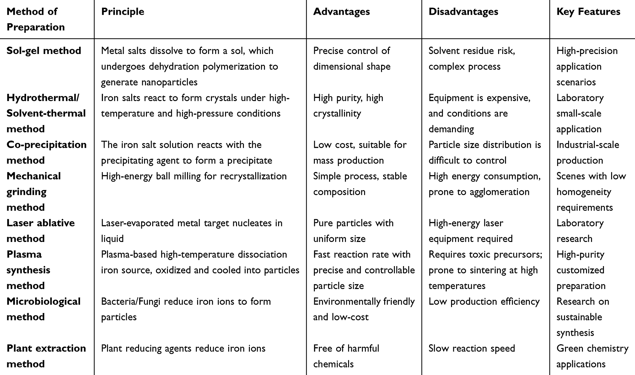 |
Table 1 Classification of Ferrosoferric Oxide Nanoparticles (Fe3O4NPs) Preparation Methods and Summary of Characteristics |
 |
Figure 1 Schematic Diagram of the Synthesis and Functionalization Mechanism of Ferrosoferric oxide nanoparticles (Fe3O4NPs). Figure provides a comprehensive representation of the three preparation pathways and surface modification strategies for Fe3O4NPs. Synthesis Routes: 1) Sol-gel method: Hydrolyze a 0.1–0.5 M mixed solution of ferric chloride/ferrous chloride at pH 3–5 to form a sol; heat-treat at 400°C for 2h to obtain Fe3O4NPs; 2) Coprecipitation method: Fe2⁺/Fe3⁺ (1:2) mixture precipitated at pH 11.5, 70–80°C with surfactant addition, particles separated by magnetic field; 3) Physical method (laser ablation): Irradiating an iron target with a 1064nm laser at 100–500mJ/pulse induces liquid-phase nucleation (particle size 5–30nm), but this method involves high equipment costs and yields <50mg/h; 4) Biological Method: Polyphenols found in lemon and seaweed extracts have been shown to reduce Fe3⁺ levels. The reaction occurs at room temperature and does not require the use of chemical solvents, thereby reducing energy consumption by 70%. The synthesis of Fe3O4NPs was conducted with a particle size of 15nm and a magnetic strength of 65 emu/g. Surface Modification: 1) PEG Modification: The particle half-life was extended to 24 hours, and the zeta potential was shifted from +25millivolts to −2millivolts; 2) Liposome Encapsulation (Phospholipid:Cholesterol = 7:3): It has been demonstrated that the target objective of achieving 85% drug loading capacity has been successfully met; 3) RGD Peptide Modification: The efficacy of targeting bone tumor cells was enhanced by a factor of 200%. |
Chemical Synthesis Methods
Chemical synthesis methods are currently among the most commonly used approaches for preparing Fe3O4NPs, primarily including sol-gel, hydrothermal, solvothermal, and coprecipitation techniques.18 These methods enable effective control over particle morphology and size and are suitable for large-scale production, but each method differs in operating conditions, equipment requirements, and product quality.24
The sol-gel method involves dissolving metal salts in a solvent to form a sol, followed by dehydration, polymerization, and thermal treatment to obtain Fe3O4NPs.25 The primary advantage of the sol-gel method lies in its ability to precisely control particle size and morphology, making it particularly suitable for applications demanding high precision in nanoparticle dimensions. However, the process involves multiple steps, is operationally complex, and carries the risk of solvent residue, which may pose challenges for biomedical applications. Additionally, sol-gel production incurs relatively high costs and is susceptible to factors such as solution concentration, temperature, and pH.26
Both hydrothermal and solvothermal methods utilize high-temperature, high-pressure environments to promote the reaction between iron salts and water or solvents, thereby generating Fe3O4NPs.27,28 In these reaction processes, water or solvents serve as reaction media and solvents. The advantages of these methods include the ability to synthesize Fe3O4NPs with high purity and crystallinity, along with precise control over particle size and morphology. However, their drawbacks lie in the requirement for expensive equipment and stringent operating conditions, which limit their application in industrial-scale production. Fe3O4NPs remains under active research for widespread utilization.29,30
The coprecipitation method is a simple, low-cost synthesis technique widely used in the preparation of Fe3O4NPs. Its fundamental principle involves mixing an iron salt solution with a precipitating agent, causing iron ions to react with the precipitating agent and form Fe3O4NPs.31 The advantages of this method include its simplicity, short reaction time, low cost, and suitability for large-scale production. However, precise control over particle size and distribution is challenging, and may be influenced by various factors such as the type of precipitant, reaction time, and temperature.32,33 Therefore, the co-precipitation method imposes stringent requirements on reaction conditions during production and necessitates further optimization to ensure product uniformity and stability.
Physical Synthesis Methods
Physical synthesis methods primarily rely on physical forces (such as mechanical grinding, laser ablation, and plasma synthesis) to prepare Fe3O4NPs. These approaches typically yield nanoparticles with high purity and allow for easy control over particle morphology and size distribution, but they often require specialized equipment and incur higher costs.
Mechanical grinding employs grinding media within high-energy ball mills to apply mechanical force to iron source mixtures. Through impact and shear forces, the raw material crystal lattice is fractured and reorganized, ultimately forming Fe3O4NPs. This method offers the advantages of a straightforward operational process, controllable particle size via adjustment of grinding time and rotational speed, and the potential for large-scale production. The resulting nanoparticles exhibit high compositional stability. However, mechanical grinding consumes substantial energy. Prolonged grinding often leads to particle agglomeration and a broad size distribution typically ranging from micrometers to submicrometers. It may also introduce impurities from the grinding media, making it more suitable for industrial applications where particle uniformity is less critical. While mechanically grinding holds practical value for low-cost magnetic nanomaterial production, the resulting products struggle to meet the stringent performance requirements of high-precision biomedical fields.
Laser ablation involves irradiating a metal target with a laser beam, utilizing its high temperature to vaporize the target material and form Fe3O4NPs in the liquid.34 The advantage of this method lies in its ability to produce highly pure, morphologically uniform Fe3O4NPs with a narrow size distribution. However, laser ablation requires stringent operating conditions, high-energy laser equipment, and a complex process, resulting in higher costs. Consequently, it is best suited for small-scale laboratory research.35,36 Research on laser ablation in the preparation of nanomaterials holds significant academic value, but its practical application is constrained by cost and scale limitations.
Plasma synthesis utilizes high temperatures (5000–10000K) generated by radiofrequency or direct-current plasma to dissociate iron sources into atomic states. These react within an oxygen-containing atmosphere and rapidly cool to form Fe3O4NPs. This method offers advantages including rapid reaction rates (millisecond scale), high product crystallinity, and precise particle size control (10–50nm) through adjustment of plasma power and gas flow rate, making it suitable for continuous production. However, plasma synthesis requires handling toxic metal-organic precursors, entails high equipment maintenance costs, and is prone to particle sintering in high-temperature environments. Consequently, it is primarily used for customized preparation of high-purity nanoparticles in laboratories. While plasma synthesis demonstrates unique advantages in fabricating high-performance magnetic materials, its industrial application remains constrained by safety management and cost considerations.
Biosynthetic Methods
With the rise of green chemistry concepts, biosynthetic approaches have gradually emerged as a significant pathway for preparing Fe3O4NPs. These methods are not only environmentally friendly and sustainable but also offer lower costs, fewer byproducts, and superior performance.37,38
Microbial methods utilize specific bacteria or fungi to convert iron sources into Fe3O4NPs.39 Certain bacteria and fungi possess the ability to reduce iron ions to Fe3O4. Therefore, Fe3O4NPs could be synthesized under controlled conditions by culturing these microorganisms.40,41 The advantage of this method lies in its eco-friendly, environmentally sustainable, and relatively low-cost nature, while also leveraging microorganisms that are widely present in nature for synthesis. However, microbial methods exhibit lower production efficiency and require strict cultivation conditions and time control, thus preventing their implementation in large-scale production.
Plant extraction methods utilize reducing agents in plant extracts to synthesize Fe3O4NPs. Natural reducing agents present in plants effectively reduce iron ions to form Fe3O4NPs.42,43 This method is characterized by its eco-friendly, low-cost, and sustainable nature, with no harmful chemicals generated during production, making it a highly promising green synthesis approach. However, the drawbacks of the plant extraction method include relatively slow reaction rates and variations in the properties of reducing agents from different plant sources. Consequently, its production efficiency and controllability still require further optimization and enhancement.
It is evident that the employment of the aforementioned diverse preparation methods has resulted in the synthesis of Fe3O4NPs, which has yielded remarkable outcomes across a range of application domains. In the context of ongoing technological advancements, these preparation methods are undergoing continuous refinement and optimisation, with the objective of facilitating more efficient and environmentally friendly production processes. The selection of these methods is contingent upon factors such as the requirements of the application, cost considerations, and the desired properties of the nanoparticles. In the future, the development of innovative synthesis processes that integrate the advantages of different preparation methods will facilitate the widespread application of Fe3O4NPs in biomedical fields, particularly in the treatment of bone metabolic diseases.
Characterization Techniques
Shape and Size (TEM, SEM)
In the context of biological medicine, the morphology and dimensions of Fe3O4NPs are pivotal factors that determine their efficacy, particularly in the domains of targeted drug delivery and tissue engineering. The size, uniformity, and dispersion of the particles directly impact their biological distribution, cell uptake, intracellular stability, and drug release properties. Consequently, it is imperative to employ appropriate analytical methods to accurately determine the morphology and dimensions of the particles.
The transmission electron microscope (TEM) utilises its superior resolution to provide a clear and detailed view of the internal crystal structure, size, and uniformity of Fe3O4NPs.44,45 The application of TEM analysis enables precise measurement of the distribution of particles and their diameter, ensuring that they reach the nanoscale level (typically 10–100nm) to satisfy the penetration requirements of bone tissue.46 In addition, TEM has been shown to reveal the interaction between particles and cells, as well as their distribution within the cell. In the context of cancer treatment research, TEM could be used to observe the interaction between Fe3O4NPs and cancer cells, thereby determining their location within the cell. This provides a foundation for the optimisation of drug delivery design.47
 |
Table 2 Comparison of Characterization Technical Parameters for Fe3O4NPs |
The scanning electron microscope (SEM) focuses on analyzing particle surface morphology and aggregation states. SEM images can be used to evaluate the dispersion of Fe3O4NPs and determine whether agglomeration occurs. Agglomerated particles impair drug delivery efficiency and the uniformity of magnetothermal effects (Table 2), necessitating improvement through surface modification techniques.48,49 The use of SEM allows for the assessment of the uniformity and agglomeration of the particles, thereby assisting researchers in determining whether agglomeration phenomena have occurred during the synthesis of the nanoscale particles.50 In addition, the combination of SEM and Energy-Dispersive X-ray Spectroscopy could be used to accurately determine the elemental composition of Fe3O4NPs. This ensures that the proportions and distribution of Fe and O are consistent with the design specifications, thereby preventing impurities from affecting their biological compatibility.51
Magnetic Properties (VSM)
The key feature of Fe3O4NPs is their super-structure magnetic anisotropy, which is essential for their application in magnetic target drug delivery, magnetic heating therapy and magnetic resonance imaging (MRI). The measurement of Fe3O4NPs’ magnetic saturation, coercivity and Hysteresis loop are the primary means by which this is achieved.52–54
Vibrating sample magnetometer (VSM) is a technique frequently employed for the purpose of evaluating the magnetic properties of nanoscale particles.55 The utilisation of VSM facilitates the measurement of Fe3O4NPs’ Magnetic field strength curve under the influence of an external magnetic field, thereby enabling the determination of critical parameters such as the magnetic saturation intensity (Ms), the coercivity (Hc), and the hysteresis loop.56,57 The maximum possible magnetisation of MsFe3O4NPs in a magnetic field is represented by Ms. The highest Ms values ensure that the particles generate a strong magnetic response when driven by an external magnetic field, thus achieving efficient targeted delivery. Fe3O4NPs typically possess high Ms, which enable them to generate a strong magnetic response when subjected to a magnetic field, thus facilitating targeted drug delivery. Hc represents the ability of the particles to retain magnetic properties after the removal of the magnetic field. Fe3O4NPs must possess low Hc values to ensure that they are devoid of magnetic properties after the removal of the magnetic field, thus preventing the accumulation of particles or damage to normal bone tissue. The hysteresis loop represents the energy dissipation during the magnetic transformation of the particles. By analysing the hysteresis loop, the efficiency of the energy conversion of Fe3O4NPs in a thermotherapy could be evaluated, and the stability of the generation of heat in a alternating magnetic field could be ensured, with energy dissipation that meets safety standards.
Surface Treatment and Functionality
The enhancement of Fe3O4NPs’ biological compatibility, targeted delivery, and drug carrier properties is contingent on surface modification and functionalisation, which directly impact their applicability and efficacy in the treatment of bone metabolic diseases.
Polyethylene glycol (PEG) modification is currently one of the most common methods for surface modification of nanoparticles. PEG possesses favourable biocompatibility and hydrophilic properties, and through PEG modification of Fe3O4NPs, it is possible to effectively avoid immune system recognition and clearance, thereby prolonging the duration of its presence in the blood.58 In addition, the modification of PEG has been demonstrated to enhance the water solubility of the particles, reduce their tendency to aggregate, and augment their biological availability.59 Nonetheless, it is important to note that the efficacy of PEG modification may be subject to alteration in accordance with the increase in the length of the PEG molecule. Consequently, it is essential to undertake a rigorous optimisation process when selecting the appropriate PEG molecular weight.60,61
Lipid-based coating is a method of coating Fe3O4NPs within the lipid body. The lipid body could simulate the structure of the cell membrane, thereby facilitating the fusion of the nanocrystals with the cell membrane and enhancing their cellular uptake capacity.62–64 Fe3O4NPs, when coated with lipids, could also be used to carry drugs, thus achieving dual effects in terms of both drug delivery and targeted therapy.63,65 This method has been demonstrated to possess both excellent biocompatibility and biodegradability, thus rendering it a highly promising surface modification strategy at present.
By attaching specific peptide molecules to the surface of Fe3O4NPs, efficient recognition and binding to target cells can be achieved. Research indicates that modifying nanoparticle surfaces with specific receptor-targeting peptide segments enables more precise targeting of cancer cells or inflammatory sites, thereby enhancing the accuracy and efficiency of drug delivery.66,67 Peptide modification not only improves nanoparticle targeting capabilities but also strengthens their interaction with cells, increasing selectivity and affinity for target cells.68
In summary, surface modification has significantly enhanced the biocompatibility, stability, targeting capability, and drug delivery performance of Fe3O4NPs, laying the foundation for their application in the treatment of bone metabolic diseases.
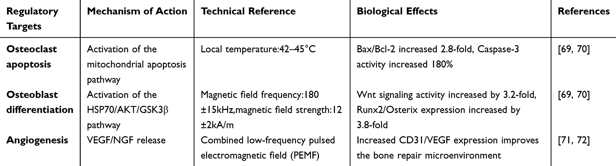 |
Table 3 Magnetic-Thermal Therapy Modulates Bone Metabolic Parameters |
Applications of Fe3O4NPs in Bone Metabolic Diseases
Treatment for Weak Bones
The core pathological mechanism of osteoporosis is an imbalance between osteoclast-mediated bone resorption and osteoblast-mediated bone formation, leading to bone mass loss and disruption of bone microarchitecture (Table 3). Among these, the RANKL-mediated signaling pathway plays a crucial role in regulating osteoclast differentiation and activity.73,74 Fe3O4NPs leveraging dual functions of magnetic-targeted delivery and magnetothermal effects, can precisely regulate bone metabolic balance, offering a novel therapeutic strategy for osteoporosis (Figure 2).75,76
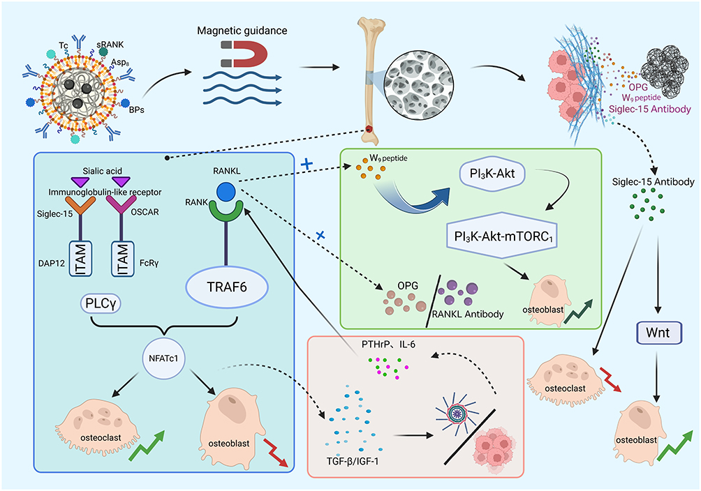 |
Figure 2 Mechanism Diagram of Fe3O4NPs-Based Targeted Therapy for Osteoporosis. As illustrated in Figure, the targeted therapy for osteoporosis utilizes three core mechanisms of Fe3O4NPs. Targeted Delivery: Fe3O4NPs modified with sRANK (RANK antagonist), Tc (tetracycline), and ASP8 (acidic oligopeptide) bind bisphosphonates (BPs) to accumulate in active bone remodeling zones, targeting osteoporotic sites under magnetic field guidance. The phenomenon of signal blockade has been observed. The release of OPG (decoy receptor) and RANKL antibodies results in a competitive binding of RANKL, while Siglec-15 antibodies serve to inhibit co-stimulatory signals. This results in the obstruction of the RANK-RANKL-TRAF6-NFATc1 pathway, thereby suppressing osteoclast differentiation. Bidirectional Regulation: The W9 peptide has been shown to activate the PI3K-Akt pathway in osteoblasts, thereby promoting Wnt signaling expression. Concurrently, it has been observed to suppress abnormal secretion of PTHrP and IL-6, which are caused by disrupted osteoclast-osteoblast coupling. This effect, ultimately, serves to restore bone metabolic balance. |
RANK-Targeted Delivery System
Receptor Activator of Nuclear Factor Kappa-B Ligand (RANKL) serves as a core regulatory factor for osteoclast differentiation with irreplaceable significance. It initiates signaling pathways by specifically binding to the RANK receptor on osteoclast precursor cells, synergistically activated by M-CSF.77 Upon binding, it recruits TRAF6 and TAK1 kinase to activate NF-κB and MAPK signaling pathways (including ERK1/2 phosphorylation cascades). Simultaneously, co-stimulatory receptors such as Siglec-15 synergistically activate calcium oscillations through ITAM motifs, ultimately inducing nuclear translocation and self-amplifying expression of the key transcription factor NFATc1. This process drives osteoclast differentiation, fusion, and maturation. NFATc1, acting as the master switch for osteoclast differentiation, directly upregulates the expression of maturation markers such as TRAP (tartrate-resistant acid phosphatase) and CTSK (cathepsin K), thereby conferring osteoclasts with bone resorption capabilities. Previous studies have demonstrated that blocking the RANKL-RANK interaction through anti-RANKL antibodies (such as denosumab) significantly reduces osteoclast differentiation rates, decreases the number of TRAP-positive cells, lowers CTSK activity, and shrinks the area of bone resorption pits.78–80 This intervention effectively slows bone resorption, increases bone density, and reduces fracture risk in osteoporosis patients, providing a clear theoretical basis for targeted osteoporosis treatment.78,80,81
Nonetheless, conventional pharmaceutical administration is frequently encumbered by issues pertaining to drug metabolism and tissue permeation, which often give rise to suboptimal bioavailability, erratic pharmacodynamics and deleterious adverse effects.82,83 To this end, bisphosphonates (such as alendronate sodium) were employed to couple Fe3O4NPs via an amidation reaction, constructing bone-targeting nanocarriers (BTNPs) that significantly enhance bone tissue enrichment efficiency.84 This design relies on the high affinity of bisphosphonates for bone matrix and external magnetic field guidance.20 The TEM revealed BTNPs exhibit uniform spherical morphology (particle size 34.9±0.5nm). Fe3O4NPs scavenge excess reactive oxygen species (ROS) to inhibit osteoclast differentiation.23 Combined with near-infrared (NIR) photothermal stimulation (40–42°C), they upregulate heat shock proteins (HSP70/HSP47) to promote osteoblast differentiation.71 In animal studies, BTNPs significantly improved bone microarchitecture in osteoporotic rats: Micro-CT revealed increased bone trabecular volume (BV/TV) and bone mineral density (BMD).72 Simultaneously, neuromagnetic stimulation increases oxytocin and estrogen secretion by regulating the paraventricular nucleus of the hypothalamus, thereby suppressing the RANKL/OPG ratio to reduce bone resorption.71,72 VEGF/NGF controlled-release scaffolds further promote angiogenesis and nerve axon regeneration, achieving coordinated reconstruction of bone, nerves, and blood vessels.23,72
Magnetothermotherapy Regulation of Bone Balance
Magnetothermotherapy utilizes Fe3O4NPs to generate localized heating effects under an external alternating magnetic field, enabling precise regulation of local bone tissue temperature. This provides a novel intervention approach for restoring bone metabolic balance. Osteoporosis is typically characterized by excessive osteoclast activity and diminished osteoblast function, leading to bone resorption exceeding bone formation and subsequent bone loss. By modulating local temperature, the functions of both osteoclasts and osteoblasts can be regulated at the molecular and cellular levels.69,74
In vitro, At the cellular level, moderate thermal stimulation with localized temperatures maintained between 42–45°C significantly induces osteoclast apoptosis by activating the mitochondrial apoptosis pathway. This is manifested by a 2.8-fold increase in the Bax/Bcl-2 ratio and an approximately 180% rise in Caspase-3 activity.69,70 Concurrently, the application of heat stimuli in osteoblastic cells has been demonstrated to activate the HSP70/AKT/GSK3β signalling pathway, thereby promoting β-catenin nuclear translocation and increasing Wnt signalling activity by approximately 3.2-fold. This, in turn, enhances the proliferative and differentiative capacities of osteoblastic cells.69,70 Technical specifications: By setting the magnetic field frequency to 180±15kHz and the magnetic field strength to 12±2kA/m, the Fe3O4NPs achieve a specific absorption rate of 250W/g. Concurrently, MR thermometry technology is employed for real-time temperature monitoring (accuracy ±0.3°C), ensuring precise control of the heating effect.69 In addition, the combination of magnetic therapy and low-frequency pulsed electromagnetic field has been shown to enhance the expression of Runx2 and Osterix, two critical genes involved in bone formation. This enhancement, observed in both genes, has been found to be approximately 3.8-fold and 3.2-fold, respectively. These findings suggest a potential for further promotion of bone formation.70
This magnetothermal regulation strategy leverages the magnetothermal conversion capability of Fe3O4NPs and the guiding advantage of external magnetic fields to achieve localized precision heating. It simultaneously suppresses osteoclast activity and reduces bone resorption while activating osteoblast function and promoting bone formation. This effectively restores the balance between osteoclasts and osteoblasts, offering a safe and efficient new therapeutic approach for improving osteoporosis.
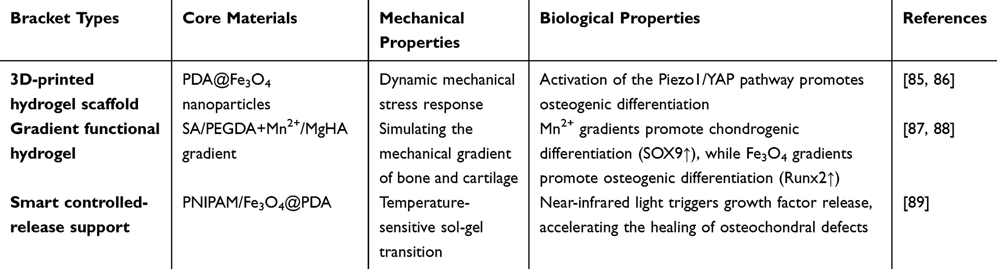 |
Table 4 Magnetic Bracket Performance Parameters |
Bone Defect Repair
Magnetic Supports and “Mechanical-Biological Signal” Regulation
The primary objective of bone defect repair is to reconstruct the bone tissue structure and restore its function. Fe3O4NPs could provide a multifaceted solution for bone defect repair by constructing magnetic supports and regulating growth factors. Their magnetic properties, in conjunction with their compatibility with biological systems, significantly enhance bone regeneration (Table 4). As demonstrated in studies,90,91 have been shown to be capable of inducing cell migration in response to an external magnetic field, thereby significantly promoting the processes of migration, proliferation and differentiation of bone cells. Concurrently, the use of magnetic substrates enables the precise spatial localisation of nanoparticles by the external magnetic field, facilitating directed tissue growth and regeneration. This process effectively controls the cell’s trajectory and direction of growth, thereby enhancing the efficacy of bone tissue regeneration (Figure 3).70,92
 |
Figure 3 Schematic Diagram of the Synergistic Regulation Mechanism for Bone Defect Repair Based on Fe3O4NPs. Figure illustrates the multidimensional regulatory mechanism of Fe3O4NPs in bone defect repair. Magnetic Scaffold Guidance: It has been demonstrated that 3D hydrogel scaffolds embedded with PDA@Fe3O4NPs undergo magnetic field-induced particle alignment, thereby generating 1–5 pN dynamic shear forces. This activation of Piezo1 channels in cell membranes has been shown to promote Ca2+ influx and nuclear translocation of Yes-associated protein (YAP)/β-catenin, resulting in a 3.2-fold increase in osteogenic gene Runx2/Osterix expression. Gradient Regulation: The application of a Mn2+ gradient (0–5 mM) in the cartilage layer has been demonstrated to activate HIF-1α, resulting in a fourfold increase in SOX9 expression. In addition, the presence of Fe3O4NPs/MgHA in the transition layer has been shown to promote osteogenic differentiation, as evidenced by a significant increase in ALP activity (180%). Finally, the bone layer: BMP-2/BMP-7 sustained release (200→50ng/mL) has been demonstrated to increase Runx2/OCN expression by 4.2-fold. Neuro-regulation: Low-frequency pulsed magnetic field stimulation has been demonstrated to induce paraventricular nucleus oxytocin release, thereby suppressing the RANKL/OPG ratio (down 30%) and reducing bone resorption. |
Building upon this foundation, a 3D-printed hydrogel composite magnetic scaffold integrating “mechanical signals” with “biological signals (mineralization)” further enhances repair efficacy. This scaffold achieves “magneto-mechanical” conversion by embedding PDA@Fe3O4NPs within the hydrogel: when exposed to an external magnetic field, the PDA@Fe3O4NPs undergo reorientation, rotation, or vibration, converting magnetic energy into mechanical force acting on cells within the scaffold. These dynamic mechanical signals activate intracellular Piezo1 channels, triggering calcium influx that subsequently activates the YAP and β-catenin signaling pathways, thereby enhancing osteogenic differentiation of stem cells. This mechanism aligns with Fe3O4NPs’ role in modulating cellular molecular signaling pathways through magnetic effects, simultaneously maintaining the magnetic scaffold’s guidance for cell alignment and reinforcing osteogenesis-related signaling activation via dynamic mechanical stimulation.85,86
The research team developed a platform of mesenchymal stem cells (MSCs) loaded with antioxidant melanin@Fe3O4NPs (MFNPs), termed Magcells. This platform integrates intercellular mechanical communication with intracellular signaling regulation through a time-varying magnetic field (TMF): Magcells formed by MFNP internalization exhibit precise magnetotaxis (rectangular trajectory rolling). Their antioxidant properties significantly enhance MSC survival in inflammatory environments and upregulate anti-inflammatory genes such as IL-14 and IL-10. Low-frequency TMF stimulation dynamically mimicking gait cycles activates adhesion gene expression, accelerates cytoskeletal reorganization, and modulates key signaling pathway modules. This ultimately significantly upregulates chondrogenic-specific gene and protein expression, synergizing with TGF-β to achieve efficient chondrogenic differentiation.93
Simultaneously, the mechanical performance of the magnetic support material is significantly enhanced by the close binding of Fe3O4NPs with the substrate material, thereby providing a stable microenvironment for the repair process and enhancing the structural stability.91 In addition, the autonomous mineralisation of the polymeric substances in the composite could be modelled by the accumulation of inorganic matrix. This not only provides a rich source of calcium, but also enables the controlled promotion of mineral transformation through biological signals. In conjunction with the physical signals, this establishes a “directed arrangement-physical stimulation-biological mineralisation” regulatory network.
Fe3O4NPs possess three key advantages in their use as a magnetic scaffold. Firstly, the cells are directed to arrange in a specific pattern by the magnetic field, thereby establishing the structural foundation for new bone formation. Secondly, the magnetic field is used to transmit dynamic mechanical signals, thus activating the relevant signalling pathways involved in bone formation. Thirdly, in combination with biological signals, the magnetic field provides a functional microenvironment for the cells. The synergy of these three factors significantly enhances the efficiency of bone repair, thereby promoting the application of magnetic scaffolding in bone tissue engineering.
Synergistic Release of Growth Factors and Gradient Functional Hydrogels
In the field of bone regeneration and repair, bone morphogenetic proteins (BMPs), particularly BMP-2 and BMP-7, have been identified as playing a pivotal role. These proteins have been shown to promote the differentiation of progenitor cells in the bone tissue, thereby facilitating the formation of new bone matrix and accelerating the healing of bone defects. However, the clinical application of BMPs is constrained by issues such as rapid metabolic clearance and the inability to maintain effective concentrations for extended periods. Consequently, the development of delivery systems that could effectively control the release of BMPs has emerged as a critical strategy to enhance their therapeutic efficacy. Fe3O4NPs serve as an ideal carrier, leveraging their exceptional surface functionalization capabilities to achieve stable binding with BMP-2/BMP-7 through modification, enabling efficient loading. Simultaneously, leveraging their magnetic responsiveness, these particles enable targeted delivery and controlled release rates of growth factors under external magnetic field regulation. This ensures sustained effective concentrations at bone defect sites, preventing adverse effects from rapid clearance or excessive release. Furthermore, their biodegradability allows gradual degradation as bone healing progresses, eliminating any detrimental impacts.87
Building upon this foundation, the gradient-functionalized hydrogel system incorporating Fe3O4NPs further expands the synergistic regulatory potential for growth factor sustained release and osteochondral repair. Magnetic field-induced gradient distribution of Fe3O4NPs within SA/PEGDA hydrogels enables the construction of scaffolds with continuous mechanical gradients, precisely mimicking the natural mechanical gradient of osteochondral tissue. This provides a matched mechanical and magnetic gradient microenvironment for full-thickness osteochondral regeneration. Notably, following secondary crosslinking with Mn2⁺ after SA, the hydrogel exhibits a reverse gradient distribution of Mn2⁺, Mg-doped hydroxyapatite (MgHA), and Fe3O4NPs: the Mn2⁺ gradient significantly promotes chondrogenic differentiation of bone marrow mesenchymal stem cells (BMSCs), manifested by markedly elevated expression levels of chondrogenic marker genes SOX9, Col-II, and ACAN (particularly pronounced in the PSMn group). Conversely, the Fe3O4NPs and MgHA gradients synergistically enhanced osteogenic differentiation, leading to high expression of osteogenic marker genes ALP, Col-I, and Runx2 in the FPSMn and FHPSMn groups (with the highest expression in FHPSMn). Simultaneously, Fe3O4NPs reversed Mn2⁺-induced inhibition of angiogenesis in human umbilical vein endothelial cells (HUVECs), promoting CD31, VEGF, and KDR expression to provide vascularization support for bone repair.88 This gradient composition complements the sustained release of growth factors mediated by Fe3O4NPs, jointly optimizing the microenvironment for stem cell differentiation and tissue regeneration.
Furthermore, the dual-crosslinked smart hydrogel scaffold (SPA5-Mg/GH/FP) constructed from PNIPAM and Fe3O4@PDA nanoparticles enables precise controlled release of growth factors. This water-soluble gel is temperature sensitive, and at room temperature it is liquid and injectable, making it suitable for the minimally invasive treatment of bone and soft tissue defects. Under NIR irradiation, controlled-release and accelerated release of growth hormone can be achieved on demand. It also has excellent biological safety and anti-inflammatory and antibacterial properties, and in a model of new Zealand rabbit joint soft tissue defect, it has been shown to significantly accelerate healing.89
Fe3O4NPs not only serve as carriers for targeted sustained release of BMP-2/BMP-7, but also establish a mechanically-compositionally synergistic regulatory network through gradient distribution. Combined with the minimally invasive and responsive drug delivery properties of smart hydrogels, they comprehensively optimize the repair efficiency of osteochondral defects, offering an innovative strategy for complex bone tissue regeneration.
Treatment of Bone Tumors
Chemotherapy-Hyperthermia Combination Therapy
Bone tumors, particularly osteosarcoma, are characterized by high invasiveness and a tendency to metastasize. Traditional chemotherapy and radiotherapy suffer from issues such as poor targeting, significant side effects, and susceptibility to drug resistance. Fe3O4NPs, with their multifunctional capabilities including magnetic-targeted delivery, magnetothermal therapy, and radiosensitization, offer a multimodal synergistic strategy for bone tumor treatment. This approach significantly enhances therapeutic efficacy while reducing adverse effects.94 Magnetothermal therapy utilizes alternating magnetic fields applied to Fe3O4NPs to precisely elevate tumor region temperatures to 42–45°C, directly inducing thermal death in tumor cells. When Fe3O4NPs are conjugated with chemotherapeutic agents such as paclitaxel or cisplatin, they not only enhance drug penetration into tumor tissues under magnetic field control to elevate local effective drug concentrations, but the thermal effect also increases tumor cells’ sensitivity to chemotherapy drugs. This approach enhances therapeutic efficacy while reducing systemic side effects and partially overcomes tumor drug resistance (Figure 4).95–97
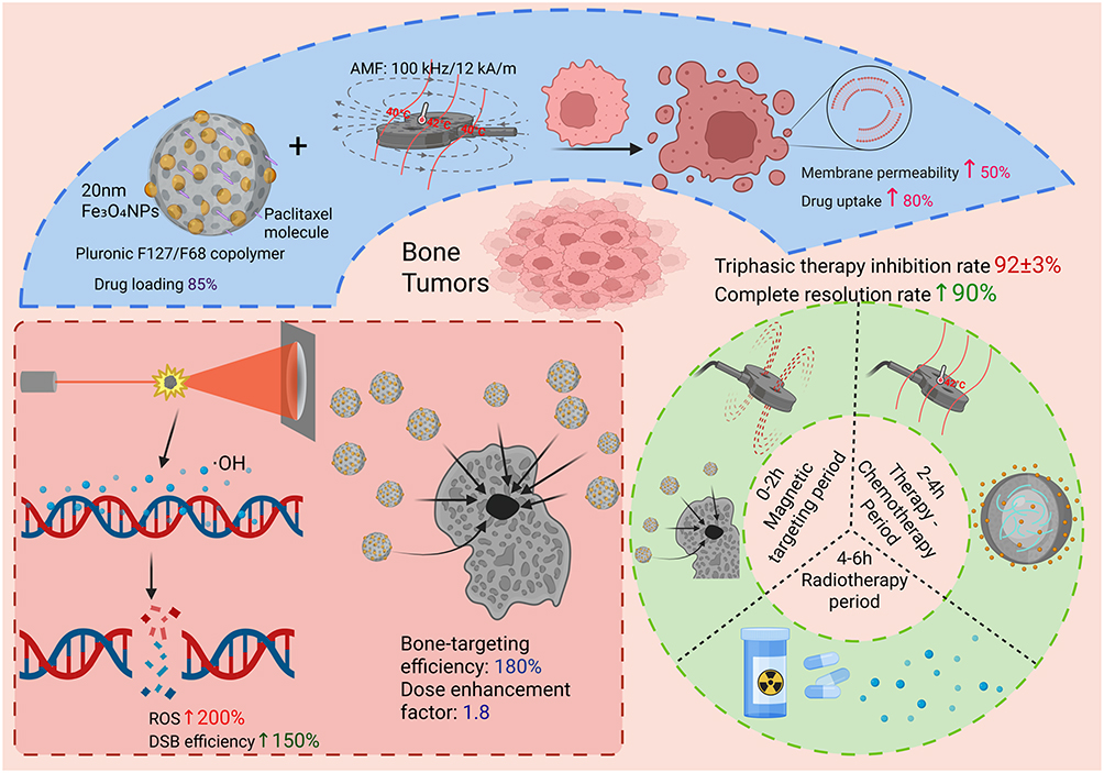 |
Figure 4 Graphical representation of the mechanism of treatment of bone tumors using Fe3O4NPs. As illustrated in Figure, the Fe3O4NPs-mediated synergistic chemotherapy-hyperthermia-radiotherapy system for bone tumors demonstrates the potential for comprehensive treatment modalities. The present study explores the synergistic effect of chemotherapy and hyperthermia. Fe3O4NPs (core) encapsulated with Pluronic F127/F68 carry paclitaxel (drug loading rate 85%), and an alternating magnetic field induces particle heating (localized 42°C), increasing tumor cell membrane permeability by 50%, drug uptake by 80%, and promoting apoptosis. Radiotherapy sensitization: The application of X-ray irradiation to Fe3O4NP clusters has been demonstrated to elicit a substantial increase in ·OH radicals, with a concomitant rise in ROS levels of up to 200%. This treatment has been observed to enhance DNA double-strand break repair efficiency by up to 150%, while concurrently increasing particle enrichment efficiency in osteolytic lesions by up to 180%. Notably, this enhancement in radiotherapy efficacy is accompanied by a dose enhancement factor of 1.8, signifying the potential for significant improvements in treatment outcomes. Time-controlled delivery: 0–2hours. The process of magnetic field-guided particle targeting, which has been demonstrated to yield peak accumulation levels of 2.5 milligrams per gram, is initiated within a time frame of 2–4 hours. The drug release was synchronized with the occurrence of hyperthermia, with 85% of the dosage being released within a 4-hour time frame. The remaining 15% was released over the following 4hours to 6hours. The combination of radiotherapy with radical effects (t1/2=4h) has been demonstrated to achieve multimodal precision therapy. |
On this foundation, a new system comprising Cu-Fe3O4NCs-AS-ALG has been developed, which has further expanded the chemotherapy-hyperthermia synergistic mechanism, providing a more effective approach for treating bone and soft tissue tumours. This system employs three mechanisms to achieve the synergistic destruction of cancer cells. Firstly, Cu-Fe3O4 nanocrystals exhibit peroxidase, catalyzing the generation of abundant ROS under high H2O2 and acidic conditions in the tumor microenvironment. This directly kills tumor cells and improves hypoxia, synergizing with the magnetothermal effect of Fe3O4NPs to intensify oxidative damage; Secondly, iron and copper ions released by NCs in acidic environments react with the sesquiterpene structure of artemisinin, generating specific carbon radicals (·C). This amplifies intracellular oxidative stress independently of tumor microenvironment constraints, enabling “targeted activation” of the chemotherapeutic agent and enhancing AS’s therapeutic efficacy. Thirdly, iron ions released from NCs induce ferroptosis via GPX4 pathway by increasing intracellular iron overload and depleting glutathione, while copper ions activate copperptosis by disrupting DLAT-mediated mitochondrial respiratory chain function. These dual death pathways synergize with thermal injury from magnetothermal therapy, significantly enhancing tumor cell killing efficiency and overcoming limitations of monotherapy approaches.98
The Cu-Fe3O4 NCs-AS-ALG hydrogel system retains the magnetothermal and drug-carrying advantages of Fe3O4NPs while achieving multidimensional synergy through multi-enzyme activity, free radical generation, and multi-pathway cell death induction. This approach integrates chemotherapy, thermotherapy, and metal ion therapy, offering a novel direction for precise and efficient treatment of bone tumors.
Radiation-Enhanced Osteolytic Lesions
Osteolytic lesions refer to the process where tumors grow within bone tissue and destroy bone structure. These lesions typically exhibit high tolerance to radiation therapy, limiting the effectiveness of conventional radiotherapy in treating them. Beyond serving as carriers for magnetothermotherapy, Fe3O4NPs can also function as radiosensitizers, enhancing the therapeutic efficacy of radiation therapy against bone tumors, particularly osteolytic lesions. The magnetic properties of Fe3O4NPs make them particularly well-suited for use in radiotherapy.
Fe3O4NPs could be magnetically targeted to tumor sites, particularly osteolytic lesions. Through the action of an external magnetic field, Fe3O4NPs could be made to accumulate a radioactive agent at the site of a neoplasm with great precision, thereby increasing the absorption of the radiation by the neoplasm and enhancing its sensitivity to the effects of the radiation. In addition to acting as a target for direction by a magnetic field, Fe3O4NPs could also cause local heating in a neoplasm through its magnetic properties, thereby increasing the sensitivity of the neoplastic cells to radiation. The mechanism of action of a radiosensitising agent could be explained by two factors. Firstly, Fe3O4NPs could interact with neoplastic cells, altering their internal environment and increasing their ability to absorb and respond to radiation. Secondly, the magnetic properties of Fe3O4 could enhance the permeability of the cell membrane, thereby increasing the ability of the radiation to penetrate the neoplastic cells. The research demonstrated that Fe3O4NPs, when used as radiosensitizers, could significantly enhance the therapeutic efficacy of radiotherapy on osteolytic lesions while reducing damage to surrounding normal tissues.99
In summary, Fe3O4NPs demonstrate high targeting specificity and precision in radiotherapy. By adjusting the intensity and direction of the external magnetic field, precise localization of tumor sites can be achieved, thereby maximizing the accuracy and efficacy of radiotherapy, minimizing side effects, and improving patient prognosis. This strategy offers a novel solution for treating bone tumors, particularly osteolytic lesions, and holds broad clinical application prospects (Table 5).
 |
Table 5 Applications of Fe3O4NPs in Bone Metabolic Diseases |
Diagnosis and Treatment Integration
Dual-Modality MRI/CT Imaging
In the early diagnosis of bone metabolic diseases, imaging techniques play a crucial role. While MRI and CT imaging each possess distinct advantages, their inherent limitations impede their effectiveness in the diagnosis of bone diseases. However, Fe3O4NPs, due to their unique characteristics of high saturation magnetism and high density, have emerged as a promising material for enhancing the contrast of MRI and CT imaging, thereby significantly improving the diagnostic accuracy of bone diseases.
In the process of MRI, the signal amplification effect is enhanced by the superparamagnetic properties of Fe3O4NPs. This is due to the fact that Fe3O4NPs exhibits a distinct magnetic response to an external magnetic field, thereby increasing the contrast and resolution of the image.100,101 The employment of Fe3O4NPs as a contrast agent in MRI imaging has been shown to enhance the visibility of soft tissue, thereby facilitating precise location of the area of interest by medical professionals. This approach is of particular significance in the diagnosis of conditions such as osteoporosis and bone defect.102 In addition, Fe3O4NPs has been shown to have a significant impact on CT scans. The process of CT imaging involves the transmission of X-rays through the human body, and the high density of Fe3O4NPs has been demonstrated to enhance the contrast of the X-rays, thereby improving the quality of the image.103 The high density of Fe3O4NPs not only provides stronger contrast in CT images, but also assists medical professionals in accurately identifying the intricate structural details of bone tissue in CT images.
The integration of Fe3O4NPs with MRI and CT imaging technologies enables the implementation of a dual-modality imaging method, thereby facilitating precise diagnosis of bone metabolism-related diseases.104 This dual-modality imaging modality is capable of simultaneously providing detailed information regarding bone tissue degradation, osteoporosis and bone defect. It assists medical professionals in accurately determining the location of the pathology, thereby providing essential reference information for the subsequent formulation of treatment plans.105 In addition, the dual-mode thermal imaging technology has the capacity to provide real-time monitoring of the target area during the treatment process. This capability enables more precise and personalised treatment, thereby enhancing the efficacy of the treatment.106
The combination of a dual-source CT and an MRI system offers distinct advantages in medical imaging. The CT component provides precise information about the structure of the body’s bones and joints, while the MRI component delivers high-resolution images of soft tissue. The integration of these two modalities enables the implementation of a “one-stop” diagnostic service, facilitating early diagnosis of bone metabolism-related conditions and providing reliable data for evaluating and monitoring subsequent treatment outcomes in real time.100
Bone Metabolism Marker Testing
The detection of bone metabolism markers is crucial for the early diagnosis of bone metabolic diseases and the evaluation of treatment efficacy. Bone resorption markers and bone formation markers serve as important biomarkers in bone metabolism processes. Changes in their concentrations reflect the metabolic activity levels of bone tissue, thus holding significant clinical importance. However, traditional marker detection methods often suffer from issues such as invasiveness, time-consuming procedures, and low sensitivity, making it difficult to meet the demands for real-time, non-invasive, and highly sensitive detection.
Fe3O4NPs possess both excellent magnetic properties and functionalisation capabilities, thus becoming a vital tool for the sensitive detection of biomarkers associated with bone metabolism.107 The surface functionalization of Fe3O4NPs with antibodies or molecular recognition elements enables the specific identification and binding of bone metabolism markers, such as CTX-1 and P1NP. The interaction of Fe3O4NPs with an external magnetic field results in alterations to their magnetic response, which could be utilised to monitor the concentration of the target substance through variations in the magnetic field. Specifically, the magnetic properties of Fe3O4NPs change during the binding process with the target substance, resulting in a corresponding alteration in the nanoscale particles’ magnetic properties. These changes could be measured using a magnetic field detection device, which converts them into electrical signals, enabling the quantitative detection of target substances in the context of bone metabolism. In comparison to conventional detection methods, Fe3O4NPs offers superior sensitivity, selectivity, and real-time monitoring capabilities, facilitating rapid and non-invasive diagnosis and treatment monitoring.
Concomitantly, by integrating intelligent sensing technology, the variation in the magnetic properties of Fe3O4NPs could be converted into an electrical signal, thereby facilitating more precise quantitative analysis. This method of detection, based on magnetic resonance technology, not only enhances the accuracy of the measurement but also offers a more efficient and convenient diagnostic approach in clinical practice. The non-invasive, rapid and accurate detection facilitated by Fe3O4NPs provides a novel solution for the early diagnosis and monitoring of bone metabolism-related diseases.
In summary, the integration of imaging technology and magnetic resonance technology has led to significant advancements in the comprehensive diagnosis of bone metabolic diseases. This multifaceted “integrated diagnosis and treatment” model has not only enhanced the early diagnosis rate of the disease, but also enabled real-time monitoring of the treatment process, providing substantial support for clinical treatment.
Biological Safety
Bone Tissue-Specific Toxicity
Fe3O4NPs has a wide range of applications in bone tissue, particularly in the treatment of bone diseases. They could be used to achieve precise treatment of bone tissue through methods such as magnetic targeting and delivery. However, the toxicity mechanism of Fe3O4NPs remains a key issue in their clinical application. After entering the body, Fe3O4NPs is usually absorbed by cells such as Macrophages and osteoblasts in the bone tissue. Research has shown that the accumulation of Fe3O4NPs in bone tissue is relatively slow, suggesting that they have some biological compatibility. However, high concentrations of Fe3O4NPs could potentially induce toxicity, particularly through oxidative stress reactions.
Simultaneously, oxidative stress represents one of the primary mechanisms underlying the toxic effects of Fe3O4NPs. These nanoparticles react with oxygen molecules within cells to generate free radicals, disrupting intracellular redox balance and subsequently causing cellular damage. This is particularly significant when cells are exposed to Fe3O4NPs over a prolonged period, as this could cause membrane damage, protein modification, and DNA damage, which could ultimately result in cell apoptosis or necrosis by necrosis. In addition, Fe3O4NPs may promote inflammation, further exacerbating tissue damage. Researchers have developed a method to reduce the accumulation and the reaction of Fe3O4NPs within cells by modifying their size and surface chemistry, thereby enhancing their biocompatibility.
In physiological conditions, bone tissue possesses a relatively strong capacity for self-repair. However, the accumulation of Fe3O4NPs has been shown to potentially suppress this reparative process, thereby affecting the normal metabolic activity of bone tissue. Therefore, by precisely controlling the dosage and distribution of Fe3O4NPs, their potential toxicity to bone tissue can be effectively reduced, thereby enhancing their specific therapeutic efficacy.
Long-Term Retention and Metabolism
The pharmacokinetics of Fe3O4NPs, including their potential for long-term retention and associated risks, represent a crucial aspect in evaluating their biological safety. Following cellular uptake, Fe3O4NPs are primarily cleared via the mononuclear phagocyte system. The MPS primarily encompasses organs such as the liver, spleen, and lymph nodes, which eliminate foreign particles through phagocytosis by macrophages. Fe3O4NPs are typically metabolized in the liver and spleen before being ultimately excreted via urine or feces. However, prolonged retention within the body may induce chronic toxic effects, including liver damage and immune system dysfunction.
In addition, the reduction-oxidation reaction of Fe3O4NPs may result in their transformation into Fe2O3, which in turn may affect their metabolic pathways. The generation of Fe2O3 may lead to an increase in the aggregation of Fe3O4NPs, thereby altering their distribution and metabolic pathways within the body. Therefore, the rate of degradation and surface properties of Fe3O4NPs must be controlled to ensure their biocompatibility and reduce their potential for harmful reactions. Research has shown that appropriate surface modification methods could promote the degradation of Fe3O4NPs and reduce their accumulation in the body over time. By modifying the biological degradation and clearance processes of Fe3O4NPs, the risk of their prolonged presence could be effectively reduced.
Surface-Modified Immunodepletion
The immune clearance of Fe3O4NPs is also a critical issue in their biosafety. After entering various tissues via the bloodstream, nanoparticles are readily recognized and cleared by the immune system. Specifically, immune cells such as macrophages and dendritic cells can identify and eliminate exogenous particles through receptor-mediated phagocytosis.
In order to reduce the duration of Fe3O4NPs within the body, researchers have developed a variety of surface modification strategies. The modification of Fe3O4NPs’ surfaces with hydrophilic polymers (eg. PEG, glucose) has been shown to reduce their retention time in the body and enhance their biological degradation. The hydrophilic properties of PEG enable the formation of a stable hydration layer on the surface of Fe3O4NPs, thereby preventing direct contact between Fe3O4NPs and immune cells or stromal cells.108 Simultaneously, the PEG chain enhances the hydrophilicity of the particles, effectively reducing aggregation.58 However, the length of the PEG molecular chain requires optimization.58,59 While high molecular weight PEGs (eg. PEG-6000) enhance stability,58 excessive modification may mask the targeting site, particularly within pathological microenvironments.59
Strategies such as lipid encapsulation and glycan modification can also effectively reduce the immune clearance of Fe3O4NPs. Encapsulating Fe3O4NPs within liposomes enables the nanoparticles to better integrate into the lipid environment within the body, thereby reducing their recognition and clearance by immune cells.65 Glycan modification further reduces immune responses and enhances targeting by attaching specific sugar molecules to the surface of Fe3O4NPs, leveraging the binding of these sugar molecules to receptors within the body.66
In summary, the surface modification technique enables Fe3O4NPs to circulate for a longer duration within the body, thereby reducing immune clearance and enhancing their efficacy in pharmaceutical delivery systems. Concurrently, surface modification could enhance the biocompatibility of Fe3O4NPs, thereby reducing their potential immune-toxicity reactions.
Challenges and Outlook
Fe3O4NPs still face multiple challenges in practical applications for treating bone metabolic diseases, requiring targeted breakthroughs to advance their clinical translation. Furthermore, Fe3O4NPs combined with polymer-based scaffolds and other drug delivery systems warrant further investigation. In terms of enhancing the efficiency of deep-tissue magnetic targeting, the current limitations of magnetic field penetration and the influence of Fe3O4NPs distribution, particle size, and surface properties on targeting outcomes require subsequent resolution. This could be achieved through the regulation of particle size, surface charge, and magnetic properties to enhance deep-tissue penetration, as well as the combination of ultrasound and photothermal treatment methods to improve targeting efficiency by exploiting the synergy of physical and chemical signals. Furthermore, the development of novel magnetic field enhancement technologies and multifunctional particle solutions is necessary.
In terms of the alignment of Fe3O4NPs with the mineralogical and degradation processes in bone tissue, the degradation rate must be balanced with the regeneration rate of bone tissue in real time. Accelerating or decelerating the degradation rate too quickly or slowly will have a negative impact on bone healing. Therefore, future research should focus on the study of mineralogical processes in bone tissue, with the aim of optimising the degradation rate through the modification of particle composition and structure. This will also allow for the enhancement of the compatibility of the degradation products with biological systems and the prevention of the formation of harmful substances. In establishing a framework for evaluating the efficacy of Fe3O4NPs in clinical applications, it is essential to consider the complexity of the structure of Fe3O4NPs and its functionality. This complexity must be evaluated comprehensively in terms of its distribution, metabolism, target efficacy, and biological safety. The evaluation should be conducted in conjunction with high-throughput screening to accelerate the process of clinical research and the selection of high-quality particles.
In the context of clinical transformation and personalised treatment, the physiological and pathological characteristics, and the pharmacokinetics of the patients must be taken into account, as these factors could influence the efficacy of Fe3O4NPs. Therefore, while focusing on the development of generic particles, it is crucial to consider the individual needs of each patient, and to develop customised particles that could be delivered using nanotechnology. This approach will enhance the efficacy of the treatment and reduce adverse effects.
Conclusion
Fe3O4NPs, with their unique magnetic response properties and excellent biocompatibility, have emerged as a highly promising platform in the treatment of bone metabolic diseases. Through their multifaceted roles in “magnetic-targeted delivery, magnetothermal regulation, structural support, and integrated diagnosis and therapy,” they address challenges in traditional treatments such as insufficient targeting, low drug utilization, and the separation of treatment and monitoring.
In osteoporosis, Fe3O4NPs achieve targeted delivery via anti-RANKL antibodies to block osteoclast activation pathways. Combined with magnetic heating effects at 42–45°C, they activate osteoblast HSP70/AKT/GSK3β signaling, establishing bidirectional regulation that “inhibits bone resorption while promoting bone formation.” In bone defect diseases, Fe3O4NPs utilize magnetic scaffolds to achieve “magnetic-to-mechanical signal conversion.” They synergize with BMP-2/BMP-7 gradient controlled release and Mn2⁺/MgHA gradient components to construct a regenerative microenvironment featuring “directed alignment-mechanical stimulation-biomineralization.” In bone tumor diseases, Fe3O4NPs integrate chemotherapy drug loading, magnetothermal killing, radiosensitization, and ferroptosis/copperptosis induction to significantly enhance tumor cell-specific killing efficiency while minimizing damage to normal bone tissue.
Regarding preparation and functional optimization, the advantages and applicable scenarios of three primary pathways—chemical synthesis (sol-gel, coprecipitation, etc)., physical synthesis (laser ablation, plasma synthesis, etc)., and biological synthesis (microbial, plant extraction methods). Surface modifications (PEG, liposomes, peptide modifications) further enhance their biocompatibility, targeting capabilities, and in vivo circulation stability, laying the material foundation for clinical translation.
However, the clinical application of Fe3O4NPs still faces several critical challenges: insufficient magnetic targeting penetration efficiency in deep bone tissues requires synergistic breakthroughs through particle size optimization and magnetic field enhancement techniques; dynamic matching between degradation kinetics and bone regeneration rates remains incomplete, necessitating further regulation of degradation characteristics via biomimetic mineralization modifications; and personalized treatment protocols lack standardized evaluation systems, making the integration of organoid models with high-throughput screening technologies a key breakthrough point.
Future research should focus on three core directions: Firstly, enhancing targeting precision for deep bone lesions by optimizing particle surface charge, magnetic parameters, and magnetic field-assisted techniques; secondly, achieving a dynamic equilibrium between Fe3O4NPs degradation and bone regeneration through biomimetic mineralization modifications that regulate degradation kinetics, thereby reducing long-term retention toxicity; thirdly, establishing a high-throughput screening system based on organoid models to develop customized particles tailored to individual patient characteristics, thereby advancing from “universal treatment” to “precision personalized therapy.”
In summary, Fe3O4NPs demonstrate significant potential in treating osteoporosis, bone defects, and bone tumors through the synergistic action of multiple mechanisms mediated by magnetic response. With ongoing improvements in their biosafety, enhanced targeting efficiency, and refined delivery systems, these nanoparticles are poised to overcome existing technological limitations. They hold promise as a core therapeutic platform for bone metabolic disorders, offering patients more effective and safer treatment options while advancing the clinical translation and development of precision medicine for bone diseases.
Funding
This research was supported by the Henan Provincial Joint Fund Project (232301420022, 242301420095); the Henan Provincial Key Scientific and Technological Project (232102310414); the Key Discipline Construction Project of Henan University of Chinese Medicine (15102040X-5-19); Henan Provincial Inheritance and Innovation Project (2022CCCX008, 2022JDZX004); Henan Provincial Base Special Project (2019JDZX2007, 2018JDZX111); and Henan Provincial Traditional Chinese Medicine Research Special Project (2025ZY3014).
Disclosure
The authors report no conflicts of interest in this work.
References
1. Wu D, Li L, Wen Z, Wang G. Romosozumab in osteoporosis: yesterday, today and tomorrow. J Transl Med. 2023;21(1):668. doi:10.1186/s12967-023-04563-z
2. Sözen T, Özışık L, Başaran NÇ. An overview and management of osteoporosis. Eur J Rheumatol. 2017;4:46–22. doi:10.5152/eurjrheum.2016.048
3. Harris K, Zagar CA, Lawrence KV. Osteoporosis: common Questions and Answers. Am Fam Physician. 2023;107:238–246.
4. Cao G-D, Pei Y-Q, Liu J, Li P, Liu P, Li X-S. Research progress on bone defect repair materials. China J Orthopaedics Traumatol. 2021;34:382–388. doi:10.12200/j.issn.1003-0034.2021.04.018
5. Eaton BR, Schwarz R, Vatner R, et al. Osteosarcoma. Pediatr Blood Cancer. 2021;68(2):e28352. doi:10.1002/pbc.28352
6. Choi JH, Ro JY. The 2020 WHO Classification of Tumors of Bone. Adv Anat Pathol. 2021;28:119–138. doi:10.1097/PAP.0000000000000293
7. PubMed. Update on aneurysmal bone cyst: pathophysiology, histology, imaging and treatment. Available from: https://pubmed.ncbi.nlm.nih.gov/35941207/.
8. Carbonell-Abella C, Carbonell JT, Martínez Martí M. Adherence in the pharmacological treatment of osteoporosis. Med Clin. 2024;162:e59–e63. doi:10.1016/j.medcli.2024.03.001
9. Cromer SJ, D’Silva KM, Yu EW, Landon J, Desai RJ, Kim SC. Secular Trends in the Pharmacologic Treatment of Osteoporosis and Malignancy-Related Bone Disease from 2009 to 2020. J Gen Intern Med. 2022;37:1917–1924. doi:10.1007/s11606-021-06938-8
10. Nifosì G, Nifosì L, Nifosì AF. Mesenchymal stem cells in the treatment of osteonecrosis of the jaw. J Korean Assoc Oral Maxillofac Surg. 2021;47(2):65–75. doi:10.5125/jkaoms.2021.47.2.65
11. Sindel D. Osteoporosis: spotlight on current approaches to pharmacological treatment. Turk J Phys Med Rehabil. 2023;69:140–152. doi:10.5606/tftrd.2023.13054
12. Zhang M, Xu F, Cao J, et al. Research advances of nanomaterials for the acceleration of fracture healing. Bioact Mater. 2024;31:368–394. doi:10.1016/j.bioactmat.2023.08.016
13. Manescu A, Giuliani A, Mohammadi S, et al. Osteogenic potential of dualblocks cultured with human periodontal ligament stem cells: in vitro and synchrotron microtomography study. J Periodontal Res. 2016;51(1):112–124. doi:10.1111/jre.12289
14. Diomede F, Merciaro I, Martinotti S, et al. miR-2861 is involved in osteogenic commitment of human periodontal ligament stem cells grown onto 3D scaffold. J Biol Regul Homeost Agents. 2016;30(4):1009–1018.
15. Sun H, Wang X, Guo Z, et al. Fe3O4 Nanoparticles That Modulate the Polarisation of Tumor-Associated Macrophages Synergize with Photothermal Therapy and Immunotherapy (PD-1/PD-L1 Inhibitors) to Enhance Anti-Tumor Therapy. Int J Nanomed. 2024;19:7185–7200. doi:10.2147/IJN.S459400
16. Zheng Y, Pan Y, Wang Z, Yan Z, Zhang L, Zhang W. Comparative study on selective profiling of cardiovascular and cerebrovascular disease-associated proteins using two statin-based magnetic separation materials. Anal Methods. 2025;17(39):8014–8022. doi:10.1039/d5ay01174d
17. Cheng G, Liu Z, Yan Z, et al. Minocycline nanoplatform penetrates the BBB and enables the targeted treatment of Parkinson’s disease with cognitive impairment. J Control Release. 2025;377:591–605. doi:10.1016/j.jconrel.2024.11.066
18. Tran H-V, Ngo NM, Medhi R, et al. Multifunctional Iron Oxide Magnetic Nanoparticles for Biomedical Applications: a Review. Materials. 2022;15:503. doi:10.3390/ma15020503
19. Wang Q, Cheng Y, Wang W, Tang X, Yang Y. Polyetherimide- and folic acid-modified Fe3 O4 nanospheres for enhanced magnetic hyperthermia performance. J Biomed Mater Res B Appl Biomater. 2023;111:795–804. doi:10.1002/jbm.b.35190
20. Wei W, Cai M, Yu S, Chen H, Luo Y, Zhang X. Effect of Magnetic Nanoparticles on Hormone Level Changes During Perimenopausal Period and Regulation of Bone Metabolism. Cell Mol Biol. 2022;68:91–96. doi:10.14715/cmb/2022.68.12.17
21. Dousti M, Parsa S, Sani F, et al. Enhancing bone regeneration: unleashing the potential of magnetic nanoparticles in a microtissue model. J Cell Mol Med. 2024;28:e70040. doi:10.1111/jcmm.70040
22. Kim M-O, Jung H, Kim S-C, Park J-K, Seo Y-K. Electromagnetic fields and nanomagnetic particles increase the osteogenic differentiation of human bone marrow-derived mesenchymal stem cells. Int J Mol Med. 2015;35:153–160. doi:10.3892/ijmm.2014.1978
23. Vasić K, Knez Ž, Leitgeb M. Multifunctional Iron Oxide Nanoparticles as Promising Magnetic Biomaterials in Drug Delivery: a Review. J Funct Biomater. 2024;15:227. doi:10.3390/jfb15080227
24. Biedrzycka A, Skwarek E, Hanna UM. Hydroxyapatite with magnetic core: synthesis methods, properties, adsorption and medical applications. Adv Colloid Interface Sci. 2021;291:102401. doi:10.1016/j.cis.2021.102401
25. Sabouri Z, Labbaf S, Karimzadeh F, Baharlou-Houreh A, McFarlane TV, Esfahani MHN. Fe3O4/bioactive glass nanostructure: a promising therapeutic platform for osteosarcoma treatment. Biomed Mater. 2021;16. doi:10.1088/1748-605X/aba7d5.
26. Gritli I, Bardaoui A, Naceur JB, et al. A facile approach for the synthesis of porous hematite and magnetite nanoparticles through sol-gel self-combustion. Turk J Chem. 2021;45:1916–1932. doi:10.3906/kim-2104-59
27. Mehrabi M, Ghasemi MF, Rasti B, Falahati M, Mirzaie A, Hasan A. Nanoporous iron oxide nanoparticle: hydrothermal fabrication, human serum albumin interaction and potential antibacterial effects. J Biomol Struct Dyn. 2021;39:2595–2606. doi:10.1080/07391102.2020.1751296
28. Angotzi MS, Mameli V, Cara C, Ardu A, Ňnanský DN, Musinu A. Oleate-Based Solvothermal Approach for Size Control of MIIFe2IIIO4 (MII ═ MnII, FeII) Colloidal Nanoparticles. J Nanosci Nanotechnol. 2019;19:4954–4963. doi:10.1166/jnn.2019.16785
29. Juang R-S, Su C-J, Wu M-C, Lu H-C, Wang S-F, Sun A-C. Fabrication of Magnetic Fe3O4 Nanoparticles with Unidirectional Extension Pattern by a Facile and Eco-Friendly Microwave-Assisted Solvothermal Method. J Nanosci Nanotechnol. 2019;19:7645–7653. doi:10.1166/jnn.2019.16846
30. PubMed. Hierarchical Core-Shell Fe3O4@mSiO2@Chitosan Nanoparticles for pH-Responsive Drug Delivery - PubMed. Available from: https://pubmed.ncbi.nlm.nih.gov/33653475/.
31. Anbarasu M, Anandan M, Chinnasamy E, Gopinath V, Balamurugan K. Synthesis and characterization of polyethylene glycol (PEG) coated Fe3O4 nanoparticles by chemical co-precipitation method for biomedical applications. Spectrochim Acta A Mol Biomol Spectrosc. 2015;135:536–539. doi:10.1016/j.saa.2014.07.059
32. Liu P, Wang T, Yang Z, Hong Y, Xie X, Hou Y. Effects of Fe3O4 nanoparticle fabrication and surface modification on Chlorella sp. harvesting efficiency. Sci Total Environ. 2020;704:135286. doi:10.1016/j.scitotenv.2019.135286
33. Wang P, Kong X, Ma L, et al. Metal(loid)s removal by zeolite-supported iron particles from mine contaminated groundwater: performance and mechanistic insights. Environ Pollut. 2022;313. doi:10.1016/j.envpol.2022.120155.
34. Al-Salih M, Samsudin S, Arshad SS. Synthesis and characterizations iron oxide carbon nanotubes nanocomposite by laser ablation for anti-microbial applications. J Genet Eng Biotechnol. 2021;19:76. doi:10.1186/s43141-021-00161-y
35. Talaikis M, Mikoliunaite L, Gkouzi A-M, et al. Multiwavelength SERS of Magneto-Plasmonic Nanoparticles Obtained by Combined Laser Ablation and Solvothermal Methods. ACS Omega. 2023;8:49396–49405. doi:10.1021/acsomega.3c08007
36. Yang Y, Li G, Wang X, Fan W, Cheng G, Si J. Femtosecond laser ablation in liquid synthesis of iron-oxidation nanoparticles with saturable absorption performance. Opt Express. 2023;31:23589–23597. doi:10.1364/OE.493436
37. Piro NS, Hamad SM, Mohammed AS, Barzinjy AA. Green Synthesis Magnetite (Fe3O4) Nanoparticles From Rhus coriaria Extract: a Characteristic Comparison With a Conventional Chemical Method. IEEE Trans NanoBiosci. 2023;22:308–317. doi:10.1109/TNB.2022.3187344
38. Mahdavi M, Namvar F, Ahmad MB, Mohamad R. Green biosynthesis and characterization of magnetic iron oxide (Fe3O4) nanoparticles using seaweed (Sargassum muticum) aqueous extract. Molecules. 2013;18:5954–5964. doi:10.3390/molecules18055954
39. Gupta M, Bandyopadhyay A, Sinha SK, et al. Heterogeneous biocatalysis by magnetic nanoparticle immobilized biomass-degrading enzymes derived from microbial cultures. J Mater Chem B. 2025;13:3644–3652. doi:10.1039/d4tb02011a
40. Kim Y, Jang H, Suh Y, Roh Y. Characterization of magnetite-organic complex nanoparticles by metal-reducing bacteria. J Nanosci Nanotechnol. 2011;11:7242–7245. doi:10.1166/jnn.2011.4868
41. Ghosh S, Sarkar B, Kaushik A, Mostafavi E. Nanobiotechnological prospects of probiotic microflora: synthesis, mechanism, and applications. Sci. Total Environ. 2022;838:156212. doi:10.1016/j.scitotenv.2022.156212
42. Gindaba GT, Demsash HD, Jayakumar M. Green synthesis, characterization, and application of metal oxide nanoparticles for mercury removal from aqueous solution. Environ Monit Assess. 2022;195:9. doi:10.1007/s10661-022-10586-8
43. Elizondo-Villarreal N, Verástegui-Domínguez L, Rodríguez-Batista R, et al. Green Synthesis of Magnetic Nanoparticles of Iron Oxide Using Aqueous Extracts of Lemon Peel Waste and Its Application in Anti-Corrosive Coatings. Materials. 2022;15:8328. doi:10.3390/ma15238328
44. Seenuvasan M, Malar CG, Preethi S, et al. Fabrication, characterization and application of pectin degrading Fe3O4-SiO2 nanobiocatalyst. Mater Sci Eng C Mater Biol Appl. 2013;33:2273–2279. doi:10.1016/j.msec.2013.01.050
45. Yang C, Dai S, Zhang X, Zhao T, Yan S, Zhao X. Electromagnetic Wave Absorption Property of Graphene with FeO4 Nanoparticles. J Nanosci Nanotechnol. 2016;16:1483–1490. doi:10.1166/jnn.2016.10707
46. Sun M, Bai X, Fu X, et al. Modification of Fe3O4 magnetic nanoparticles for antibiotic detection. Sci Rep. 2025;15:4751. doi:10.1038/s41598-025-87901-z
47. Nwoko KC, Raab A, Cheyne L, Dawson D, Krupp E, Feldmann J. Matrix-dependent size modifications of iron oxide nanoparticles (Ferumoxytol) spiked into rat blood cells and plasma: characterisation with TEM, AF4-UV-MALS-ICP-MS/MS and spICP-MS. J Chromatogr B Analyt Technol Biomed Life Sci. 2019;1124:356–365. doi:10.1016/j.jchromb.2019.06.029
48. Hamidian H, Tavakoli T. Preparation of a new Fe3O4/starch-g-polyester nanocomposite hydrogel and a study on swelling and drug delivery properties. Carbohydr Polym. 2016;144:140–148. doi:10.1016/j.carbpol.2016.02.048
49. Li X, Zhang F, Ma C, Saul E, He N. Green synthesis of uniform magnetite (Fe3O4) nanoparticles and micron cubes. J Nanosci Nanotechnol. 2012;12:2939–2942. doi:10.1166/jnn.2012.5684
50. Di H-W, Luo Y-L, Xu F, Chen Y-S, Nan Y-F. Fabrication and caffeine release from Fe3O4/P(MAA-co-NVP) magnetic microspheres with controllable core-shell architecture. J Biomater Sci Polym Ed. 2011;22:557–576. doi:10.1163/092050610X487891
51. Wang X, Ge M, He X. Effect of green synthesized Fe3O4NP priming on alfalfa seed germination under drought stress. Plants. 2025;14:1236. doi:10.3390/plants14081236
52. J M, P J, H A, et al. Superstructure magnetic anisotropy in Fe3O4 nanoparticle chains. Nat Commun. 2025;16. doi:10.1038/s41467-025-60888-x.
53. Lubambo AF, Ono L, Drago V, et al. Tuning Fe3O4 nanoparticle dispersion through pH in PVA/guar gum/electrospun membranes. Carbohydr Polym. 2015;134:775–783. doi:10.1016/j.carbpol.2015.08.013
54. Kong X, Gao R, He X, Chen L, Zhang Y. Synthesis and characterization of the core-shell magnetic molecularly imprinted polymers (Fe3O4@MIPs) adsorbents for effective extraction and determination of sulfonamides in the poultry feed. J Chromatogr A. 2012;1245:8–16. doi:10.1016/j.chroma.2012.04.061
55. Sun J-Z, Sun Y-C, Sun L. Synthesis of surface modified Fe3O4 super paramagnetic nanoparticles for ultra sound examination and magnetic resonance imaging for cancer treatment. J Photochem Photobiol B. 2019;197:111547. doi:10.1016/j.jphotobiol.2019.111547
56. Lu W, Ling M, Jia M, Huang P, Li C, Yan B. Facile synthesis and characterization of polyethylenimine-coated Fe3O4 superparamagnetic nanoparticles for cancer cell separation. Mol Med Rep. 2014;9:1080–1084. doi:10.3892/mmr.2014.1906
57. Chen F, Chen Q, Fang S, et al. Multifunctional nanocomposites constructed from Fe3O4-Au nanoparticle cores and a porous silica shell in the solution phase. Dalton Trans. 2011;40:10857–10864. doi:10.1039/c1dt10374a
58. Micurova A, Kluknavsky M, Liskova S, et al. Differences in Distribution and Biological Effects of F3O4@PEG Nanoparticles in Normotensive and Hypertensive Rats-Focus on Vascular Function and Liver. Biomedicines. 2021;9:1855. doi:10.3390/biomedicines9121855
59. Chang M, Chang Y-J, Chao PY, Yu Q. Exosome purification based on PEG-coated Fe3O4 nanoparticles. PLoS One. 2018;13:e0199438. doi:10.1371/journal.pone.0199438
60. Ebadi M, Zain ARM, Aziz THTA, Mohammadi H, Tee CAT, Yusop MR. Formulation and Characterization of Fe3O4@PEG Nanoparticles Loaded Sorafenib; Molecular Studies and Evaluation of Cytotoxicity in Liver Cancer Cell Lines. Polymers. 2023;15:971. doi:10.3390/polym15040971
61. Sun Y, Davis EW. Multi-Stimuli-Responsive Janus Hollow Polydopamine Nanotubes. Langmuir. 2022;38:9777–9789. doi:10.1021/acs.langmuir.2c00564
62. PubMed. Liposome-Boosted Peroxidase-Mimicking Nanozymes Breaking the pH Limit. Available from: https://pubmed.ncbi.nlm.nih.gov/33027544/.
63. Chowdhury AD, Sharmin S, Nasrin F, et al. Use of Target-Specific Liposome and Magnetic Nanoparticle Conjugation for the Amplified Detection of Norovirus. ACS Appl Bio Mater. 2020;3:3560–3568. doi:10.1021/acsabm.0c00213
64. Chen M, Huang H, Pan Y, et al. Preparation of layering-structured magnetic fluorescent liposomes and labeling of HepG2 cells. Biomed Mater Eng. 2022;33:147–158. doi:10.3233/BME-228000
65. Wang T, Wu Q, Wang Z, Hu X, Mao X. Engineering hetero-structural iron nanozyme decorated liposome with a self-cascade catalysis performance. Biomater Sci. 2023;11:6167–6176. doi:10.1039/d3bm00885a
66. Dowaidar M, Abdelhamid HN, Hällbrink M, et al. Magnetic Nanoparticle Assisted Self-assembly of Cell Penetrating Peptides-Oligonucleotides Complexes for Gene Delivery. Sci Rep. 2017;7:9159. doi:10.1038/s41598-017-09803-z
67. Wei Y, Yin G, Ma C, et al. Synthesis and cellular compatibility of biomineralized Fe3O4 nanoparticles in tumor cells targeting peptides. Colloids Surf B Biointerfaces. 2013;107:180–188. doi:10.1016/j.colsurfb.2013.01.058
68. Chen C, Chen Y, Zhang L, et al. Dual-targeting nanozyme for tumor activatable photo-chemodynamic theranostics. J Nanobiotechnology. 2022;20:466. doi:10.1186/s12951-022-01662-9
69. Xu C, Wang Z, Liu Y, et al. Extracellular vesicles derived from bone marrow mesenchymal stem cells loaded on magnetic nanoparticles delay the progression of diabetic osteoporosis via delivery of miR-150-5p. Cell Biol Toxicol. 2023;39:1257–1274. doi:10.1007/s10565-022-09744-y
70. Thong PQ, Huong LTT, Tu ND, et al. Multifunctional nanocarriers of Fe3O4@PLA-PEG/curcumin for MRI, magnetic hyperthermia and drug delivery. Nanomedicine. 2022;17:1677–1693. doi:10.2217/nnm-2022-0070
71. Zhao Y, Liu J, Hu L, et al. Novel “hot spring”-mimetic scaffolds for sequential neurovascular network reconstruction and osteoporosis reversion. Biomaterials. 2025;320:123191. doi:10.1016/j.biomaterials.2025.123191
72. Zheng L, Zhuang Z, Li Y, et al. Bone targeting antioxidative nano-iron oxide for treating postmenopausal osteoporosis. Bioact Mater. 2022;14:250–261. doi:10.1016/j.bioactmat.2021.11.012
73. Honma M, Ikebuchi Y, Suzuki H. RANKL as a key figure in bridging between the bone and immune system: its physiological functions and potential as a pharmacological target. Pharmacol Ther. 2021;218:107682. doi:10.1016/j.pharmthera.2020.107682
74. Lee M-S, Su C-M, Yeh J-C, Wu P-R, Tsai T-Y, Lou S-L. Synthesis of composite magnetic nanoparticles Fe3O4 with alendronate for osteoporosis treatment. Int J Nanomed. 2016;11:4583–4594. doi:10.2147/IJN.S112415
75. Yu C, Yang W, Yang L, et al. Synergistic effect of magneto-mechanical bioengineered stem cells and magnetic field to alleviate osteoporosis. ACS Appl Mater Interfaces. 2023;15:19976–19988. doi:10.1021/acsami.3c01139
76. Yang J, Wu J, Guo Z, Zhang G, Zhang H. Iron oxide nanoparticles combined with static magnetic fields in bone remodeling. Cells. 2022;11:3298. doi:10.3390/cells11203298
77. Takayanagi H. RANKL as the master regulator of osteoclast differentiation. J Bone Miner Metab. 2021;39:13–18. doi:10.1007/s00774-020-01191-1
78. Zhang Y, Liang J, Liu P, Wang Q, Liu L, Zhao H. The RANK/RANKL/OPG system and tumor bone metastasis: potential mechanisms and therapeutic strategies, Front. Endocrinol. 2022;13:1063815. doi:10.3389/fendo.2022.1063815
79. Udagawa N, Koide M, Nakamura M, et al. Osteoclast differentiation by RANKL and OPG signaling pathways. J Bone Miner Metab. 2021;39:19–26. doi:10.1007/s00774-020-01162-6
80. Yasuda H. Discovery of the RANKL/RANK/OPG system. J Bone Miner Metab. 2021;39:2–11. doi:10.1007/s00774-020-01175-1
81. Celik B, Leal AF, Tomatsu S. Potential Targeting Mechanisms for Bone-Directed Therapies. Int J Mol Sci. 2024;25:8339. doi:10.3390/ijms25158339
82. Sandomierski M, Zielińska M, Buchwald T, Patalas A, Voelkel A. Controlled release of the drug for osteoporosis from the surface of titanium implants coated with calcium titanate. J Biomed Mater Res B Appl Biomater. 2022;110:431–437. doi:10.1002/jbm.b.34919
83. Alatzoglou F-EG, Vassaki M, Nirgianaki K, et al. Surface-Modified Silica Hydrogels for the Programmable Release of Bisphosphonate Anti-Osteoporosis Drugs: the Case of Etidronate. Materials. 2023;16:3379. doi:10.3390/ma16093379
84. Kalinkovich A, Livshits G. Biased and allosteric modulation of bone cell-expressing G protein-coupled receptors as a novel approach to osteoporosis therapy. Pharmacol Res. 2021;171:105794. doi:10.1016/j.phrs.2021.105794
85. Yang L, Li W, Ding X, Zhao Y, Qian X, Shang L. Biomimetic Mineralized Organic–Inorganic Hybrid Scaffolds From Microfluidic 3D Printing for Bone Repair. Adv Funct Mater. 2025;35:2410927. doi:10.1002/adfm.202410927
86. Guo X, Tao Z, Dai Z, et al. Magnetically guided mechanoactive mineralization scaffolds for enhanced bone regeneration. Adv Funct Mater. 2025;2503903. doi:10.1002/adfm.202503903
87. Zhang G, Zhen C, Yang J, et al. 1–2 T static magnetic field combined with Ferumoxytol prevent unloading-induced bone loss by regulating iron metabolism in osteoclastogenesis. J Orthop Translat. 2023;38:126–140. doi:10.1016/j.jot.2022.10.007
88. Xu J, Cui Y, Li P, et al. Continuous mechanical-gradient hydrogel with on-demand distributed Mn2+/mg-doped hydroxyapatite@Fe3O4 for functional osteochondral regeneration. Bioact Mater. 2025;49:608–626. doi:10.1016/j.bioactmat.2025.03.013
89. Chen T, Zhang Z, Zhou X, et al. Fe3O4@PDA nanoparticle-doped smart hydrogel scaffold for osteochondral defect repair by synergistical stimulation. Adv Funct Mater. 2025. doi:10.1002/adfm.202501354
90. Xie Y, Wang X, Wang Z, Feng J, Li D. Exosomes from magnetic particles-primed mesenchymal stem cells enhance neural differentiation of PC12 cells. Heliyon. 2023;9:e21075. doi:10.1016/j.heliyon.2023.e21075
91. Santos LF, Mendes MC, Pereira JA, et al. Remote-controlled magnetic stimulation of cell-based bioengineered tissues for in situ bone regeneration. Adv Mater. 2025;e2500657. doi:10.1002/adma.202500657
92. Wiley Online Library. Precipitation-Based Silk Fibroin Fast Gelling, Highly Adhesive, and Magnetic Nanocomposite Hydrogel for Repair of Irregular Bone Defects - Zou - 2023 - Advanced Functional Materials. doi:10.1002/adfm.202302442.
93. Deng C, Li Z, Lu L, et al. Sophisticated magneto-mechanical actuation promotes in situ stem cell assembly and chondrogenesis for treating osteoarthritis. ACS Nano. 2023;17:21690–21707. doi:10.1021/acsnano.3c06909
94. Yang W, Deng C, Shi X, et al. Structural and Molecular Fusion MRI Nanoprobe for Differential Diagnosis of Malignant Tumors and Follow-Up Chemodynamic Therapy. ACS Nano. 2023;17:4009–4022. doi:10.1021/acsnano.2c12874
95. Lu Y, Huang C, Fu W, et al. Design of the distribution of iron oxide (Fe3O4) nano-particle drug in realistic cholangiocarcinoma model and the simulation of temperature increase during magnetic induction hyperthermia. Pharmacol Res. 2024;207:107333. doi:10.1016/j.phrs.2024.107333
96. Khodaei A, Jahanmard F, Hosseini HRM, et al. Controlled temperature-mediated curcumin release from magneto-thermal nanocarriers to kill bone tumors. Bioact Mater. 2022;11:107–117. doi:10.1016/j.bioactmat.2021.09.028
97. Yuan P, Min Y, Zhao Z. Multifunctional nanoparticles for the treatment and diagnosis of osteosarcoma. Biomater Adv. 2023;151:213466. doi:10.1016/j.bioadv.2023.213466
98. Zhang Y, Zhang N, Xing J, et al. In situ hydrogel based on cu-Fe3O4 nanoclusters exploits oxidative stress and the ferroptosis/cuproptosis pathway for chemodynamic therapy. Biomaterials. 2024;311:122675. doi:10.1016/j.biomaterials.2024.122675
99. Zhu W, Cheng X, Xu P, et al. Radiotherapy-driven nanoprobes targeting for visualizing tumor infiltration dynamics and inducing ferroptosis in myeloid-derived suppressor cells. J Am Chem Soc. 2024;146:22455–22468. doi:10.1021/jacs.4c05650
100. Navarro G, Gómez-Autet M, Morales P, et al. Homodimerization of CB2 cannabinoid receptor triggered by a bivalent ligand enhances cellular signaling. Pharmacol Res. 2024;208:107363. doi:10.1016/j.phrs.2024.107363
101. Zou L, Zhang Y, Cheraga N, et al. M2 Macrophage Membrane-Camouflaged Fe3 O4 -Cy7 Nanoparticles with Reduced Immunogenicity for Targeted NIR/MR Imaging of Atherosclerosis. Small. 2024;20:e2304110. doi:10.1002/smll.202304110
102. Wu B, Deng K, Lu S-T, et al. Reduction-active Fe3O4-loaded micelles with aggregation- enhanced MRI contrast for differential diagnosis of Neroglioma. Biomaterials. 2021;268:120531. doi:10.1016/j.biomaterials.2020.120531
103. Cai J, Miao YQ, Li L, Fan HM. Facile Preparation of Gold-Decorated Fe3O4 Nanoparticles for CT and MR Dual-Modal Imaging. Int J Mol Sci. 2018;19:4049. doi:10.3390/ijms19124049
104. Das RS, Maiti D, Kar S, et al. Design of Water-Soluble Rotaxane-Capped Superparamagnetic, Ultrasmall Fe3O4 Nanoparticles for Targeted NIR Fluorescence Imaging in Combination with Magnetic Resonance Imaging. J Am Chem Soc. 2023;145:20451–20461. doi:10.1021/jacs.3c06232
105. Hong Y, Han Y, Wu J, et al. Chitosan modified Fe3O4/KGN self-assembled nanoprobes for osteochondral MR diagnose and regeneration. Theranostics. 2020;10:5565–5577. doi:10.7150/thno.43569
106. Wiley Online Library. Dendrimer-Assisted Formation of Fe3O4/Au Nanocomposite Particles for Targeted Dual Mode CT/MR Imaging of Tumors - Cai - 2015 - Small -. doi:10.1002/smll.201500856.
107. Li Y, Chen P, Wu Z, et al. A nonradiographic strategy to real-time monitor the position of three-dimensional-printed medical orthopedic implants by embedding superparamagnetic Fe3O4 particles. Interdisciplinary Mate. 2024;3:133–149. doi:10.1002/idm2.12133
108. Kakavoulia M-A, Karakota M, Kaloyianni M, et al. The cytotoxicity effect of a bis-MPA-based dendron, a bis-MPA-PEG dendrimer and a magnetite nanoparticle on stimulated and non-stimulated human blood lymphocytes. Toxicol In Vitro. 2022;82:105377. doi:10.1016/j.tiv.2022.105377
 © 2026 The Author(s). This work is published and licensed by Dove Medical Press Limited. The
full terms of this license are available at https://www.dovepress.com/terms
and incorporate the Creative Commons Attribution
- Non Commercial (unported, 4.0) License.
By accessing the work you hereby accept the Terms. Non-commercial uses of the work are permitted
without any further permission from Dove Medical Press Limited, provided the work is properly
attributed. For permission for commercial use of this work, please see paragraphs 4.2 and 5 of our Terms.
© 2026 The Author(s). This work is published and licensed by Dove Medical Press Limited. The
full terms of this license are available at https://www.dovepress.com/terms
and incorporate the Creative Commons Attribution
- Non Commercial (unported, 4.0) License.
By accessing the work you hereby accept the Terms. Non-commercial uses of the work are permitted
without any further permission from Dove Medical Press Limited, provided the work is properly
attributed. For permission for commercial use of this work, please see paragraphs 4.2 and 5 of our Terms.