Back to Journals » Advances in Medical Education and Practice » Volume 16
Research on the Application and Effect of a Virtual Simulation Platform in Undergraduate Education in Oral Radiology Courses
Authors Tang B ![]() , Wang K
, Wang K ![]() , Liu Y, Liu L, Ren J, You M
, Liu Y, Liu L, Ren J, You M
Received 16 June 2025
Accepted for publication 9 October 2025
Published 18 October 2025 Volume 2025:16 Pages 1929—1937
DOI https://doi.org/10.2147/AMEP.S544415
Checked for plagiarism Yes
Review by Single anonymous peer review
Peer reviewer comments 2
Editor who approved publication: Dr Md Anwarul Azim Majumder
Bei Tang,* Kaili Wang,* Yuanyuan Liu, Li Liu, Jiayin Ren, Meng You
State Key Laboratory of Oral Diseases & National Center for Stomatology & National Clinical Research Center for Oral Diseases & Department of Oral Medical Imaging, West China Hospital of Stomatology, Sichuan University, Chengdu, 610041, People’s Republic of China
*These authors contributed equally to this work
Correspondence: Meng You, State Key Laboratory of Oral Diseases & National Center for Stomatology & National Clinical Research Center for Oral Diseases & Department of Oral Medical Imaging, West China Hospital of Stomatology, Sichuan University, No. 14, Sec.3, Ren Min Nan Lu, Chengdu, Sichuan, 610041, People’s Republic of China, Tel +86 28 85501467, Email [email protected]
Purpose: With the advancement of educational technology and the continuous evolution of teaching philosophies, undergraduate teaching methods have undergone significant changes. In recent years, interactive teaching, inquiry-based learning, and problem-based learning (PBL) have been increasingly integrated into undergraduate education, emphasizing student participation and practical experience. Virtual simulation teaching, an emerging educational approach, leverages computer and virtual reality technologies to simulate real clinical environments and procedures, allowing students to practice and operate in a virtual context. This study investigates the application and effectiveness of a virtual simulation platform in undergraduate oral radiology education.
Methods: The study involved 80 undergraduate students, who were enrolled in an oral radiology course. The students were divided into two groups: Group A received traditional classroom teaching, while Group B supplemented traditional teaching with the Oral Radiology Virtual Reality (ORVR) system. The ORVR system consisted of four modules: Knowledge Base, Image Library, Virtual Radiographic Imaging, and Virtual Clinical Training. The effectiveness of the virtual simulation teaching was evaluated through system usage data, theoretical exam scores, and a questionnaire survey.
Results: The results showed that Group B scored significantly higher than Group A in both imaging diagnosis and case analysis exams. The questionnaire survey revealed that over 90% of the students in Group B found the virtual simulation system innovative and engaging, helping them better understand the principles of oral imaging techniques, enhance theoretical knowledge, and improve diagnostic abilities. Although user satisfaction with the system indicated room for improvement, the overall satisfaction with the teaching process was high.
Conclusion: The application of virtual simulation teaching in undergraduate oral radiology education improves students’ practical skills and clinical reasoning abilities while stimulating their interest and initiative in learning. This approach supports cultivating high-quality dental professionals and represents a significant advancement in dental education.
Keywords: education technology, oral radiology education, virtual simulation teaching, virtual reality training, undergraduate dental student
Introduction
Oral radiology is a crucial component of dental education, encompassing the comprehensive study of diagnostic techniques and methods for diseases affecting the oral and maxillofacial regions. Through imaging diagnostics, early detection and accurate assessment of diseases are achievable, which can guide clinical treatment and improve outcomes. This course not only enhances students’ professional knowledge but also lays a solid foundation for their future clinical practice. Unlike other theoretical courses, the study of imaging diagnostics is more clinically oriented. Therefore, teaching methods should not be limited to traditional theoretical or classroom-based approaches. Instead, a more diverse range of methods should be employed to allow students to experience a “real clinical environment”.
With advancements in educational technology and the continuous evolution of teaching philosophies, undergraduate teaching methods have undergone significant changes.1 Traditional teaching models involve primarily lectures, where students passively receive knowledge. However, this process has gradually shown its limitations in engaging students and fostering their initiative. In recent years, interactive teaching, case-based learning (CBL), and problem-based learning (PBL) have been increasingly integrated into undergraduate education.2,3
These methods emphasize student participation and practical experience, enhancing students’ interest in and ability to apply knowledge through discussions, case analyses, and group collaborations.4 Virtual simulation teaching is also an emerging educational approach that leverages computer and virtual reality technologies to simulate real clinical environments and procedures, allowing students to practice and operate in a virtual context.5 This teaching method has distinct advantages in medical education. First, virtual simulation teaching transcends the limitations of time and space, enabling students to learn and practice anytime and anywhere. Second, it provides a safe learning environment where students can repeatedly operate and practice without the risk of harming real patients. Moreover, virtual simulation teaching enhances learning efficiency by offering intuitive three-dimensional demonstrations and interactive operations, aiding students in better understanding complex anatomical structures and pathological changes.6
Virtual simulation teaching systems have been increasingly integrated into dental education, offering innovative and effective ways to enhance student learning and clinical practice.7,8 Studies have shown that this method significantly improves students’ ability to retain knowledge and apply it effectively in clinical settings. Traditional teaching methods in dental education often face limitations such as the availability of clinical cases, ethical concerns, and the risk of exposure to harmful materials or conditions. Virtual simulation overcomes these challenges by providing a safe, controlled environment where students can practice as many times as needed to achieve proficiency.9 A systematic review by Algarni et al10 highlighted the effectiveness of virtual simulation in overcoming these limitations and enhancing the learning experience for dental students. Through the review and analysis of the literature, the author believes that VR simulators enhance the overall learning ability of dental students and should be regarded as an integral component of the current curriculum.
Considering the advantages of virtual simulation teaching methods, they are highly suitable for application in the undergraduate teaching of oral radiology. Our group has developed a virtual simulation training system (ORVR) for oral radiology education and applied it to undergraduate teaching for the first time. However, the effectiveness of virtual simulation in oral radiology teaching has not yet been evaluated, and whether an online virtual system can achieve the expected teaching results has created great challenges for practical teaching. In this study, the researchers aimed to evaluate the effectiveness of the teaching mode by integrating the virtual simulation system into the classroom teaching process.
Materials and Methods
Study Design
In December 2023, 80 undergraduate students from the West China School of Stomatology, Sichuan University, who were enrolled in the oral radiology course, participated in this study. The students were divided into two groups, Group A and Group B, each consisting of 40 students.
Teaching Implementation
Both groups received classroom theoretical lectures and practical sessions of the same duration and with the same instructors. Additionally, the students in Group B engaged with the ORVR system after class to complete the designated content. They had to meet the required learning duration before undergoing assessment.
ORVR System
The system consists of four modules: the knowledge base, the image library, virtual radiographic imaging module and virtual clinical training. An example of the partial user interface of the virtual simulation system is shown in Figure 1.
 |
Figure 1 Operation interface of the ORVR system component. (A) Knowledge base, providing structured theoretical information for reference. (B) Image library, containing a collection of representative radiographic cases. (C) Virtual Radiographic Imaging module, demonstrating the shooting process of the periapical film, with the red dashed circle highlighting the target tooth and the green arrows indicating the positioning and projection directions during image acquisition. (D) Virtual clinical training module, illustrating the simulated clinical examination process for learners. |
Knowledge Base (Figure 1A): Including theoretical content such as the principles of X-ray imaging and the clinical application of commonly used X-ray examinations.
Image Library (Figure 1B): Covering the normal anatomical imaging of commonly used intraoral and extraoral films, imaging and analysis of common oral diseases, including image interpretation tests.
Virtual Radiographic Imaging Module (Figure 1C): This is one of the main interactive modules of the system, including the commonly used oral imaging examination techniques, such as periapical film, panoramic film, and CBCT. The shooting process includes patient reception, checking the request form, selecting radiation protection equipment, positioning, equipment operation, and image capture. Students follow the instructions to systematically learn the entire process of taking a radiology image for a patient.
The Virtual Clinical Training Module (Figure 1D): Including 12 typical cases covering common oral and maxillofacial diseases. Students should complete the whole process as follows: (1) history taking, (2) virtual clinical examination, (3) imaging examination selection, (4) virtual image examination, and (5) disease diagnosis. Errors during the training process were recorded by the system and analyzed later.
Data Collection and Statistical Analysis
The teaching effectiveness evaluation of the ORVR system consisted of three parts.
System Usage Data: By utilizing the system’s background data, the usage patterns of the students in Group B were analyzed. The data include the number of training sessions in the virtual training part, identifying common errors during the process. By analyzing common errors, teachers can determine students’ learning status and conduct dynamic analysis of their learning progress.
Theoretical Exam Score: This theory test assesses students’ knowledge of basic theoretical concepts and their ability to analyze clinical cases. The examination content includes the imaging diagnosis (40 points) and case analysis (15 points) of common oral and maxillofacial diseases.
Questionnaire Survey: After the theoretical assessment, a questionnaire survey was conducted to evaluate the students’ opinions on this teaching method. The respondents used the Likert scale11 to evaluate their levels of satisfaction and perceived burden related to the use of a visual learning tool, levels of the visual system helping improve learning effectiveness, and whether the teaching assessment setting was reasonable and comprehensive. The evaluation ratings and scores were classified as strongly agree (5), agree (4), partially agree (3), disagree (2), or strongly disagree (1). The study research team devised the testing and questionnaire content. Scores of 4 or 5 are considered student approval of the teaching method.
Statistical Analysis
Statistical Package for the Social Sciences (SPSS; version 21.0; SPSS Inc., Chicago, IL, USA) software was used for statistical analysis, and measurement data are expressed as the mean ± standard error (x± s). Mann–Whitney U-tests, rank-sum tests, and two in-dependent samples t tests were used for the overall understanding of the results of the questionnaire. P < 0.05 was considered statistically significant.
Results
Analysis of the System Usage Data
The system usage data were obtained mainly from the Virtual Radiographic Imaging Module and Virtual Clinical Training Module. The data of the virtual radiographic imaging shown in Table 1 include the accumulated training times and the statistics of the error-prone points. The results revealed that the total number of trainings was 566; periapical film and panoramic radiography were the key training contents for the students. The error prone points were concentrated in the adjustment of key technical parameters such as the “tube angle” and “canine line position”. There are few technical parameter adjustment steps in the process of obtaining cephalometric lateral radiographs and CT images, and most students can achieve full marks through few practice times.
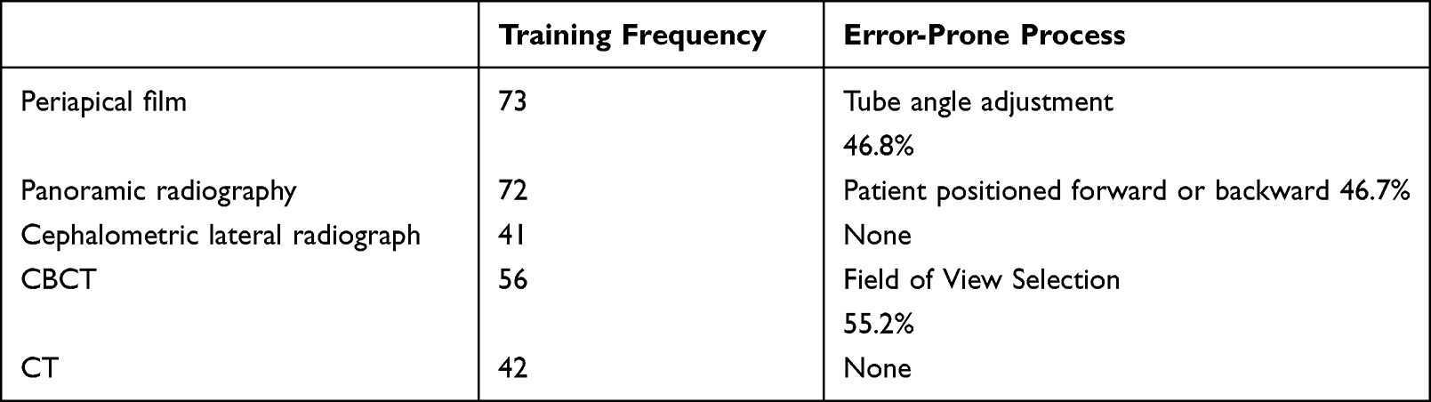 |
Table 1 Usage Data of the Virtual Radiographic Imaging Module |
Twelve specific cases and training content are shown in Table 2. In the score loss statistics table of the five steps of case training, the two parts with the highest error rates are “virtual image examination” and “disease diagnosis”, while the error rates for history taking, virtual clinical examination, and imaging examination selection are all less than 10%. The score loss rate in the diagnostic process varies among different cases, as shown in Figure 2. Among the 12 cases, the cases with high diagnostic error rates were osteomyelitis of the jawbone (20%) and deep caries of the posterior teeth (10%).
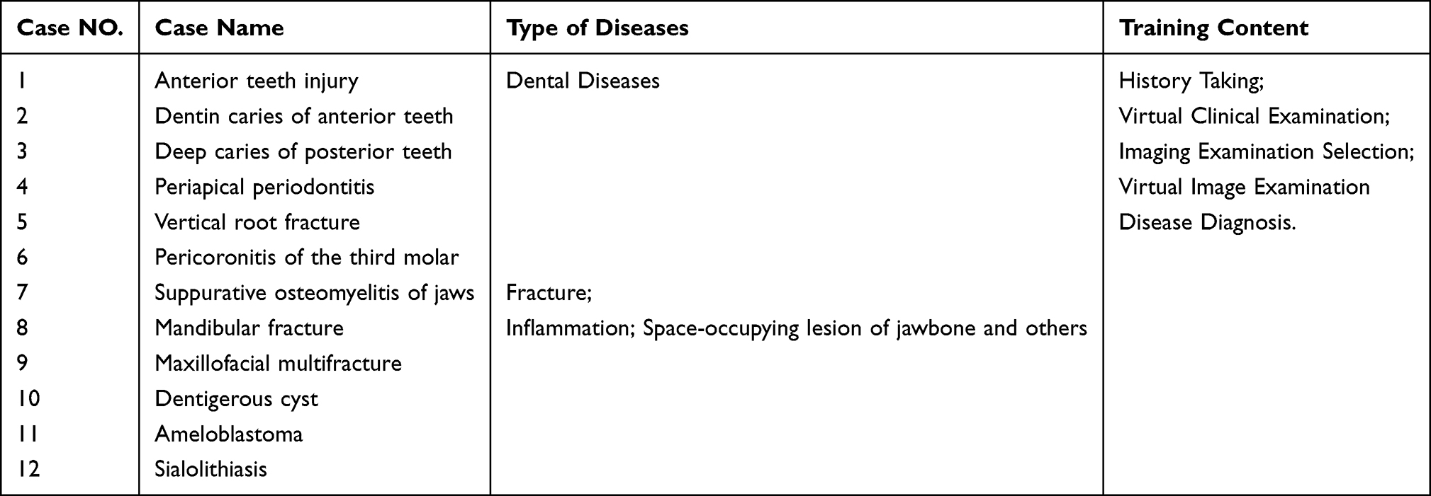 |
Table 2 The Case Study in the Virtual Clinical Training Module |
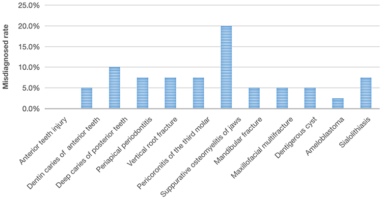 |
Figure 2 The score loss rate in the diagnostic process varies among different cases. |
Analysis of the Final Exam
The theoretical exam scores of Group A and Group B are shown in Table 3. Compared with Group A, Group B scored higher on both the imaging diagnostic exam (29.83+4.01 vs 26.62+6.18) and the case analysis (9.50+1.84 vs 8.12+3.13). These differences were statistically significant, as presented in Table 3.
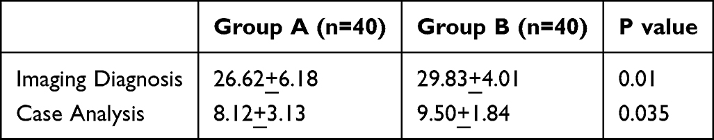 |
Table 3 Comparison of the Scores of the Two Groups of Theoretical Exams |
Analysis of the Questionnaire Survey
At the end of the semester, 40 students from Group B participated in a questionnaire survey to assess their satisfaction with the ORVR system in the teaching process. A total of 40 questionnaires were collected, resulting in a response rate of 100%. As shown in Table 4, over 90% of the students indicated that the training module involving video capture in the ORVR system was innovative and engaging, helping them better understand the principles of oral imaging technology. Additionally, all the students felt that the case training module within the ORVR system was rich in case studies, closely related to clinical practice, which helped reinforce theoretical knowledge and enhance classroom learning outcomes. Certainly, as the system is still in its initial stages, the user satisfaction rate is 50%, indicating room for improvement. However, the system has significantly increased students’ interest in learning (75%). Additionally, 92.5% of the students felt that the final assessment was comprehensive and reasonable, and 97.5% rated the overall teaching process as satisfactory. Some students suggested that the ORVR system could include more explanations of key concepts, expanding on clinical and other specialized knowledge areas. Further analysis of the main questionnaire indicators is shown in Figure 3, with average scores for most of the indicators exceeding 4, except for user satisfaction with the system.
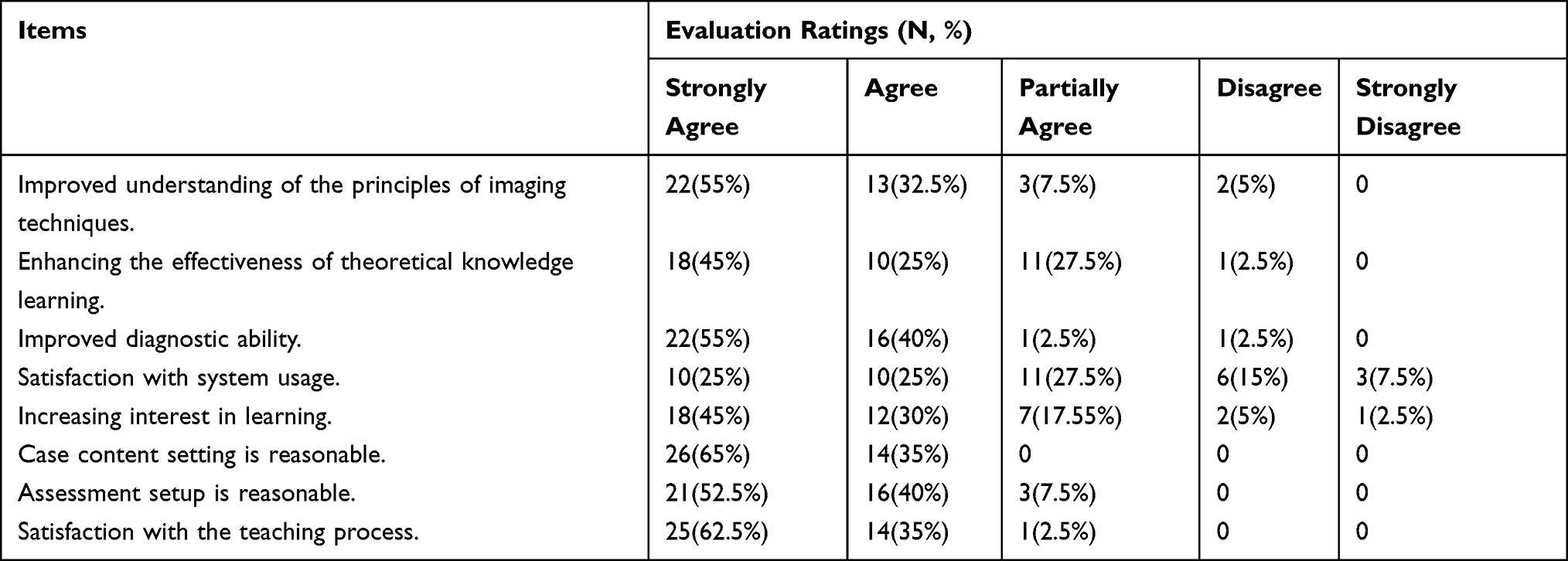 |
Table 4 Satisfaction Evaluation of Students From Group B |
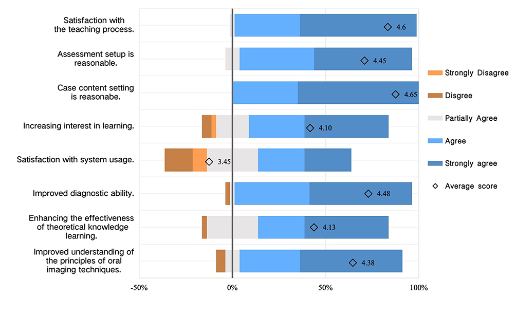 |
Figure 3 Statistical analysis of key issues in the questionnaire survey. |
Discussion
With the rapid advancement of medical imaging technology, imaging examinations have become a primary source of clinical diagnostic evidence. The interpretation of these examinations directly influences the determination of treatment plans and patient prognosis. Therefore, basic imaging diagnostic skills are essential for oral clinicians. The main teaching objective of the “Oral and Maxillofacial Imaging Diagnostics” course is not only to ensure that students master the operational methods and principles of commonly used oral imaging techniques but also, more importantly, to help students learn to use these imaging methods judiciously in an environment heavily exposed to medical images. This course aims to train diagnostic skills and establish clinical diagnostic thinking. The undergraduate curriculum of oral and maxillofacial imaging diagnostics is rich and covers a wide range of oral diseases. The traditional teaching mode requires students to absorb a large amount of content in a short time, leaving them with little opportunity to review and digest theoretical content effectively, which impacts the quality of classroom teaching and reduces students’ learning interest. Moreover, practical training in imaging techniques involves ethical issues related to radiation safety, preventing students from conducting repeated practice on human subjects. This lack of hands-on experience hinders their understanding of the principles and implementation of these technologies, as well as their grasp of indications and diagnostic imaging.
The rapid development of computers and the internet have profoundly impacted medical education.12 Many medical institutions worldwide are using online teaching platforms to enhance their courses and facilitate learning.13 The COVID-19 pandemic and subsequent isolation measures further accelerated the transition to online education.14–18 Consequently, there is a pressing demand for hybrid teaching methods that fuse online and offline elements. Therefore, we have integrated the virtual simulation teaching platform into theoretical lectures to increase teaching effectiveness.19 The virtual simulation training system for oral medical imaging developed by our teaching and research office is the first reported virtual simulation system in the field of oral imaging diagnostics. This system combines simulated images with real case images, simulates clinical work scenarios, and offers strong interactivity with the capability for repeated operations. It reinforces theoretical knowledge through foundational knowledge and image reading modules. The online knowledge base and image reading training break the constraints of time and location, allowing students to make better use of their spare time for image reading and self-assessment. Students can use this platform for shooting training and comprehensive training on typical clinical cases, effectively avoiding ethical and radiation safety issues. By analyzing students’ training data, we found that most errors in radiographic imaging training were due to inaccurate memory of key technical parameters, such as incorrect vertical angles in periapical radiography or improper positioning of the canine line in panoramic tomography. This suggests that focusing on these issues during experimental courses and providing targeted analysis and explanations may help reduce such errors during clinical internships. Additionally, the system creates a virtual environment for the photo room and equipment, enabling students to familiarize themselves with the equipment and the environment before entering clinical practice. This comprehensive virtual simulation training provides a solid foundation for students’ future clinical internships, enhancing their confidence while reducing anxiety when they first enter clinical settings.20
The teaching tasks of oral and maxillofacial imaging diagnostics extend beyond imparting theoretical knowledge to students; they also involve converting disease theory into clinical diagnostic capabilities through experimental classes and other teaching methods. Teachers need to engage students in independently establishing linear connections between knowledge points, constructing knowledge networks, and cultivating clinical thinking and diagnostic abilities. The case training module in this system is closely integrated with clinical practice, featuring reasonable and diverse cases that simulate real clinical scenarios and training students in comprehensive disease diagnosis and treatment thinking. In the five-step process, students must accurately and comprehensively collect patient history, conduct clinical examinations, correctly select and implement imaging examination methods, and diagnose virtual patients on the basis of patient history, clinical examination, and imaging results. For students who have just begun professional courses and have not yet entered clinical environments, this learning method is realistic and comprehensive. Analysis of student training data revealed that the highest error rate in image reading and diagnosis was in cases of suppurative osteomyelitis of the jaw. In real clinical practice, jaw infections present diverse clinical and imaging features, making differentiation from malignant tumors or other space-occupying lesions challenging. The application of this system highlights students’ weak areas in theoretical learning, allowing teachers to focus on these diseases in both theoretical and practical teaching and providing detailed analysis of diagnostic points and differential diagnosis, which is very helpful in enhancing classroom learning outcomes.21
The differences in final theoretical exam scores further indicate that the application of the ORVR system can effectively enhance theoretical learning outcomes. There were differences in the scores of the image reading and case analysis questions between the two groups. Through imaging technique training, students gain a better understanding of imaging principles and strengthen their recognition and interpretation of images. The case training module required students to develop comprehensive thinking skills, guiding them through an autonomous learning process of encountering problems, consulting information, and solving problems. This process improved their learning interest and transformed theoretical knowledge into personal knowledge reserves. The students’ understanding of the theoretical lecture content was subtly deepened, thus improving their imaging diagnosis and case analysis abilities.22
This study utilized a five-point Likert scale to evaluate the teaching process and the effectiveness of the virtual simulation system. The results revealed that the average scores of all the items exceeded 4.0, indicating that the students acknowledged all the aspects of the evaluation content. Most students believed that their diagnostic abilities improved through this course and that the virtual simulation teaching system effectively enhanced learning interest and efficiency, fostering a better integration of knowledge. The case training module, in particular, received the highest level of satisfaction because of its ability to improve diagnostic ability. This outcome aligns with the teaching and research office’s initial goal of focusing on enhancing students’ diagnostic skills. However, owing to issues such as page lag and incompatibility with certain browsers, the satisfaction with the virtual simulation system was relatively lower. Additionally, since imaging technique training is not the primary focus of internship teaching compared with diagnostic ability, some students showed less enthusiasm for shooting training, leading to limited improvement in their shooting skills. This is an area that the teaching and research office should focus on when further refining internship teaching management.
Conclusion
In summary, the application of virtual simulation teaching in undergraduate education in oral and maxillofacial imaging diagnostics not only improves students’ practical skills and clinical reasoning abilities but also stimulates their interest in and initiative in learning, thereby providing strong support for cultivating high-quality dental professionals.
Institutional Review Board Statement
The study was conducted in accordance with the Declaration of Helsinki, and approved by the Ethics Committee at West China Hospital of Stomatology, Sichuan University (No. WCHSIRB-CT-2022-344). All participants provided written informed consent before participating in the study.
Acknowledgments
We would like to thank all participants who were willing to participate in the study.
Funding
This research was funded by: A Stratified Controlled Study on Ultrasound-Assisted Precise Determination of Mandibular Protrusion Amount in Repositioning Splints, grant number “LCYJ-MS-202307”; Construction and Demonstration of a Multimodal Clinical Teaching Resource Platform Based on T-PACS, grant number “SCU11265”; Construction of a Multimodal Experimental Teaching System for Oral Radiology Empowered by AI and Reform of the Teaching Model, grant number “SCU2025052”.
Disclosure
The authors report no conflicts of interest in this work.
References
1. Shelton PG, Corral I, Kyle B. Advancements in undergraduate medical education: meeting the challenges of an evolving world of education, healthcare, and technology. Psychiatr Q. 2017;88(2):225–234. doi:10.1007/s11126-016-9471-x
2. Sun L, Liu D, Lian J, Yang M. Application of flipped classroom combined with virtual simulation platform in clinical biochemistry practical course. BMC Med Educ. 2023;23(1):771. doi:10.1186/s12909-023-04735-x
3. Xu G, Wang S. Application of problem-based learning combined with a virtual simulation training platform in clinical biochemistry teaching during the COVID-19 pandemic. Front Med. 2022;9:985128.
4. Karimian Z, Barkhor A, Mehrabi M, et al. Which virtual education methods do e-students prefer? Design and validation of Virtual Education Preferences Questionnaire (VEPQ). BMC. Med Educ. 2023;23:722. doi:10.1186/s12909-023-04687-2
5. Raja M, Priya GGL. Conceptual origins, technological advancements, and impacts of using virtual reality technology in education. Webology. 2021;18(2):116–134. doi:10.14704/WEB/V18I2/WEB18311
6. Dutra V, Madarapu S, Arriola-Guillén LA, Liedke GS, Wong P, Wood ZM. Use of virtual reality as a radiology teaching tool for dental students. Oral Surg Oral Med Oral Pathol Oral Radiol. 2025;139(3):e68. doi:10.1016/j.oooo.2024.11.004
7. Lu J, Zhang L, Ye L. Construction and application of dental virtual reality simulation teaching system in China: necessity and strategies. Eur J Dent Educ. 2023;
8. Ren Q, Wang Y, Zheng Q, Ye L, Zhou XD, Zhang LL. Survey of student attitudes towards digital simulation technologies at a dental school in China. Eur J Dent Educ. 2017;21(3):180–186. doi:10.1111/eje.12198
9. Daud A, Matoug-Elwerfelli M, Daas H, Zahra D, Ali K. Enhancing learning experiences in pre-clinical restorative dentistry: the impact of virtual reality haptic simulators. BMC Med Educ. 2023;23(1):948. doi:10.1186/s12909-023-04904-y
10. Algarni YA, Saini RS, Vaddamanu SK, et al. The impact of virtual reality simulation on dental education: a systematic review of learning outcomes and student engagement. J Dent Educ. 2024;88(11):1549–1562. doi:10.1002/jdd.13619
11. Likert R. A technique for the measurement of attitudes. Arch Psychol. 1932;22(140):55.
12. Shang R, Qin Y. Research on humanistic quality higher medical education based on internet of things and intelligent computing. Comput Intell Neurosci. 2022;2022:1–11. doi:10.1155/2022/8633190
13. Fu Y, Chu F, Lu X, et al. Assessment and evaluation of online education and virtual simulation technology in dental education: a cross-sectional survey. BMC Med Educ. 2024;24(1):191. doi:10.1186/s12909-024-05171-1
14. Chen K, Lin J, Wang Z, Chen K, Yang J. Teaching performance of chemistry teachers in chinese mainland during the COVID-19 pandemic: a content analysis study. J Chem Educ. 2023;100(4):1466–1475. doi:10.1021/acs.jchemed.2c00873
15. Ng CF, Lim K, Yee CH, Chiu PKF, Teoh JYC, Lai FPT. Time for change? Feasibility of introducing micromodules into medical student education: a randomised controlled trial. Hong Kong Med J. 2023;29(3):208–213. doi:10.12809/hkmj219267
16. Linling Z, Abdullah R. The impact of COVID-19 pandemic on flipped classroom for EFL courses: a systematic literature review. Sage Open. 2023;13(1):21582440221148149. doi:10.1177/21582440221148149
17. Lo CK, Hew KF. Design principles for fully online flipped learning in health professions education: a systematic review of research during the COVID-19 pandemic. BMC Med Educ. 2022;22:720. doi:10.1186/s12909-022-03782-0
18. Feng Y, Zhao B, Zheng J, et al. Online flipped classroom with team-based learning promoted learning activity in a clinical laboratory immunology class: response to the COVID-19 pandemic. BMC Med Educ. 2022;22(1):836. doi:10.1186/s12909-022-03917-3
19. Meng Y, Song J, Yu X, Xu X, Zhang H. Design and evaluation of blended teaching in the smart classroom combined with virtual simulation training in basic nursing courses. BMC Med Educ. 2023;23(1):752. doi:10.1186/s12909-023-04721-3
20. Rodrigues P, Nicolau F, Norte M, et al. Preclinical dental students self-assessment of an improved operative dentistry virtual reality simulator with haptic feedback. Sci Rep. 2023;13(1):2823. doi:10.1038/s41598-023-29537-5
21. Ho KC, Huang TS, Lin JC, Chiang HK. The online interactive visual learning improves learning effectiveness and satisfaction of physicians with postgraduate year during the COVID-19 pandemic in Taiwan. BMC Med Educ. 2023;23(1):713. doi:10.1186/s12909-023-04639-w
22. Sung H, Kim M, Park J, Shin N, Han Y. Effectiveness of virtual reality in healthcare education: systematic review and meta-analysis. Sustainability. 2024;16(19):8520. doi:10.3390/su16198520
 © 2025 The Author(s). This work is published and licensed by Dove Medical Press Limited. The
full terms of this license are available at https://www.dovepress.com/terms
and incorporate the Creative Commons Attribution
- Non Commercial (unported, 4.0) License.
By accessing the work you hereby accept the Terms. Non-commercial uses of the work are permitted
without any further permission from Dove Medical Press Limited, provided the work is properly
attributed. For permission for commercial use of this work, please see paragraphs 4.2 and 5 of our Terms.
© 2025 The Author(s). This work is published and licensed by Dove Medical Press Limited. The
full terms of this license are available at https://www.dovepress.com/terms
and incorporate the Creative Commons Attribution
- Non Commercial (unported, 4.0) License.
By accessing the work you hereby accept the Terms. Non-commercial uses of the work are permitted
without any further permission from Dove Medical Press Limited, provided the work is properly
attributed. For permission for commercial use of this work, please see paragraphs 4.2 and 5 of our Terms.