
Back to Journals » Drug Design, Development and Therapy » Volume 19
Repurposing Artemisinin-Based Drugs from Antimalarial to Pan-Therapeutic: Pharmacological Promise and Therapeutic Challenges
Authors Wu D, Zeng H, Tan J, Xu Q, Khan FA, Pandupuspitasari NS, Yang S, Huang C
Received 6 June 2025
Accepted for publication 28 September 2025
Published 11 October 2025 Volume 2025:19 Pages 9199—9226
DOI https://doi.org/10.2147/DDDT.S545163
Checked for plagiarism Yes
Review by Single anonymous peer review
Peer reviewer comments 2
Editor who approved publication: Dr Muzammal Hussain
Di Wu,1,* Hong Zeng,2,* Jiachen Tan,1 Qinzheng Xu,1,3 Faheem Ahmed Khan,4 Nuruliarizki Shinta Pandupuspitasari,5 Shengqiang Yang,2 Chunjie Huang1
1School of Medicine, Nantong University, Nantong, 226001, People’s Republic of China; 2School of Basic Medicine, Youjiang Medical University for Nationalities, Baise, 533000, People’s Republic of China; 3Department of Obstetrics and Gynecology, Affiliated Hospital of Nantong University, Nantong, 226001, People’s Republic of China; 4Research Center for Animal Husbandry, National Research and Innovation Agency, Jakarta Pusat, 10340, Indonesia; 5Faculty of Animal and Agricultural Sciences, Universitas Diponegoro, Semarang, 50275, Indonesia
*These authors contributed equally to this work
Correspondence: Shengqiang Yang, Email [email protected] Chunjie Huang, Email [email protected]
Abstract: The sourcing of pharmaceutical agents from natural products remains a cornerstone of drug discovery. For millennia, herbal remedies have served as foundational therapeutic interventions, yet their mechanisms of action often remain elusive. Artemisinin (ART, or “Qinghaosu”) and its derivatives, collectively known as ARTs, exemplify this paradigm. Originating from traditional Chinese medicine, these well-established antimalarial compounds exhibit potent cytotoxicity, positioning them as promising anti-cancer agents. ARTs operate through multiple cancer cell-intrinsic mechanisms and demonstrate synergistic effects with conventional therapies, even against chemoresistant malignancies. Beyond oncology, the therapeutic portfolio of ART-based drugs is rapidly expanding. This review synthesizes recent advancements in the application of ARTs for treating various cancers, Alzheimer’s disease, and reproductive disorders. Furthermore, we explore the frontier of ART drug development, focusing on the rational design of ART-derived molecular hybrids and the engineering of nano-enabled delivery systems. These innovative strategies are pivotal for enhancing anti-cancer efficacy and achieving cancer-specific targeting. Ultimately, this work aims to guide the innovation of ART-based precision medicine, revitalizing the exploration and development of natural products in modern pharmacotherapy.
Keywords: artemisinin, malignancy, neurodegenerative disease, reproductive disorder, drug delivery
Introduction
Naturally occurring substances and medicinal plants constitute a significant reservoir of biologically active compounds that are utilized as potent therapeutic modalities across various diseases.1–11 Artemisinin (ART) and its derivatives, collectively known as ARTs, are bioactive sesquiterpene lactones extracted from Artemisia annua, a traditional herbal medicine with antioxidant and anti-inflammatory properties. ART-based combination therapy has been established as the standard treatment for severe malaria, leading to the awarding of the 2015 Nobel Prize in Medicine to the Chinese scientist Tu You-You for her contributions to the isolation and discovery of ART.
Since the 1970s, several ART derivatives, including dihydroartemisinin (DHA) and artesunate (AS), have been identified, exhibiting enhanced solubility and bioavailability12 (Figure 1). ARTs have short in vivo half-lives and are capable of diffusing across physiological barriers, such as the blood–brain barrier, which contributes to their advantageous safety and pharmacokinetics in diseases treatment. They contain a tetracyclic core with an endoperoxide bridge (C-O-O-C), which is essential for their cytotoxic properties. Interacting with heme causes the cleavage of the endoperoxide bridge by heme iron (II) oxide, leading to the generation of peroxy free radicals and reactive oxygen species (ROS), which inflict damage on vulnerable biomolecules, including DNA, thereby initiating cell death.
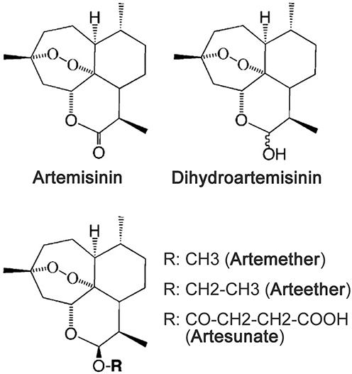 |
Figure 1 Chemical structure of artemisinin and its derivatives. |
ARTs also engage with a diverse array of cellular proteins, thereby participating in multiple biological processes, including apoptosis, autophagy, ferroptosis, glycolysis, and the inflammatory response.13–18 This endows ARTs with potent pharmacological activities under disease conditions. Compelling evidence suggests that ARTs exhibit selective cytotoxicity toward malignant cells since being first reported in 1993, as most cancer cells express higher levels of transferrin receptors (TfRs) on their surface, featuring an increased influx of iron ions via transferrin mechanism.16,19–21 Notably, the therapeutic effectiveness even in certain cancers featuring chemoresistance can be significantly improved by the combination of ARTs with chemotherapeutic drugs.
In this review, we provide a comprehensive summary of the recent advancements in the therapeutic properties of ARTs in malignancies, neurodegenerative disorder, and reproductive disease, with an emphasis on their underlying molecular mechanisms and pharmacological effects. We further discuss the recent development of ART-derived hybrids and nanoenabled delivery systems that aim at enhancing the anti-cancer efficacy and cancer-specific targeting property of ARTs, which innovates the ART-based therapeutic applications.
Therapeutic Activities of ARTs in Malignancies
Despite the emergence of chemotherapy in combination with immunotherapy as a promising therapeutic strategy, cancer heterogeneity, along with both primary and acquired resistance to these agents, significantly impedes their efficacy in cancer treatment.22 ART-based drugs demonstrate anti-cancer effects on various malignancies via multiple mechanisms known to be cancer cell intrinsic. Therefore, the combinatorial treatment with ART-based drugs and other molecular-targeted drugs holds the promise of achieving better outcomes in cancer treatment (Table 1).
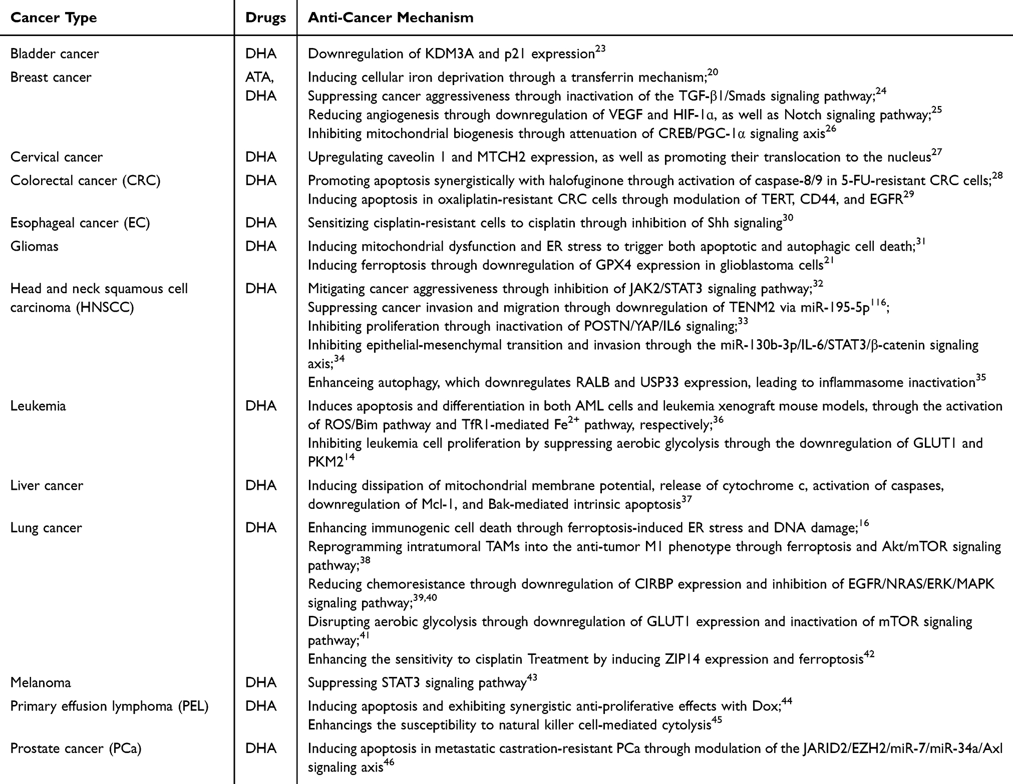 |
Table 1 The Anti-Cancer Therapeutic Effects ARTs on Malignancies |
The anti-cancer properties of ARTs are primarily attributed to their capacity to induce ferroptosis, a form of programmed iron-dependent cell death induced by overwhelming lipid peroxidation. Ferroptosis functions as a tumor-suppressive mechanism, which is associated with the activities of several tumor suppressors, including p53, BRCA1-associated protein 1 (BAP1), and fumarate hydratase.47–49 The p53 protein can inhibit ferroptosis through the transcriptional induction of CDKN1A or by interacting with dipeptidyl peptidase 4 (DPP4). Conversely, p53 can promote ferroptosis by transcriptionally repressing the cystine/glutamate antiporter SLC7A11/xCT, which uptakes cysteine for glutathione synthesis.50 The xCT transporter represents a targetable vulnerability in solid tumors; its deficiency can induce ferroptosis, thereby inhibiting metastasis and enhancing the efficacy of anti-PD1 immunotherapy across various cancer types.51–54 Either deprivation of cysteine or inhibition of glutathione peroxidase 4 (GPX4)—a critical enzyme for peroxide clearance—has been shown to increase sensitivity of aggressive mesenchymal cancer cells to ferroptosis.48–50,55 Ferroptosis has been demonstrated to enhance therapeutic efficacy of chemotherapy and immunotherapy.54,56 The pro-ferroptotic activity of ARTs artemisinins cancer is further by the evidence indicating these compounds significantly alter the expression of iron-related genes.57 DHA increases the susceptibility to GPX4 inhibition-induced ferroptosis in a set of cancer cells that are otherwise highly resistant to ferroptosis, primarily through regulation of iron homeostasis.13 Mechanistically, it induces the lysosomal degradation of ferritin, a nano-cage complex responsible for iron storage, via an autophagy-independent mechanism, which elevates cellular levels of free iron. The free iron subsequently stimulates the binding of iron-regulatory proteins (IRPs) to those transcripts containing iron-responsive element (IRE) sequence, thereby creating a positive feedback that further promotes free iron accumulation13 (Figure 2).
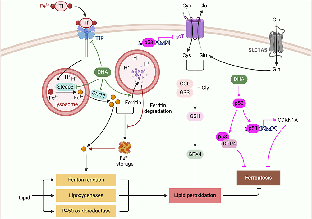 |
Figure 2 ARTs in modulation of ferroptosis. |
Breast Cancer
In breast cancer, DHA demonstrates anti-cancer properties by inducing cellular iron deprivation through a ROS-independent mechanism.20 Iron (Fe(III)), when bound to transferrin (Tf), is internalized via transferrin receptor 1 (TfR1)-mediated endocytosis. Within the acidic environment of lysosomes, Fe(III) is released and reduced to Fe(II) by the ferrireductase Steap3, followed by its translocation into the cytosol through the divalent metal transporter 1 (DMT1). DHA not only diminishes the levels of cell-surface TfR1 by promoting its palmitoylation and internalization but also down-regulates the expression of Steap3 and DMT1, leading to iron depletion in breast cancer MCF7 cells.20 Similar phenomena have been observed in hepatocellular carcinoma (HCC) HepG2 cells, suggesting that ARTs may serve as promising candidates for the development of iron-based cancer therapies. Moreover, DHA exhibits more potent anti-breast cancer activity compared to ART, primarily through the inactivation of the TGF-β1/Smads signaling pathway.24 The TGF-β1-induced aggressiveness of MCF-7 cells is mitigated by DHA treatment. However, the therapeutic application of DHA should be approached with caution due to its potential hepatotoxicity, despite its favorable tolerance observed in the MCF-7 xenograft mouse model.24
The anti-breast cancer efficacy of ARTs is also attributable to their ability to inhibit angiogenesis within the tumor microenvironment (TME) (Figure 3). Current Food and Drug Administration (FDA)-approved anti-angiogenic agents primarily target the signaling pathway of vascular growth factor A (VEGFA, also known as VEGF). These agents widely utilized, in combination with immunotherapy and/or chemotherapy for the treatment of various malignancies.58 In the hypoxic TME, hypoxia-inducible factor 1 (HIF-1), particularly HIF-1α promotes transcription and stabilization VEGF mRNA, thereby enhancing VEGF expression and angiogenesis.25 ART can reduce the vascular density in tumor tissues and impair the proliferation of breast tumor xenografts in the nude mice, by down-regulating the expression of angiogenesis-related factors VEGF and HIF-1ɑ.25 Notch signal-related factors Notch1, Delta-like ligand-4 (Dll4), and Jagged ligand-1 (Jag-1), which are known to promote angiogenesis and cancer aggressiveness, are also down-regulated by ART in a breast tumor xenograft mouse model.25,36,59
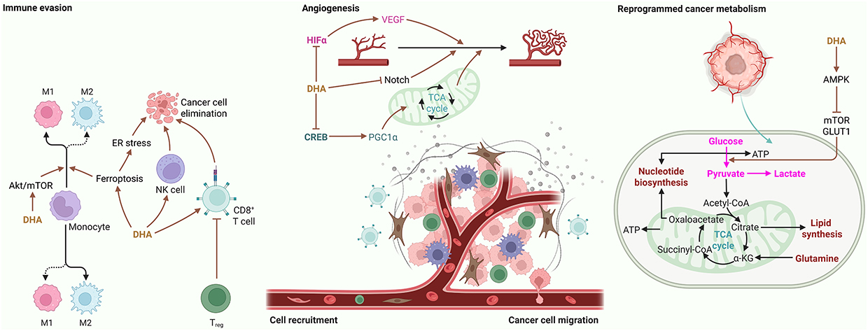 |
Figure 3 ARTs induce the reprogramming of TME to exhibit anti-cancer efficacy. Note that the immune evasion in TME is featured by: 1) low immunogenicity of cancer cells; 2) polarization of TAM into pro-tumor M1 type; 3) insufficient infiltration of cytotoxic T cells; and 4) recruitment of pro-tumorigenic regulatory T (Treg) cells and pre-mature dendritic cells. |
In another study, it is observed that ART enhances the sensitivity of breast cancer cells to the chemotherapeutic agent vinorelbine (NVB), a drug commonly utilized for its anti-angiogenic properties, by inhibiting mitochondrial biogenesis.26 Mitochondrial metabolism plays a crucial role in angiogenesis and tumor progression, which is stimulated by VEFG and peroxisome proliferator-activated receptor γ coactivator 1α (PGC-1α).60–64 Both VEGF and PGC-1α are subject to regulation by the cAMP response element-binding protein (CREB), which is notably overexpressed in various malignancies, including mesothelioma, breast cancer, and lung cancer.26,65 The combination of ART and NVB significantly inhibits tumor growth by targeting the CREB/PGC-1α signaling axis in a breast tumor xenograft mouse model.26
Cervical Cancer
In cervical cancer, DHA induces anti-cancer effects by upregulating caveolin 1 (Cav1) and mitochondrial carrier homolog 2 (MTCH2), as well as promoting their translocation to the nucleus.27 Cav1 contributes to p53 activation by facilitating its nuclear localization and stabilization; furthermore, the loss of Cav1 has been linked to tumorigenesis across various cancer types.27,66 In addition, MTCH2 contributes to apoptosis through its direct interaction with and recruitment of truncated Bid (t-Bid).67 Cav1 also negatively regulates antioxidant signaling by directly interacting with and inhibiting nuclear factor erythroid 2-related factor 2 (Nrf2).68 Consequently, the sensitivity of cervical cancer cells to DHA can be augmented by the overexpression of Cav1 and MTCH2. This study indicates that DHA functions as a p53 activator, thereby inhibiting cancer cell viability.27
Lung Cancer
Lung cancer stands as one of the three malignancies with the most unfavorable prognosis.69 ARTs exhibit anti-lung cancer activity also by enhancing anti-tumor immunity within the TME (Figure 3). The immunosuppressive nature of TME is characterized by the low immunogenicity of cancer cells, the insufficient infiltration of cytotoxic T cells, the high abundance of regulatory T cells, and the predominance of immature dendritic cells and pro-tumor M2-type tumor-associated macrophages (TAMs). These factors compromises the effectiveness and safety of anti-cancer immunotherapy.70–72 DHA is shown to exhibit promotive effects on immunogenic cell death through ferroptosis-induced endoplasmic reticulum stress and DNA damage, thereby reshaping the TME for renovation of anti-cancer immunity in lung cancer while exhibiting low toxicity to normal cells.16,73,74 It also induces ferroptosis in TAMs, which causes DNA damage and NF-κB activation, leading to phenotypic remodeling of intratumoral TAMs into the anti-tumor M1 phenotype.38 The immunogenicity-promoting activities of DHA can be subverted by ferroptosis inhibition while it is enhanced by exogenous iron ions, which is reminiscent of the finding that higher levels of ions are observed in malignant cells, including lung cancer cells.16,38,75 DHA has also been proposed as an M1 stimulus through the Akt/mTOR signaling pathway in a Lewis lung carcinoma (LLC) model. Additionally, DHA enhances the proinflammatory response and phagocytic capacity in TAMs, and the M1-related phenotypes induced by DHA can be inhibited through the blockade of mTOR signaling.15
The NRAS gene, an oncogene of the N-Ras family, exhibits significantly elevated expression levels in lung cancer. Mutations in NRAS lead to malignant proliferation via the ERK/MAPK signaling axis and inhibit DNA damage response, thereby contributing to chemoresistance in various cancers.39,76–79 Recent studies have indicated that ARTs possess anti-lung cancer potency by acting as direct inhibitors of epidermal growth factor receptor (EGFR), thereby impeding the NRAS/ERK/MAPK signaling pathway. This finding aligns with previous reports suggesting that DHA is more effective in cancers harboring mutations in EGFR or NRAS.39,80
Patients with non-small cell lung cancer (NSCLC) have only 26% of 5-year survival rate.81 NSCLC cells feature high propensity of developing radioresistance following fractional radiation (2 Gy per day for consecutive 20 days). DHA can reduce the radiation-induced mitophagy and radioresistance of NSCLC via inhibition of cold-inducible RNA binding protein (CIRBP) which shows highly expression in radioresistant NSCLC.40 DHA also induces apoptosis in NSCLC cells by disrupting aerobic glycolysis, primarily through the inactivation of mechanistic target of rapamycin (mTOR) and downregulation of glucose transporter GLUT1. Reactivation mTOR or overexpression GLUT1 significantly mitigates DHA-induced apoptosis in NSCLC cells by restoring glycolytic metabolism that is attenuated by DHA. Notably, DHA exhibits a synergistic effect with the glycolytic inhibitor 2-Deoxy-D-glucose (2DG) by activating caspases-3, −8, and −9, as well as promoting the release of cytochrome c, thereby enhancing apoptosis in NSCLC cells while demonstrating no significant toxicity to normal lung fibroblast cells.41,82
Liver Cancer
Liver cancer is a common malignancy characterized by rapid progression, early recurrence, and poor prognosis.83 DHA induces the dissipation of mitochondrial membrane potential (ΔΨm), release of cytochrome c, activation of caspases, downregulation of Mcl-1, and Bak-mediated intrinsic apoptosis in hepatocarcinoma (HCC) cells. These phenomena can be diminished by Bax deficiency and enhanced by Mcl-1 deficiency.37 Although the expression of Puma and Noxa remains unchanged following DHA treatment, apoptosis induced by DHA is suppressed when Puma, Noxa, or Bim is silenced.37 Furthermore, artesunate and DHA exhibit a synergistic effect with the heat shock protein 70 (HSP70) inhibitor VER-155,008 to induce apoptosis in HCC cells.84 HSPs, frequently overexpressed in human cancers, are essential for maintaining cellular homeostasis.85 HSP70 subfamily includes the constitutively expressed heat shock cognate 70 kDa protein (HSC70), the heat shock-inducible HSP70, and the glucose-regulated 78 kDa protein (GRP78). HSC70 involves in the tumorigenesis-promoting autophagy and GRP78 modulates unfolded protein response for tumorigenesis, while HSP70, with significant upregulation in HCC, drives tumor aggressiveness and chemotherapy resistance.84–88
Wang et al (2024) have identified rhein, an active compound derived from Rheum undulatum L., as a novel HSP70 inhibitor that enhances the sensitivity of HCC cells to artesunate or DHA.89 Importantly, rhein also suppresses CRC by inactivating the mTOR signaling, inhibits nasopharyngeal carcinoma by inducing Ca2+-dependent mitochondrial death signaling, and inhibits renal cell carcinoma by attenuating MAPK/NF-κB signaling.90–92 mTOR complex 1 (mTORC1) is known to promote the biosynthesis of macromolecules required for cellular growth and homeostasis.93 DHA activates AMP-activated protein kinase (AMPK) for mTORC1 inhibition, thereby impairing the proliferation and viability of rhabdomyosarcoma cells but not normal cells. These inhibitory effects can be mimiced by artesunate and be diminished by both genetic and pharmacological inhibition of AMPK.94
Colorectal Cancer
Colorectal cancer (CRC) ranks as the second leading cause of cancer-related mortality globally. The primary chemotherapeutic agent employed in CRC treatment is 5-Fluorouracil (5-FU); however, the sensitivity of CRC cells to 5-FU is diminished by p53 knockout.95 The tumor suppressor p53, which is encoded by the TP53 gene, functions as a transcription factor with over 4000 binding sites within the human genome.96 Loss-of-function mutations in TP53 are frequently observed across various malignancies, including breast cancer and CRC, and are correlated with unfavorable prognoses and chemoresistance.97,98 Yao et al (2018) demonstrate that DHA effectively restores 5-FU sensitivity in TP53-deficient CRC cells.95 Furthermore, Gong et al (2022) report a synergistic effect between ART and halofuginone (HF), a bioactive compound derived from the traditional Chinese herb Dichroa febrifuga Lour (Changshan in Chinese), in promoting caspase-dependent apoptosis via activation of caspase-8/9 in 5-FU-resistant CRC cells, both in vitro and in vivo.28,99 Interestingly, ART alone activates only caspase-9; however, when caspase-9 is inhibited, ART activates caspase-8 via induction of autophagy. In this scenario, with defective apoptosis, CRC cells switch to autophagic cell death in response to combination treatment with ART and HF.28 β-Lapachone (β-Lap), a NAD(P)H:quinone oxidoreductase 1 (NQO1) bioactive phytochemical derived from the lapacho tree, induces NQO1-mediated tumour-specific cell necrosis and has been tested in clinical trials for the treatment of solid tumors.100–102 The polyphenols extracted from Korean Artemisia annua L. potentiates the anti-cancer efficacy of β-lapachone in both parental and oxaliplatin-resistant CRC cells by inducing DNA damage and apoptosis through modulation of telomerase reverse transcriptase (TERT), CD44, and EGFR.29
Gliomas
Gliomas are the primary brain tumours originating from neural stem or progenitor cells that harbor tumour-initiating genetic alterations.103 DHA is reported to exert anti-glioblastoma activity by inducing mitochondrial dysfunction and endoplasmic reticulum (ER) stress, leading to both apoptotic and autophagic cell death in human glioblastoma cells.31 Compared to normal astrocytes, glioblastoma cells feature an elevated expression of TfR, a type II transmembrane protein involved in iron uptake, and the associated elevation in cellular iron levels contributes to DHA-induced ferroptosis in glioblastoma.21 Despite with constant expression of SLC7A11/xCT and acyl-CoA synthetase long-chain family member 4 (ACSL4), the expression of GPX4 in glioblastoma cells is significantly decreased by DHA.21 Of note, ACSL4 is an executor of ferroptosis with preferential expression in basal-like breast cancer cells and dictates ferroptosis sensitivity by shaping cellular lipid composition.104 While with ferroptosis-inducing activity, DHA is reported to initiate the HSP family A member 5 (HSPA5)-mediated ferroptosis inhibition in glioma cells.105 HSPA5 is an ER chaperone involved in lipid metabolism. DHA-induced unfolded protein response in the ER upregulates HSPA5 expression via the protein kinase R-like ER kinase (PERK)-activating transcription factor 4 (ATF4) signaling pathway, which thereby increases the expression and activity of GPX4, protecting glioma cells from ferroptosis. Genetic and pharmacological inhibition of PERK-ATF4-HSPA5-GPX4 pathway enhances the susceptibility of glioma cells to DHA-induced ferroptosis.105
A recent genome-wide screening revealed that porphyrin (heme) biosynthesis is essential for the cytotoxicity of DHA in eukaryotic cells.106 Pharmacological inhibition of porphyrin biosynthesis significantly decreases susceptibility to DHA, while pharmacological activation of porphyrin biosynthesis produces the opposite effect. 5-Aminolevulinic acid (5-ALA), a clinically approved enhancer of porphyrin synthesis, sensitizes multidrug-resistant, patient-derived high-grade gliomas to DHA. Furthermore, a combinatorial treatment involving 5-ALA and artesunate demonstrates a potent synergistic anticancer effect in reducing glioblastoma growth in patient-derived xenograft mouse models.106 This study thus identifies a novel combination therapy for human cancers, including drug-resistant glioblastomas.
Hematologic Cancer
In primary effusion lymphoma (PEL), a rare and aggressive B-cell lymphoma for which effective therapies are lacking, DHA has been shown to induce apoptotic cell death and importantly, it exhibits synergistic anti-proliferative effects when combined with the chemotherapeutic agent doxorubicin.44 Additionally, ART enhances the susceptibility of cancer cells to natural killer (NK) cell-mediated cytolysis. NK cells serve as the sentinel of tumor surveillance, and immunotherapy targeting NK cells has demonstrated significant anti-cancer efficacy.45,107,108 Interestingly, ART does not induce apoptosis in hematopoietic cancer cells; rather, it sensitizes these cells to apoptosis in the context of NK cell cytolysis, while preserving both the tumor recognition capabilities and the overall architecture of NK cells.45
Leukemia encompasses a group of aggressive hematopoietic malignancies that are characterized by genetic alterations and chromosomal abnormalities affecting the proliferation and differentiation of hematopoietic stem and precursor cells (HSPCs).109 Acute myeloid leukemia (AML) is specifically marked by the accumulation of undifferentiated myeloid blasts. Although numerous anti-AML therapies are available, particularly the differentiation therapies utilizing all-trans retinoic acid and arsenic trioxide, the prognosis for most other subtypes of AML remains unfavorable.110–114 Artesunate has been proposed as a novel chemotherapeutic agent for AML, as it induces apoptosis and differentiation in both AML cells and leukemia xenograft mouse models, while exhibiting minimal cytotoxicity towards healthy HSPCs, bone marrow mononuclear cells, and experimental animals.36 The apoptosis induced by artesunate is associated with the activation of ROS/Bim pathway, whereas the TfR1-mediated Fe2+ pathway constitutes a mechanism of artesunate-induced differentiation.36 Furthermore, DHA has been demonstrated to inhibit leukemia cell proliferation by suppressing aerobic glycolysis through the downregulation of GLUT1 and pyruvate kinase M2 (PKM2).14
Prostate Cancer
Prostate cancer (PCa) is the most prevalent solid malignancy among aging males. Although effective therapeutic strategies exist for local PCa, such as androgen ablation therapy, there are limited pharmacological options available for hormone-refractory metastatic PCa.115 The receptor tyrosine kinase Axl is frequently upregulated in various malignancies, including prostate and breast cancers, and is associated with poor overall survival rates and increased chemoresistance.116–120 Axl expression in PCa has been found to be inversely correlated with the expression levels of miR-7 and miR-34a.46 DHA demonstrates its anti-cancer effects on metastatic castration-resistant PCa by targeting the Jumonji, AT-rich interaction domain containing 2 (JARID2)/enhancer of zeste homolog 2 (EZH2)-miR-7/miR-34a-Axl signaling axis, which leads to apoptosis of PCa cells and inhibition of tumor growth. Mechanistically, DHA downregulates JARID2 and EZH2, both components of the polycomb repressive complex 2 (PRC2), which subsequently leads to the upregulation of miR-7 and miR-34a, thereby inhibiting Axl translation. Notably, DHA has been shown to synergize with docetaxel to enhance the survival rate of PCa xenograft mice.46 Furthermore, by activating the EGFR/PKC/mTOR signaling axis, Axl confers the chemoresistance to PI3Kα inhibition in head and neck squamous cell carcinoma (HNSCC) and esophageal squamous cell carcinoma (ESCC).120
Head and Neck Cancer
HNSCC is the primary form of head and neck cancer and is associated with a poor prognosis, ranking seventh in global cancer prevalence.121 DHA has been shown to mitigate the aggressiveness of HNSCC by inhibiting Janus Kinase 2 (JAK2)/signal transducer and activator of transcription 3 (STAT3) signaling pathway.32 Notably, suppression of STAT3 signaling pathway also underlies the mechanism by which DHA inhibits the proliferation, migration, and pulmonary metastases of melanoma.43 Recent findings indicate that DHA can suppress HNSCC invasion and migration through the downregulation of TENM2 via miR-195-5p.122 DHA also exhibits anti-proliferative and pro-apoptotic effects on laryngeal squamous cell carcinoma (LSCC), a type of HNSCC, by inactivation of periostin (POSTN)/Yes-associated protein (YAP)/IL-6 signaling which is implicated in the tumorigenesis of various malignancies, including CRC.33,123,124 Additionally, DHA inhibits epithelial–mesenchymal transition (EMT) and LSCC invasion via the miR-130b-3p/IL-6/STAT3/β-catenin signaling axis.34
In laryngeal squamous cell carcinoma (LSCC), the tumor-suppressive effects of DHA is mediated through the inhibition of inflammasome.35 The smoldering inflammatory TME constitutes an incubator for the progression and therapy resistance in nearly all cancer types.125 Inflammasome activates caspase-1 and promotes the production of pro-inflammatory cytokines interleukin (IL)-1β and IL-18. It is shown that a lower expression of the inflammasome component absent in melanoma 2 (AIM2), a PYHIN protein, is associated with a poorer survival rate in patients with hypopharyngeal squamous cell carcinoma (HSCC); while the overexpression of interferon gamma inducible protein 16 (IFI16), another PYHIN protein, is positively correlated with caspase-1-mediated pyroptosis in LSCC.35,126,127 DHA enhances autophagy, which downregulates the expression of RAS like proto-oncogene B (RALB) and ubiquitin-specific protease 33 (USP33), leading to the inactivation of IFI16/caspase-1 inflammasome. Consequently, IL-1β production in the serum and TME is compromised in Hep-2 xenograft mouse model.35
Esophageal Cancer
Esophageal cancer (EC) is one of the most lethal cancers and its mortality rate ranks among the top ten in China. DHA has been shown to induce an autophagy-dependent cell cycle arrest in esophageal cancer Eca109 cells.123 A common subtype of EC is ESCC—one of the most incurable malignancies due to the prevalence of drug resistance.128 Hyperactivation of sonic hedgehog (Shh) signaling is a contributing factor to carcinogenesis, cancer stemness maintenance, and chemoresistance, and is associated with poor outcomes in ESCC patients undergoing platinum-based chemotherapy,30,129–131 Importantly, continuous chemotherapy with cisplatin can activate Shh signaling to enhance the cancer stem cell-like phenotype of ESCC cells. By acting as an inhibitor of Shh signaling, DHA attenuates the cisplatin-activated Shh signaling and sensitizes cisplatin-resistant ESCC to cisplatin.30 Notably, by inducing ZIP14 expression and ferroptosis, DHA also enhances cisplatin sensitivity in lung cancer, which may optimize the treatment outcomes of lung cancer chemotherapy42 (Figure 4).
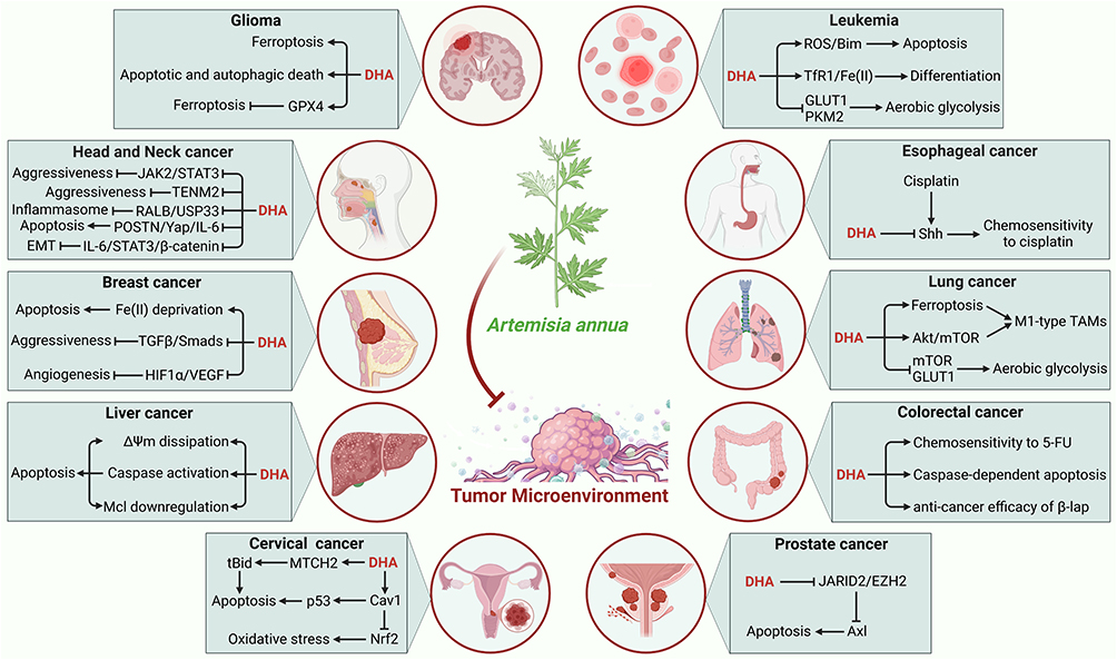 |
Figure 4 Representative therapeutic mechanisms of ARTs in various malignancies. |
Clinical Trials on ARTs in Cancer
Despite the well-documented anti-cancer effects of ARTs in basic research, only a limited number of compounds have progressed to clinical trials. In 2011, Jansen et al conducted a pilot study to investigate the clinical benefits, safety, and tumor marker effects of the orally administered ART derivative, Artenimol-R, in patients with advanced cervical cancer. The study observed that, following a 28-day treatment period, clinical remission was achieved, with a median time for the disappearance of symptoms being 7 days. Importantly, no adverse events of grades 3 or 4 were reported, suggesting an improvement in clinical symptoms and good tolerability of Artenimol-R in patients with advanced cervical cancer.132
In 2014, Ericsson et al conducted an investigation into the pharmacokinetics of AS and DHA following long-term oral administration of AS in a cohort of 23 patients with metastatic breast cancer. The study demonstrated that the population pharmacokinetics of AS and DHA can be accurately characterized by a combined drug-metabolite model without any covariates, alongside a significant increase in the elimination rate of DHA over time. Furthermore, the estimated saliva/plasma ratio of DHA aligns closely with the reported unbound fraction of DHA in human plasma, while the concentrations of AS in saliva exhibit a poor correlation with plasma concentrations.133
In 2014, Krishna et al investigated the anticancer activity of AS in a cohort of 23 CRC patients. Following a 14-day regimen of oral administration of 200 mg AS, Bayesian analysis indicated a probability of 0.89 for a reduction in Ki67 expression and 0.79 for an increase in CD31 expression, and no death reported in AS group, in contrast to three deaths in the placebo group. Furthermore, the relapse-free survival rate following AS treatment was significantly higher than that of the placebo group (0.89 vs 0.5 at 42 months), suggesting that AS possesses anti-CRC activity with a favorable tolerability.134
In 2016, König et al investigated the safety and tolerability of a four-week AS therapy in patients with breast cancer. They showed that daily doses of 100 mg, 150 mg, and 200 mg were well tolerated among individuals with metastatic or locally advanced breast cancer. Furthermore, it was observed that any dose-limiting adverse events (DL-AEs) were completely reversible after the therapy.135
In 2017, von Hagens et al recruited 23 patients with metastatic breast cancer and divided them into three groups based on the administration of oral AS at doses of 100 mg/day, 150 mg/day, and 200 mg/day, with the objective of defining the well-tolerated dose for use as add-on therapy. Throughout the trial period of 4 ± 1 weeks, three patients experienced a total of six DL-AEs, suggesting that an oral dose of up to 200 mg/day is both safe and well tolerated.136 In a subsequent follow-up study, 25 AEs grade ≥ 2 and at least possibly related to long-term AS therapy were documented, with the distribution of events being 2, 6, and 17 across the 100 mg, 150 mg, and 200 mg/day dose groups, respectively. Thus, they concluded that long-term administration of oral AS at doses of up to 200 mg/day over a cumulative treatment duration of 1115 days does not present significant safety concerns for patients with metastatic breast cancer.137
In 2018, Deeken et al conducted a Phase I trial to explore the maximum tolerated dose (MTD) and dose-limiting toxicities (DLTs) of intravenous AS in 19 patients with advanced solid malignancies. The administration of intravenous AS was performed on the first and eighth days of a 21-day cycle. The results indicated that DLTs were observed at doses of 12, 18, and 25 mg/kg, while the MTD was determined to be 18 mg/kg.138
ART-Based Hybrid Entity
Efforts that improve the therapeutic effectiveness of ARTs have led to the discovery of ART-derived hybrid entities that exhibit more favorable druggability. Compared to native pharmacophore, ART hybrids posses many advantages, including enhanced therapeutic activity, improved pharmacokinetic profile, less cross-resistance, and reduced side effects139–141 (Table 2).
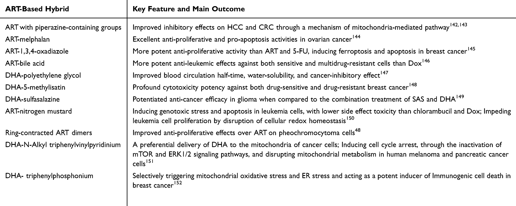 |
Table 2 ART-Based Hybrid Entities Exhibit Improved Therapeutic Effectiveness |
ART with piperazine-containing groups at 10-position carbon (C10) exhibit improved inhibitory effects on hepatocellular cancer cells, while introducing fluorine atoms into piperazine-containing groups to ART improves its anti-cancer activity in multidrug-resistant breast cancer cells, pheochromocytoma cells, and CRC cells.142,143 The six-membered cyclic carbamate ART derivatives containing piperazine and fluorine groups show excellent anti-cancer effects compared to ART and DHA, with lower cytotoxicity in normal liver cells than Dox. Among them, compound 12h exhibits potent anti-cancer activities in CRC cells but not in glioblastoma cells and lung adenocarcinoma cells, via a mechanism of mitochondria-mediated pathway.143 These findings envision further endeavors to develop novel ART-based entities with less side effects and improved disease-mitigating activities.
Incorporating the pharmacophore of melphalan into the basic scaffold of ART with a succinic linker lead to the discovery of a compound ARS4 that exerts excellent anti-proliferative and pro-apoptosis activities in ovarian cancer while with low cytotoxicity to normal cells. Further structure optimization produces multiple ARS4 derivatives exhibiting cytotoxicity potency against liver and ovarian cancer models.144 A series of synthesized 1,3,4-oxadiazole-ART hybrids also show more potent anti-proliferative activities against five human cancer cell lines than ART and 5-FU. Among them the A8 compound induces ferroptosis and apoptosis in breast cancer MCF-7 cells.145 In another study, a series of bile acid-ART hybrids show promising anti-leukemic effects against both sensitive and multidrug-resistant cells, with amide hybrid 25 being the most active compound that is more potent than the chemotherapeutic drug doxorubicin (Dox).146
DHA in conjugation with multiarm polyethylene glycol shows improved blood circulation half-time, water-solubility, and cancer-inhibitory effect of native DHA, while the hybrid of DHA-5-methylisatin tethered by two-carbon linker exhibits profound cytotoxicity potency against both drug-sensitive and drug-resistant breast cancer cells.147,148 A hybrid of clinically established agents sulfasalazine (SAS) and DHA, termed AC254, demonstrates potentiated chemotherapeutic efficacy in human glioma cells when compared to the combination treatment of native SAS and DHA, with a minor cytotoxicity towards primary neurons and astrocytes.149
Nitrogen mustard derivatives, chlorambucil, melphalan, and cyclophosphamide, are extensively used DNA-alkylating agents in cancer chemotherapy.153 Synthesis of nitrogen mustard-ART hybrids obtains a lead compound 5a that effectively causes DNA damage-mediated genotoxic stress and Bcl2 downregulation-mediated apoptosis in leukemia cells, with lower toxicity towards normal liver cells than chlorambucil and Dox.150 5a also hinders leukemia cell proliferation by disrupting cellular redox signaling, due to the activation of glutathione peroxidase (GPx) which catalyzes the peroxidation of glutathione (GSH) to deplete ROS.150,154 In addition, the ring-contracted ART dimers linked by a phosphate ester moiety exhibit improved anti-proliferative effects over ART, with compound 8b shows prominent anti-cancer activity in pheochromocytoma cells, via inducing arrested cell cycle and caspased-dependent apoptosis.48
Given the significant difference of mitochondria inner membrane potential in cancer cells compared to normal cells, conjugating DHA with N-Alkyl triphenylvinylpyridinium (TPVP), a mitochondria-accumulating agent, leads to a preferential delivery of DHA to the mitochondria of cancer versus normal cells.151,155 TPVP-DHA induces G1 cell cycle arrest, via the inactivation of mTOR and ERK1/2 signaling pathways, and disrupt mitochondrial metabolism, in human melanoma and pancreatic cancer cells in a dose-dependent way.151 Conjugating triphenylphosphonium (TPP) to DHA leads to the development of a mitochondria-targeting DHA (TPP-DHA) that selectively triggers mitochondrial oxidative stress and ER stress, thus acting as a potent inducer of Immunogenic cell death (ICD).152 ICD occurs in stressed or dying cells and triggers anti-cancer immunity by the release of damage-associated molecular patterns (DAMPs), including calreticulin (CRT) which acts as “eat-me” signals to facilitate the phagocytosis of dendritic cells (DCs), ATP which acts as “find me” signal for recruiting DCs to sites of ICD, and high-mobility group box 1 (HMGB1) which is liberated to the extracellular environment that enhances DCs’ antigen presentation effect.156,157 TPP-DHA-treated cancer cell vaccine effectively establishes an immunogenic TME, which strengthens the efficacy of anti-breast cancer.152
Nano-Enabled Delivery System
Since ARTs exert cytotoxicity against both normal cells and cancer cells via ROS-mediated DNA damage, their side effects on normal cells cannot be ignored. Thus, the effectiveness of ARTs in cancer therapy could be improved by site-specific drug delivery as well as the synergistic effects of extra components. Nanoenabled delivery system, including liposome and nanoparticle, enables an active and passive drug release strategy that offer enhanced permeability, prolonged retation, and site-specific accumulation of drugs for disease treatment158–160161(Table 3).
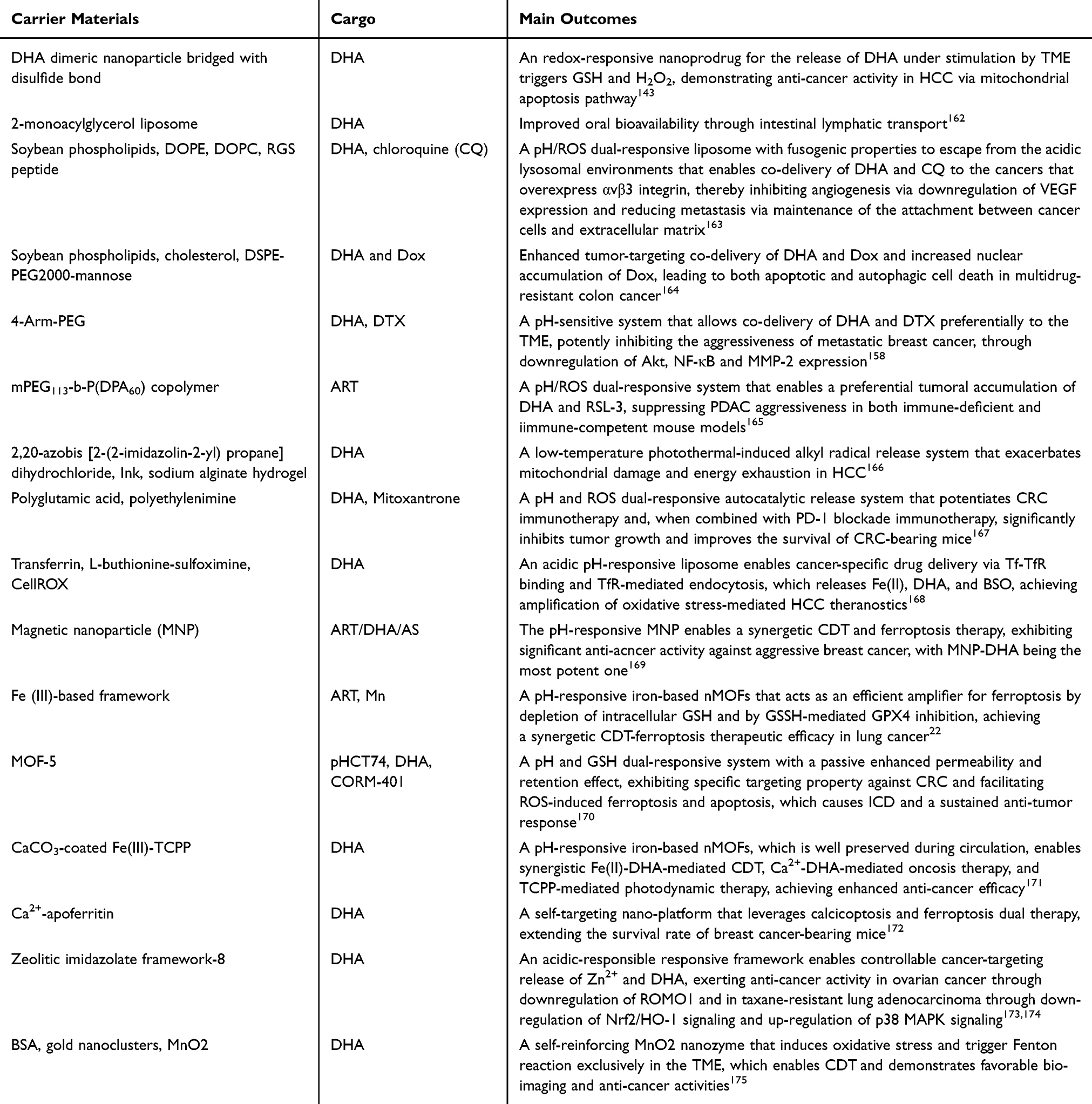 |
Table 3 A Summary of the Representative Nana-Enabled Drug Delivery System |
DHA loaded 2-monoacylglycerol mimetic liposomes promote DHA accumulation in mesenteric lymphatic nodes, significantly improving its oral bioavailability through intestinal lymphatic transport.162 Kang et al design a mannosylated LNP composed of soybean phospholipids (SPC), cholesterol, and DSPE-PEG2000-mannose (referred to as Man-lip) for the co-delivery of DHA and Dox in multidrug-resistant colon cancer.164 This Man-lip demonstrates effective endocytosis by drug-resistant human colon cancer HCT8/ADR cells, which exhibit overexpression of the mannose receptor CD206. In comparison to treatments involving Dox alone or the combination of DHA and Dox, Man-lip exhibits enhanced tumor-targeting drug delivery and increases the nuclear accumulation of Dox, leading to both apoptotic and autophagic cell death in drug-resistant colon cancer cells.164 Zhao et al introduce a self-targeting biomineralized apoferritin-based nano-platform (Ca/DHA@AFn) that leverages calcicoptosis and ferroptosis dual therapy, circumventing the insufficient intracellular ROS and sub-optimal drug accumulation within breast cancer, which hinders the efficacy of ferroptosis. Ca/DHA@AFn demonstrates a uniform distribution and high biocompatibility, exerting synergistic anti-cancer effects when compared to single-modality treatments, which thereby extends the survival rate of mice bearing breast cancer.172
Glycolysis Inhibition
Cancer cells engage active metabolic rewiring, with a preference for aerobic glycolysis (also known as the Warburg effect), to create a conducive microenvironment for their maximal survival.176 The observed upregulation in GLUTs in cancer cells is correlated with cancer aggressiveness, making GLUTs attractive targets of starvation therapy for cancer treatment.177 By encapsulating DHA, 2,20-azobis [2-(2-imidazolin-2-yl) propane] dihydrochloride (AI), and Ink into the sodium alginate hydrogel, Su et al develop a low-temperature photothermal-induced alkyl radical ((R•)) release system DHA/AI/Ink@ALG that exhibits anti-cancer potency in hepatocellular carcinoma.166 Upon intratumoral injection followed by in-situ irradiation with 1064 nm laser, AI rapidly decomposes into R• which not only exacerbates the mitochondrial damage synergistically with DHA-induced oxidative stress, leading to impaired ATP production, but also facilitates DHA-induced GLUT1 inhibition, leading to hindered glucose uptake.166 Thus, this thermocontrolled starvation strategy affords efficient tumor-suppressive effects by achieving energy exhaustion in tumor cells.
pH Responsible
Cancer cells feature a hallmark of “reversed” pH gradient” with an intracellular pH that is higher than the extracellular pH, allowing the innovation of pH-dependent cytotoxicity against cancer cells, from virtually non-cytotoxic at physiological pH to highly toxic in acidic microenvironment.178,179 By developing a pH-sensitive nanocarrier drug delivery system, 4-Arm-PEG-DTX-DHA, that allows acid-triggered co-delivery of DHA and docetaxel preferentially to the acidic TME, Tao et al observe excellent inhibitory effects on the proliferation and aggressiveness of metastatic breast cancer cells.158 Docetaxel (DTX) is an effective chemotherapy drug for breast cancer, but its therapeutic efficacy is unfavorable in metastatic breast cancer.180 The anti-metastatic activity of 4-Arm-PEG-DTX-DHA is associated with decreased expression of phosphorylated protein kinase B (Akt), NF-κB and matrix metallopeptidase 2 (MMP-2).158
Zeolitic imidazolate framework-8 (ZIF-8) stands as an ideal drug carrier due to its pH responsiveness, hydrothermal stability, excellent monodispersity, and high drug loading capacity.181 Yan et al develop an acidic-responsible responsive metal-organic framework loading DHA in the core of ZIF-8. ZIF-DHA collapses in the TME, enabling a controllable cancer-targeting release of Zn2+ and DHA, which exerts preferable pro-apoptosis and anti-cancer activity in ovarian cancer cells by downregulation of the reactive oxygen species modulator 1 (ROMO1).173 ZIF-DHA also demonstrates enhanced anti-cancer effects of DHA in taxane-resistant human lung adenocarcinoma (A549-TAX) cells. This is achieved via down-regulation of Nrf2/HO-1 signaling as well as up-regulation of p38 MAPK signaling while with reduced side effects on normal cells.174
An acidic pH-responsive, Tf-decorated liposomal nanocarrier that encapsulates DHA, L-buthionine-sulfoximine (BSO), and CellROX (Tf-DBC NP) is implemented for efficient tumor eradication in HCC via amplification of oxidative stress, without inducing obvious oxidative injury to normal liver cells.168 Tf-DBC NP achieves cancer-specific drug delivery via Tf-TfR binding and TfR-mediated endocytosis. The acidic environment within lysosomes activates Tf-DBC NP to release Fe(II), DHA, and BSO. Fe(II) can catalyze DHA to produce ROS, while BSO is a potent inhibitor of GSH synthesis. In addition, CellROX is a fluorescent probe for oxidative stress imaging, enabling in situ monition of the therapeutic efficacy. Tf-DBC NP thus offers a paradigm to achieve amplification of oxidative stress-mediated cancer theranostics.168
ROS/GSH Responsible
Cancer cells typically exhibit elevated levels of GSH and ROS, giving rise to the heterogeneity of TME in terms of oxidative and reductive stress.182 Li et al design an redox-responsive DHA dimeric nanoprodrug bridged with disulfide bond (DHA2-SS) for the release of DHA under stimulation by TME triggers GSH and H2O2.143 DHA2-SS can self-assemble into nanoparticles with high DHA content. Under oxidative stress, the disulfide bond of DHA2-SS is oxidized to hydrophilic sulfoxide or sulphone; while under reductive stress, the occurrence of sulfhydryl-disulfide bond exchange generates a thiol group. These reactions facilitate the hydrolysis of the adjacent ester bond, leading to DHA release. Compared with free DHA, DHA2-SS demonstrates more preferable anti-cancer activity in human hepatoma HepG2 cells xenograft mouse model by inducing apoptosis via mitochondrial apoptosis pathway, as well as glycolysis inhibition via down-regulation of PI3K/AKT/HIF-1α signaling.143
Nanoscale metal-organic framework (nMOF), which is created by inorganic metal nodes with organic bridging ligands, has been emerged as functional materials for diverse biomedical applications, due to its nontoxic nature, favorable biocompatibility and biodegradability, and controllable drug delivery.22,171 Wang et al develop a cubic manganese oxide nanozyme (BSA-AuNC-MnO2@DHA) that integrates bovine serum albumin (BSA)-coated gold nanoclusters (AuNC) with MnO2 to encapsulate DHA, which confers favorable bio-imaging and anti-cancer activities.175 MnO2 nanozymes are promising ROS-generating nanomedicines in chemodynamic therapy (CDT) which utilizes iron-initiated Fenton or Fenton-like reactions to eradicate tumor cells via converting less-active H2O2 into highly toxic ˙OH.183 ·OH can irreversibly cause accumulation of lipid peroxide (LPO), leading to ferroptosis. Considering CDT is activated by the TME triggers, such as low pH and elevated ROS, CDT-mediated ROS production is exclusively achieved at tumor region and thus, exerting low toxicity and side effects on normal tissues.184 As a self-reinforcing nanoreactor, MnO2 features both Fenton-like Mn2+ delivery and GSH depletion activities in the TME while with biodegradability.185,186 BSA–AuNC–MnO2@DHA is shown to suppress tumor growth in LLC model by amplifying DHA-induced oxidative stress with exceptional biocompatibility and safety.175
Similarly, the pH-responsive iron-based nMOFs also effectively release Fe(II) to trigger tumor-specific Fenton reaction under acidic TME for tumor eradication. Wan et al develop a nanoplatform for the programmed delivery of DHA at tumor site using Fe-TCPP [(4,4,4,4-(porphine-5,10,15,20-tetrayl) tetrakis(benzoic acid)] nMOF.171 Having a CaCO3 mineralized coating, the nanoplatform is preserved during circulation187. When arriving at the tumor site, the weakly acidic microenvironment and high levels of GSH trigger DHA release and TCPP activation, enabling the synergistic Fe(II)-DHA-mediated CDT, Ca2+-DHA-mediated oncosis therapy, and TCPP-mediated photodynamic therapy, which achieves enhanced anti-cancer efficiency but with negligible toxicity.171
Yang et al develop an iron-based nMOFs for the co-release of DHA and iron ions (MIL-101@DHA) under the acid TME. The generated ˙OH thereupon causes ferroptosis and apoptosis in LLC cells.22 In addition, DHA@MIL-101 can act as an efficient amplifier for ferroptosis by depletion of intracellular GSH (DHA@MIL-101 + GSH→Fe(II) + GSSH) and by GSSH-mediated GPX4 inhibition. The prominent anti-cancer activity of MIL-101@DHA with minimal systemic toxicity in LCC xenograft mouse model suggests this nanoreactor is a promising synergetic CDT-ferroptosis therapy for the treatment of lung cancer.22
By loading ART-based drugs into magnetic nanoparticle (MNP), Guo et al provide a framework to improve the therapeutic efficacy of CDT. Blank MNP is almost nontoxic, whereas MNP-ART, MNP-DHA, and MNP-AS demonstrate significant anti-cancer effects on drug-resistant breast cancer MCF-7/ADR cells, with MNP-DHA being the most potent one.169 The pH-responsive MNP produces Fe(II) within the acidic TME, which thereby catalyzes DHA to produce ROS, leading to cell death. Furthermore, MNP-DHA exhibits inhibitory activity against another two aggressive breast cancer MDA-MB-231 cells and MDA-MB-453 cells, making MNP-DHA a promising therapeutic for the treatment of intractable breast cancers.169
pH and ROS/GSH Dual Responsible
By engineering a pH/ROS dual-responsive nanoenabled system PDBA@RSL-3, a preferential tumoral accumulation of DHA and RSL-3, a GPX4 inhibitor, is achieved.165 DHA interacts with free ions to product carbon radicals and depletes intracellular glutathione, thereby synergizing with RSL-3 to potentiate ferroptotic therapy of pancreatic ductal adenocarcinoma (PDAC), one of the most lethal malignancies with resistance to conventional therapies, in both immune-deficient mouse models.165,188,189 Importantly, PDBA@RSL-3 in combination with the programmed cell death protein 1 (PD-1) blockade immunotherapy affords attractive suppressive effect on PDAC aggressiveness in immune-competent mouse model, providing a novel hint for innovation of ferroptosis-based immunotherapy for PDAC treatment.156,165,190
Peng et al develop a pH/ROS dual-responsive lipid nanoparticle (LNP) for the co-delivery of DHA and chloroquine phosphate (CQ), an effective autophagy inhibitor. DHA and CQ are encapsulated within a lipid layer composed of soybean phospholipids (SPC), 1,2-dioleoyl-sn-glycero-3-phosphoethanolamine (DOPE), and 1,2-dioleoyl-sn-glycero-3-phosphocholine (DOPC).163 SPC is susceptible to oxidation, leading to the formation of lipid peroxides, which increases the hydrophilicity of the lipid layer and compromises its structural integrity. DOPE imparts fusogenic properties to the liposomes, enabling them to escape from lysosomes in acidic environments.163,191 Coating LNPs with cyclic arginylglycylaspartic acid (RGD) peptide, a specific ligand for the αvβ3 integrin, enhances the capability of the LNPs towards cancers that overexpress αvβ3 integrin, including CRC.47 The resulting RGD-modified LNPs (RLNP/DC) exhibits enhanced anti-cancer effects of combination therapy with DHA and CQ in CRC. This is achieved by inducing oxidative stress via DHA-induced ROS generation and CQ-mediated autophagy suppression, inhibiting angiogenesis via downregulation of VEGF expression, and reducing metastasis via maintenance of the attachment between paxillin in cancer cells and integrin on extracellular matrix (ECM) via upregulation of paxillin expression.163
Su et al construct a pH and ROS dual-responsive autocatalytic release system (TPDM/PGA) by conjugating DHA and mitoxantrone to ROS-responsive polyethylenimine (TP) through a ROS-cleavable linker. This system is subsequently coated with pH-responsive polyglutamic acid (PGA). The dissociation of PGA within the TME facilitates its deep penetration and cellular internalization. Meanwhile, the ROS-responsive release of DHA and mitoxantrone creates a positive feedback to exacerbate oxidative stress. TPDM/PGA potentiates CRC immunotherapy and, when combined with PD-1 blockade immunotherapy, it significantly inhibits tumor growth and improves the survival of CRC-bearing mice.167
Yang et al develop a pH/GSH dual-responsive nanoenabled delivery system, pHCT74/MOF-5@DHA&CORM-401 (PMDC NPs), which enables efficient release of cargo, including DHA and CORM-401, in the TME with a passive enhanced permeability and retention effect. In addition, PMDC NPs exhibit specific targeting property against CRC due to the pHCT74 peptide-mediated active targeting of overexpressed α-enolase on CRC cells. Mechanistically, DHA stimulates CORM-401 to release CO, facilitating ROS-induced ferroptosis and apoptosis, which causes ICD and a sustained anti-tumor response.170 These findings indicate that the combined use of immunotherapy and gas therapy represents a promising therapeutic strategy for the treatment of CRC.
Therapeutic Activities of ARTs in Alzheimer’s Disease
Alzheimer’s disease (AD) is a neurodegenerative disorder characterized by progressive cognitive impairment, the histopathological features of which is the presence of extracellular β-amyloid (Aβ)-containing plaques and intracellular hyperphosphorylated tau-containing neurofibrillary tangles.192,193 Aβ is generated from amyloid precursor protein (APP) via the amyloidogenic pathway, which is catalyzed by β-secretase (BACE1) and γ-secretase. The aggregation of Aβ can disrupt intercellular communication, induce neuroinflammation, and initiate tauopathy, all of which contribute to AD pathogenesis. ARTs are shown to beneficially mitigate AD pathology by regulating a wide spectrum of cellular processes, including Aβ deposition and the inflammatory response (Figure 5).
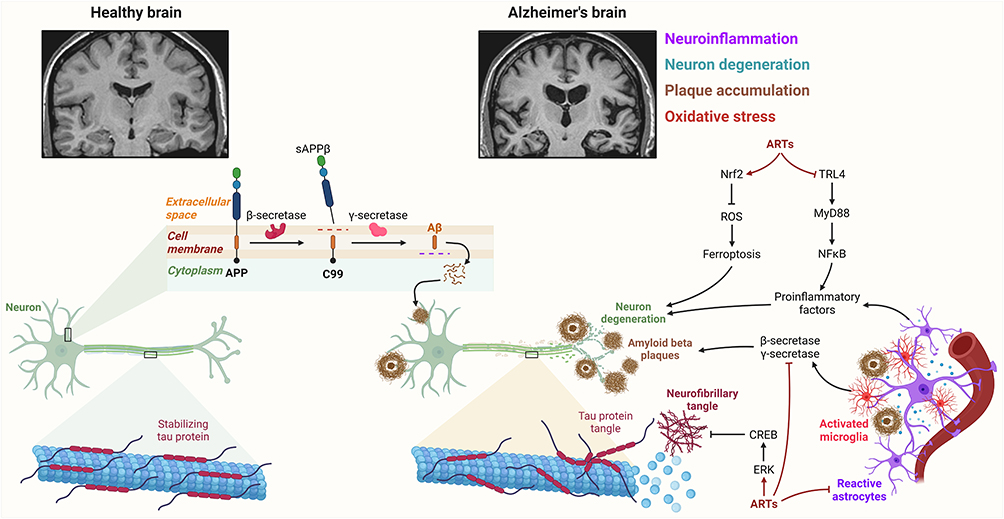 |
Figure 5 Therapeutic potential of ARTs in Alzheimer’s disease by amelioration of amyloidopathy, tauopathy, and neuroinflammation. |
Reduction of Amyloidogenic Components’ Expression
The administration of ART over a consecutive period of 30 days has been shown to decrease the brain expression of BACE1, resulting in the amelioration of neuritic plaque burden by approximately 61% in the hippocampus and 48% in the cortex of the APPswe/PS1dE9 (APP/PS1) transgenic mouse model of AD.194 In a separate investigation, treatment with ART or AS for three months significantly diminished Aβ plaque load in the cortex and hippocampus of APP/PS1 mice, as evidenced by a decrease in the abundance of APP C-terminal fragments (APP-CTFs) and soluble Aβ in the hippocampus, suggesting a blocking of APP cleavage.195,196 Notably, the preventive effects against amyloidosis were not observed at a high dosage of 100 mg/kg/day, highlighting the importance of considering dose-dependent effects in therapeutic applications.195 Furthermore, oral administration of DHA for three months also led to a reduction in the presence of amyloid plaques in the cortex and hippocampus of male APP/PS1 mice, attributed to the downregulation of APP and BACE1 expression, without evident toxicity to the liver and kidneys.197,198 The phenotypic traits associated with AD were similarly improved by AS, as indicated by a reduction in brain deposition of amyloid plaques in 5xFAD mice, another model of AD.199 However, the brain expressions of amyloidogenic components, including APP, APP-CTFs, and BACE1, as well as Aβ-degrading enzymes such as neprilysin and insulin-degrading enzyme in 5xFAD mice, remained unchanged following AS treatment. This observation suggests the presence of alternative mechanisms involved in the elimination of amyloid plaques in AS-treated 5xFAD mice.199 Collectively, these findings provide compelling evidence for the therapeutic potential of ARTs in mitigating AD’s amyloid pathology.
Amelioration of Tau Pathology
It is well-established that regional interactions between Aβ and tau drive the propagation of tauopathy, which subsequently exacerbates amyloidopathy in AD.200,201 Tau is a microtubule-associated protein that facilitates the organization and stabilization of microtubules, exhibiting high expression levels in axonal structures. In the context of AD, tau undergoes hyperphosphorylation, resulting in the formation of insoluble aggregates that can be transferred intercellularly. This process leads to the impairment of axonal transport, thereby compromising neuronal function and synaptic plasticity. Li et al (2019) documented the beneficial effects of ART following a four-week treatment regimen, which resulted in a reduction of amyloidopathy and tau pathology in the cortex of the 3xTg mouse model of AD. Furthermore, ART has been shown to ameliorate Aβ-induced cognitive impairments by downregulating the expression levels of Aβ, BACE1, and tau in N2a cells.202 In 3xTg mice, treatment with ART or extracts from Artemisia annua significantly reduced the levels of Aβ deposition, neurofibrillary tangles, and tau hyperphosphorylation in both the cortex and hippocampus, without altering the total tau expression. This effect was observed in a dose-dependent manner and was mediated through the activation of the extracellular regulated protein kinases (ERK)/cAMP-response element binding protein (CREB) signaling pathways.194,203
Suppression of Inflammatory Response
Elevated levels of inflammatory markers in patients with AD and the involvement of AD risk genes in innate immunity suggest that neuroinflammation is a significant aspect of AD pathology.204,205 Pro-inflammatory cytokines not only enhance amyloidopathy by upregulating BACE1 and γ-secretase but also exacerbate tau pathology by stimulating atypical cyclin-dependent kinase 5 (CDK5)-mediated tau hyperphosphorylation.206,207 With important regulatory activities in neuroinflammation in central nervous system (CNS), glia cells including microglia and astrocyte exhibit spatial and temporal phenotypic variations in different settings.205 Microglia cells are macrophages that represent less than 10% of brain cells. The activation of microglia at early stage of disease pathogenesis has neuroprotective effects by removal of harmful stimuli, while its persistent activation induced by the neurotoxic Aβ stimulates NF-κB signaling to incite the activation of NLRP3 inflammasome, thereby driving neurotoxicity. Inhibition of NLRP3 has been shown to ameliorate cognitive deficits in AD models.208,209 Activated microglia also release several astrocyte-activating signals, including tumor necrosis factor alpha (TNF-α), interleukin-1, alpha (IL-1α), and complement component 1q (C1q).210,211 Astrocytes, the most abundant glial cells in the brain, reside at the parenchymal part of the blood–brain barrier (BBB) that maintain the structural and functional integrity of BBB. Astrocytes possess the ability for Aβ clearance, which confers neuroprotective effects. However, when stimulated by neurotoxic Aβ or activated microglia, they exacerbate neuronal amyloidopathy by upregulating the expression of BACE1 and γ-secretase.212,213 These findings indicate that modulation of glial activity may serve as a intervention strategy for AD.
Recent studies have demonstrated the anti-inflammatory effects of ARTs in AD mouse models. Specifically, Artemisia annua extract has been shown to reduce neuroinflammation in 12-month-old 3xTg mice by lowering the production of inflammatory cytokines while alleviating tau and amyloid pathologies.203 In a separate study, ART treatment administered to 5-month-old APPswe/PS1dE9 mice over a 30-day period resulted in a reduction of NF-κB p65 nuclear translocation and an increase in I-κBα expression, leading to decreases of 37.16% and 34.46% in the production of IL-6 and TNF-α, respectively. Furthermore, ART treatment decreased NALP3 expression by 37% and reduced the production of caspase-1 p20 and IL-1β by 50% and 29.73%, respectively.214 In 3xTg mice, intraperitoneal injection (i.p). of ART was found to attenuate the activation of glial cells and the expression of inflammatory factors.194 In APPswe/PS1dE9 mice, administration of AS from 2 to 6 months of age effectively reduced the expression of TNF-α, IL-6, and IL-1β, while also improving alterations in mitochondrial dynamics.196 Additionally, AS (32 mg/kg/day, i.p.) has been shown to suppress neuroinflammation in 5XFAD mice, evidenced by reduced activation of both microglia and astrocytes.199 In non-transgenic AD models, ART treatment was associated with an increase in IL-10 and a decrease in TNF-α in Aβ25-35-injected mice, while also significantly reducing glial activation in Aβ1-42-injected mice.215,216 Notably, ART has demonstrated a protective effect on dopaminergic neurons by reducing microglial activation through the Toll-like receptor 4 (TLR4)/Myeloid differentiation factor 88 (MyD88)/NF-κB signaling pathway in a 1-methyl-4-phenyl-1,2,3,6-tetrahydropyridine (MPTP)-induced Parkinson’s disease mouse model.217
Mitigation of Oxidative Stress
Aβ can stimulate the production of reactive oxygen species (ROS) and ARTs can exhibit neuroprotective effects by mitigating oxidative stress in hippocampal neurons.175,197,218,219 Ferroptosis is an iron-dependent form of non-apoptotic programmed cell death induced by overwhelming lipid peroxidation of biological membranes. It has important implications in a wide spectrum of diseases, including neurodegenerative disorders and type 2 diabetes mellitus (T2DM).23,220,221 Epidemiological study suggests that individuals with type 2 diabetes are at approximate 60% greater risk for the development of dementia, and the diabetic cognitive deficits can be improved by ferroptosis inhibition.222,223 ART effectively ameliorates neuropathological changes and cognitive impairments in T2DM mice by activation of Nrf2 to inhibit neuronal ferroptosis in the hippocampal CA1 region.175
ARTs in Improvement of Reproductive Health
Recently, ARTs have been identified as effective anti-polycystic ovary syndrome (PCOS) agents in both animal models and human patients. PCOS stands as the leading cause of female infertility, with an estimated heritability of 70% and a prevalence of ~15% in women of reproductive age. It is associated with a diverse lifetime risk of comorbidities, including hyperandrogenemia, hyperinsulinemia, and gynecological pathologies.224 While accumulating evidence suggests that androgen and anti-Müllerian hormone (AMH) are contributing factors to PCOS through genetic or epigenetic mechanisms,225–227 the etiology of PCOS remains largely unknown, making current interventions for PCOS are nonspecific and symptomatic. Recently, ARTs have been identified as effective anti-PCOS agents in both animal models and human patients. They work by inducing the degradation of cytochrome P450 11A1 (CYP11A1), which suppresses ovarian androgen synthesis.18 ARTs can directly target lon peptidase 1 (LONP1), enhancing LONP1-CYP11A1 interaction and facilitating LONP1-driven CYP11A1 degradation. The phenotypes of this process can be replicated by LONP1 overexpression.18 These findings present a novel therapeutic strategy for alleviating and treating PCOS.
Potential Reproductive Toxicity of ARTs: Evidence From Animal and Cohort Studies
Malaria in pregnancy is prevalent in malaria-endemic areas, being a clinically changeling infectious disease without effective drugs, partially due to drug resistance, to improve pregnancy outcomes.228,229 Evidence from non-clinic studies suggests that single or consecutive treatment of ARTs in the first trimester of pregnancy is associated with embryolethality, due to the loss of circulating embryonic primitive erythroblasts, and teratogenic effects.230,231 Cohort studies, however, suggest that first-trimester exposure to ARTs induces no adverse pregnancy outcomes in women with or without malaria infection.228,230,232–234 ART-based combination therapies (ACTs), such as artesunate-amodiaquine, artesunate-mefloquine, artemether-lumefantrine, and DHA-piperaquine are proposed as favorable treatments for uncomplicated P falciparum malaria in the first trimester, compared to quinine-based therapy.229,232–234 The adverse pregnancy outcomes, including miscarriage, stillbirth, low birth weight, small for gestational age and congenital anomalies and cardiovascular defects in newborns, are less seen in artemether-lumefantrine-exposed pregnancies, compared with that in quinine-exposed pregnancies, after anti-malarial treatment in the first, second, and third trimester.228,232–238 It is accepted that the benefit of 3-d ATCs in the early pregnancy is likely to outweigh the unfavorable outcomes of partially treated malaria, which occurs with quinine treatment due to the known low tolerability and associated poor compliance to the 7-d regimen.228,239
The potential reproductive toxicity of ART should be considered seriously, as relatively low dosage is applied for only a few days for malaria therapy. Thus, drug accumulation over a prolonged period of treatment, which frequently arises toxic side effects, is less possible. Indeed, the toxic effects are documented following prolonged treatment. In non-pregnant female rats, exposure to ART (7, 35, 70 mg/kg) for 7 consecutive days significantly increases the levels of hydrogen peroxide and malondialdehyde while decreasing the activity of catalase, glutathione peroxidase and superoxide dismutase in the erythrocytes and uterus, but not in the ovarian. The decreased follicle-stimulating hormone levels and increased levels of progesterone are also seen in ART-exposed females.240 These findings indicate ART is a endocrine-disrupting chemical with oxidative toxicity. Similar oxidative toxicities are likewise observed in the epididymis, but not in the testes, of the male rats treated with ART (35 mg/kg) for 7 consecutive days.241 While exhibiting no endocrine toxicity against the brain-pituitary-testicular axis, ART incurs severe degeneration in epididymis and impairment in sperm quality and quantity.241
Conclusion
Natural products serve as the foundation for discovering and developing novel therapeutic drugs, with a long history of global use for both preventive and therapeutic purposes.242–252 ARTs represent a profoundly valuable class of natural compounds that have evolved from a traditional remedy into front-line anti-malarial drugs, now demonstrating significant potential for repurposing in oncology, neurology, and reproductive medicine253. The combination of ARTs with other pharmacological agents can produce synergistic therapeutic effects but may also lead to antagonistic toxicities. However, the successful translation of this potential into safe and effective clinical therapies is contingent upon overcoming critical challenges. A fundamental gap exists in our understanding of the therapeutic mechanisms of ART-based drugs at the molecular level, which hinders the rational design of optimized treatments and the prediction of off-target effects. Furthermore, while short-term use is well-tolerated, the safety profile of long-term, high-dose regimens-essential for treating chronic diseases like cancer-remains inadequately documented. The development of ART-based therapies must therefore be guided by rigorous preclinical and clinical safety evaluations, particularly as novel delivery systems like nanomaterials are explored.
Current Challenges and Future Perspectives
The development of ART-based drugs is constrained by several key limitations, including an ambiguous understanding of their molecular mechanisms, which hinders treatment optimization and prediction of off-target effects. Furthermore, their safety profile is incomplete, with a notable lack of documentation on the long-term side effects, especially at the high doses used in oncology. The application of nano-enabled delivery systems introduces additional concerns regarding the toxicity of the carriers themselves over extended treatments. Finally, while combination therapies offer promise, they also carry the risk of antagonistic interactions that could diminish efficacy or exacerbate toxicity.
Future research needs to focus on addressing current challenges to crack the full paradigm of ART-based therapy, primarily through detailed investigations to elucidate its ambiguous molecular mechanisms and provide a foundation for rational drug design. This should be coupled with laboratory-based mechanistic studies and advanced preclinical and long-term clinical trials to establish safety parameters for chronic use, particularly at high doses. Also, efforts should be devoted toward developing safer, more biocompatible nanocarriers for delivery and exploring novel formulations to improve bioavailability and targeting specificity. Finally, the integration of artificial intelligence for screening combination therapies and personalizing treatment regimens presents a powerful strategy to maximize synergistic efficacy while circumventing toxicity and antagonistic effects for individual patients.
Declaration of Generative AI in Scientific Writing
The authors declare that no generative AI was used in scientific writing upon preparation of the paper.
Data Sharing Statement
Data sharing is not applicable to this article because no new data were created or analyzed in this study.
Acknowledgment
We sincerely thank all the scientists dedicated to the understanding of ARTs in disease treatment.
Author Contributions
All authors made a significant contribution to the work reported, whether that is in the conception, study design, execution, acquisition of data, analysis and interpretation, or in all these areas; took part in drafting, revising or critically reviewing the article; gave final approval of the version to be published; have agreed on the journal to which the article has been submitted; and agree to be accountable for all aspects of the work.
Funding
This work was supported by the Natural Science Foundation of Jiangsu Province (No. BK20250952) and the Natural Science Foundation of the Higher Education Institutions of Jiangsu Province (No. 24KJB180015).
Disclosure
The authors declare that no known competing financial interests or personal relationships that could have appeared to influence the work reported in this paper.
References
1. Wang Z, Lu Z, Chen Y, et al. Targeting the AKT-P53/CREB pathway with epicatechin for improved prognosis of traumatic brain injury. CNS Neurosci Ther. 2024;30(2):e14364. doi:10.1111/cns.14364
2. Zhang Y, Shen L, Xie J, et al. Pushen capsule treatment promotes functional recovery after ischemic stroke. Phytomedicine. 2023;111:154664. doi:10.1016/j.phymed.2023.154664
3. Liu ZB, Guo S, Dong Q. Nobiletin suppresses IL-21/IL-21 receptor-mediated inflammatory response in MH7A fibroblast-like synoviocytes (FLS): an implication in rheumatoid arthritis. Eur J Pharmacol. 2020;875:172939. doi:10.1016/j.ejphar.2020.172939
4. Cao RY, Zhang Y, Feng Z, et al. The effective role of natural product berberine in modulating oxidative stress and inflammation related atherosclerosis: novel insights into the gut-heart axis evidenced by genetic sequencing analysis. Front Pharmacol. 2021;12:764994. doi:10.3389/fphar.2021.764994
5. Fan Z, Cai L, Wang S, Wang J, Chen B. Baicalin prevents myocardial ischemia/reperfusion injury through inhibiting ACSL4 mediated ferroptosis. Front Pharmacol. 2021;12:628988. doi:10.3389/fphar.2021.628988
6. Hua YQ, Zeng Y, Xu J, Xu XL. Naringenin alleviates nonalcoholic steatohepatitis in middle-aged Apoe-/- mice: role of SIRT1. Phytomedicine. 2021;81:153412. doi:10.1016/j.phymed.2020.153412
7. Xia B, Li Q, Wu J, et al. Sinomenine confers protection against myocardial ischemia reperfusion injury by preventing oxidative stress, cellular apoptosis, and inflammation. Front Pharmacol. 2022;13:922484. doi:10.3389/fphar.2022.922484
8. Xia M, Zhang Y, Wu H, et al. Forsythoside B attenuates neuro-inflammation and neuronal apoptosis by inhibition of NF-κB and p38-MAPK signaling pathways through activating Nrf2 post spinal cord injury. Int Immunopharmacol. 2022;111:109120. doi:10.1016/j.intimp.2022.109120
9. Zhang H, Sun Y, Yau S, et al. Synergistic effects of two naturally occurring iridoids in eliciting a rapid antidepressant action by up-regulating hippocampal PACAP signalling. Br J Pharmacol. 2022;179(16):4078–4091. doi:10.1111/bph.15847
10. Zhang L, Zhang H, Gu J, et al. Glabridin inhibits liver fibrosis and hepatic stellate cells activation through suppression of inflammation and oxidative stress by activating PPARγ in carbon tetrachloride-treated mice. Int Immunopharmacol. 2022;113(Pt B):109433. doi:10.1016/j.intimp.2022.109433
11. Xu L, Bai E, Zhu Y, Qin J, Du X, Huang H. pH-Responsive hydrogel as a potential oral delivery system of baicalin for prolonging gastroprotective activity. Pharmaceutics. 2023;15(1):257. doi:10.3390/pharmaceutics15010257
12. O’Neill PM, Posner GH. A medicinal chemistry perspective on artemisinin and related endoperoxides. J Med Chem. 2004;47(12):2945–2964. doi:10.1021/jm030571c
13. Chen GQ, Benthani FA, Wu J, Liang D, Bian ZX, Jiang X. Artemisinin compounds sensitize cancer cells to ferroptosis by regulating iron homeostasis. Cell Death Differ. 2020;27(1):242–254. doi:10.1038/s41418-019-0352-3
14. Gao P, Shen S, Li X, et al. Dihydroartemisinin inhibits the proliferation of leukemia cells K562 by suppressing PKM2 and GLUT1 mediated aerobic glycolysis. Drug Des Devel Ther. 2020;14:2091–2100. doi:10.2147/DDDT.S248872
15. Xiao X, Li Y, Wang Y, et al. Dihydroartemisinin inhibits Lewis lung carcinoma progression by inducing macrophages M1 polarization via AKT/mTOR pathway. Int Immunopharmacol. 2022;103:108427. doi:10.1016/j.intimp.2021.108427
16. Han N, Yang ZY, Xie ZX, et al. Dihydroartemisinin elicits immunogenic death through ferroptosis-triggered ER stress and DNA damage for lung cancer immunotherapy. Phytomedicine. 2023;112:154682. doi:10.1016/j.phymed.2023.154682
17. Liu Y, Jiang JJ, Du SY, et al. Artemisinins ameliorate polycystic ovarian syndrome by mediating LONP1-CYP11A1 interaction. Science. 2024;384(6701):eadk5382. doi:10.1126/science.adk5382
18. Liu Y, Li H, Luo Z, et al. Artesunate, a new antimalarial clinical drug, exhibits potent anti-AML activity by targeting the ROS/Bim and TFRC/Fe2+ pathways. Br J Pharmacol. 2023;180(6):701–720. doi:10.1111/bph.15986
19. Woerdenbag HJ, Moskal TA, Pras N, et al. Cytotoxicity of artemisinin-related endoperoxides to Ehrlich ascites tumor cells. J Nat Prod. 1993;56(6):849–856. doi:10.1021/np50096a007
20. Ba Q, Zhou N, Duan J, et al. Dihydroartemisinin exerts its anticancer activity through depleting cellular iron via transferrin receptor-1. PLoS One. 2012;7(8):e42703. doi:10.1371/journal.pone.0042703
21. Yi R, Wang H, Deng C, et al. Dihydroartemisinin initiates ferroptosis in glioblastoma through GPX4 inhibition. Biosci Rep. 2020;40(6):BSR20193314. doi:10.1042/BSR20193314
22. Yang X, Xu X, Wang M, et al. A nanoreactor boosts chemodynamic therapy and ferroptosis for synergistic cancer therapy using molecular amplifier dihydroartemisinin. J Nanobiotechnology. 2022;20(1):230. doi:10.1186/s12951-022-01455-0
23. Wang T, Luo R, Li W, et al. Dihydroartemisinin suppresses bladder cancer cell invasion and migration by regulating KDM3A and p21. J Cancer. 2020;11(5):1115–1124. doi:10.7150/jca.36174
24. Li Y, Zhou X, Liu J, et al. Dihydroartemisinin inhibits the tumorigenesis and metastasis of breast cancer via downregulating CIZ1 expression associated with TGF-β1 signaling. Life Sci. 2020;248:117454. doi:10.1016/j.lfs.2020.117454
25. Dong J, Chen Y, Yang W, Zhang X, Li L. Antitumor and anti-angiogenic effects of artemisinin on breast tumor xenografts in nude mice. Res Vet Sci. 2020;129:66–69. doi:10.1016/j.rvsc.2020.01.005
26. Tsui KH, Wu MY, Lin LT, et al. Disruption of mitochondrial homeostasis with artemisinin unravels anti-angiogenesis effects via auto-paracrine mechanisms. Theranostics. 2019;9(22):6631–6645. doi:10.7150/thno.33353
27. Zhang T, Hu Y, Wang T, Cai P. Dihydroartemisinin inhibits the viability of cervical cancer cells by upregulating caveolin 1 and mitochondrial carrier homolog 2: involvement of p53 activation and NAD(P)H:quinone oxidoreductase 1 downregulation. Int J Mol Med. 2017;40(1):21–30. doi:10.3892/ijmm.2017.2980
28. Gong RH, Yang DJ, Kwan HY, Lyu AP, Chen GQ, Bian ZX. Cell death mechanisms induced by synergistic effects of halofuginone and artemisinin in colorectal cancer cells. Int J Med Sci. 2022;19(1):175–185. doi:10.7150/ijms.66737
29. Jung EJ, Kim HJ, Shin SC, et al. Artemisia annua L. polyphenols enhance the anticancer effect of β-lapachone in oxaliplatin-resistant HCT116 colorectal cancer cells. Int J Mol Sci. 2023;24(24):17505. doi:10.3390/ijms242417505
30. Cui W, Fang T, Duan Z, et al. Dihydroartemisinin sensitizes esophageal squamous cell carcinoma to cisplatin by inhibiting sonic hedgehog signaling. Front Cell Dev Biol. 2020;8:596788. doi:10.3389/fcell.2020.596788
31. Qu C, Ma J, Liu X, et al. Dihydroartemisinin exerts anti-tumor activity by inducing mitochondrion and endoplasmic reticulum apoptosis and autophagic cell death in human glioblastoma cells. Front Cell Neurosci. 2017;11:310. doi:10.3389/fncel.2017.00310
32. Jia L, Song Q, Zhou C, et al. Dihydroartemisinin as a putative STAT3 inhibitor, suppresses the growth of head and neck squamous cell carcinoma by targeting Jak2/STAT3 signaling. PLoS One. 2016;11(1):e0147157. doi:10.1371/journal.pone.0147157
33. Zhang X, Li R, Xu C, Li X. Regulation of dihydroartemisinin on the pathological progression of laryngeal carcinoma through the periostin/YAP/IL-6 pathway. Heliyon. 2024;10(6):e27494. doi:10.1016/j.heliyon.2024.e27494
34. Sun Y, Lu X, Li H, Li X. Dihydroartemisinin inhibits IL-6-induced epithelial-mesenchymal transition in laryngeal squamous cell carcinoma via the miR-130b-3p/STAT3/β-catenin signaling pathway. J Int Med Res. 2021;49(11):3000605211009494. doi:10.1177/03000605211009494
35. Shi X, Li S, Wang L, et al. RalB degradation by dihydroartemisinin induces autophagy and IFI16/caspase-1 inflammasome depression in the human laryngeal squamous cell carcinoma. Chin Med. 2020;15:64. doi:10.1186/s13020-020-00340-y
36. Liu ZL, Chen HH, Zheng LL, Sun LP, Shi L. Angiogenic signaling pathways and anti-angiogenic therapy for cancer. Signal Transduct Target Ther. 2023;8(1):198. doi:10.1038/s41392-023-01460-1
37. Qin G, Zhao C, Zhang L, et al. Dihydroartemisinin induces apoptosis preferentially via a Bim-mediated intrinsic pathway in hepatocarcinoma cells. Apoptosis. 2015;20(8):1072–1086. doi:10.1007/s10495-015-1132-2
38. Li LG, Peng XC, Yu TT, et al. Dihydroartemisinin remodels macrophage into an M1 phenotype via ferroptosis-mediated DNA damage. Front Pharmacol. 2022;13:949835. doi:10.3389/fphar.2022.949835
39. Li LG, Peng XC, Yang ZY, et al. Dihydroartemisinin-driven selective anti-lung cancer proliferation by binding to EGFR and inhibition of NRAS signaling pathway-induced DNA damage. Sci Rep. 2024;14(1):11704. doi:10.1038/s41598-024-62126-8
40. Wu S, Li Z, Li H, Liao K. Dihydroartemisinin reduces Irradiation-Induced mitophagy and radioresistance in lung cancer A549 cells via CIRBP inhibition. Life. 2022;12(8):1129. doi:10.3390/life12081129
41. Wu S, Zhang K, Liang Y, et al. Nano-enabled tumor systematic energy exhaustion via zinc (II) interference mediated glycolysis inhibition and specific GLUT1 depletion. Adv Sci. 2022;9(7):e2103534. doi:10.1002/advs.202103534
42. Yang Z, Zhou Z, Meng Q, et al. Dihydroartemisinin sensitizes lung cancer cells to cisplatin treatment by upregulating ZIP14 expression and inducing ferroptosis. Cancer Med. 2024;13(19):e70271. doi:10.1002/cam4.70271
43. Zhang Q, Jin L, Jin Q, et al. Inhibitory effect of dihydroartemisinin on the proliferation and migration of melanoma cells and experimental lung metastasis from melanoma in mice. Front Pharmacol. 2021;12:727275. doi:10.3389/fphar.2021.727275
44. Phikulsod P, Kariya R, Panaampon J, Okada S. Dihydroartemisinin induced apoptosis and synergized with chemotherapy in pleural effusion lymphoma cells. Anticancer Res. 2023;43(3):1139–1148. doi:10.21873/anticanres.16259
45. Lu Z, Bi J, Wan X. Artemisinin sensitizes tumor cells to NK cell-mediated cytolysis. Biochem Biophys Res Commun. 2020;524(2):418–423. doi:10.1016/j.bbrc.2020.01.094
46. Paccez JD, Duncan K, Sekar D, et al. Dihydroartemisinin inhibits prostate cancer via JARID2/miR-7/miR-34a-dependent downregulation of Axl. Oncogenesis. 2019;8(3):14. doi:10.1038/s41389-019-0122-6
47. Wang Y, Yin S, Zhang L, et al. A tumor-activatable particle with antimetastatic potential in breast cancer via inhibiting the autophagy-dependent disassembly of focal adhesion. Biomaterials. 2018;168:1–9. doi:10.1016/j.biomaterials.2017.10.039
48. Zhang Y, Shi J, Liu X, et al. BAP1 links metabolic regulation of ferroptosis to tumour suppression. Nat Cell Biol. 2018;20(10):1181–1192. doi:10.1038/s41556-018-0178-0
49. Gao M, Yi J, Zhu J, et al. Role of mitochondria in ferroptosis. Mol Cell. 2019;73(2):354–363.e3. doi:10.1016/j.molcel.2018.10.042
50. Dixon SJ, Pratt DA. Ferroptosis: a flexible constellation of related biochemical mechanisms. Mol Cell. 2023;83(7):1030–1042. doi:10.1016/j.molcel.2023.03.005
51. Chafe SC, Vizeacoumar FS, Venkateswaran G, et al. Genome-wide synthetic lethal screen unveils novel CAIX-NFS1/xCT axis as a targetable vulnerability in hypoxic solid tumors. Sci Adv. 2021;7(35):eabj0364. doi:10.1126/sciadv.abj0364
52. Tian S, Chu Y, Hu J, et al. Tumour-associated neutrophils secrete AGR2 to promote colorectal cancer metastasis via its receptor CD98hc-xCT. Gut. 2022;71(12):2489–2501. doi:10.1136/gutjnl-2021-325137
53. Ruiu R, Cossu C, Iacoviello A, et al. Cystine/glutamate antiporter xCT deficiency reduces metastasis without impairing immune system function in breast cancer mouse models. J Exp Clin Cancer Res. 2023;42(1):254. doi:10.1186/s13046-023-02830-x
54. Tang B, Zhu J, Wang Y, et al. Targeted xCT-mediated ferroptosis and protumoral polarization of macrophages is effective against HCC and enhances the efficacy of the Anti-PD-1/L1 response. Adv Sci. 2023;10(2):e2203973. doi:10.1002/advs.202203973
55. Viswanathan VS, Ryan MJ, Dhruv HD, et al. Dependency of a therapy-resistant state of cancer cells on a lipid peroxidase pathway. Nature. 2017;547(7664):453–457. doi:10.1038/nature23007
56. Xie Y, Hou W, Song X, et al. Ferroptosis: process and function. Cell Death Differ. 2016;23(3):369–379. doi:10.1038/cdd.2015.158
57. Ooko E, Saeed ME, Kadioglu O, et al. Artemisinin derivatives induce iron-dependent cell death (ferroptosis) in tumor cells. Phytomedicine. 2015;22(11):1045–1054. doi:10.1016/j.phymed.2015.08.002
58. Pérez-Gutiérrez L, Ferrara N. Biology and therapeutic targeting of vascular endothelial growth factor A. Nat Rev Mol Cell Biol. 2023;24(11):816–834. doi:10.1038/s41580-023-00631-w
59. Radtke F, Raj K. The role of notch in tumorigenesis: oncogene or tumour suppressor? Nat Rev Cancer. 2003;3(10):756–767. doi:10.1038/nrc1186
60. Lagouge M, Argmann C, Gerhart-Hines Z, et al. Resveratrol improves mitochondrial function and protects against metabolic disease by activating SIRT1 and PGC-1alpha. Cell. 2006;127(6):1109–1122. doi:10.1016/j.cell.2006.11.013
61. Guo D, Wang Q, Li C, Wang Y, Chen X. VEGF stimulated the angiogenesis by promoting the mitochondrial functions. Oncotarget. 2017;8(44):77020–77027. doi:10.18632/oncotarget.20331
62. Porporato PE, Filigheddu N, Pedro JMB, Kroemer G, Galluzzi L. Mitochondrial metabolism and cancer. Cell Res. 2018;28(3):265–280. doi:10.1038/cr.2017.155
63. Diebold LP, Gil HJ, Gao P, Martinez CA, Weinberg SE, Chandel NS. Mitochondrial complex III is necessary for endothelial cell proliferation during angiogenesis. Nat Metab. 2019;1(1):158–171. doi:10.1038/s42255-018-0011-x
64. Raggi C, Taddei ML, Sacco E, et al. Mitochondrial oxidative metabolism contributes to a cancer stem cell phenotype in cholangiocarcinoma. J Hepatol. 2021;74(6):1373–1385. doi:10.1016/j.jhep.2020.12.031
65. Yamada T, Amann JM, Fukuda K, et al. Akt kinase-interacting protein 1 signals through CREB to drive diffuse malignant mesothelioma. Cancer Res. 2015;75(19):4188–4197. doi:10.1158/0008-5472.CAN-15-0858
66. Bélanger MM, Roussel E, Couet J. Caveolin-1 is down-regulated in human lung carcinoma and acts as a candidate tumor suppressor gene. Chest. 2004;125(5_suppl):106S. doi:10.1378/chest.125.5_suppl.106s
67. Zaltsman Y, Shachnai L, Yivgi-Ohana N, et al. MTCH2/MIMP is a major facilitator of tBID recruitment to mitochondria. Nat Cell Biol. 2010;12(6):553–562. doi:10.1038/ncb2057
68. Volonte D, Liu Z, Musille PM, et al. Inhibition of nuclear factor-erythroid 2-related factor (Nrf2) by caveolin-1 promotes stress-induced premature senescence. Mol Biol Cell. 2013;24(12):1852–1862. doi:10.1091/mbc.E12-09-0666
69. Yang D, Liu Y, Bai C, Wang X, Powell CA. Epidemiology of lung cancer and lung cancer screening programs in China and the United States. Cancer Lett. 2020;468:82–87. doi:10.1016/j.canlet.2019.10.009
70. Lavin Y, Kobayashi S, Leader A, et al. Innate immune landscape in early lung adenocarcinoma by paired single-cell analyses. Cell. 2017;169(4):750–765.e17. doi:10.1016/j.cell.2017.04.014
71. Singhal S, Stadanlick J, Annunziata MJ, et al. Human tumor-associated monocytes/macrophages and their regulation of T cell responses in early-stage lung cancer. Sci Transl Med. 2019;11(479):eaat1500. doi:10.1126/scitranslmed.aat1500
72. Chen S, Saeed AFUH, Liu Q, et al. Macrophages in immunoregulation and therapeutics. Signal Transduct Target Ther. 2023;8(1):207. doi:10.1038/s41392-023-01452-1
73. Liu P, Zhao L, Pol J, et al. Crizotinib-induced immunogenic cell death in non-small cell lung cancer. Nat Commun. 2019;10(1):1486. doi:10.1038/s41467-019-09415-3
74. Han N, Li LG, Peng XC, et al. Ferroptosis triggered by dihydroartemisinin facilitates chlorin e6 induced photodynamic therapy against lung cancer through inhibiting GPX4 and enhancing ROS. Eur J Pharmacol. 2022;919:174797. doi:10.1016/j.ejphar.2022.174797
75. Torti SV, Manz DH, Paul BT, Blanchette-Farra N, Torti FM. Iron and cancer. Annu Rev Nutr. 2018;38:97–125. doi:10.1146/annurev-nutr-082117-051732
76. Grabocka E, Pylayeva-Gupta Y, Jones MJ, et al. Wild-type H- and N-Ras promote mutant K-Ras-driven tumorigenesis by modulating the DNA damage response. Cancer Cell. 2014;25(2):243–256. doi:10.1016/j.ccr.2014.01.005
77. Colomer C, Margalef P, Villanueva A, et al. IKKα kinase regulates the DNA damage response and drives chemo-resistance in cancer. Mol Cell. 2019;75(4):669–682.e5. doi:10.1016/j.molcel.2019.05.036
78. Adam C, Fusi L, Weiss N, et al. Efficient suppression of NRAS-driven melanoma by co-inhibition of ERK1/2 and ERK5 MAPK pathways. J Invest Dermatol. 2020;140(12):2455–2465.e10. doi:10.1016/j.jid.2020.03.972
79. Lai LP, Fer N, Burgan W, et al. Classical RAS proteins are not essential for paradoxical ERK activation induced by RAF inhibitors. Proc Natl Acad Sci U S A. 2022;119(5):e2113491119. doi:10.1073/pnas.2113491119
80. Cai X, Miao J, Sun R, et al. Dihydroartemisinin overcomes the resistance to osimertinib in EGFR-mutant non-small-cell lung cancer. Pharmacol Res. 2021;170:105701. doi:10.1016/j.phrs.2021.105701
81. Ettinger DS, Wood DE, Aisner DL, et al. NCCN guidelines insights: non-small cell lung cancer, version 2.2021. J Natl Compr Canc Netw. 2021;19(3):254–266. doi:10.6004/jnccn.2021.0013
82. Mi YJ, Geng GJ, Zou ZZ, et al. Dihydroartemisinin inhibits glucose uptake and cooperates with glycolysis inhibitor to induce apoptosis in non-small cell lung carcinoma cells. PLoS One. 2015;10(3):e0120426. doi:10.1371/journal.pone.0120426
83. Sun Y, Zhang W, Bi X, et al. Systemic therapy for hepatocellular carcinoma: Chinese consensus-based interdisciplinary expert statements. Liver Cancer. 2022;11(3):192–208. doi:10.1159/000521596
84. Wang F, Xu C, Li G, Lv P, Gu J. Incomplete radiofrequency ablation induced chemoresistance by up-regulating heat shock protein 70 in hepatocellular carcinoma. Exp Cell Res. 2021;409(2):112910. doi:10.1016/j.yexcr.2021.112910
85. Gomez-Pastor R, Burchfiel ET, Thiele DJ. Regulation of heat shock transcription factors and their roles in physiology and disease. Nat Rev Mol Cell Biol. 2018;19(1):4–19. doi:10.1038/nrm.2017.73
86. Li H, Li Y, Liu D, et al. Extracellular HSP70/HSP70-PCs promote epithelial-mesenchymal transition of hepatocarcinoma cells. PLoS One. 2013;8(12):e84759. doi:10.1371/journal.pone.0084759
87. Rangel DF, Dubeau L, Park R, et al. Endoplasmic reticulum chaperone GRP78/BiP is critical for mutant kras-driven lung tumorigenesis. Oncogene. 2021;40(20):3624–3632. doi:10.1038/s41388-021-01791-9
88. Hubert V, Weiss S, Rees AJ, Kain R. Modulating chaperone-mediated autophagy and its clinical applications in cancer. Cells. 2022;11(16):2562. doi:10.3390/cells11162562
89. Wang J, Zhang J, Guo Z, et al. Targeting HSP70 chaperones by rhein sensitizes liver cancer to artemisinin derivatives. Phytomedicine. 2024;122:155156. doi:10.1016/j.phymed.2023.155156
90. Lin ML, Chen SS, Lu YC, et al. Rhein induces apoptosis through induction of endoplasmic reticulum stress and Ca2+-dependent mitochondrial death pathway in human nasopharyngeal carcinoma cells. Anticancer Res. 2007;27(5A):3313–3322.
91. Ma YL, Chen F, Shi J. Rhein inhibits malignant phenotypes of human renal cell carcinoma by impacting on MAPK/NF-κB signaling pathways. Onco Targets Ther. 2018;11:1385–1394. doi:10.2147/OTT.S153798
92. Zhang H, Yi JK, Huang H, et al. Rhein suppresses colorectal cancer cell growth by inhibiting the mTOR pathway in vitro and in vivo. Cancers. 2021;13(9):2176. doi:10.3390/cancers13092176
93. Goul C, Peruzzo R, Zoncu R. The molecular basis of nutrient sensing and signalling by mTORC1 in metabolism regulation and disease. Nat Rev Mol Cell Biol. 2023;24(12):857–875. doi:10.1038/s41580-023-00641-8
94. Luo J, Odaka Y, Huang Z, et al. Dihydroartemisinin inhibits mTORC1 signaling by activating the AMPK pathway in rhabdomyosarcoma tumor cells. Cells. 2021;10(6):1363. doi:10.3390/cells10061363
95. Yao Z, Bhandari A, Wang Y, et al. Dihydroartemisinin potentiates antitumor activity of 5-fluorouracil against a resistant colorectal cancer cell line. Biochem Biophys Res Commun. 2018;501(3):636–642. doi:10.1016/j.bbrc.2018.05.026
96. Wang X, Simpson ER, Brown KA. p53: protection against tumor growth beyond effects on cell cycle and apoptosis. Cancer Res. 2015;75(23):5001–5007. doi:10.1158/0008-5472.CAN-15-0563
97. Liebl MC, Hofmann TG. The role of p53 signaling in colorectal cancer. Cancers. 2021;13(9):2125. doi:10.3390/cancers13092125
98. Armbruster H, Schotte T, Götting I, et al. Aberrant p53 immunostaining patterns in breast carcinoma of no special type strongly correlate with presence and type of TP53 mutations. Virchows Arch. 2024;485(4):631–642. doi:10.1007/s00428-024-03897-3
99. Dekker E, Tanis PJ, Vleugels JLA, Kasi PM, Wallace MB. Colorectal cancer. Lancet. 2019;394(10207):1467–1480. doi:10.1016/S0140-6736(19)32319-0
100. Chakrabarti G, Silvers MA, Ilcheva M, et al. Tumor-selective use of DNA base excision repair inhibition in pancreatic cancer using the NQO1 bioactivatable drug, β-lapachone. Sci Rep. 2015;5:17066. doi:10.1038/srep17066
101. Gerber DE, Beg MS, Fattah F, et al. Phase 1 study of ARQ 761, a β-lapachone analogue that promotes NQO1-mediated programmed cancer cell necrosis. Br J Cancer. 2018;119(8):928–936. doi:10.1038/s41416-018-0278-4
102. Chen Y, Wu R, Li X, et al. β-Lapachone, an NQO1 bioactivatable drug, prevents lung tumorigenesis in mice. Eur J Pharmacol. 2024;973:176511. doi:10.1016/j.ejphar.2024.176511
103. Weller M, Wen PY, Chang SM, et al. Glioma. Nat Rev Dis Primers. 2024;10(1):33. doi:10.1038/s41572-024-00516-y
104. Doll S, Proneth B, Tyurina YY, et al. ACSL4 dictates ferroptosis sensitivity by shaping cellular lipid composition. Nat Chem Biol. 2017;13(1):91–98. doi:10.1038/nchembio.2239
105. Chen Y, Mi Y, Zhang X, et al. Dihydroartemisinin-induced unfolded protein response feedback attenuates ferroptosis via PERK/ATF4/HSPA5 pathway in glioma cells. J Exp Clin Cancer Res. 2019;38(1):402. doi:10.1186/s13046-019-1413-7
106. Taubenschmid-Stowers J, Orthofer M, Laemmerer A, et al. A whole-genome scan for artemisinin cytotoxicity reveals a novel therapy for human brain tumors. EMBO Mol Med. 2023;15(3):e16959. doi:10.15252/emmm.202216959
107. Myers JA, Miller JS. Exploring the NK cell platform for cancer immunotherapy. Nat Rev Clin Oncol. 2021;18(2):85–100. doi:10.1038/s41571-020-0426-7
108. Liu S, Galat V, Galat Y, Lee YKA, Wainwright D, Wu J. NK cell-based cancer immunotherapy: from basic biology to clinical development. J Hematol Oncol. 2021;14(1):7. doi:10.1186/s13045-020-01014-w
109. Whiteley AE, Price TT, Cantelli G, Sipkins DA. Leukaemia: a model metastatic disease. Nat Rev Cancer. 2021;21(7):461–475. doi:10.1038/s41568-021-00355-z
110. de Thé H. Differentiation therapy revisited. Nat Rev Cancer. 2018;18(2):117–127. doi:10.1038/nrc.2017.103
111. Riether C, Pabst T, Höpner S, et al. Targeting CD70 with cusatuzumab eliminates acute myeloid leukemia stem cells in patients treated with hypomethylating agents. Nat Med. 2020;26(9):1459–1467. doi:10.1038/s41591-020-0910-8
112. Yankova E, Blackaby W, Albertella M, et al. Small-molecule inhibition of METTL3 as a strategy against myeloid leukaemia. Nature. 2021;593(7860):597–601. doi:10.1038/s41586-021-03536-w
113. Thol F, Döhner H, Ganser A. How I treat refractory and relapsed acute myeloid leukemia. Blood. 2024;143(1):11–20. doi:10.1182/blood.2023022481
114. Wu D, Khan FA, Zhang K, et al. Retinoic acid signaling in development and differentiation commitment and its regulatory topology. Chem Biol Interact. 2024;387:110773. doi:10.1016/j.cbi.2023.110773
115. Fizazi K, Scher HI, Molina A, et al. Abiraterone acetate for treatment of metastatic castration-resistant prostate cancer: final overall survival analysis of the COU-AA-301 randomised, double-blind, placebo-controlled Phase 3 study. Lancet Oncol. 2012;13(10):983–992. doi:10.1016/S1470-2045(12)70379-0
116. Zhang Y, Knyazev PG, Cheburkin YV, et al. AXL is a potential target for therapeutic intervention in breast cancer progression. Cancer Res. 2008;68(6):1905–1915. doi:10.1158/0008-5472.CAN-07-2661
117. Holland SJ, Pan A, Franci C, et al. R428, a selective small molecule inhibitor of Axl kinase, blocks tumor spread and prolongs survival in models of metastatic breast cancer. Cancer Res. 2010;70(4):1544–1554. doi:10.1158/0008-5472.CAN-09-2997
118. Zhang Z, Lee JC, Lin L, et al. Activation of the AXL kinase causes resistance to EGFR-targeted therapy in lung cancer. Nat Genet. 2012;44(8):852–860. doi:10.1038/ng.2330
119. Paccez JD, Vasques GJ, Correa RG, et al. The receptor tyrosine kinase Axl is an essential regulator of prostate cancer proliferation and tumor growth and represents a new therapeutic target. Oncogene. 2013;32(6):689–698. doi:10.1038/onc.2012.89
120. Elkabets M, Pazarentzos E, Juric D, et al. AXL mediates resistance to PI3Kα inhibition by activating the EGFR/PKC/mTOR axis in head and neck and esophageal squamous cell carcinomas. Cancer Cell. 2015;27(4):533–546. doi:10.1016/j.ccell.2015.03.010
121. Mody MD, Rocco JW, Yom SS, Haddad RI, Saba NF. Head and neck cancer. Lancet. 2021;398(10318):2289–2299. doi:10.1016/S0140-6736(21)01550-6
122. Wu X, Yang C, Li Z, et al. Dihydroartemisinin inhibits HNSCC invasion and migration by controlling miR-195-5p expression. Heliyon. 2024;10(11):e32522. doi:10.1016/j.heliyon.2024.e32522
123. Ma Q, Liao H, Xu L, et al. Autophagy-dependent cell cycle arrest in esophageal cancer cells exposed to dihydroartemisinin. Chin Med. 2020;15:37. doi:10.1186/s13020-020-00318-w
124. Zhao Z, Zhang Y, Guo E, Zhang Y, Wang Y. Periostin secreted from podoplanin-positive cancer-associated fibroblasts promotes metastasis of gastric cancer by regulating cancer stem cells via AKT and YAP signaling pathway. Mol Carcinog. 2023;62(5):685–699. doi:10.1002/mc.23517
125. Denk D, Greten FR. Inflammation: the incubator of the tumor microenvironment. Trends Cancer. 2022;8(11):901–914. doi:10.1016/j.trecan.2022.07.002
126. Unterholzner L, Keating SE, Baran M, et al. IFI16 is an innate immune sensor for intracellular DNA. Nat Immunol. 2010;11(11):997–1004. doi:10.1038/ni.1932
127. Li Z, Shi X, Li H, Wang W, Li X. Low expression of AIM2 combined with high expression of p-STAT3 is associated with poor prognosis in hypopharyngeal squamous cell carcinoma. Oncol Rep. 2019;41(4):2396–2408. doi:10.3892/or.2019.7029
128. Arnold M, Soerjomataram I, Ferlay J, Forman D. Global incidence of oesophageal cancer by histological subtype in 2012. Gut. 2015;64(3):381–387. doi:10.1136/gutjnl-2014-308124
129. Zahreddine HA, Culjkovic-Kraljacic B, Assouline S, et al. The sonic hedgehog factor GLI1 imparts drug resistance through inducible glucuronidation. Nature. 2014;511(7507):90–93. doi:10.1038/nature13283
130. Zhu R, Gires O, Zhu L, et al. TSPAN8 promotes cancer cell stemness via activation of sonic Hedgehog signaling. Nat Commun. 2019;10(1):2863. doi:10.1038/s41467-019-10739-3
131. Wu J, Tan HY, Chan YT, et al. PARD3 drives tumorigenesis through activating sonic hedgehog signalling in tumour-initiating cells in liver cancer. J Exp Clin Cancer Res. 2024;43(1):42. doi:10.1186/s13046-024-02967-3
132. Jansen FH, Adoubi I, JC KC, et al. First study of oral Artenimol-R in advanced cervical cancer: clinical benefit, tolerability and tumor markers. Anticancer Res. 2011;31(12):4417–4422.
133. Ericsson T, Blank A, Von Hagens C, Ashton M, Äbelö A. Population pharmacokinetics of artesunate and dihydroartemisinin during long-term oral administration of artesunate to patients with metastatic breast cancer. Eur J Clin Pharmacol. 2014;70(12):1453–1463. doi:10.1007/s00228-014-1754-2
134. Krishna S, Ganapathi S, Ster IC, et al. A randomised, double blind, placebo-controlled pilot study of oral artesunate therapy for colorectal cancer. EBioMedicine. 2014;2(1):82–90. doi:10.1016/j.ebiom.2014.11.010
135. König M, von Hagens C, Hoth S, et al. Investigation of ototoxicity of artesunate as add-on therapy in patients with metastatic or locally advanced breast cancer: new audiological results from a prospective, open, uncontrolled, monocentric phase I study. Cancer Chemother Pharmacol. 2016;77(2):413–427. doi:10.1007/s00280-016-2960-7
136. von Hagens C, Walter-Sack I, Goeckenjan M, et al. Long-term add-on therapy (compassionate use) with oral artesunate in patients with metastatic breast cancer after participating in a phase I study (ARTIC M33/2). Phytomedicine. 2019;54:140–148. doi:10.1016/j.phymed.2018.09.178
137. von Hagens C, Walter-Sack I, Goeckenjan M, et al. Prospective open uncontrolled phase I study to define a well-tolerated dose of oral artesunate as add-on therapy in patients with metastatic breast cancer (ARTIC M33/2). Breast Cancer Res Treat. 2017;164(2):359–369. doi:10.1007/s10549-017-4261-1
138. Deeken JF, Wang H, Hartley M, et al. A phase I study of intravenous artesunate in patients with advanced solid tumor malignancies. Cancer Chemother Pharmacol. 2018;81(3):587–596. doi:10.1007/s00280-018-3533-8
139. Shaveta, Mishra S, Singh P. Hybrid molecules: the privileged scaffolds for various pharmaceuticals. Eur J Med Chem. 2016;124:500–536. doi:10.1016/j.ejmech.2016.08.03
140. Nayak S, Gaonkar SL, Musad EA, Al Dawsar AM. 1,3,4-Oxadiazole-containing hybrids as potential anticancer agents: recent developments, mechanism of action and structure-activity relationships. J Saudi Chem Soc. 2021;25:101284. doi:10.1016/j.jscs.2021.101284
141. Chen Y, Zhao TY, Luo C. Emerging small molecule-engineered hybrid nanomedicines for cancer therapy. Chem Eng J. 2022;435:135160. doi:10.1016/j.cej.2022.135160
142. Sun Q, Wang J, Li Y, et al. Synthesis and evaluation of cytotoxic activities of artemisinin derivatives. Chem Biol Drug Des. 2017;90(5):1019–1028. doi:10.1111/cbdd.13016
143. Li Y, Pei Q, Cui B, et al. A redox-responsive dihydroartemisinin dimeric nanoprodrug for enhanced antitumor activity. J Nanobiotechnology. 2021;19(1):441. doi:10.1186/s12951-021-01200-z
144. Zhou Y, Li X, Chen K, et al. Structural optimization and biological evaluation for novel artemisinin derivatives against liver and ovarian cancers. Eur J Med Chem. 2021;211:113000. doi:10.1016/j.ejmech.2020.113000
145. Zhong Y, Li Z, Jiang X, et al. Identification of novel artemisinin hybrids induce apoptosis and ferroptosis in MCF-7 cells. Int J Mol Sci. 2022;23(24):15768. doi:10.3390/ijms232415768
146. Letis AS, Seo EJ, Nikolaropoulos SS, Efferth T, Giannis A, Fousteris MA. Synthesis and cytotoxic activity of new artemisinin hybrid molecules against human leukemia cells. Bioorg Med Chem. 2017;25(13):3357–3367. doi:10.1016/j.bmc.2017.04.021
147. Dai L, Wang L, Deng L, et al. Novel multiarm polyethylene glycol-dihydroartemisinin conjugates enhancing therapeutic efficacy in non-small-cell lung cancer. Sci Rep. 2014;4:5871. doi:10.1038/srep05871
148. Yao Y, Wang H, Xu J, Gao F, Cao W. The anti-breast cancer activity of dihydroartemisinin-5-methylisatin hybrids tethered via different carbon spacers. Molecules. 2022;27(22):7994. doi:10.3390/molecules27227994
149. Ackermann A, Çapcı A, Buchfelder M, Tsogoeva SB, Savaskan N. Chemical hybridization of sulfasalazine and dihydroartemisinin promotes brain tumor cell death. Sci Rep. 2021;11(1):20766. doi:10.1038/s41598-021-99960-z
150. Dai T, Lin L, Chen H, et al. Novel nitrogen mustard-artemisinin hybrids with potent anti-leukemia action through DNA damage and activation of GPx. Eur J Med Chem. 2022;244:114783. doi:10.1016/j.ejmech.2022.114783
151. Varmazyad M, Modi MM, Kalen AL, et al. N-alkyl triphenylvinylpyridinium conjugated dihydroartemisinin perturbs mitochondrial functions resulting in enhanced cancer versus normal cell toxicity. Free Radic Biol Med. 2021;165:421–434. doi:10.1016/j.freeradbiomed.2021.01.050
152. Zhao H, Li K, Wang D, et al. A mitochondria-targeting dihydroartemisinin derivative as a reactive oxygen species -based immunogenic cell death inducer. iScience. 2023;27(1):108702. doi:10.1016/j.isci.2023.108702
153. Highley MS, Landuyt B, Prenen H, Harper PG, De Bruijn EA. The nitrogen mustards. Pharmacol Rev. 2022;74(3):552–599. doi:10.1124/pharmrev.120.000121
154. Schieber M, Chandel NS. ROS function in redox signaling and oxidative stress. Curr Biol. 2014;24(10):R453–R462. doi:10.1016/j.cub.2014.03.034
155. Murphy MP. Targeting lipophilic cations to mitochondria. Biochim Biophys Acta. 2008;1777(7–8):1028–1031. doi:10.1016/j.bbabio.2008.03.029
156. Kornepati AVR, Vadlamudi RK, Curiel TJ. Programmed death ligand 1 signals in cancer cells. Nat Rev Cancer. 2022;22(3):174–189. doi:10.1038/s41568-021-00431-4
157. Wang Y, Wang W, Gu R, et al. In situ vaccination with mitochondria-targeting immunogenic death inducer elicits CD8+ T cell-dependent antitumor immunity to boost tumor immunotherapy. Adv Sci. 2023;10(20):e2300286. doi:10.1002/advs.202300286
158. Tao J, Tan Z, Diao L, et al. Co-delivery of dihydroartemisinin and docetaxel in pH-sensitive nanoparticles for treating metastatic breast cancer via the NF-κB/MMP-2 signal pathway. RSC Adv. 2018;8(39):21735–21744. doi:10.1039/c8ra02833h
159. Wan X, Min Y, Bludau H, et al. Drug combination synergy in worm-like polymeric micelles improves treatment outcome for small cell and non-small cell lung cancer. ACS Nano. 2018;12(3):2426–2439. doi:10.1021/acsnano.7b07878
160. Zheng D, Yan J, Liu X, et al. Artesunate nanoplatform targets the Serine-MAPK axis in Cancer-Associated fibroblasts to reverse photothermal resistance in triple-negative breast cancer. Adv Mater. 2025;37:e2502617. doi:10.1002/adma.202502617
161. Ni Y, Deng P, Yin R, Zhu Z, Ling C, Ma M, Wang J, Li S and Liu R. (2023). Effect and mechanism of paclitaxel loaded on magnetic Fe 3 O 4 @mSiO 2 -NH 2 -FA nanocomposites to MCF-7 cells. Drug Delivery, 30(1), 64–82. 10.1080/10717544.2022.2154411
162. Zheng B, Pan F, Shi M, et al. 2-monoacylglycerol mimetic liposomes to promote intestinal lymphatic transport for improving oral bioavailability of dihydroartemisinin. Int J Nanomedicine. 2024;19:5273–5295. doi:10.2147/IJN.S462374
163. Peng J, Wang Q, Zhou J, et al. Targeted lipid nanoparticles encapsulating dihydroartemisinin and chloroquine phosphate for suppressing the proliferation and liver metastasis of colorectal cancer. Front Pharmacol. 2021;12:720777. doi:10.3389/fphar.2021.720777
164. Kang XJ, Wang HY, Peng HG, et al. Codelivery of dihydroartemisinin and doxorubicin in mannosylated liposomes for drug-resistant colon cancer therapy. Acta Pharmacol Sin. 2017;38(6):885–896. doi:10.1038/aps.2017.10
165. Wang Y, Chen F, Zhou H, et al. Redox dyshomeostasis with dual stimuli-activatable dihydroartemisinin nanoparticles to potentiate ferroptotic therapy of pancreatic cancer. Small Methods. 2023;7(5):e2200888. doi:10.1002/smtd.202200888
166. Su X, Ouyang B, Liu Y, et al. Low-temperature photothermal-induced alkyl radical release facilitates dihydroartemisinin-triggered “valve-off” starvation therapy. Asian J Pharm Sci. 2023;18(5):100850. doi:10.1016/j.ajps.2023.100850
167. Su Q, Wang Z, Li P, Wei X, Xiao J, Duan X. pH and ROS dual-responsive autocatalytic release system potentiates immunotherapy of colorectal cancer. Adv Healthc Mater. 2024;13(29):e2401126. doi:10.1002/adhm.202401126
168. Yu X, Lu M, Luo Y, et al. A cancer-specific activatable theranostic nanodrug for enhanced therapeutic efficacy via amplification of oxidative stress. Theranostics. 2020;10(1):371–383. doi:10.7150/thno.39412
169. Guo S, Yao X, Jiang Q, et al. Dihydroartemisinin-loaded magnetic nanoparticles for enhanced chemodynamic therapy. Front Pharmacol. 2020;11:226. doi:10.3389/fphar.2020.00226
170. Yang C, Ming H, Li B, et al. A pH and glutathione-responsive carbon monoxide-driven nano-herb delivery system for enhanced immunotherapy in colorectal cancer. J Control Release. 2024;376:659–677. doi:10.1016/j.jconrel.2024.10.043
171. Wan X, Zhong H, Pan W, et al. Programmed release of dihydroartemisinin for synergistic cancer therapy using a CaCO3 mineralized metal-organic framework. Angew Chem Int Ed Engl. 2019;58(40):14134–14139. doi:10.1002/anie.201907388
172. Zhao S, Liu P, Li Y. Biomineralized apoferritin nanoparticles delivering dihydroartemisinin and calcium for synergistic breast cancer therapy. Sci Rep. 2024;14(1):29402. doi:10.1038/s41598-024-80735-1
173. Yan Y, Yang X, Han N, et al. Metal-organic framework-encapsulated dihydroartemisinin nanoparticles induces apoptotic cell death in ovarian cancer by blocking ROMO1-mediated ROS production. J Nanobiotechnology. 2023;21(1):204. doi:10.1186/s12951-023-01959-3
174. Yan X, Zhao X, Fan M, et al. Acidic Environment-Responsive metal organic Framework-Mediated dihydroartemisinin delivery for triggering production of reactive oxygen species in Drug-Resistant lung cancer. Int J Nanomedicine. 2024;19:3847–3859. doi:10.2147/IJN.S451042
175. Wang AT, Wen X, Duan S, Tian J, Liu L, Zhang W. A gold cluster fused manganese dioxide nanocube loaded with dihydroartemisinin for effective cancer treatment via amplified oxidative stress. RSC Adv. 2024;14(38):27703–27711. doi:10.1039/d4ra03164d
176. Wu D, Zhang K, Khan FA, et al. A comprehensive review on signaling attributes of serine and serine metabolism in health and disease. Int J Biol Macromol. 2024;260(Pt 2):129607. doi:10.1016/j.ijbiomac.2024.129607
177. Yadav D, Yadav A, Bhattacharya S, Dagar A, Kumar V, Rani R. GLUT and HK: two primary and essential key players in tumor glycolysis. Semin Cancer Biol. 2024;100:17–27. doi:10.1016/j.semcancer.2024.03.001
178. Webb BA, Chimenti M, Jacobson MP, Barber DL. Dysregulated pH: a perfect storm for cancer progression. Nat Rev Cancer. 2011;11(9):671–677. doi:10.1038/nrc3110
179. Tapia D, Pérez Y, Carreira-Barral I, et al. Tuning pH-dependent cytotoxicity in cancer cells by peripheral fluorine substitution on pseudopeptidic cages. Cell Reports Physical Science. 2024;6(8):102778.
180. Martin M, Pienkowski T, Mackey J, et al. Adjuvant docetaxel for node-positive breast cancer. N Engl J Med. 2005;352(22):2302–2313. doi:10.1056/NEJMoa043681
181. Sun Y, Zheng L, Yang Y, et al. Metal-Organic framework nanocarriers for drug delivery in biomedical applications. Nanomicro Lett. 2020;12(1):103. doi:10.1007/s40820-020-00423-3
182. Sharma A, Lee MG, Won M, et al. Targeting heterogeneous tumors using a multifunctional molecular prodrug. J Am Chem Soc. 2019;141(39):15611–15618. doi:10.1021/jacs.9b07171
183. Lin LS, Song J, Song L, et al. Simultaneous Fenton-like ion delivery and glutathione depletion by MnO2-based nanoagent to enhance chemodynamic therapy. Angew Chem Int Ed Engl. 2018;57(18):4902–4906. doi:10.1002/anie.201712027
184. Chen Y, Deng J, Liu F, et al. Energy-free, singlet oxygen-based chemodynamic therapy for selective tumor treatment without dark toxicity. Adv Healthc Mater. 2019;8(18):e1900366. doi:10.1002/adhm.201900366
185. Tang Z, Liu Y, He M, Bu W. Chemodynamic therapy: tumour microenvironment-mediated fenton and fenton-like reactions. Angew Chem Int Ed Engl. 2019;58(4):946–956. doi:10.1002/anie.201805664
186. Sisakhtnezhad S, Rahimi M, Mohammadi S. Biomedical applications of MnO2 nanomaterials as nanozyme-based theranostics. Biomed Pharmacother. 2023;163:114833. doi:10.1016/j.biopha.2023.114833
187. Xu L, Du X, Zhou Y, Cao X, Shen Y, Zhu H and Huang H. (2023). Polyaspartic Acid-Stabilized CaCO 3 -Containing In Situ Hydrogel for Protection and Treatment of Gastric Ulcer Polyaspartic Acid-Stabilized CaCO3Containing In Situ Hydrogel for Protection and Treatment of Gastric Ulcer. Mol. Pharmaceutics, 20(4), 2105–2118. 10.1021/acs.molpharmaceut.2c0106210.1021/acs.molpharmaceut.2c01062.s001
188. Du J, Wang X, Li Y, et al. DHA exhibits synergistic therapeutic efficacy with cisplatin to induce ferroptosis in pancreatic ductal adenocarcinoma via modulation of iron metabolism. Cell Death Dis. 2021;12(7):705. doi:10.1038/s41419-021-03996-y
189. Halbrook CJ, Lyssiotis CA, Pasca Di Magliano M, Maitra A. Pancreatic cancer: advances and challenges. Cell. 2023;186(8):1729–1754. doi:10.1016/j.cell.2023.02.014
190. Badgley MA, Kremer DM, Maurer HC, et al. Cysteine depletion induces pancreatic tumor ferroptosis in mice. Science. 2020;368(6486):85–89. doi:10.1126/science.aaw9872
191. Simões S, Moreira JN, Fonseca C, Düzgüneş N, de Lima MC. On the formulation of pH-sensitive liposomes with long circulation times. Adv Drug Deliv Rev. 2004;56(7):947–965. doi:10.1016/j.addr.2003.10.038
192. Knopman DS, Amieva H, Petersen RC, et al. Alzheimer disease. Nat Rev Dis Primers. 2021;7(1):33. doi:10.1038/s41572-021-00269-y
193. Su J, Xiao Y, Wei L, Lei H, Sun F, Wang W, Li S, Wang X, Zheng J and Wang J. (2024). A new tau dephosphorylation-targeting chimera for the treatment of tauopathies. Acta Pharmacol Sin, 45(11), 2267–2276. 10.1038/s41401-024-01326-4
194. Zhao X, Li S, Gaur U, Zheng W. Artemisinin improved neuronal functions in alzheimer’s disease animal model 3xtg mice and neuronal cells via stimulating the ERK/CREB signaling pathway. Aging Dis. 2020;11(4):801–819. doi:10.14336/AD.2019.0813
195. Kiss E, Kins S, Zöller Y, et al. Artesunate restores the levels of inhibitory synapse proteins and reduces amyloid-β and C-terminal fragments (CTFs) of the amyloid precursor protein in an AD-mouse model. Mol Cell Neurosci. 2021;113:103624. doi:10.1016/j.mcn.2021.103624
196. Qin YR, Ma CQ, Jiang JH, et al. Artesunate restores mitochondrial fusion-fission dynamics and alleviates neuronal injury in Alzheimer’s disease models. J Neurochem. 2022;162(3):290–304. doi:10.1111/jnc.15620
197. Zhao Y, Long Z, Ding Y, et al. Dihydroartemisinin ameliorates learning and memory in alzheimer’s disease through promoting autophagosome-lysosome fusion and autolysosomal degradation for Aβ clearance. Front Aging Neurosci. 2020;12:47. doi:10.3389/fnagi.2020.00047
198. Xiao M, Xiang W, Chen Y, et al. DHA ameliorates cognitive ability, reduces amyloid deposition, and nerve fiber production in alzheimer’s disease. Front Nutr. 2022;9:852433. doi:10.3389/fnut.2022.852433
199. Kisler K, Sagare AP, Lazic D, et al. Anti-malaria drug artesunate prevents development of amyloid-β pathology in mice by upregulating PICALM at the blood-brain barrier. Mol Neurodegener. 2023;18(1):7. doi:10.1186/s13024-023-00597-5
200. Fyfe I. Influence of amyloid-β on tau spread in alzheimer disease explained. Nat Rev Neurol. 2022;18(6):318. doi:10.1038/s41582-022-00667-0
201. Lee WJ, Brown JA, Kim HR, et al. Regional Aβ-tau interactions promote onset and acceleration of alzheimer’s disease tau spreading. Neuron. 2022;110(12):1932–1943.e5. doi:10.1016/j.neuron.2022.03.034
202. Li S, Zhao X, Lazarovici P, Zheng W. Artemether activation of AMPK/GSK3β(ser9)/Nrf2 signaling confers neuroprotection towards β-amyloid-induced neurotoxicity in 3xTg alzheimer’s mouse model. Oxid Med Cell Longev. 2019;2019:1862437. doi:10.1155/2019/1862437
203. Zhou W, Lei B, Yang C, et al. Artemisia annua extract improves the cognitive deficits and reverses the pathological changes of alzheimer’s disease via regulating YAP signaling. Int J Mol Sci. 2023;24(6):5259. doi:10.3390/ijms24065259
204. Heneka MT, Carson MJ, El Khoury J, et al. Neuroinflammation in alzheimer’s disease. Lancet Neurol. 2015;14(4):388–405. doi:10.1016/S1474-4422(15)70016-5
205. Leng F, Edison P. Neuroinflammation and microglial activation in Alzheimer disease: where do we go from here? Nat Rev Neurol. 2021;17(3):157–172. doi:10.1038/s41582-020-00435-y
206. Di Benedetto G, Burgaletto C, Bellanca CM, Munafò A, Bernardini R, Cantarella G. Role of microglia and astrocytes in Alzheimer’s disease: from neuroinflammation to Ca2+ homeostasis dysregulation. Cells. 2022;11(17):2728. doi:10.3390/cells11172728
207. Kiss E, Kins S, Gorgas K, Venczel Szakács KH, Kirsch J, Kuhse J. Another use for a proven drug: experimental evidence for the potential of artemisinin and its derivatives to treat Alzheimer’s disease. Int J Mol Sci. 2024;25(8):4165. doi:10.3390/ijms25084165
208. Simon E, Obst J, Gomez-Nicola D. The evolving dialogue of microglia and neurons in alzheimer’s disease: microglia as necessary transducers of pathology. Neuroscience. 2019;405:24–34. doi:10.1016/j.neuroscience.2018.01.059
209. Jha D, Bakker ENTP, Kumar R. Mechanistic and therapeutic role of NLRP3 inflammasome in the pathogenesis of alzheimer’s disease. J Neurochem. 2023;168(10):3574–3598. doi:10.1111/jnc.15788
210. Burgaletto C, Munafò A, Di Benedetto G, et al. The immune system on the TRAIL of alzheimer’s disease. J Neuroinflammation. 2020;17(1):298. doi:10.1186/s12974-020-01968-1
211. Liddelow SA, Guttenplan KA, Clarke LE, et al. Neurotoxic reactive astrocytes are induced by activated microglia. Nature. 2017;541(7638):481–487. doi:10.1038/nature21029
212. Zhao J, O’Connor T, Vassar R. The contribution of activated astrocytes to Aβ production: implications for alzheimer’s disease pathogenesis. J Neuroinflammation. 2011;8:150. doi:10.1186/1742-2094-8-150
213. Monterey MD, Wei H, Wu X, Wu JQ. The many faces of astrocytes in alzheimer’s disease. Front Neurol. 2021;12:619626. doi:10.3389/fneur.2021.619626
214. Shi JQ, Zhang CC, Sun XL, et al. Antimalarial drug artemisinin extenuates amyloidogenesis and neuroinflammation in APPswe/PS1dE9 transgenic mice via inhibition of nuclear factor-κB and NLRP3 inflammasome activation. CNS Neurosci Ther. 2013;19(4):262–268. doi:10.1111/cns.12066
215. Qiang W, Cai W, Yang Q, et al. Artemisinin B improves learning and memory impairment in AD dementia mice by suppressing neuroinflammation. Neuroscience. 2018;395:1–12. doi:10.1016/j.neuroscience.2018.10.041
216. Zhao X, Huang X, Yang C, Jiang Y, Zhou W, Zheng W. Artemisinin attenuates amyloid-induced brain inflammation and memory impairments by modulating TLR4/NF-κB signaling. Int J Mol Sci. 2022;23(11):6354. doi:10.3390/ijms23116354
217. Lv J, Zhu J, Wang P, et al. Artemisinin exerts a protective effect in the MPTP mouse model of Parkinson’s disease by inhibiting microglial activation via the TLR4/Myd88/NF-κB pathway. CNS Neurosci Ther. 2023;29(4):1012–1023. doi:10.1111/cns.14063
218. Albasher G, Aljarba N, Al Sultan N, Alqahtani WS, Alkahtani S. Evaluation of the neuro-protective effect of Artemisia judaica extract in a murine diabetic model. J Food Biochem. 2020;44(8):e13337. doi:10.1111/jfbc.13337
219. Shao C, Liu Y, Chen Z, et al. 3D two-photon brain imaging reveals dihydroartemisinin exerts antiepileptic effects by modulating iron homeostasis. Cell Chem Biol. 2022;29(1):43–56.e12. doi:10.1016/j.chembiol.2021.12.006
220. Bao WD, Pang P, Zhou XT, et al. Loss of ferroportin induces memory impairment by promoting ferroptosis in Alzheimer’s disease. Cell Death Differ. 2021;28(5):1548–1562. doi:10.1038/s41418-020-00685-9
221. Jiang X, Stockwell BR, Conrad M. Ferroptosis: mechanisms, biology and role in disease. Nat Rev Mol Cell Biol. 2021;22(4):266–282. doi:10.1038/s41580-020-00324-8
222. Chatterjee S, Peters SA, Woodward M, et al. Type 2 diabetes as a risk factor for dementia in women compared with men: a pooled analysis of 2.3 million people comprising more than 100,000 cases of dementia. Diabetes Care. 2016;39(2):300–307. doi:10.2337/dc15-1588
223. Xie Z, Wang X, Luo X, et al. Activated AMPK mitigates diabetes-related cognitive dysfunction by inhibiting hippocampal ferroptosis. Biochem Pharmacol. 2023;207:115374. doi:10.1016/j.bcp.2022.115374
224. Stener-Victorin E, Deng Q. Epigenetic inheritance of polycystic ovary syndrome - challenges and opportunities for treatment. Nat Rev Endocrinol. 2021;17(9):521–533. doi:10.1038/s41574-021-00517-x
225. Risal S, Pei Y, Lu H, et al. Prenatal androgen exposure and transgenerational susceptibility to polycystic ovary syndrome. Nat Med. 2019;25(12):1894–1904. doi:10.1038/s41591-019-0666-1
226. Dapas M, Lin FTJ, Nadkarni GN, et al. Distinct subtypes of polycystic ovary syndrome with novel genetic associations: an unsupervised, phenotypic clustering analysis. PLoS Med. 2020;17(6):e1003132. doi:10.1371/journal.pmed.1003132
227. Mimouni NEH, Paiva I, Barbotin AL, et al. Polycystic ovary syndrome is transmitted via a transgenerational epigenetic process. Cell Metab. 2021;33(3):513–530.e8. doi:10.1016/j.cmet.2021.01.004
228. Dellicour S, Sevene E, McGready R, et al. First-trimester artemisinin derivatives and quinine treatments and the risk of adverse pregnancy outcomes in Africa and Asia: a meta-analysis of observational studies. PLoS Med. 2017;14(5):e1002290. doi:10.1371/journal.pmed.1002290
229. Fried M, Duffy PE. Malaria during pregnancy. Cold Spring Harb Perspect Med. 2017;7(6):a025551. doi:10.1101/cshperspect.a025551
230. Clark RL. Embryotoxicity of the artemisinin antimalarials and potential consequences for use in women in the first trimester. Reprod Toxicol. 2009;28(3):285–296. doi:10.1016/j.reprotox.2009.05.002
231. Gomes C, Boareto AC, Dalsenter PR. Clinical and non-clinical safety of artemisinin derivatives in pregnancy. Reprod Toxicol. 2016;65:194–203. doi:10.1016/j.reprotox.2016.08.003
232. Saito M, Briand V, Min AM, McGready R. Deleterious effects of malaria in pregnancy on the developing fetus: a review on prevention and treatment with antimalarial drugs. Lancet Child Adolesc Health. 2020;4(10):761–774. doi:10.1016/S2352-4642(20)30099-7
233. Saito M, Mansoor R, Kennon K, et al. Efficacy and tolerability of artemisinin-based and quinine-based treatments for uncomplicated falciparum malaria in pregnancy: a systematic review and individual patient data meta-analysis. Lancet Infect Dis. 2020;20(8):943–952. doi:10.1016/S1473-3099(20)30064-5
234. Saito M, McGready R, Tinto H, et al. Pregnancy outcomes after first-trimester treatment with artemisinin derivatives versus non-artemisinin antimalarials: a systematic review and individual patient data meta-analysis. Lancet. 2023;401(10371):118–130. doi:10.1016/S0140-6736(22)01881-5
235. Nambozi M, Tinto H, Mwapasa V, et al. Artemisinin-based combination therapy during pregnancy: outcome of pregnancy and infant mortality: a cohort study. Malar J. 2019;18(1):105. doi:10.1186/s12936-019-2737-7
236. Augusto O, Stergachis A, Dellicour S, et al. First trimester use of artemisinin-based combination therapy and the risk of low birth weight and small for gestational age. Malar J. 2020;19(1):144. doi:10.1186/s12936-020-03210-y
237. D’Alessandro S, Menegola E, Parapini S, Taramelli D, Basilico N. Safety of artemisinin derivatives in the first trimester of pregnancy: a controversial story. Molecules. 2020;25(15):3505. doi:10.3390/molecules25153505
238. Bangirana P, Conroy AL, Opoka RO, et al. Effect of malaria and malaria chemoprevention regimens in pregnancy and childhood on neurodevelopmental and behavioral outcomes in children at 12, 24, and 36 months: a randomized clinical trial. Clin Infect Dis. 2023;76(4):600–608. doi:10.1093/cid/ciac815
239. Tarning J. Treatment of malaria in pregnancy. N Engl J Med. 2016;374(10):981–982. doi:10.1056/NEJMe1601193
240. Farombi EO, Abolaji AO, Adedara IA, Maduako I, Omodanisi I. Artemisinin induces hormonal imbalance and oxidative damage in the erythrocytes and uterus but not in the ovary of rats. Hum Exp Toxicol. 2015;34(1):83–92. doi:10.1177/0960327114532385
241. Farombi EO, Adedara IA, Abolaji AO, Anamelechi JP, Sangodele JO. Sperm characteristics, antioxidant status and hormonal profile in rats treated with artemisinin. Andrologia. 2014;46(8):893–901. doi:10.1111/and.12170
242. Li J, Xiang H, Huang C, Lu J. Pharmacological actions of myricetin in the nervous system: a comprehensive review of preclinical studies in animals and cell models. Front Pharmacol. 2021;12:797298. doi:10.3389/fphar.2021.797298
243. Zhu Z, Hu R, Li J, et al. Alpinetin exerts anti-inflammatory, anti-oxidative and anti-androgenic effects through activating the Nrf2 pathway and inhibiting NLRP3 pathway in carbon tetrachloride-induced liver fibrosis. Int Immunopharmacol. 2021;96:107660. doi:10.1016/j.intimp.2021.107660
244. Zhang J, Cai Z, Yang M, Tong L, Zhang Y. Inhibition of tanshinone IIA on renin activity protected against osteoporosis in diabetic mice. Pharm Biol. 2020;58(1):219–224. doi:10.1080/13880209.2020.1738502
245. Wang ZY, Liu J, Zhu Z, et al. Traditional Chinese medicine compounds regulate autophagy for treating demonstrativeness disease: a mechanism review. Biomed Pharmacother. 2021;133:110968. doi:10.1016/j.biopha.2020.110968
246. Liu D, Dong C, Wang F, et al. Active post-transcriptional regulation and ACLY-mediated acetyl-CoA synthesis as a pivotal target of Shuang-Huang-Sheng-Bai formula for lung adenocarcinoma treatment. Phytomedicine. 2023;113:154732. doi:10.1016/j.phymed.2023.154732
247. Liu H, Wang J, Zhang Q, Geng L, Yang Y, Wu N. Protective effect of fucoidan against MPP+-induced SH-SY5Y cells apoptosis by affecting the PI3K/Akt pathway. Mar Drugs. 2020;18(6):333. doi:10.3390/md18060333
248. Zhu P, Qian J, Xu Z, et al. Piperlonguminine and piperine analogues as TrxR inhibitors that promote ROS and autophagy and regulate p38 and Akt/mTOR signaling. J Nat Prod. 2020;83(10):3041–3049. doi:10.1021/acs.jnatprod.0c00599
249. Liu A, Hu J, Yeh TS, et al. Neuroprotective strategies for stroke by natural products: advances and perspectives. Curr Neuropharmacol. 2023;21(11):2283–2309. doi:10.2174/1570159X21666230717144752
250. Zhang R, Zhu Z, Ma Y, et al. Rhizoma alismatis decoction improved mitochondrial dysfunction to alleviate SASP by enhancing autophagy flux and apoptosis in hyperlipidemia acute pancreatitis. Phytomedicine. 2024;129:155629. doi:10.1016/j.phymed.2024.155629
251. Song F, Wang CG, Wang TL, et al. Enhancement of gemcitabine sensitivity in intrahepatic cholangiocarcinoma through Saikosaponin-a mediated modulation of the p-AKT/BCL-6/ABCA1 axis. Phytomedicine. 2024;133:155944. doi:10.1016/j.phymed.2024.155944
252. Wu D, Huang C, Guan K. Mechanistic and therapeutic perspectives of miRNA-PTEN signaling axis in cancer therapy resistance. Biochem Pharmacol. 2024;226:116406. doi:10.1016/j.bcp.2024.116406
253. Chang Y, Lyu T, Luan X, et al. Artesunate-multiple pharmacological effects beyond treating malaria. Eur J Med Chem. 2025;286:117292. doi:10.1016/j.ejmech.2025.117292
 © 2025 The Author(s). This work is published and licensed by Dove Medical Press Limited. The
full terms of this license are available at https://www.dovepress.com/terms
and incorporate the Creative Commons Attribution
- Non Commercial (unported, 4.0) License.
By accessing the work you hereby accept the Terms. Non-commercial uses of the work are permitted
without any further permission from Dove Medical Press Limited, provided the work is properly
attributed. For permission for commercial use of this work, please see paragraphs 4.2 and 5 of our Terms.
© 2025 The Author(s). This work is published and licensed by Dove Medical Press Limited. The
full terms of this license are available at https://www.dovepress.com/terms
and incorporate the Creative Commons Attribution
- Non Commercial (unported, 4.0) License.
By accessing the work you hereby accept the Terms. Non-commercial uses of the work are permitted
without any further permission from Dove Medical Press Limited, provided the work is properly
attributed. For permission for commercial use of this work, please see paragraphs 4.2 and 5 of our Terms.