Back to Journals » International Journal of Women's Health » Volume 15
Recurrent Partial Hydatidiform Mole: A Case Report of Seven Consecutive Molar Pregnancies
Authors Salima S , Wibowo MH, Dewayani BM , Nisa AS, Alkaff FF
Received 26 May 2023
Accepted for publication 27 July 2023
Published 3 August 2023 Volume 2023:15 Pages 1239—1244
DOI https://doi.org/10.2147/IJWH.S421386
Checked for plagiarism Yes
Review by Single anonymous peer review
Peer reviewer comments 2
Editor who approved publication: Dr Everett Magann
Siti Salima,1 Mulyohadi Hadi Wibowo,1 Birgitta M Dewayani,2 Aisyah Shofiatun Nisa,1 Firas Farisi Alkaff3,4
1Department of Obstetrics and Gynecology, Faculty of Medicine, Universitas Padjadjaran-Dr. Hasan Sadikin Hospital, Bandung, Indonesia; 2Department of Pathology of Anatomy, Faculty of Medicine, Universitas Padjadjaran-Dr. Hasan Sadikin Hospital, Bandung, Indonesia; 3Division of Pharmacology and Therapy, Department of Anatomy, Histology, and Pharmacology, Faculty of Medicine Universitas Airlangga, Surabaya, Indonesia; 4Division of Nephrology, Department of Internal Medicine, University Medical Center Groningen, Groningen, the Netherlands
Correspondence: Siti Salima, Department of Obstetrics and Gynecology, Faculty of Medicine, Universitas Padjadjaran-Dr. Hasan Sadikin Hospital, Jl. Pasteur 38, Bandung, West Java, 40161, Indonesia, Tel +62811223830, Email [email protected] Firas Farisi Alkaff, Division of Pharmacology and Therapy, Department of Anatomy, Histology, and Pharmacology, Faculty of Medicine Universitas Airlangga, Surabaya, Indonesia, Tel +6281330101993, Email [email protected]; [email protected]
Abstract: Hydatidiform mole (HM) is an aberrant pregnancy characterized by atypical trophoblastic hyperplasia, hydropic chorionic villi, and deprived fetal development. There are two types of HM, ie, complete (CHM) and partial (PHM). Both CHM and PHM can recur; however, the recurrence of PHM is very scarce compared to CHM. In this report, we present a case of a 33-year-old woman with recurrent PHM for 7 times without any normal pregnancy in-between. PHM was determined by histology examination. The patient underwent suction curettage and was followed up with serial β-hCG levels. Recurrent PHM, although rare, is associated with an increased incidence of malignancy. A series of clinical and β-hCG evaluation should be warranted because of the possibility of gestational trophoblastic neoplasia development.
Keywords: hydatidiform mole, pregnancy, partial hydatidiform mole, case report
Introduction
Molar pregnancy, also known as hydatidiform mole (HM), is an abnormal pregnancy characterized by hydropic chorionic villi, atypical trophoblastic hyperplasia, and stunted fetal growth.1 HM can be categorized into complete (CHM) or partial (PHM). CHM is usually diploid, without the presence of fetal tissue, and tends to have very high levels of serum beta-human chorionic gonadotrophin (β-hCG). In contrast, PHM is typically triploid, with the presence of fetal tissue, and the β-hCG serum level tends to be within the normal range for the gestational age or even lower.2
The incidence of HM varies according to ethnic group. In developed countries such as England, HM occurs in 1–3 pregnancies out of 1000 pregnancies, whereas, in Japan, it occurs in 1 out of 500 pregnancies.3,4 Indonesia has a fairly high incidence of HM, ie, 1 in 80 pregnancies.4 Some of the risk factors considered to be associated with the incidence of HM are extreme maternal age, diet, gravidity, and contraception use.5
The risk of HM in subsequent pregnancies is known to increase in cases of previous HM. Recurrences occur in 1.3% to 2% of women who have had HM and rise to 15% in women who have had two consecutive HM.6 HM can occur repeatedly in cases of CHM or PHM. However, women who experience more than 2 recurrent molar pregnancies usually have the complete type.6 In this report, we present a case of PHM that was recurred consecutively for 7 times, without any normal pregnancy or spontaneous abortion in-between.
Case Description
A 33-year-old woman came to the outpatient clinic with chief complaints of late menstruation and positive β-hCG test. This was the seventh pregnancy, and the gestational age was approximately 10 weeks based on the last menstrual period. The patient had history of recurrent PHM 6 times before. The first PHM occurred when the patient was 25 years old. At that time, the patient underwent suction curettage on 14 weeks of pregnancies. The diagnosis of PHM was established by histological evaluation where a partial mole with over-proliferation of trophoblast cells was found. The results of the β-hCG examination returned to normal following curettage. The second, third, and fourth pregnancies also turned out to be PHM, and the patient underwent suction curettage to remove the PHM. The β-hCG was also high in the beginning and returned to normal after the curettage procedure was performed.
On the fifth pregnancy, an ultrasound examination at 8 weeks gestation showed an abnormal gestational sac with aberrant morphology. The suction curettage was performed, and PHM was diagnosed based on the histology evaluation. In this pregnancy, the β-hCG initially decreased but increased again after 3 months. Based on this, the patient was diagnosed with Gestational Trophoblastic Neoplasia stage I and treated with methotrexate 50 mg for 3 cycles. Two months afterwards, the β-hCG was surge above the limit of quantification (>300,000 milli-international units per milliliter). The patient was then treated with 6 cycles of EMACO (Etoposide, Methotrexate, Actinomycin D, Cyclophosphamide and Vincristine) chemotherapy and followed by 2 cycles of consolidating chemotherapy. Follow-up evaluation five months after treatment revealed that the β-hCG level was within the normal limit. Two years later, the patient was pregnant for the sixth time, and again the patient had to undergo curettage because of the abnormal finding from the USG, which then revealed to be a recurrent PHM.
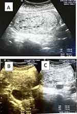
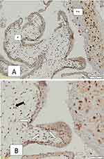
None of the family members had ever been diagnosed with PHM. In the current pregnancy, USG evaluation showed a snowstorm appearance with enlarged uterus (Figure 1). Based on that, the patient was suspected to have a recurrent PHM. The patient then underwent a curettage procedure. Histological evaluation afterwards confirmed the diagnosis of PHM (Figure 2). Patient’s genetic could not be assessed because it was not covered by the patient’s medical insurance and the patient did not want to pay using her own money. The patient was then advised to undergo in-vitro fertilization (IVF) procedure to avoid the recurrence of PHM again in the future, but the patient refused. The timeline of this case is presented in Figure 3.
 |
Figure 1 The USG examination before curettage. (A) An enlarged uterus filled with a heterogeneous mass and multiple anechoic spaces implied a snowstorm appearance; (B) lutein cyst; (C) fibroid. |
 |
Figure 3 Timeline of the patient. |
Discussion
The prevalence of recurrent HM varies between countries. In Western countries, approximately 1% to 2% of patients with HM develop the second one.7 In the Middle Eastern region, the prevalence of recurrent HM is reported to be higher than in Western countries, with the recurrent rates ranging between 2.5% and 9.4%.8
Women with histologically confirmed PHM are at risk of recurrent HM in a subsequent pregnancy. If this occurs, most cases will be of the same type of HM as in the previous pregnancy. The recurrence of PHM for the subsequent pregnancy has been reported to be approximately 1.7%.6 We performed a literature search on PubMed without a publication year limit on 1 September 2023 using the following search terms: (“hydatidiform mole” OR “molar pregnancy”) AND “recurrent” AND “partial”. The inclusion criteria were as follows: (1) Case report or series, (2) confirmed PHM based on the histology evaluation, (3) the recurrence occurs in at least 3 consecutive pregnancies. Non-English language publications were excluded. From the search, we found 4 reported cases; however, none of the reported cases had the number of recurrence as high as our patient (Table 1).9–12
 |
Table 1 Reported Recurrent Partial Hydatidiform Mole from the Literature |
The pathophysiology of HM is related to abnormality of trophoblastic proliferation in the formation of the placenta. However, there is no definitive cause of this abnormality of trophoblastic proliferation. Nevertheless, a genetic mutation is strongly suspected to be the cause of HM. CHM occurs in about 75% of HM and usually had a diploid androgenetic with a karyotype of 46 XX that comes only from the father. There are also those who argue that a CHM occurs as a result of fertilization of an empty ovum with diploid sperm, whereas PHM occurs due to fertilization of haploid ovum and diploid sperm. Because of that, the karyotype of PHM is usually 46, XXY or 46, XXX.13
It was suggested that defective oocytes are responsible for the recurrence of HM.14 The concentrations of certain coagulation factors are altered in women with HM compared to normal pregnancies. Specifically, higher concentrations of fibrinogen degradation products and fibrinogen factor VII are observed in women with HM. On other hand, the concentrations of prothrombin, plasminogen, factor X, and plasminogen activator are lower in women with HM compared to normal pregnancies. Hypercoagulability refers to an increased tendency of the blood to clot, often observed in women with HM due to a decrease in platelet count. It will contribute to an increased risk of clot formation in the blood vessels.15
In patients with family history of recurrent HM, autosomal recessive genetic defects are suspected. This suggest that certain genetic factors inherited from both parents might contribute to the occurrence of HM in some cases. Partial hydatidiform moles are characterized by having one set of maternal chromosomes and two sets of paternal chromosomes. This genetic configuration can lead to an abnormal embryonic development and the formation of a partial mole.16
The treatment approach for molar pregnancies mainly depends on whether the patients wish to preserve their fertility. In patients who still desire to have children in the future, the treatment is by evacuating the molar pregnancy by suction and curettage. For patients who have no desire to be pregnant, a hysterectomy is the treatment option since this procedure eliminates the risk of future molar pregnancies.
For patients who undergo suction and curettage, a serial measurement of β-hCG levels should be done to monitor the patient’s recovery and detect the occurrence of persistent gestational trophoblast (PTG) after molar pregnancy evacuation. It is because the PTG has the potential to become gestational trophoblastic neoplasia (GTN). In majority of cases, β-hCG levels will return to normal levels within 2 months after the evacuation procedure. However, approximately 1–5% of patients with PHM will develop persistent disease that requires further treatment.17
Assisted reproductive technology such as IVF is considered to be one of the options for patients with recurrent HM.13 Genetic analysis will be carried out on sperm cells, ovum and embryos before the implantation process. Some experts suggest that in cases with RHM it is necessary to do DNA testing if there is a genetic mutation in NLRP7 or KHDC3L, then oocyte donation needs to be done to increase the chances of a normal pregnancy.18 It is because genetic mutations of NLRP7 and KHDC3L are considered to have a role in the occurrence of increased incidence of RHM.8,19
Conclusions
The incidence of recurrent PHM is rare. When this occurs, counseling should be given to the patients to select for other fertilization methods in order to prevent recurrent PHM and to be able to have a normal pregnancy.
Informed Consent Statement
The patient has been given an explanation regarding the details of the case and pictures which will be published in the case report. Institutional approval was waived because this was a case report and the patient has given consent to be published in the report.
Acknowledgments
We would like to thank Dr. Hasan Sadikin Hospital, Bandung, Indonesia, for the support in this case.
Author Contributions
All authors made a significant contribution to the work reported, whether that is in the conception, study design, execution, acquisition of data, analysis and interpretation, or in all these areas; took part in drafting, revising or critically reviewing the article; gave final approval of the version to be published; have agreed on the journal to which the article has been submitted; and agree to be accountable for all aspects of the work.
Disclosure
The authors declare that they have no conflicts of interest.
References
1. Lurain JR. Gestational trophoblastic disease I: epidemiology, pathology, clinical presentation and diagnosis of gestational trophoblastic disease, and management of hydatidiform mole. Am J Obstet Gynecol. 2010;203(6):531–539. doi:10.1016/j.ajog.2010.06.073
2. Cavaliere A, Ermito S, Dinatale A, et al. Management of molar pregnancy. J Prenat Med. 2009;3(1):15.
3. Sebire N, Seckl M. Gestational trophoblastic disease: current management of hydatidiform mole. BMJ. 2008;337:a1193. doi:10.1136/bmj.a1193
4. Steigrad SJ. Epidemiology of gestational trophoblastic diseases. Best Pract Res Clin Obstet Gynaecol. 2003;17(6):837–847. doi:10.1016/S1521-6934(03)00049-X
5. Sebire N, Foskett M, Fisher RA, et al. Risk of partial and complete hydatidiform molar pregnancy in relation to maternal age. BJOG. 2002;109(1):99–102. doi:10.1111/j.1471-0528.2002.t01-1-01037.x
6. Eagles N, Sebire NJ, Short D, et al. Risk of recurrent molar pregnancies following complete and partial hydatidiform moles. Hum Reprod. 2015;30(9):2055–2063. doi:10.1093/humrep/dev169
7. Dean J, Rosenblat O, Jones A. Update on gestational trophoblastic disease. Cancer. 2022;24(3):7–15.
8. Nguyen NMP, Slim R. Genetics and epigenetics of recurrent hydatidiform moles: basic science and genetic counselling. Curr Obstet Gynecol Rep. 2014;3(1):55–64. doi:10.1007/s13669-013-0076-1
9. Narayan H, Mansour P, McDougall W. Recurrent consecutive partial molar pregnancy. Gynecol Oncol. 1992;46(1):122–127. doi:10.1016/0090-8258(92)90209-2
10. Koc S, Ozdegirmenci O, Tulunay G, et al. Recurrent partial hydatidiform mole: a report of a patient with three consecutive molar pregnancies. Int J Gynecol Cancer. 2006;16(2). doi:10.1136/ijgc-00009577-200603000-00082
11. Guha Sarkar P, Dalmia S, Khatri P. A case report on recurrent partial moles in three consecutive pregnancies. J Obstet Gynaecol. 2022;42(5):1591–1592. doi:10.1080/01443615.2021.1997958
12. Helwani MN, Seoud M, Zahed L, et al. A familial case of recurrent hydatidiform molar pregnancies with biparental genomic contribution. Hum Genet. 1999;105(1–2):112–115. doi:10.1007/s004399900088
13. Williams D, Hodgetts V, Gupta J. Recurrent hydatidiform moles. Eur J Obstetr Gynecol Reprod Biol. 2010;150(1):3–7. doi:10.1016/j.ejogrb.2010.01.003
14. Hemida R, van Doorn H, Fisher R. A novel genetic mutation in a patient with recurrent biparental complete hydatidiform mole: a brief report. Int J Gynecol Cancer. 2016;26(7):1351–1353. doi:10.1097/IGC.0000000000000755
15. Khaza’leh F, Haloub K, Freij M. Recurrent hydatidiform molar pregnancy: a case report of 5 consecutive molar pregnancies complicated by HELLP and DIC, and review of literature. Open J Obstetr Gynecol. 2015;5(12):731. doi:10.4236/ojog.2015.512102
16. Oikonomidis P, Pergialiotis B, Pitsouni E, et al. Repetitive complete molar pregnancy in a 54-year-old patient in a time distance of eighteen years from the first incident: case report and mini review. Case Rep Med. 2011;2011:1–4. doi:10.1155/2011/351267
17. Lurain J, Seckl M, Schink J. Gestational trophoblastic disease. In: Textbook of Uncommon Cancer. Wiley Online Library; 2017:653–662.
18. Kalogiannidis I, Kalinderi K, Kalinderis M, et al. Recurrent complete hydatidiform mole: where we are, is there a safe gestational horizon? Opinion and mini-review. J Assist Reprod Genet. 2018;35(6):967–973. doi:10.1007/s10815-018-1202-9
19. Rezaei M, Nguyen NMP, Foroughinia L, et al. Two novel mutations in the KHDC3L gene in Asian patients with recurrent hydatidiform mole. Hum Genome Var. 2016;3(1):16027. doi:10.1038/hgv.2016.27
 © 2023 The Author(s). This work is published and licensed by Dove Medical Press Limited. The full terms of this license are available at https://www.dovepress.com/terms.php and incorporate the Creative Commons Attribution - Non Commercial (unported, v3.0) License.
By accessing the work you hereby accept the Terms. Non-commercial uses of the work are permitted without any further permission from Dove Medical Press Limited, provided the work is properly attributed. For permission for commercial use of this work, please see paragraphs 4.2 and 5 of our Terms.

