Back to Journals » International Journal of Nanomedicine » Volume 20
Recent Progress in Peptide-Based Fluorescent Probes Biomedical Applications: A Review
Authors Zhong X ![]() , Xie Y
, Xie Y ![]() , Chen Y, Lu Y, Hou M
, Chen Y, Lu Y, Hou M ![]()
Received 20 March 2025
Accepted for publication 28 July 2025
Published 3 September 2025 Volume 2025:20 Pages 10751—10770
DOI https://doi.org/10.2147/IJN.S529323
Checked for plagiarism Yes
Review by Single anonymous peer review
Peer reviewer comments 4
Editor who approved publication: Prof. Dr. Anderson Oliveira Lobo
Xue Zhong,1,2,* Yuanlong Xie,1,* Yuen Chen,2 Yushen Lu,2 Mingming Hou3
1Department of Spine Surgery and Musculoskeletal Tumor, Zhongnan Hospital of Wuhan University, Wuhan, Hubei, People’s Republic of China; 2Second Clinical College, Wuhan University, Wuhan, Hubei, People’s Republic of China; 3Department of Orthopedics, Honghui Hospital, Xi’an Jiaotong University, Xi’an, Shaanxi, People’s Republic of China
*These authors contributed equally to this work
Correspondence: Mingming Hou, Department of Orthopedics, Honghui Hospital, Xi’an Jiaotong University, Xi’an, Shaanxi, People’s Republic of China, Email [email protected]
Abstract: Peptide-based fluorescent probes have found widespread applications in biomedical research, including bio-imaging, disease diagnosis, drug discovery, and image-guided surgery. Their favorable properties—such as small molecular size, low toxicity, minimal immunogenicity, and high targeting specificity—have contributed to their growing utility in both basic research and translational medicine. This review provides a comprehensive overview of recent advances in peptide-based fluorescent probes, emphasizing design strategies, biological targets, and diverse functional applications. Key areas of focus include the integration of molecular targeting with imaging capabilities, the emergence of multimodal imaging techniques, and the development of activatable probes responsive to specific biological stimuli. Applications are discussed in the context of tumor cell membrane recognition, subcellular organelle targeting, non-cancer disease diagnosis, and detection of both metal ions and non-metal ions. Notably, responsive probes for reactive oxygen species (ROS) and other biologically relevant non-metal ions are also highlighted, underscoring their diagnostic and therapeutic potential. The review also addresses key limitations—such as poor in vivo stability, limited targeting accuracy, and delivery efficiency—and outlines future directions including smart peptide probe platforms, self-reporting systems, and high-throughput screening based on peptide libraries to accelerate next-generation probe development.
Keywords: peptide fluorescent probes, drug-targeted delivery, bio-imaging, tumor cell identification, organelle targeting, metal ion detection, multimodal imaging
Introduction
Intraoperative navigation, surgical therapy, and tumor diagnostics all benefit significantly from advanced techniques capable of visualizing molecular and anatomical features in vivo. While conventional imaging methods such as positron emission tomography (PET), single-photon emission computed tomography (SPECT), magnetic resonance imaging (MRI), and ultrasound remain widely used, they suffer from limitations in spatial and temporal resolution, sensitivity, and molecular-level specificity, making them suboptimal for real-time dynamic monitoring.
In contrast, molecular imaging using targeted probes provides a more accurate approach for visualizing complex biological processes at the cellular or subcellular level. For example, Tang et al synthesized a B7H3-targeted IRDye800CW probe that enabled precise labeling of osteosarcoma tissues and facilitated real-time intraoperative identification and resection of tumor margins.1 Peptide molecules have recently emerged as a particularly promising class of imaging agents due to their small size, excellent target specificity, low immunogenicity, good permeability, and favorable biocompatibility. These properties make peptides ideal carriers for targeted fluorescent probes. Compared to antibodies and nanoparticles, peptide-based fluorescent probes offer several significant advantages, including lower immunogenicity, faster renal clearance, better tumor penetration, and easier synthesis and modification. In contrast to small-molecule probes, peptides exhibit enhanced specificity and affinity due to their ability to engage in multivalent interactions and form stable secondary structures that improve target recognition. These properties make peptides particularly attractive for in vivo imaging applications where both biocompatibility and precision are critical.In parallel, advances in second near-infrared (NIR-II, 1000–1700 nm) fluorophores have further expanded the utility of in vivo imaging. For instance, Tang et al reported an intramolecular repulsive interaction (IRI)-based strategy to boost the luminescence efficiency of NIR-II aggregation-induced emission (AIE) materials.2 A recent study also highlighted the multifunctional potential of peptide-based fluorescent probes in both tumor targeting and tumor microenvironment modulation, further expanding their biomedical applications,3 Despite the clinical potential of traditional dyes such as sodium fluorescein and indocyanine green (ICG),4,5 their poor targeting capacity remains a significant drawback. Peptide-based fluorescent probes—typically composed of a targeting peptide, a linker, and a fluorescent moiety—offer a viable solution, combining minimal immunogenicity, high targeting efficiency, and real-time visualization potential.6
This review aims to provide a comprehensive overview of recent progress in peptide-targeted fluorescent probes, with a specific focus on their applications in tumor imaging, organelle targeting, non-tumor disease diagnosis, metal ion detection, multimodal imaging, and drug-targeted delivery. We also highlight emerging strategies such as activatable probes and intelligent designs, and discuss existing challenges and translational potential. A general classification of peptide-based fluorescent probes is provided in Figure 1, serving as a conceptual framework for the topics covered.
 |
Figure 1 The classification of the peptide based fluorescent probes or drugs mentioned in this review. Created in BioRender. Peng, R. (2025) https://BioRender.com/w88u512. |
Peptide Fluorescent Probes for Biological Targeting and Imaging
Peptide Fluorescent Targeting Tumor Cell Membrane Receptors
One of the key tactics for accurately diagnosing and treating malignant tumors is fluorescence navigation, particularly ICG-based near-infrared fluorescence navigation technology, which significantly improves surgical oncology precision and can raise patient survival and prognosis. Tumor-associated targets such as integrins, HER2, SSTR, and GRPR serve as important foundations for the development of peptide-based fluorescent probes due to their highly specific overexpression in various cancer cells. Integrins—especially αvβ3—promote tumor angiogenesis and migration; HER2 is highly expressed in breast and gastric cancers; SSTR2 is commonly found in neuroendocrine tumors; and GRPR is involved in cell proliferation in cancers such as pancreatic cancer, all demonstrating strong potential for imaging applications.7,8 There are several potential therapeutic applications for using these receptors to create tailored peptides and targeted peptide fluorescence probes. Peptide-based fluorescent probes targeting integrins, HER2, and GRPR demonstrate strong binding affinity, with examples such as KSP*-Cy5.5 showing a dissociation constant of 21 nM for HER2. These probes also exhibit excellent imaging stability—for instance, cRGD signals persist for over 24 hours, and the imaging window of ICG-Herceptide extends up to 8 hours. Compared to traditional dyes, peptide probes offer higher signal-to-noise ratios and lower background signals, with mechanisms like aggregation/assembly-induced retention (AIR) further enhancing tumor site accumulation.9,10 Additionally, multiple studies have confirmed their low toxicity and good biocompatibility, supporting their promising clinical potential.
Peptide Probes Targeting Integrins
Integrins are a broad family of transmembrane cell adhesion molecules that are classified as collagen receptors, leukocyte-specific receptors, arginine-glycine-aspartate (RGD)-binding receptors, and laminin receptors.11 Malignant neovascular and tumor cells exhibit markedly elevated expression of these molecules.12
Because of its excellent binding specificity and stability, the tripeptide RGD and its cyclized variants (such as cRGDyK and cRGDfK) have found extensive application in targeted imaging and treatment.13,14 By creating a cyclized structure, cRGD makes the peptide more rigid, which enhances its stability and specificity of binding with integrin αvβ3.
It has been demonstrated that αvβ3-positive glioma U87MG cells may efficiently absorb cRGD-PEG-siEGFR, which is produced by attaching the cyclic peptide via polyethylene glycol to a short interfering RNA that targets the epidermal growth factor receptor (EGFR), to provide anti-tumor effects.15
Regarding tumor imaging, the imaging probe of the NIR fluorescent cyclic RGD peptide produced by combining an aminated RGD peptide with a near-infrared (NIR) dye was able to enable targeted imaging of tumor endothelial cells with high expression of integrins.16
MMP2/9 enzymes are generally recognized as being involved in cancer metastasis and are regarded as biomarkers for malignant tumors. Based on this concept, an MMP2/9-responsive fluorescent probe has been designed for the selective detection of malignant tumor cells with high MMP2/9 expression and the conditional activation of reactive oxygen species (ROS) generation, enabling the integration of imaging and photodynamic therapy (PDT).
Utilizing the aggregation/assembly-induced retention (AIR) effect and the tumor-specific excretion-retarded (TER) effect, a near-infrared probe A has been developed to target integrin receptors on the surface of human renal cell carcinoma (RCC) and liver tumor cells. This probe is designed by integrating a targeting motif (RGD), an enzyme-responsive peptide ligand (PLGYLG), a self-assembly motif (YLGFFC), and a near-infrared signaling molecule (Cy). Initially, probe A selectively binds to the highly expressed αvβ3 integrin in RCC, after which it is cleaved by MMP2/9 enzymes. The resulting fragments spontaneously self-assemble into nanofibers, facilitating high-performance detection of human RCC.17
MMP2/9 enzymes are widely recognized for their involvement in cancer metastasis and are considered biomarkers for malignant tumors. Accordingly, a responsive fluorescent probe incorporating an MMP2/9-cleavable peptide linker (Pro-Leu-Gly-Val-Arg-Gly) has been designed to enable the selective detection of malignant tumor cells with elevated MMP2/9 expression.18
Long-term cell labeling may be possible with dual-targeted fluorescence probes. With a peptide composition made up of cNGR (targeted cycling peptide motif), CPP (cell-penetrating peptide), and NLS (nuclear localization signal sequence), the dual-targeted multifunctional fluorescence probe (TCNTP) may be utilized for behavioral investigations of cancer cells. In order to provide effective nucleus-specific imaging and long-term, low-toxicity monitoring of cancer cells, this probe combines a flexible delivery mechanism with the aggregation-induced luminescence of a tetrastyrene derivative.19
Table 1 Summary of Tumor-Related Peptide Fluorescent Probes.
 |
Table 1 Summary of Tumor-Related Peptide Fluorescent Probes |
Peptide Fluorescent Probes Targeting Receptor 2 for Human Epidermal Growth Factor
The oncogene ERBB2 encodes the receptor known as HER2 (Human Epidermal Growth Factor Receptor 2), which is expressed on the surface of a wide range of tumor cells, including colorectal, liver, gastric, breast, and non-small cell lung (NSCLC) cancers, among many others.8,25,26
A newly discovered peptide, Herceptide (RSLWSDFYASASRGP), was employed as the targeting moiety. The probe ICG-Herceptide, synthesized by conjugating Herceptide with ICG—a NIR(II) near-infrared fluorescent dye—exhibits excellent targeting ability, high specificity, and low biotoxicity.In in vivo mouse experiments, the probe effectively delineated the boundary between the tumor and adjacent tissues, providing valuable assistance in tumor resection.20
Based on the targeting peptides RSLWSDFY, KSPNPRF, and RSLWSDFYKSPNPRF, three new HER2 targeting probes were created: DOTA-ZC01-ICG, DOTA-KSP-ICG, and DOTA-ZC02-ICG. When it came to HER2-positive tumors, DOTA-ZC02-ICG outperformed the others in terms of affinity and specificity and produced good bimodal imaging.21 A peptide with the sequence KSPNPRF, designed by Bishnu P. Joshi et al, was conjugated to the fluorescent dyes FITC or Cy5.5 via the linker GGGSK using Fmoc-mediated solid-phase synthesis, resulting in a fluorescent probe named KSP*-Cy5.5. This probe holds great potential as a powerful tool for the early detection of colon tumors.9
Peptide Fluorescent Probes Targeting the Growth Inhibitory Receptor
The growth inhibitory receptor (SSTR) is expressed in different parts of the brain, stomach, pituitary, endocrine, and adrenal glands at variable levels.27 SSTR subtype 2 (SSTR2) is an ideal target for imaging and treating persons with neuroendocrine tumors (NETs) and neuroendocrine tumors in general.28
In their investigation of cyclic peptides, Genady Kostenich et al developed 19 probes, nine of which were tested in vivo on animals. The most specific of these probes was linker 3207–86. The basic peptide sequence of this probe is (D)Phe-Cys*-Phe-Trp-(D)Trp-Lys-Thr-Phe-Gly(S2)*-NH2 (the “*” marks the cyclization site). The linker is a four-carbon γ-aminobutyric acid, whereas fluorescein is the luminous moiety. Isothiocyanate works as a polypeptide binding agent. The polypeptide contains two critical D-type amino acids in a unique configuration: (D)Phe and (D)Trp, which improve the polypeptide’s stability and ability to bind to SSTR. In polypeptides, amino acids like Lys (lysine) and Thr (threonine) play important roles in the SSTR binding sites. The arrangement of these amino acids increases the peptide’s affinity for the growth inhibitor receptor.22
Kai Licha et al used automated solid-phase synthesis to create growth inhibitor receptor-specific peptides with the sequence H2N-(D-Phe)-cyclo.[Cys-Phe-(D-Trp)-Lys-Thr-Cys]-Thr-OH contains carboxylated indole-dicarbon or indole-tricarbon cyanine dyes attached to the N-terminal end to improve fluorescence properties. Key amino acid residues in the peptide sequence (eg, D-Trp, Phe, Lys, etc.) contribute considerably to receptor binding and have a high binding affinity for growth inhibitory receptor isoforms (especially isoforms 2 and 5), providing the probe excellent specificity.23
Peptide Fluorescent Probes Targeting Gastrin-Releasing Peptide-Specific Receptors
In vivo, the gastrin-releasing peptide receptor (GRPr) regulates cancer and immune cells by promoting proliferation and inflammation.A wide range of malignancies and inflammatory disorders have been linked to abnormal GRPr expression. Therefore, it is critical to design probes that can detect and modulate GRPr function.29
Tu et al identified GB-6, a six-amino-acid peptide with the sequence: Gln-5-Htp-β-Ala-Nva-Gly-His-NH2.This peptide was ligated with an MPA dye via PEG4 to create a new peptide probe, and in vitro competitive binding tests in liver-transferred mice confirmed that MPA-PEG4-GB-6 and GRPR were specific binding partners. In vivo near-infrared fluorescence imaging revealed that GRPR-positive SW1990 tumors produced prominent NIR red fluorescence signals within 1 hour, which increased by 6 hours and remained visible throughout 12 hours, indicating efficient accumulation of MPA-PEG4-GB-6 in the tumor.24
Peptide Fluorescent Probes Targeting Organelles
The Golgi apparatus, endoplasmic reticulum, lysosomes, and mitochondria are among the endomembrane system organelles found in eukaryotic cells. When these organelles are damaged, they malfunction, which can result in a number of significant ailments, including as cancer, metabolic issues, heart disease, and neurological disorders.30–32 In order to target cellular organelles and their activities, peptide fluorescent probes have been developed in recent years.
Probes Targeting Mitochondria
AIDS, cancer, liver damage, neurological illnesses, cardiovascular disease, and Alzheimer’s disease can all result from abnormal biothiol levels.33–35 One such example of a biothiol is glutathione (GSH), which is widely distributed in mitochondria.
Through a specific strategy of linking the targeted TAT peptide (RRQRRKKRG) to the fluorescent moieties naphthyridine imide and rhodamine B by a natural chemical linkage (NCL) reaction, the study by Su et al offers a novel idea for the simultaneous detection of biothiols and differentiation of GSH, Cys, and Hcy in mitochondria of living cells using two-photon excitation. This could provide a useful tool for the study and diagnosis of hyperhomocysteinemia, neural tube defects, and cardiovascular disease.36 As shown in Figure 2, two-photon confocal fluorescence imaging revealed distinct cellular responses when HeLa cells were treated with the TAT-based fluorescent probe (5 μM) under various experimental conditions,and Figure 3 reveals its subcellular localization in HeLa cells co-treated with Cys (100 μM) and organelle trackers.
 |
Figure 2 Two-photon confocal fluorescence imaging of HeLa cells treated with the TAT-based fluorescent probe (5 μM) under different conditions: (A) PBS; (B) NEM (1 mM); (C) Cys (100 μM); (D) GSH (100 μM); (E) Hcy (100 μM). (1) Bright-field images; (2) Channel 1: λex = 820 nm, λem = 520 ± 10 nm; (3) Channel 2: λex = 820 nm, λem = 585 ± 10 nm; (4) Channel 3: λex = 545 nm, λem = 585 ± 10 nm. Reprinted from Talanta, 218, Su P, Zhu Z, Tian Y, et al. A TAT peptide-based ratiometric two-photon fluorescent probe for detecting biothiols and sequentially distinguishing GSH in mitochondria, 121127, ©Copyright 2020 with permission from Elsevier. [DOI: 10.1016/j.talanta.2020.121907].36 |
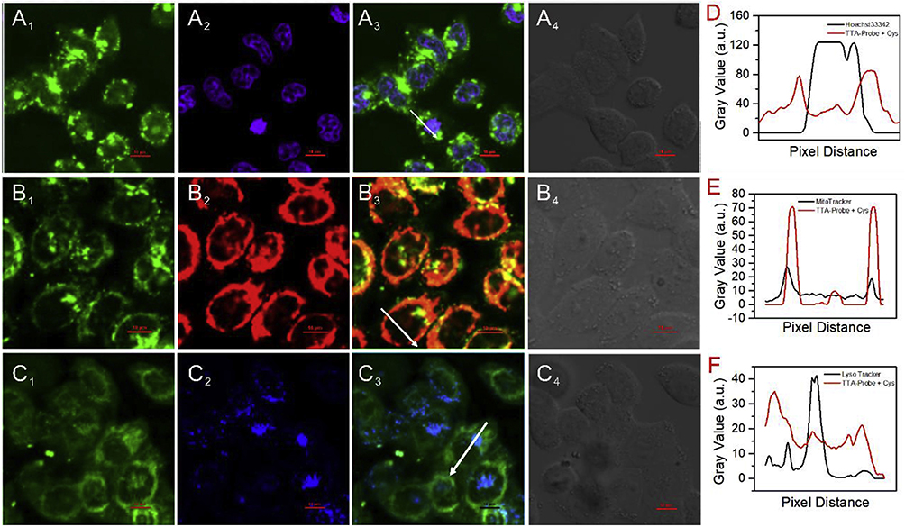 |
Figure 3 Subcellular localization of the TAT-probe (5 μM) in HeLa cells co-incubated with Cys (100 μM) and (A) Hoechst 33342 (100 nM); (B) MitoTracker Red; (C) LysoTracker (100 nM) for 30 min at 37 °C. (1) TAT-probe fluorescence (λex = 820 nm, λem = 520 ± 10 nm); (2) Counterstaining channel; (3) Merged images; (4) Bright-field images. (D–F) Intensity profiles along the white arrows in merged images. Reprinted from Talanta, 218, Su P, Zhu Z, Tian Y, et al. A TAT peptide-based ratiometric two-photon fluorescent probe for detecting biothiols and sequentially distinguishing GSH in mitochondria, 121127, ©Copyright 2020 with permission from Elsevier. [DOI: 10.1016/j.talanta.2020.121907].36 |
Aggregation-induced luminescence molecules (AIEgen) and peptides served as the basis for Chen et al’s probe design. They discovered that the functional units’ arrangement order significantly impacted the probe’s performance. The combination of mitochondrial malfunctioning elements (M elements, HLAHLAHHLAHLAH) and tumor cell uptake elements (T elements, RGDGPLGVRGRKKRRQRRR) allowed the AIEgen peptide probes with TPE derivatives as the backbone to accomplish targeted functionalities. It was discovered that the T-M-AIE structure was more cytotoxic and more suited for functional intervention, whilst the T-AIE-M structure showed greater tumor cell selectivity and was appropriate for targeted imaging.30
Probes for Targeting the Nucleus
Disease phenotypes are strongly associated with changes in the nucleus’s structure, and when it malfunctions, it can lead to conditions including Parkinson’s, cancer, and retinitis pigmentosa.37 As a result, nuclear tracking, nuclear treatment and illness diagnostics depend on fluorescent imaging probes that target the nucleus.
Cargo proteins can penetrate the nuclear membrane through negatively charged nuclear pore complexes mediated by nuclear localization signal peptides (NLS), which often comprise lengthy amino acid sequences of positively charged lysines or arginines.38 NLS peptides can be used in this context as particular peptides that target the nucleus.
The photonic and two-photon fluorescence features of carbon dots (CDs), which are made from tryptophan and formic acid using a one-step hydrothermal process, have garnered a lot of interest in the bio-imaging community.The TAT peptide (GRKKRRQRRRPQ) is a nuclear localization signal peptide capable of traversing both the cellular and nuclear membranes. By coupling carbon dots with TAT peptides to form imaging probes (TAT-CDs), one-photon and two-photon nuclear-targeted fluorescence imaging can be achieved, offering valuable applications in disease diagnosis and therapeutic research.39
Using citric acid, PEG, and ethylenediamine to create CDs@PEG that can precisely release intense blue fluorescence and then covalently attaching NLS peptides (PKKKRKVG) to the carbon dots to create NLS-CDs for nucleus-targeted imaging is another technique for creating probes.40
Targeting hydrogen peroxide in the nucleus was made possible by the innovative use of the ratiometric fluorescent probe NP1. The nuclear localization signal peptide (NLS, sequence VQRKRQKLMP-NH2) effectively directs the probe into the nucleus when combined with NP1, allowing for the accurate detection of hydrogen peroxide in the nucleus.41
Peptide Fluorescent Probes for Targeting the Golgi Apparatus
The Golgi is critical for maintaining cellular homeostase, and any dysfunction of the copper-transporting ATPase in the Golgi can disrupt copper homeostase, leading to neurodegenerative diseases, cancer, and more.42,43
In order to detect Cu+ in the Golgi apparatus, the Dansyl-labeled tripeptide probe Dns-LLC was created. Its unique structure provides a potent tool for the study of Golgi function by binding Cu ions via sulfhydryl groups and working in concert with dansulfonyl and amide groups to produce persistent fluorescent signals.44
Furin protease is a Golgi-transported preprotein convertase, and new evidence indicates that Furin plays a key role in neurodegenerative and neuropsychiatric disorders, cancer, and infectious diseases.45 Furin can be targeted by taking advantage of its specific cleavage of substrates (Arg-X-Arg/Lys− Arg↓) in the Golgi.46
Due to the diffusive nature of the fluorescent molecules, existing probes against furin protease are challenging to detect and image in situ. Li et al responded by creating the HPQF probe, which uses the self-destructing linker piperidin-2-ylmethanamine to connect the RVRR peptide to Cl-HPQ. When transformed by furin, HPQF releases free Cl-HPQ and starts to precipitate while emitting bright solid-state fluorescence, enabling the HPQF probe to detect furin activity in living cells in real time and for long-term in situ imaging.47
The classical near-infrared fluorescent molecule HD, an RVRR peptide, and a self-eliminating linker make up another RVRR peptide-based fluorescent probe, HD-F (not only allows for in situ detection of furin in the Golgi but also visualization of the variations in furin expression levels between cells), which is perfectly suited for tracking changes in furin expression levels of hypoxia-inducible factor-1 stabilized by CoCl2.48
Li et al reported the first instance of the self-assembly of a soluble amphiphilic peptide, C-3, which selectively detects furin in living cells with high furin expression. C-3 comprises the RVRRFFF sequence and a nitrobenzoxadiazole (NBD) fluorophore. The RVRR sequence enhances membrane permeability and hydrophilicity, enabling a rapid response to furin in aqueous solution within 5 minutes. The FFF tripeptide sequence promotes self-assembly by increasing hydrophobicity and π−π interactions. Furthermore, the self-assembled C-3 remains in living cells for an extended period, allowing continuous intracellular furin detection, making it well-suited for tracer analysis.49
In order to precisely destroy cancer cells, Tan et al’s study focuses on focusing on the Golgi apparatus in cancer cells. The three primary components of the team’s newly created phosphorothioate peptide (pS1) are the phosphorothioate moiety, D-diphenylalanine (D-ff), and the fluorescent moiety NBD. pS1 was quickly dephosphorylated by alkaline phosphatase (ALP) to produce self-assembled thiophosphopeptide (S1), which accumulated in the Golgi apparatus and killed cancer cells.50 Thiophosphate groups were substituted for other sulfur-containing groups in the follow-up study to create a fluorescent peptide thioester probe. These thiopeptides were hydrolyzed by particular enzymes in the Golgi after entering the cells, and the resulting dimers were subsequently enriched in the GA and the ER, interfering with the protein transport process and ultimately causing the death of cancer cells.51
Peptide Fluorescent Probes for Targeting Peroxisomes
Serious metabolic disorders (PBDs) are caused by deficiencies or dysfunctions in peroxisomes, which are membrane-enclosed organelles found in the majority of eukaryotic cells.52
Edward H.W. Pap et al created a novel probe based on the peroxisome targeting sequence (PTS1) and the membrane permeable peptide CKGGAKL to address the problems of peroxisome targeting and cell membrane penetration. This probe was then acetylated at the N-terminal end to improve membrane penetration and combined with the fluorescent motifs BODIPY and SNAFL to increase its affinity with the cell membrane. The results of experiments demonstrated that this The probe was quickly absorbed by cells and concentrated in peroxisomes.53
Table 2 Summary of Organelle-Targeting Peptide Fluorescent Probes.
 |
Table 2 Summary of Organelle-Targeting Peptide Fluorescent Probes |
Multimodal Imaging Modalities Based on Peptide Fluorescent Probes
Due to its safety, high spatial resolution, and real-time performance, fluorescence imaging has become a widely used imaging technique for clinical tumor detection and image-guided surgery, in contrast to more conventional anatomical and molecular imaging methods like X-CT, PET, MRI, PAI, and USI.54 Combining different imaging modalities has become a mainstream trend in disease diagnosis.55 The process of creating images by combining chemical data from several platforms is known as multimodal imaging.56 They are dual-labeled with a fluorescent contrast agent and a radioactive material, and they have specific locations. These substances can be utilized for surgically guided tumor removal and allow for intraoperative and whole-body imaging with a single dosage, Optical imaging offers high resolution but shallow penetration, whereas MRI provides deep tissue imaging with excellent anatomical detail. Combining modalities allows for comprehensive diagnostic information.57 Multimodal probes enable more accurate tumor localization, intraoperative guidance, and real-time therapy monitoring, which are crucial for improving surgical outcomes and personalized treatment.58 Despite their promise, challenges such as probe synthesis complexity, cost, and in vivo pharmacokinetics must be addressed for successful clinical translation.59 For fluorescent probes involved in multimodal imaging, we have also created a schematic diagram (Figure 4).
 |
Figure 4 The structures of peptide-based fluorescent probes involved in multimodal imaging. Created in BioRender. Peng, R. (2025) https://BioRender.com/k77a932. |
Combined with CT Imaging Modalities
There is significant promise for usage in the field of thrombus identification when peptide probes are combined with CT imaging methods that use nanoparticles as imaging contrast agents.60,61
For dual-modal thrombus imaging employing near-infrared fluorescence (NIRF) and micro-computed tomography (micro-CT), Kwon et al synthesized a thrombin-activatable fluorescent peptide (TAP) and integrated it into silica-coated gold nanoparticles (TAP-SiO2@AuNPs). The TAP molecules restore the NIRF signal by precisely targeting thrombin activity. Its potential for thrombus detection and treatment was highlighted by experimental results that showed considerable buildup at thrombus sites and good imaging performance. Figure 5 illustrates62 thrombus detection through TAP-SiO2@AuNPs, showing thrombin-activated multimodal imaging capability.
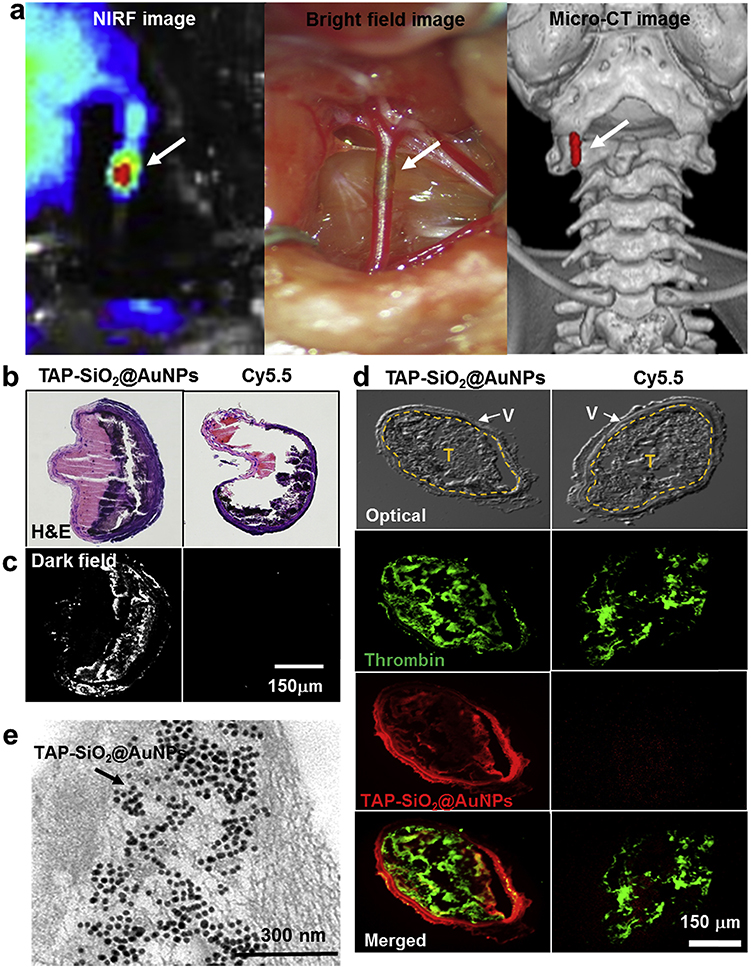 |
Figure 5 Multimodal thrombus imaging using thrombin-activatable fluorescent peptide-incorporated gold nanoparticles (TAP-SiO2@AuNPs). (a) Dual-modality in vivo imaging (NIRF, bright-field, and micro-CT) of an in situ thrombotic mouse model treated with TAP-SiO2@AuNPs, showing thrombus accumulation (white arrows). (b) H&E staining of dissected common carotid arteries (CCA) from mice treated with TAP-SiO2@AuNPs or Cy5.5. (c) Dark-field imaging of the same samples. (d) Immunofluorescence imaging of thrombin activity (green) and nanoparticle distribution (red) in vessel cross-sections, with merged views showing thrombus localization. “V” indicates vessel endothelium, “T” indicates thrombus. (e) Transmission electron microscopy (TEM) image of the dissected CCA, confirming the presence of TAP-SiO2@AuNPs (black dots). Reprinted from Biomaterials, 150, Kwon SP, Jeon S, Lee SH, et al. Thrombin-activatable fluorescent peptide incorporated gold nanoparticles for dual optical/computed tomography thrombus imaging. 125–136. ©Copyright 2018, with permission from Elsevier. [DOI: 10.1016/j.biomaterials.2017.10.038].62 |
Combined with PET Imaging Modality
Although PET imaging is renowned for its great sensitivity,63 it is challenging to offer accurate anatomical information due to its lack of resolution restriction.64 The diagnosis, staging, and border determination of malignant tumors have been successfully enhanced by integrating preoperative positron emission tomography (PET) and molecular probes with near-infrared fluorescence (NIRF)-guided imaging, which has increased the accuracy of surgical resection.65
In a single probe, Peter S. Conti et al integrated fluorescence and PET imaging. After attaching the c(RGDyK)2 peptide (denoted as RGD2) to a Cy5.5 fluorescent dye with the chelator BaAn(Boc)Sar and labeling with 64Cu, PET and fluorescence imaging were carried out. According to the experimental results, the probe had outstanding intraoperative imaging ability and clinical translational potential, and it could efficiently and specifically recognize and bind to the integrin αvβ3 receptor in PET and fluorescence imaging.66
Ann-Christin Baranski et al employed the chelator HBED-CC to conjugate 68Ga with the peptide Glu-urea-Lys(Ahx), which was subsequently coupled with fluorescent dyes (IRDye800CW, DyLight800) to synthesize the PSMA-targeting probe 68Ga-Glu-urea-Lys-HBED-CC-IRDye800CW. This probe enables PET/fluorescence dual-modality imaging, offering a valuable tool for precise tumor visualization.67
Combined with PAI Imaging Modality
In preclinical research, photoacoustic imaging (PAI) has demonstrated significant potential in delivering structural, functional, and molecular information.68,69 Recent years have also seen significant developments in the application of PAI in conjunction with other imaging modalities, particularly in tumor therapy. For instance, target-dependent photoacoustic signals can be produced using photoactivated probes.
Jelena Levi et al developed an activatable photoacoustic probe targeting matrix metalloproteinase-2 (MMP-2) for precise enzymatic activity detection. The probe utilizes the ACPP peptide (Ceeee[Ahx]PLGLAGrrrrrK), where, before cleavage, the photoacoustic signals of BHQ3 and Alexa750 cancel each other out. Upon MMP-2-mediated cleavage of the PLGLAG sequence, the BHQ3-labeled CPP portion accumulates inside cells, while Alexa750 diffuses away, resulting in photoacoustic signals only at 675 nm. Experimental results confirm that this probe is suitable for tumor microenvironment imaging and localization.70
Combined with MRI Imaging Modalities
MRI/fluorescence imaging modalities have been developed and have shown complementary imaging properties including high sensitivity and high spatial resolution for precise tumor localization, strong penetration, powerful imaging modalities, and a wide range of applications.71,72
In order to target the integrin αvβ3 receptor and the laminin receptor on B16F10 melanoma cells, respectively, the tripeptide RGD and the pentapeptide YIGSR were affixed to gadolinium diethylenetriaminepentaacetic acid (Gd-DTPA) and rhodamine B (RhB). According to the experimental findings, the probes outperformed the traditional Gd-DTPA and both demonstrated low cytotoxicity, quick imaging, and long-lasting effects.73
Through genetic engineering, the RGD-RFP-LBT-Gd molecular probe was created in E. coli. Its structure consists of a short LBT peptide that can bind Gd³+, a red fluorescent protein, and a tumor-targeting RGD peptide. The stability of the RGD-RFP-LBT-Gd probe is good. Subsequent in vitro and in vivo studies have demonstrated the probe’s strong fluorescent signal for imaging tumor locations, high resolution MRI imaging, low cytotoxicity, high tumor cell uptake, and effective tumor targeting.72
Table 3 Summary of Multimodal Imaging Probes Based on Peptides.
 |
Table 3 Summary of Peptide-Based Fluorescent Probes for Multimodal Imaging |
Peptide Fluorescent Probes for the Diagnosis and Treatment of Non-Tumor Diseases
Heterotopic Ossification
Heterotopic ossification (HO) is defined as the occurrence of extraskeletal bone in soft tissues74 and is commonly seen after joint replacement, spinal cord injury, traumatic brain injury, blast injury, elbow and acetabular fractures, and thermal injury.75 Early diagnosis and treatment is important to inhibit the progression of heterotopic ossification.
In order to endocytose chondrocytes and penetrate the cartilage matrix, Wang et al used the enrichment property of type II collagen (Col2a1) during the cartilage formation stage of HO. They created a peptide probe, WL-808, with the peptide sequence of WYRGRL. This probe was linked to the fluorescent dye IR-808 via a linker. According to in vitro tests, the probe demonstrated good specificity and minimal biotoxicity. The probe showed persistent and selective fluorescence in animals in vivo.76
The MMP family plays an important role in extracellular matrix (ECM) degradation, and its expression is altered in diseases such as rheumatoid arthritis, chronic obstructive pulmonary disease, and bone formation.77–79
A fluorescent probe targeting MMP-2/9 was developed based on the MMP-targeting peptide M2 (sequence: DOTA-KKAHWGFTLD), with the HWGF motif playing a central role in the peptide’s binding to MMP. The probe was double-labeled with IRDye 800CW dye and the radioisotope 68Ga. In vivo experiments in mice demonstrated that the probe exhibits excellent stability and specificity, effectively targeting the MMP-9 expression region associated with HO, showing significant potential for clinical application.80
Rheumatoid Arthritis
For rheumatoid arthritis (RA), methotrexate (MTX) is the recommended medication.81 However, its clinical usefulness is limited due to its short residence period at the target site and higher systemic adverse effects.
While carrying the fluorescent dye ICG for imaging, ultrasound-imaging liposomes (iELPs) were modified with a functionalized iRGD peptide (CRGDKGPDC) to enable targeted delivery of MTX. The selective enrichment of iELPs in arthritic joints was greatly improved by the iRGD peptide linkage in a mouse model of RA, increasing medication efficacy and decreasing adverse effects. According to histological analysis, mice’s joint tissues treated with iELPs and sonicated had significantly lower levels of inflammatory cell infiltration and angiogenesis.82
Compared to normal, RA patients’ synovial tissue contains more DNA fragments linked to apoptosis.83 Histones are transferred to the plasma membrane and liberated from the nucleus during apoptosis.84 Additionally, apoptosis-targeting peptide (ApoPep-1) with the sequence CQRPPR can bind histones on the surface of apoptotic cells with specificity,85 A fluorescent imaging probe called Cyclic ApoPep-1 (CApoPep-1) was created by In Seop So et al based on this notion in order to visualize and measure apoptosis in a chronic arthritis model. In in vitro, in vivo, and ex vivo detection, it shows higher sensitivity and specificity and is more stable than linear ApoPep-1.86
It has been demonstrated that pathologic diseases including osteoarthritis and rheumatoid arthritis exhibit overexpression of matrix metalloproteinases.87 To solve this issue, a diagnostic kit (MMP-D-KIT) was created that immobilizes an MMP-13-specific probe made up of the dye Cy5.5, the bursting agent BHQ-3, and the MMP-13-specific peptide GPLGVRGGKGG, which allows MMP-13 to precisely cleave the peptide link between G and V. There are numerous uses for MMP-D-KIT in detecting MMP in various biological materials due to its excellent sensitivity, linear response, and specificity when compared to other techniques.88
Alzheimer’s Disease
In the past, imaging methods including MRI, PET, CT, and SPECT were typically used to diagnose Alzheimer’s disease (AD) in order to find amyloid plaques.89,90 However, these techniques are frequently constrained by their low safety, limited spatial resolution, and time commitment. Therefore, it is particularly crucial to develop more modern, highly sensitive and selective diagnostic probes for the detection of these critical biomarkers.91
Aβ42-specific flexible ultrasensitive polyvalent-directed peptide polymer (PDPP) were constructed by linking two Aβ42-specific peptides to a flexible poly D-lysine hydrobromide (PDL) backbone, which significantly enhanced the sensitivity and specificity for the detection of Aβ42. The P1 peptide (Ac-FRHMTEQGCGK) was hydrophilic, and the P2 peptide (Ac-IPLPFYNGCGK) was hydrophobic, and both were labeled with fluorescein isothiocyanate (FITC) for enhanced detection. Both peptides were biased to bind the hydrophilic N-terminus and hydrophobic C-terminus of Aβ42, respectively, and their properties matched the Aβ42 target site, significantly increasing the binding affinity. Compared with the single peptide forms (P1-PDPP or P2-PDPP), PDPP showed an approximately 10³ to 105-fold enhancement in binding affinity and exhibited low biotoxicity.
In addition, PDPP combined with the ZnO nanoporous system for the detection of Aβ42 in cerebrospinal fluid not only enlarged the detection range, but also significantly reduced the limit of detection (LOD) and improved the sensitivity by about 104 times, while enhancing the fluorescence signal intensity. This system shows great potential for application in the early diagnosis of Alzheimer’s disease.92
Detection of Thrombophilia
Arterial thrombus plays a key role in acute coronary syndromes and stroke,93 therefore, in vivo techniques for detecting thrombus are of great significance.
Magnetic resonance imaging (MRI) has become one of the most promising non-invasive imaging techniques for detecting and characterizing thrombi in vivo.94 EP-2104R is a fibrin-targeted MRI contrast agent composed of a Gd(III) core chelated by DOTA and a fibrin-targeting peptide, which enables selective binding to fibrin within thrombi without competing with fibrinogen or collagen. Its small molecular structure provides excellent thrombus penetration and imaging signal. Additionally, its high affinity and slow clearance contribute to superior performance in MRI detection of both acute and chronic thrombi. Compared to conventional Gd-DTPA, EP-2104R offers enhanced thrombus visualization and is suitable for early thrombus detection and age differentiation.95
In another study, novel radiolabeled probes (68Ga-FBP14, 111In-FBP15, and 99mTc-FBP16) were developed on the basis of existing gadolinium-based probes (eg, EP-2104R) and validated in a rat thrombus model.
The results showed that 68Ga-FBP14 and 111In-FBP15 possessed high thrombus target/background ratios and rapid blood clearance properties in PET/SPECT imaging, which could effectively differentiate between thrombus and non-target tissues, and the target specificity was further confirmed by triple isotope validation. However, 99mTc-FBP16 performed poorly due to low target uptake and easy aggregation.96
Overall, 68Ga-FBP14 and 111In-FBP15 provide new tools for noninvasive imaging of thrombus and show good potential for clinical translation. The development of these probes provides important support for early diagnosis and precision treatment of cardiovascular diseases.
Table 4 Summary of Peptide Fluorescent Probes for Disease Diagnosis (Non-Tumor).
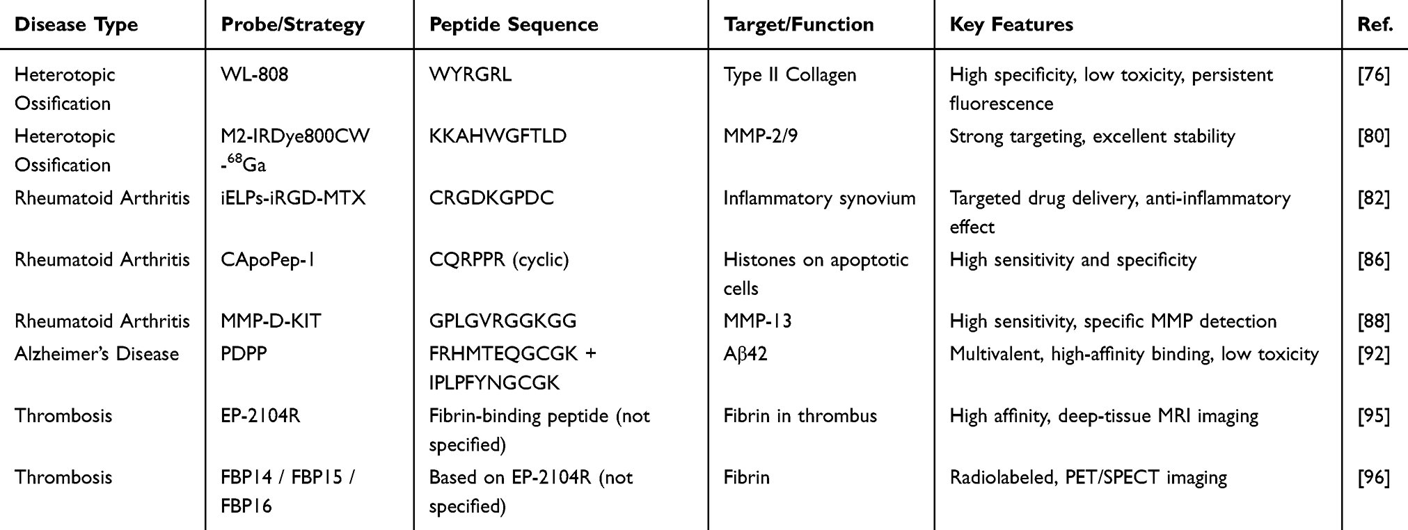 |
Table 4 Summary of Peptide-Based Fluorescent Probes for Non-Tumor Disease Diagnosis and Treatment |
Peptide Fluorescent Probes for Metal Ion Detection
Metal ions such as Hg²+, Pb²+, Cu²+, Cd²+, and Zn²+ play critical roles in various physiological and pathological processes, and their dysregulation is closely linked to diseases. Copper ions (Cu²+) and zinc ions (Zn²+) play important roles in various physiological and pathological processes in the human body. Cu²+ is involved in angiogenesis and promotes tumor invasion and metastasis. Zn²+ is a key factor in enzymatic activity and immune responses, and its homeostatic imbalance has been closely linked to diseases such as Alzheimer’s disease and prostate cancer.Mercury ions (Hg²+), lead ions (Pb²+), and cadmium ions (Cd²+) are typical toxic metals whose intake or accumulation poses serious threats to human health. Hg²+ and Pb²+ exhibit significant neurotoxicity, leading to cognitive impairment and neurodegenerative disorders. Cd²+ is closely associated with nephrotoxicity and carcinogenicity, causing lesions in various organs and tissues.97–108
Probes for Biologically Relevant Metal Ions
A fluorescent probe, L, incorporating a targeting peptide (Pro-Gly-His-Trp-NH2), was developed for the efficient detection of Zn²+ ions, demonstrating robust detection performance. Each Zn²+ ion was found to coordinate with two probe molecules through two amide groups and two imidazole groups, forming a stable complex. This coordination event induces a proportional fluorescence response via fluorescence resonance energy transfer (FRET), enabling a highly selective and sensitive approach for Zn²+ detection.109
The FLH probe offers an efficient and accurate approach for detecting copper ions (Cu²+). Its lysine backbone, along with amino groups in the side chain, not only facilitates the attachment of functional groups but also provides substantial design flexibility. FLH binds to Cu²+ through histidine residues located on both termini, exhibiting a distinct fluorescence on/off response, high selectivity, rapid detection capability, and excellent recyclability.110
FGGH (FITC-Gly-Gly-His-NH2) is a peptide-based fluorescent probe designed for the highly selective detection of Cu²+. Its design is inspired by the amino-terminal Cu(II)- and Ni(II)-binding (ATCUN) motif found in metalloproteins, allowing for specific recognition through a square planar coordination involving the amino-terminal nitrogen, two peptide nitrogen atoms, and the imidazole nitrogen. According to the Hard-Soft Acid-Base (HSAB) theory, the imidazole nitrogen in histidine (His) and the nitrogen in glycine (Gly) act as soft bases, while Cu²+ is a soft acid, enabling stable binding under physiological conditions. This makes FGGH an efficient sensing platform for Cu²+ detection.111
Probes for Toxic Metal Ions
Common methods for mercury ion detection include atomic absorption spectrometry (AAS), voltammetry, and chromatography; however, these techniques often suffer from significant interferences and lengthy processing times.112
In order to overcome these constraints, the fluorescent probe DGRK was created by conjugating a monoacyl fluorophore with the tripeptide Gly-Arg-Lys-NH2. This probe successfully overcomes the limitations of traditional mercury ion detection techniques in terms of specificity and efficiency thanks to its quick response, high stability, great selectivity, and low biotoxicity. According to experimental findings, Hg²+ exhibits a strong binding affinity with DGRK and forms coordination bonds with the tripeptide’s amino and amino-nitrogen atoms. These results demonstrate DGRK’s potential as a viable option for mercury ion detection.113
Traditional lead ion detection methods often involve prolonged analysis times and complex sample pretreatment procedures.114 In response to these challenges, recent studies have focused on developing more efficient and convenient detection strategies. A novel ratiometric fluorescent probe has garnered significant attention due to its superior performance. This probe utilizes the peptide moiety Gly-Ser-Met-COOH, in which carboxyl and hydroxyl groups serve as hard ligands and thioether groups as soft ligands, facilitating selective recognition and binding of Pb²+. This structural configuration substantially enhances the sensitivity and specificity of lead ion detection, making it a promising tool for practical applications.115
Wang et al designed a fluorescent switching probe, L, for the highly selective detection of Cd²+ ions. This probe is constructed using a dansyl fluorophore conjugated to a tetrapeptide (Dansyl-Glu-Pro-Gly-Cys). The Cd²+ ion coordinates with the probe through two distinct ligand sites—the oxygen atom of glutamic acid (Glu) and the sulfur atom of cysteine (Cys)—resulting in the formation of a stable coordination complex.116
Table 5 Summary of Peptide Fluorescent Probes for Metal Ion Detection.
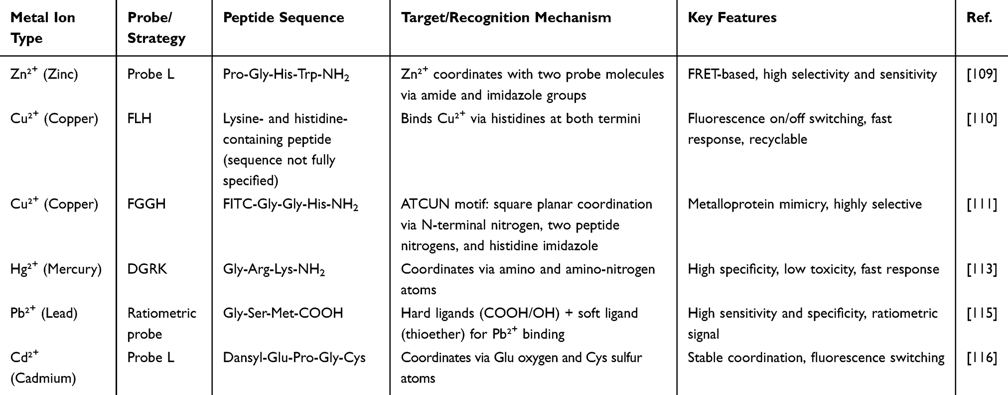 |
Table 5 Summary of Peptide-Based Fluorescent Probes for Metal Ion Detection |
Peptide-Coupled Drugs for Targeted Drug Delivery
The conjugation of payloads with targeted ligands enhances the therapeutic index of highly potent or toxic cytotoxic drugs while mitigating the adverse effects associated with their administration.117 Antibody-drug conjugates (ADCs) have emerged as a well-established therapeutic strategy for the selective delivery of cytotoxic agents to tumor sites, improving treatment efficacy and minimizing systemic toxicity.118 peptide-drug conjugates (PDCs) offer an alternative approach with distinct advantages. PDCs are easier to synthesize and allow for structural modifications that facilitate rational drug design, improving bioavailability, affinity, and stability. Additionally, peptides tend to exhibit lower immunogenicity, making them a promising platform for therapeutic development.
Lutathera (¹77Lu-dotatate) is a peptide-drug conjugate (PDC) composed of a somatostatin analog linked to a radionuclide via DOTA. It received FDA approval in January 2018 for the treatment of patients with gastroenteropancreatic neuroendocrine tumors (GEP-NETs) that express somatostatin receptors.119
TH1902 is a drug-peptide conjugate composed of docetaxel and the peptide TH19P01 (Ac-GVRAKAGVRN(Nle)FKSESY). It targets the Sortilin 1 (SORT1) receptor and is being developed for the treatment of various advanced solid tumors that are SORT1-positive. Currently, it is in Phase I clinical trials and has been granted Fast Track designation by the FDA. In addition, when TH19P01 is combined with doxorubicin, the resulting drug, TH1904, demonstrates high tolerance and efficacy in the human ovarian ES-2 tumor xenograft mouse model. This study is currently in the preclinical research stage.120
117Lu-PSMA-617 is a peptide receptor radionuclide therapy (PRRT) agent, formed by conjugating PSMA-617 with 117Lu-dotatate via a DOTA chelator. PSMA-617 consists of a Glu-urea binding motif and a DOTA chelator, enabling high-affinity binding to prostate-specific membrane antigen (PSMA).121 Upon binding to PSMA on the cell membrane, Lu-PSMA-617 undergoes internalization, leading to the release of β-particle radiation, which induces DNA damage and subsequent tumor cell death.122 The VISION Phase III trial evaluated the efficacy of 117Lu-PSMA-617 in combination with standard therapy for PSMA-positive metastatic castration-resistant prostate cancer (mCRPC). The trial successfully met its primary endpoints, demonstrating significant improvements in overall survival (OS) and progression-free survival (PFS)123
GRN1005 is a peptide-drug conjugate targeting low-density lipoprotein receptor-associated protein-1 (LRP-1), consisting of paclitaxel combined with Angiopep-2, which facilitates blood-brain barrier (BBB) penetration and reach therapeutic concentrations in tumor tissue.Phase I clinical trials demonstrated that GRN1005 was well tolerated in patients with recurrent glioma, with a maximum tolerated dose (MTD) of 650 mg/m² administered every three weeks. The most common toxicities were neutropenia and fatigue, with no central nervous system (CNS) toxicity observed.Pharmacokinetic analysis showed dose-proportional exposure, with some patients exhibiting disease stabilization or remission. GRN1005 is currently undergoing Phase II clinical trials.124
Conclusion
Peptide-based fluorescent probes, owing to their small size, excellent biocompatibility, and high targeting specificity, have shown great promise in tumor diagnosis, image-guided surgery, metal ion detection, and targeted drug delivery. By selectively binding to tumor-associated biomarkers and incorporating diverse fluorescence mechanisms, these probes enable real-time, precise visualization of pathological processes, offering significant potential for clinical translation. However, challenges such as enzymatic degradation, short circulation time, and off-target effects still hinder their in vivo stability and imaging accuracy. Future research should focus on enhancing probe stability, prolonging half-life, and developing multifunctional, activatable systems capable of responding to complex disease microenvironments. The continued optimization of peptide design and synthetic strategies will be critical to advancing their clinical utility in precision oncology and beyond.
Acknowledgment
The illustrations in this manuscript were created with the support of Li Dongping using the BioRender platform. Their contribution is gratefully acknowledged.
Author Contributions
All authors made a significant contribution to the work reported, whether that is in the conception, study design, execution, acquisition of data, analysis and interpretation, or in all these areas; took part in drafting, revising or critically reviewing the article; gave final approval of the version to be published; have agreed on the journal to which the article has been submitted; and agree to be accountable for all aspects of the work.
Funding
This work was supported by the Natural Science Foundation of Wuhan (Project number: 2024040801020356) and the Hubei Provincial Natural Science Foundation of China (Project number: 2023AFB824).
Disclosure
The authors confirm that there are no financial interests or personal relationships that could have influenced the work presented in this paper. All authors state that they have no relevant financial or personal connections to disclose.
References
1. Zeng F, Li C, Wang H. et al. Intraoperative resection guidance and rapid pathological diagnosis of osteosarcoma using B7H3 targeted probe under NIR-II fluorescence imaging. Adv Sci. 2024;11:e2310167.
2. Ma F, Gao Z, Jia Q, et al. Intramolecular repulsive interactions enable high efficiency of NIR-II aggregation-induced emission luminogens for high-contrast glioblastoma imaging. ACS Nano. 2025;19(1):1676–1688. doi:10.1021/acsnano.4c15387
3. An H, Hou D, Zhang N, et al. A self-assembled fluorescent contrast agent targeting XIAP for image-guided surgery of bladder cancer. Nano Today. 2024;56:102313. doi:10.1016/j.nantod.2024.102313
4. He S, Zhong A, Lei J, et al. Application of indocyanine green fluorescence imaging in assisting biopsy of musculoskeletal Tumors. Cancers;2023. 15. doi:10.3390/cancers16010015
5. Huang H, He S, Wei R, et al. Near-infrared (NIR) imaging with indocyanine green (ICG) may assist in intraoperative decision making and improving surgical margin in bone and soft tissue tumor surgery. J Surg Oncol. 2023;128(4):612–627. doi:10.1002/jso.27306
6. Cai M, Wen Z, Li H, et al. Peptide-based fluorescent probes for the diagnosis of tumor and image-guided surgery. Biosens Bioelectron. 2025;276:117255. doi:10.1016/j.bios.2025.117255
7. Jin H, Varner J. Integrins: roles in cancer development and as treatment targets. Br J Cancer. 2004;90(3):561–565. doi:10.1038/sj.bjc.6601576
8. Uy NF, Merkhofer CM, Baik CS. HER2 in non-small cell lung cancer: a review of emerging therapies. Cancers. 2022;15(1):14. doi:10.3390/cancers15010014
9. Joshi BP, Zhou J, Pant A, et al. Design and synthesis of near-infrared peptide for in vivo molecular imaging of HER2. Bioconjugate Chem. 2016;27(2):481–494. doi:10.1021/acs.bioconjchem.5b00565
10. Wang W, Wu K, Vellaisamy K, Leung C, Ma D. Peptide-conjugated long-lived theranostic imaging for targeting GRPr in cancer and immune cells. Angew Chem Int Edit. 2020;59(41):17897–17902. doi:10.1002/anie.202007920
11. Desgrosellier JS, Cheresh DA. Integrins in cancer: biological implications and therapeutic opportunities. Nat Rev Cancer. 2010;10(1):9–22. doi:10.1038/nrc2748
12. Cooper J, Giancotti FG. Integrin signaling in cancer: mechanotransduction, stemness, epithelial plasticity, and therapeutic resistance. Cancer Cell. 2019;35(3):347–367. doi:10.1016/j.ccell.2019.01.007
13. Danhier F, Le Breton A, Préat V. RGD-based strategies to target alpha(v) beta(3) integrin in cancer therapy and diagnosis. Mol Pharm. 2012;9(11):2961–2973. doi:10.1021/mp3002733
14. Scott JI, Deng Q, Vendrell M. Near-infrared fluorescent probes for the detection of cancer-associated proteases. Acs Chem Biol. 2021;16(8):1304–1317. doi:10.1021/acschembio.1c00223
15. Su Q, Chen J, Liu Z, Fan Y, He S. A pH-sensitive cRGD-PEG-siRNA conjugated compound targeting glioblastoma. Bioconjugate Chem. 2024;35(11):1732–1743. doi:10.1021/acs.bioconjchem.4c00255
16. Liu L, Lin G, Yin F, Law WC, Yong KT. Near-infrared fluorescent peptide probes for imaging of tumor in vivo and their biotoxicity evaluation. J Biomed Mater Res A. 2016;104(4):910–916. doi:10.1002/jbm.a.35628
17. An HW, Hou D, Zheng R, et al. A near-infrared peptide probe with tumor-specific excretion-retarded effect for image-guided surgery of renal cell carcinoma. ACS Nano. 2020;14(1):927–936. doi:10.1021/acsnano.9b08209
18. Ma Y, Persi L, Yan S, Sung SL, Baumgartner M, Yamakoshi Y. Conditionally activated ROS generation by MMP2/9-specific C 60 -based fluorescence probes. Small. 2025;21(8):e2403307. doi:10.1002/smll.202403307
19. Cheng Y, Sun C, Ou X, Liu B, Lou X, Xia F. Dual-targeted peptide-conjugated multifunctional fluorescent probe with AIEgen for efficient nucleus-specific imaging and long-term tracing of cancer cells. Chem Sci. 2017;8(6):4571–4578. doi:10.1039/C7SC00402H
20. Cao R, Li R, Shi H, Liu H, Cheng Z. Novel HER2-targeted peptide for NIR-II imaging of tumor. Mol Pharm. 2023;20(2):1394–1403. doi:10.1021/acs.molpharmaceut.2c00964
21. Cao R, Li R, Lai C, Shi H, Liu H, Cheng Z. Development of a novel HER2-targeted peptide probe for dual-modal imaging of tumors. J Med Chem. 2023;66(11):7523–7533. doi:10.1021/acs.jmedchem.3c00347
22. Kostenich G, Livnah N, Bonasera TA, et al. Targeting small-cell lung cancer with novel fluorescent analogs of somatostatin. Lung Cancer. 2005;50(3):319–328. doi:10.1016/j.lungcan.2005.07.009
23. Licha K, Hessenius C, Becker A, et al. Synthesis, characterization, and biological properties of cyanine-labeled somatostatin analogues as receptor-targeted fluorescent probes. Bioconjugate Chem. 2001;12(1):44–50. doi:10.1021/bc000040s
24. Tu Y, Han Z, Pan R, et al. Novel GRPR-targeting peptide for pancreatic cancer molecular imaging in orthotopic and liver metastasis mouse models. Anal Chem. 2023;95(30):11429–11439. doi:10.1021/acs.analchem.3c01765
25. Marchio C, Annaratone L, Marques A, Casorzo L, Berrino E, Sapino A. Evolving concepts in HER2 evaluation in breast cancer: heterogeneity, HER2-low carcinomas and beyond. Semin Cancer Biol. 2021;72:123–135. doi:10.1016/j.semcancer.2020.02.016
26. Raghav K, Moasser MM. Molecular pathways and mechanisms of HER2 in cancer therapy. Clin Cancer Res. 2023;29(13):2351–2361. doi:10.1158/1078-0432.CCR-22-0283
27. Patel YC. Somatostatin and its receptor family. Front Neuroendocrinol. 1999;20(3):157–198. doi:10.1006/frne.1999.0183
28. Fani M, Nicolas GP, Wild D. Somatostatin receptor antagonists for imaging and therapy. J Nucl Med. 2017;58(Supplement 2):61S–66S. doi:10.2967/jnumed.116.186783
29. Wang W, Wu KJ, Vellaisamy K, Leung CH, Ma DL. Peptide-conjugated long-lived theranostic imaging for targeting GRPr in cancer and immune cells. Angew Chem Int Edit. 2020;59(41):17897–17902.
30. Chen B, Yuan H, Zhang W, Hu J, Lou X, Xia F. AIEgen-peptide bioprobes for the imaging of organelles. Biosensors-Basel. 2022;13(1):12. doi:10.3390/bios13010012
31. Qiu K, Chen Y, Rees TW, Ji L, Chao H. Organelle-targeting metal complexes: from molecular design to bio-applications. Coordin Chem Rev. 2019;378:66–86. doi:10.1016/j.ccr.2017.10.022
32. Schwarz DS, Blower MD. The endoplasmic reticulum: structure, function and response to cellular signaling. Cell Mol Life Sci. 2016;73(1):79–94. doi:10.1007/s00018-015-2052-6
33. Nunnari J, Suomalainen A. Mitochondria: in sickness and in health. Cell. 2012;148(6):1145–1159. doi:10.1016/j.cell.2012.02.035
34. Zhang J, Bao X, Zhou J, et al. A mitochondria-targeted turn-on fluorescent probe for the detection of glutathione in living cells. Biosens Bioelectron. 2016;85:164–170. doi:10.1016/j.bios.2016.05.005
35. Shahrokhian S. Lead phthalocyanine as a selective carrier for preparation of a cysteine-selective electrode. Anal Chem. 2001;73(24):5972–5978. doi:10.1021/ac010541m
36. Su P, Zhu Z, Tian Y, et al. A TAT peptide-based ratiometric two-photon fluorescent probe for detecting biothiols and sequentially distinguishing GSH in mitochondria. Talanta. 2020;218:121127. doi:10.1016/j.talanta.2020.121127
37. Galganski L, Urbanek MO, Krzyzosiak WJ. Nuclear speckles: molecular organization, biological function and role in disease. Nucleic Acids Res. 2017;45(18):10350–10368. doi:10.1093/nar/gkx759
38. Goswami R, Gupta A, Bednova O, et al. Nuclear localization signal-tagged systems: relevant nuclear import principles in the context of current therapeutic design. Chem Soc Rev. 2024;53(1):204–226. doi:10.1039/D1CS00269D
39. Song Y, Li X, Cong S, Zhao H, Tan M. Nuclear-targeted of TAT peptide-conjugated carbon dots for both one-and two-photon fluorescence imaging. Colloid Surf B. 2019;180:449–456. doi:10.1016/j.colsurfb.2019.05.015
40. Yang L, Jiang W, Qiu L, et al. One pot synthesis of highly luminescent polyethylene glycol anchored carbon dots functionalized with a nuclear localization signal peptide for cell nucleus imaging. Nanoscale. 2015;7(14):6104–6113. doi:10.1039/C5NR01080B
41. Wen Y, Liu K, Yang H, et al. A highly sensitive ratiometric fluorescent probe for the detection of cytoplasmic and nuclear hydrogen peroxide. Anal Chem. 2014;86:9970–9976.
42. Li J, Wang Y. Golgi metal ion homeostasis in human health and diseases. Cells-Basel. 2022;11.
43. Desai V, Kaler SG. Role of copper in human neurological disorders. Am J Clin Nutr. 2008;88(3):855S–858S. doi:10.1093/ajcn/88.3.855S
44. Jung KH, Oh ET, Park HJ, Lee KH. Development of new peptide-based receptor of fluorescent probe with femtomolar affinity for Cu(+) and detection of Cu(+) in golgi apparatus. Biosens Bioelectron. 2016;85:437–444. doi:10.1016/j.bios.2016.04.101
45. Zhang Y, Gao X, Bai X, Yao S, Chang YZ, Gao G. The emerging role of furin in neurodegenerative and neuropsychiatric diseases. Transl Neurodegener. 2022;11:39.
46. Hosaka M, Nagahama M, Kim WS, et al. Arg-X-Lys/Arg-Arg motif as a signal for precursor cleavage catalyzed by furin within the constitutive secretory pathway. J Biol Chem. 1991;266:12127–12130.
47. Li K, Hu XX, Liu HW, et al. in situ imaging of furin activity with a highly stable probe by releasing of precipitating fluorochrome. Anal Chem. 2018;90(19):11680–11687. doi:10.1021/acs.analchem.8b03335
48. Zhu L, Liu HW, Yang Y, et al. Near-infrared fluorescent furin probe for revealing the role of furin in cellular carcinogenesis and specific cancer imaging. Anal Chem. 2019;91(15):9682–9689. doi:10.1021/acs.analchem.9b01220
49. Li X, Cao C, Wei P, et al. Self-assembly of amphiphilic peptides for recognizing high furin-expressing cancer cells. ACS Appl Mater Inter. 2019;11(13):12327–12334. doi:10.1021/acsami.9b01281
50. Tan W, Zhang Q, Wang J, Yi M, He H, Xu B. Enzymatic assemblies of thiophosphopeptides instantly target golgi apparatus and selectively kill cancer cells*. Angew Chem Int Edit. 2021;60(23):12796–12801. doi:10.1002/anie.202102601
51. Tan W, Zhang Q, Quinones-Frias MC, et al. Enzyme-responsive peptide thioesters for targeting golgi apparatus. J Am Chem Soc. 2022;144(15):6709–6713. doi:10.1021/jacs.2c02238
52. Schrader M, Fahimi HD. Peroxisomes and oxidative stress. Biochim Biophys Acta. 2006;1763(12):1755–1766. doi:10.1016/j.bbamcr.2006.09.006
53. Pap EH, Dansen TB, Wirtz KW. Peptide-based targeting of fluorophores to peroxisomes in living cells. Trends Cell Biol. 2001;11(1):10–12. doi:10.1016/S0962-8924(00)01829-8
54. Wang C, Wang Z, Zhao T, et al. Optical molecular imaging for tumor detection and image-guided surgery. Biomaterials. 2018;157:62–75. doi:10.1016/j.biomaterials.2017.12.002
55. Wang J, Wang Z, Zhong Y, et al. Central metal-derived co-assembly of biomimetic GdTPP/ZnTPP porphyrin nanocomposites for enhanced dual-modal imaging-guided photodynamic therapy. Biomaterials. 2020;229:119576. doi:10.1016/j.biomaterials.2019.119576
56. Tuck M, Blanc L, Touti R, et al. Multimodal imaging based on vibrational spectroscopies and mass spectrometry imaging applied to biological tissue: a multiscale and multiomics review. Anal Chem. 2021;93(1):445–477. doi:10.1021/acs.analchem.0c04595
57. Ghosh SC, Azhdarinia A. Advances in the development of multimodal imaging agents for nuclear/near-infrared fluorescence imaging. Curr Med Chem. 2015;22(29):3390–3404. doi:10.2174/0929867322666150904111214
58. Graziotto ME, Kidman CJ, Adair LD, James SA, Harris HH, New EJ. Towards multimodal cellular imaging: optical and X-ray fluorescence. Chem Soc Rev. 2023;52(23):8295–8318. doi:10.1039/D3CS00509G
59. Zhao R, Lan D, Xia B, Dong M, Mu J, Zhao Y. PET-based dual-modal probes for in vivo imaging. Small. 2025;21:e2409713.
60. Kinsella JM, Jimenez RE, Karmali PP, et al. X-ray computed tomography imaging of breast cancer by using targeted peptide-labeled bismuth sulfide nanoparticles. Angew Chem Int Edit. 2011;50(51):12308–12311. doi:10.1002/anie.201104507
61. Smith BR, Gambhir SS. Nanomaterials for in vivo imaging. Chem Rev. 2017;117(3):901–986. doi:10.1021/acs.chemrev.6b00073
62. Kwon SP, Jeon S, Lee SH, et al. Thrombin-activatable fluorescent peptide incorporated gold nanoparticles for dual optical/computed tomography thrombus imaging. Biomaterials. 2018;150:125–136. doi:10.1016/j.biomaterials.2017.10.017
63. Xie L, Zhao J, Li Y, Bai J. PET brain imaging in neurological disorders. Phys Life Rev. 2024;49:100–111. doi:10.1016/j.plrev.2024.03.007
64. Wang W, Ke S, Kwon S, et al. A new optical and nuclear dual-labeled imaging agent targeting interleukin 11 receptor alpha-chain. Bioconjugate Chem. 2007;18(2):397–402. doi:10.1021/bc0602679
65. Houghton JL, Zeglis BM, Abdel-Atti D, et al. Site-specifically labeled CA19.9-targeted immunoconjugates for the PET, NIRF, and multimodal PET/NIRF imaging of pancreatic cancer. Proc Natl Acad Sci USA. 2015;112(52):15850–15855. doi:10.1073/pnas.1506542112
66. Liu S, Li D, Huang CW, et al. Efficient construction of PET/fluorescence probe based on sarcophagine cage: an opportunity to integrate diagnosis with treatment. Mol Imaging Biol. 2012;14(6):718–724. doi:10.1007/s11307-012-0557-z
67. Baranski AC, Schafer M, Bauder-Wust U, et al. PSMA-11-derived dual-labeled PSMA inhibitors for preoperative PET imaging and precise fluorescence-guided surgery of prostate cancer. J Nucl Med. 2018;59(4):639–645. doi:10.2967/jnumed.117.201293
68. Mallidi S, Luke GP, Emelianov S. Photoacoustic imaging in cancer detection, diagnosis, and treatment guidance. Trends Biotechnol. 2011;29(5):213–221. doi:10.1016/j.tibtech.2011.01.006
69. Zhang HF, Maslov K, Stoica G, Wang LV. Functional photoacoustic microscopy for high-resolution and noninvasive in vivo imaging. Nat Biotechnol. 2006;24(7):848–851. doi:10.1038/nbt1220
70. Levi J, Kothapalli SR, Ma TJ, Hartman K, Khuri-Yakub BT, Gambhir SS. Design, synthesis, and imaging of an activatable photoacoustic probe. J Am Chem Soc. 2010;132(32):11264–11269. doi:10.1021/ja104000a
71. Wang Y, Yang T, Ke H, et al. Smart albumin-biomineralized nanocomposites for multimodal imaging and photothermal tumor ablation. Adv Mater. 2015;27(26):3874–3882. doi:10.1002/adma.201500229
72. Zhao H, Zhao H, Jiao Y, et al. Biosynthetic molecular imaging probe for tumor-targeted dual-modal fluorescence/magnetic resonance imaging. Biomaterials. 2020;256:120220. doi:10.1016/j.biomaterials.2020.120220
73. Zheng SY, Tang WQ, Zhang M, et al. Dual-modal polypeptide-containing contrast agents for magnetic resonance/fluorescence imaging. Bioorg Chem. 2022;129:106161. doi:10.1016/j.bioorg.2022.106161
74. Xu Y, Huang M, He W, et al. Heterotopic ossification: clinical features, basic researches, and mechanical stimulations. Front Cell Dev Biol. 2022;10:770931. doi:10.3389/fcell.2022.770931
75. Ranganathan K, Loder S, Agarwal S, et al. Heterotopic ossification: basic-science principles and clinical correlates. J Bone Joint Surg. 2015;97(13):1101–1111. doi:10.2106/JBJS.N.01056
76. Wang Z, Yi X, Yi W, et al. Early diagnosis of heterotopic ossification with a NIR fluorescent probe by targeting type II collagen. J Mater Chem B. 2023;11(8):1684–1691. doi:10.1039/D2TB02157A
77. Muroski ME, Roycik MD, Newcomer RG, et al. Matrix metalloproteinase-9/gelatinase B is a putative therapeutic target of chronic obstructive pulmonary disease and multiple sclerosis. Curr Pharm Biotechno. 2008;9(1):34–46. doi:10.2174/138920108783497631
78. Chang YH, Lin IL, Tsay GJ, et al. Elevated circulatory MMP-2 and MMP-9 levels and activities in patients with rheumatoid arthritis and systemic lupus erythematosus. Clin Biochem. 2008;41(12):955–959. doi:10.1016/j.clinbiochem.2008.04.012
79. Manduca P, Castagnino A, Lombardini D, et al. Role of MT1-MMP in the osteogenic differentiation. Bone. 2009;44(2):251–265. doi:10.1016/j.bone.2008.10.046
80. Azhdarinia A, Wilganowski N, Robinson H, et al. Characterization of chemical, radiochemical and optical properties of a dual-labeled MMP-9 targeting peptide. Bioorgan Med Chem. 2011;19(12):3769–3776. doi:10.1016/j.bmc.2011.04.054
81. Pincus T, Yazici Y, Sokka T, Aletaha D, Smolen JS. Methotrexate as the “anchor drug” for the treatment of early rheumatoid arthritis. Clin Exp Rheumatol. 2003;21:S179–85.
82. Wu H, He Y, Wu H, et al. Near-infrared fluorescence imaging-guided focused ultrasound-mediated therapy against rheumatoid arthritis by MTX-ICG-loaded iRGD-modified echogenic liposomes. Theranostics. 2020;10(22):10092–10105. doi:10.7150/thno.44865
83. Firestein GS, Yeo M, Zvaifler NJ. Apoptosis in rheumatoid arthritis synovium. J Clin Invest. 1995;96(3):1631–1638. doi:10.1172/JCI118202
84. Ohsawa S, Hamada S, Yoshida H, Miura M. Caspase-mediated changes in histone H1 in early apoptosis: prolonged caspase activation in developing olfactory sensory neurons. Cell Death Differ. 2008;15(9):1429–1439. doi:10.1038/cdd.2008.71
85. Wang K, Purushotham S, Lee JY, et al. In vivo imaging of tumor apoptosis using histone H1-targeting peptide. J Control Release. 2010;148(3):283–291. doi:10.1016/j.jconrel.2010.09.010
86. So IS, Kang JH, Hong JW, et al. A novel apoptosis probe, cyclic ApoPep-1, for in vivo imaging with multimodal applications in chronic inflammatory arthritis. Apoptosis. 2021;26(3–4):209–218. doi:10.1007/s10495-021-01659-z
87. McCawley LJ, Matrisian LM. Matrix metalloproteinases: multifunctional contributors to tumor progression. Mol Med Today. 2000;6(4):149–156. doi:10.1016/S1357-4310(00)01686-5
88. Ryu JH, Lee A, Lee S, et al. “One-step” detection of matrix metalloproteinase activity using a fluorogenic peptide probe-immobilized diagnostic kit. Bioconjugate Chem. 2010;21(7):1378–1384. doi:10.1021/bc100008b
89. Scheltens P, De Strooper B, Kivipelto M, et al. Alzheimer’s disease. Lancet. 2021;397(10284):1577–1590. doi:10.1016/S0140-6736(20)32205-4
90. Sun BL, Li WW, Zhu C, et al. Clinical research on Alzheimer’s disease: progress and perspectives. Neurosci Bull. 2018;34(6):1111–1118. doi:10.1007/s12264-018-0249-z
91. Luo Z, Xu H, Liu L, Ohulchanskyy TY, Qu J. Optical Imaging of beta-amyloid plaques in Alzheimer’s disease. Biosensors-Basel. 2021;12(1):11. doi:10.3390/bios12010011
92. Lee SC, Park HH, Kim SH, Koh SH, Han SH, Yoon MY. Ultrasensitive fluorescence detection of Alzheimer’s disease based on polyvalent directed peptide polymer coupled to a nanoporous ZnO nanoplatform. Anal Chem. 2019;91(9):5573–5581. doi:10.1021/acs.analchem.8b03735
93. Ruggeri ZM. Platelets in atherothrombosis. Nat Med. 2002;8(11):1227–1234. doi:10.1038/nm1102-1227
94. Corti R, Osende JI, Fayad ZA, et al. In vivo noninvasive detection and age definition of arterial thrombus by MRI. J Am Coll Cardiol. 2002;39(8):1366–1373. doi:10.1016/S0735-1097(02)01754-0
95. Sirol M, Fuster V, Badimon JJ, et al. Chronic thrombus detection with in vivo magnetic resonance imaging and a fibrin-targeted contrast agent. Circulation. 2005;112(11):1594–1600. doi:10.1161/CIRCULATIONAHA.104.522110
96. Oliveira BL, Blasi F, Rietz TA, Rotile NJ, Day H, Caravan P. Multimodal molecular imaging reveals high target uptake and specificity of 111 in- and 68 Ga-labeled fibrin-binding probes for thrombus detection in rats. J Nucl Med. 2015;56(10):1587–1592. doi:10.2967/jnumed.115.160754
97. Medeiros DM. Perspectives on the role and relevance of copper in cardiac disease. Biol Trace Elem Res. 2017;176(1):10–19. doi:10.1007/s12011-016-0807-z
98. Jaiser SR, Winston GP. Copper deficiency myelopathy. J Neurol. 2010;257(6):869–881. doi:10.1007/s00415-010-5511-x
99. Wang X, Zhou M, Liu Y, Si Z. Cope with copper: from copper linked mechanisms to copper-based clinical cancer therapies. Cancer Letters. 2023;561:216157. doi:10.1016/j.canlet.2023.216157
100. Walter K. What is lead poisoning? JAMA-J Am Med Assoc. 2023;329(12):1040. doi:10.1001/jama.2023.1543
101. Langford N, Ferner R. Toxicity of mercury. J Hum Hypertens. 1999;13(10):651–656. doi:10.1038/sj.jhh.1000896
102. Li WC, Tse HF. Health risk and significance of mercury in the environment. Environ Sci Pollut R. 2015;22(1):192–201. doi:10.1007/s11356-014-3544-x
103. Carter KP, Young AM, Palmer AE. Fluorescent sensors for measuring metal ions in living systems. Chem Rev. 2014;114(8):4564–4601. doi:10.1021/cr400546e
104. Zhu Y, Deng D, Xu L, et al. Ultrasensitive detection of lead ions based on a DNA-labelled DNAzyme sensor. Anal Methods-UK. 2015;7(2):662–666. doi:10.1039/C4AY02654C
105. Peng X, Du J, Fan J, et al. A selective fluorescent sensor for imaging Cd 2+ in living cells. J Am Chem Soc. 2007;129(6):1500–1501. doi:10.1021/ja0643319
106. Taki M, Desaki M, Ojida A, et al. Fluorescence imaging of intracellular cadmium using a dual-excitation ratiometric chemosensor. J Am Chem Soc. 2008;130(38):12564–12565. doi:10.1021/ja803429z
107. Wang B, Fang T, Chen H, Tian N, Li PK-T. Zinc and central nervous system disorders. Nutrients. 2023;16(1):15. doi:10.3390/nu16010015
108. Kambe T, Tsuji T, Hashimoto A, Itsumura N. The physiological, biochemical, and molecular roles of zinc transporters in zinc homeostasis and metabolism. Physiol Rev. 2015;95(3):749–784. doi:10.1152/physrev.00035.2014
109. Wang P, Wang Q, Guo Z, et al. A bifunctional peptide-based fluorescent probe for ratiometric and “turn-on” detection of Zn(II) ions and its application in living cells. Spectrochim Acta Part A. 2022;268:120653. doi:10.1016/j.saa.2021.120653
110. Wang P, Xue S, Zhou D, et al. Peptide-based colorimetric and fluorescent dual-functional probe for sequential detection of copper(II) and cyanide ions and its application in real water samples, test strips and living cells. Spectrochim Acta A. 2022;276:121222. doi:10.1016/j.saa.2022.121222
111. An Y, Li L, Li L, Sun Y, Li B, Wang P. Peptide-based probe for colorimetric and fluorescent detection of Cu(2+) and S(2-) in environmental and biological systems. J Hazard Mater. 2024;465:133192. doi:10.1016/j.jhazmat.2023.133192
112. Narayanan KB, Han SS. Highly selective and quantitative colorimetric detection of mercury(II) ions by carrageenan-functionalized Ag/AgCl nanoparticles. Carbohyd Polym. 2017;160:90–96. doi:10.1016/j.carbpol.2016.12.055
113. Mehta PK, Jeon J, Ryu K, Park S, Lee K. Ratiometric fluorescent detection of lead ions in aquatic environment and living cells using a fluorescent peptide-based probe. J Hazard Mater. 2022;427:128161. doi:10.1016/j.jhazmat.2021.128161
114. Tang H, Chang W, Xue H, et al. Engineered DNA molecular machine for ultrasensitive detection of environmental lead pollution. J Hazard Mater. 2023;459:132306. doi:10.1016/j.jhazmat.2023.132306
115. Mehta PK, Jeon J, Ryu K, Park SH, Lee KH. Ratiometric fluorescent detection of lead ions in aquatic environment and living cells using a fluorescent peptide-based probe. J Hazard Mater. 2022;427:128161.
116. Wang P, Zhou D, Chen B. A fluorescent dansyl-based peptide probe for highly selective and sensitive detect Cd2+ ions and its application in living cell imaging. Spectrochim Acta Part A. 2019;207:276–283. doi:10.1016/j.saa.2018.09.029
117. Goldenberg DM, Cardillo TM, Govindan SV, Rossi EA, Sharkey RM. Trop-2 is a novel target for solid cancer therapy with sacituzumab govitecan (IMMU-132), an antibody-drug conjugate (ADC). Oncotarget. 2015;6(26):22496–22512. doi:10.18632/oncotarget.4318
118. Chau CH, Steeg PS, Figg WD. Antibody-drug conjugates for cancer. Lancet. 2019;394(10200):793–804. doi:10.1016/S0140-6736(19)31774-X
119. Das S, Al-Toubah T, El-Haddad G, Strosberg J. 177 Lu-DOTATATE for the treatment of gastroenteropancreatic neuroendocrine tumors. Expert Rev Gastroent. 2019;13(11):1023–1031. doi:10.1080/17474124.2019.1685381
120. Demeule M, Charfi C, Currie JC, et al. TH1902, a new docetaxel-peptide conjugate for the treatment of sortilin-positive triple-negative breast cancer. Cancer Sci. 2021;112(10):4317–4334. doi:10.1111/cas.15086
121. Afshar-Oromieh A, Babich JW, Kratochwil C, et al. The rise of PSMA ligands for diagnosis and therapy of prostate cancer. J Nucl Med. 2016;57(Supplement 3):79S–89S. doi:10.2967/jnumed.115.170720
122. Sachpekidis C, Alberts I, Rominger A, Afshar-Oromieh A. PSMA radioligand therapy in prostate cancer: overview, latest advances and remaining challenges. Immunotherapy-UK. 2019;11(15):1267–1271. doi:10.2217/imt-2019-0146
123. Morris MJ, De Bono JS, Chi KN, et al. Phase III study of lutetium-177-PSMA-617 in patients with metastatic castration-resistant prostate cancer (VISION). J Clin Oncol. 2021;39(18_suppl):LBA4. doi:10.1200/JCO.2021.39.15_suppl.LBA4
124. Drappatz J, Brenner A, Wong ET, et al. Phase I study of GRN1005 in recurrent malignant glioma. Clin Cancer Res. 2013;19(6):1567–1576. doi:10.1158/1078-0432.CCR-12-2481
 © 2025 The Author(s). This work is published and licensed by Dove Medical Press Limited. The
full terms of this license are available at https://www.dovepress.com/terms
and incorporate the Creative Commons Attribution
- Non Commercial (unported, 4.0) License.
By accessing the work you hereby accept the Terms. Non-commercial uses of the work are permitted
without any further permission from Dove Medical Press Limited, provided the work is properly
attributed. For permission for commercial use of this work, please see paragraphs 4.2 and 5 of our Terms.
© 2025 The Author(s). This work is published and licensed by Dove Medical Press Limited. The
full terms of this license are available at https://www.dovepress.com/terms
and incorporate the Creative Commons Attribution
- Non Commercial (unported, 4.0) License.
By accessing the work you hereby accept the Terms. Non-commercial uses of the work are permitted
without any further permission from Dove Medical Press Limited, provided the work is properly
attributed. For permission for commercial use of this work, please see paragraphs 4.2 and 5 of our Terms.