
Back to Journals » International Journal of Nanomedicine » Volume 21
Recent Advances and Future Perspectives on Chitosan–Sago-Derived Sodium Starch Glycolate as a Biopolymeric Carrier for Alpha-Mangostin Nanoparticles in Wound Healing
Authors Putra ON, Musfiroh I  , Gazzali AM, Chaidir C, Paramitasari D, Pudjianto K, Kusnadi K, Fakih TM , Muchtaridi M
Received 18 April 2026
Accepted for publication 1 June 2026
Published 8 July 2026 Volume 2026:21 616342
DOI https://doi.org/10.2147/IJN.S616342
Checked for plagiarism Yes
Review by Single anonymous peer review
Peer reviewer comments 2
Editor who approved publication: Dr Kamakhya Misra
Okta Nama Putra,1,2 Ida Musfiroh,3 Amirah Mohd Gazzali,4 Chaidir Chaidir,5 Derina Paramitasari,6 Karjawan Pudjianto,6 Kusnadi Kusnadi,7 Taufik Muhammad Fakih,3,8 Muchtaridi Muchtaridi3,9
1Doctoral Program of Pharmacy, Faculty of Pharmacy, Universitas Padjadjaran, Sumedang, West Java, Indonesia; 2Bureau of Organization and Human Resources, National Research and Innovation Agency (BRIN), Jakarta Pusat, Indonesia; 3Department of Pharmaceutical Analysis and Medicinal Chemistry, Faculty of Pharmacy, Universitas Padjadjaran, Sumedang, West Java, Indonesia; 4School of Pharmaceutical Sciences, Universiti Sains Malaysia, Penang, Malaysia; 5Research Center for Pharmaceutical Ingredient and Traditional Medicine, National Research and Innovation Agency (BRIN), Cibinong, West Java, Indonesia; 6Research Center for Process Technology, National Research and Innovation Agency (BRIN), South Tangerang, Indonesia; 7Department of Pharmacy, Universitas Harkat Negeri, Tegal, Central Java, Indonesia; 8Department of Pharmacy, Faculty of Mathematics and Natural Sciences, Universitas Islam Bandung, Bandung, West Java, Indonesia; 9Research Collaboration Centre for Radiopharmaceuticals Theranostics, National Research and Innovation Agency (BRIN), Sumedang, West Java, Indonesia
Correspondence: Muchtaridi Muchtaridi, Department of Pharmaceutical Analysis and Medicinal Chemistry, Faculty of Pharmacy, Universitas Padjadjaran, Sumedang, West Java, Indonesia, Tel +62 813-9449-5569, Email [email protected]
Background: Chronic wounds remain a major global health challenge, characterized by persistent inflammation, biofilm formation, oxidative stress, and impaired angiogenesis. Despite extensive research on biopolymers and phytochemicals for wound management, an integrated framework that combines agro-derived excipients with nanomedicine-based delivery systems is still limited.
Objective: This review introduces a hybrid carrier strategy combining chitosan and sago-derived sodium starch glycolate (SSG) for the nanoformulation of α-mangostin (α-MG) in chronic wound therapy. The concept integrates biomaterials science and phytopharmaceutical delivery to address both biological and formulation-related challenges.
Methods: A structured literature search was conducted using Scopus, PubMed, and Google Scholar to identify peer-reviewed studies on chitosan-based dressings, SSG functional properties, and α-MG nanoformulations. The findings were critically synthesized to evaluate physicochemical characteristics, biological activities, and translational considerations.
Results: Chitosan provides hemostatic, antibiofilm, and immunomodulatory properties due to its cationic nature, while sago-derived SSG functions as a swelling matrix that regulates hydration and maintains moisture balance. Incorporation of α-MG, known for its antibacterial and antioxidant activity but limited by poor solubility, enables a biphasic delivery profile with an initial antimicrobial effect followed by sustained anti-inflammatory activity. Preclinical evidence indicates improved wound closure, reduced infection risk, and enhanced tissue regeneration.
Conclusion: Chitosan–SSG hybrid systems represent a promising and sustainable platform for α-MG delivery in wound care. This review highlights their synergistic potential and outlines future directions for translational development in chronic wound management.
Keywords: chitosan–SSG, alpha-mangostin, biopolymer carrier, nanoparticle delivery, chronic wounds
Introduction
Chronic wounds remain a persistent global health burden, with estimates in high-income settings indicating that approximately 1–2% of the population will experience a chronic, non-healing lesion associated with substantial and recurrent healthcare costs.1 The socioeconomic impact extends beyond direct medical expenses to include long-term morbidity and diminished quality of life. Chronic wounds are particularly prevalent among patients with diabetes, vascular insufficiency, and advanced age. The biological process of wound repair is highly regulated and involves four overlapping phases: hemostasis, inflammation, proliferation, and remodeling. Successful therapies typically support and modulate these interconnected stages. In chronic wounds, however, healing deviates from this orderly progression. Persistent microbial biofilm formation shields pathogens from host immunity and antimicrobial agents.2 Sustained oxidative stress damages cellular components and disrupts signaling pathways essential for tissue regeneration. Dysregulated immune responses further prolong inflammation and delay re-epithelialization.2 These pathological conditions collectively impair angiogenesis and extracellular matrix remodeling. Recent syntheses emphasize the central role of biofilm-driven chronicity in non-healing wounds.3
Conventional dressings often fail to address these multifactorial barriers simultaneously. Passive and conventional dressings (gauze, films, foams, and some hydrogel systems) may provide wound coverage, physical protection, and moisture management. However, conventional dressings may lead to moisture imbalance, wound drying, dressing adherence, scab formation, and delayed healing and may not contain bioactive components such as antimicrobial and antioxidant agents.4 In addition, simple or single-function hydrogels may be insufficient for chronic wounds characterized by bacterial infection, biofilm formation, excessive inflammation, elevated reactive oxygen species, and exudate imbalance.5 Therefore, advanced wound dressings are increasingly designed to integrate moisture regulation, infection control, biofilm inhibition, oxidative-stress modulation, inflammatory regulation, sustained delivery, and tissue-regenerative support, although the exact functional combination depends on the material system and therapeutic target.5 Multifunctional biomaterials capable of addressing microbial, oxidative, inflammatory, and hydration-related barriers are therefore highly desirable for chronic wound management.
Over the past decade, nanotechnology and polysaccharide-based hydrogels have significantly advanced wound-care strategies.6 Recent wound-healing nanomaterials include polymeric nanoparticles, lipid nanoparticles, metal and metal oxide nanoparticles, nanofibers, nanosheets, nanocomposite dressings, and injectable or self-healing hydrogel systems.7,8 These platforms are increasingly designed not only for drug delivery but also for antibacterial activity, antibiofilm effects, oxidative stress modulation, inflammation regulation, angiogenic stimulation, and stage-responsive tissue repair. Nano-enabled systems enhance local bioavailability while minimizing systemic exposure. They also protect labile bioactives from premature degradation. Controlled and site-responsive release improves therapeutic precision within the wound bed. Hydrogels derived from natural polymers maintain a moist environment that promotes cellular migration and matrix deposition.9 Their high water content enhances oxygen permeability and tissue compatibility. Conductive, injectable, and self-healing hydrogels have further expanded therapeutic possibilities by improving wound conformability, mechanical resilience, and localized therapeutic retention.5,7,8 The structural versatility of polysaccharides enables fine-tuning of mechanical strength and swelling behavior. Starch-based hydrogels have gained attention as sustainable and biodegradable matrices.10 Their renewable origin aligns with growing interest in environmentally responsible pharmaceutical materials. Chemical modification allows modulation of cross-link density and hydration dynamics, which influences drug loading efficiency and release kinetics. Biocompatibility and biodegradability further support their suitability for topical applications.10 The convergence of nanotechnology and natural polymers therefore offers promising opportunities for multifunctional wound-care platforms.
However, for rational development of multifunctional wound dressings, comparison with practical benchmark ranges reported for commercial dressings and topical nanocarriers is required. The physicochemical variability of commercial dressings for exuding wounds is large, with equilibrium swelling capacities ranging from ca. 1.5 to 23.2 g/g in water, 2.1 to 17.6 g/g in phosphate-buffered saline, and 2.9 to 20.8 g/g in simulated wound fluid, and reported moisture-vapor transmission rate (MVTR) values may vary from 40 to 930 g/m2/24 h depending on dressing type and test conditions.11 In a physiological setting, the water vapor loss from normal skin is about 204 g/m2/24 h, while the loss from wounded tissue could reach significantly higher levels, from about 279 to 5138 g/m2/24 h, and more recent research on wound dressings indicates that an MVTR up to about 2000–2500 g/m2/24 h can still provide the moist healing environment when adapted to the level of wound exudate. The values suggest that the swelling index and WVTR should not be examined in isolation but as linked fluid-handling properties that must be tuned to wound exudate, dressing structure, and simulated wound fluid conditions. Particle size is another important criterion for topical nanoparticle delivery, affecting dispersion, contact surface, local retention, drug release, and interaction with wound tissues. Generally, chitosan-based nanoparticles for wound-healing applications are thought of as nanoscale systems under 1000 nm, while drug-delivery nanocarriers are usually designed within the 10–400 nm range; α-mangostin-loaded chitosan-based systems were also reported in this submicron range, such as chitosan/alginate nanoparticles and chitosan/collagen nanoparticles incorporated into hydrogel matrices.12–14 Hence, this current review contextualizes chitosan-sago-derived SSG hybrid carriers not only in terms of their biological functions but also in terms of benchmark windows for WVTR, swelling/absorption capacity, and topical nanoparticle size.
Among natural biopolymers, chitosan is distinguished by its cationic amine groups that facilitate electrostatic interactions with negatively charged biological surfaces.15,16 Protonated amines promote coagulation and rapid hemostasis at wound sites. Chitosan exhibits intrinsic antimicrobial activity through membrane disruption mechanisms. Its bioadhesive properties enhance retention at injured tissue surfaces. The polymer also supports granulation tissue formation and accelerates re-epithelialization.16 Chitosan has been incorporated into films, sponges, fibers, hydrogels, and nanoparticulate systems. Recent reviews highlight its translational relevance in modern wound dressings.17 Chitosan-based nanocarriers are increasingly explored for wound-healing delivery because they combine biocompatibility, biodegradability, antimicrobial activity, hemostatic potential, immune modulation, and controlled-release capability.12 Electrostatic interactions improve encapsulation efficiency for hydrophobic compounds. Chitosan-based systems can also be engineered through ionic gelation, polyelectrolyte complexation, and related approaches to produce nanoscale carriers.12 However, variability in molecular weight and degree of deacetylation can influence performance. Standardization remains an important consideration for clinical translation.17 Cross-linking strategies are often employed to enhance stability and swelling control. Compatibility with other polysaccharides expands formulation versatility. Hybridization approaches may further amplify chitosan’s biological and mechanical functions.
Sodium starch glycolate (SSG) is a chemically modified, cross-linked carboxymethyl starch recognized for its rapid swelling capacity.18 Traditionally used as a superdisintegrant in oral solid dosage forms, SSG exhibits strong wicking and hydration behavior. Its ability to absorb water rapidly facilitates matrix expansion. Similar swelling dynamics may be advantageous in topical wound applications. Controlled hydration can maintain optimal moisture balance and manage exudate levels. Excess fluid control is critical to prevent maceration and secondary infection. SSG has a unique combination of carboxymethyl substitution, cross-linked structure, rapid swelling, high liquid absorption, and a reduced tendency to completely dissolve in aqueous media in comparison to native starch, pregelatinized starch, and non-cross-linked carboxymethyl starch. These properties are relevant for wound applications in that a topical carrier should absorb fluid, retain hydration, maintain structural integrity, and facilitate localized drug loading.18,19
Sago-derived SSG has been investigated and optimized by adjusting the synthesis conditions, substitution degree, and formulation-related performance. The use of sago as a regional starch source supports biomass valorization and sustainable pharmaceutical excipient development. Recent studies on sago-starch-derived SSG have demonstrated that its physicochemical and formulation performance can be systematically optimized, including through response surface methodology.18,20 These findings provide a rational basis for selecting sago-derived SSG over less-modified starch derivatives in the present review. In this context, sago-derived SSG is not selected merely as a generic starch excipient, but as a swelling-active, cross-linked, biodegradable, and locally valorized matrix component that may complement the bioadhesive, antimicrobial, hemostatic, and nanoparticle-forming functions of chitosan.10
Alpha-mangostin (α-MG), a principal xanthone derived from Garcinia mangostana pericarp, exhibits antibacterial, anti-inflammatory, and antioxidant activities relevant to wound repair.21 Its antimicrobial properties target both Gram-positive and Gram-negative pathogens. Antioxidant activity mitigates oxidative stress within inflamed tissues. Anti-inflammatory effects contribute to cytokine modulation and improved healing outcomes. Our previous study, α-MG α has antiproliferative activity against human breast cancer on MCF714,22,23 and MDA-MB-231 cell lines.24 However, poor aqueous solubility limits its bioavailability and clinical translation.21,25 Nanoformulation strategies have been developed to overcome these limitations.26 Encapsulation in lipid carriers and polymeric nanoparticles enhances stability and dispersion. Hydroxypropyl-β-cyclodextrin complexation incorporated into sodium carboxymethylcellulose hydrogel improved α-MG solubility and enhanced wound healing activity in mice.27 Alginate/chitosan hydrogel films containing α-MG have also been developed for the treatment of recurrent aphthous stomatitis and evaluated in rats.28 Propolis-based nanostructured lipid carriers of α-MG improved release behavior, cytocompatibility, and diabetic wound-healing performance in alloxan-induced mice. Importantly, this study evaluated a nanostructured lipid carrier formulation rather than a hydrogel formulation.26 In a separate study, α-MG-loaded propolis-based nanostructured lipid carriers were incorporated into a hydrogel film-forming spray to provide uniform topical application and sustained delivery for diabetic wound repair.29 Injectable self-healing quaternized chitosan/oxidized pectin hydrogels incorporating α-MG β-cyclodextrin inclusion complexes have further demonstrated the relevance of combining α-MG complexation with advanced hydrogel matrices.8 More recently, α-MG-loaded chitosan/collagen nanoparticles incorporated into hydrogel matrices were reported to improve topical wound-healing performance.13 Collectively, these studies indicate that carrier design strongly influences α-MG solubility, release behavior, local retention, biocompatibility, and wound-healing outcomes.
Improved formulations have demonstrated accelerated wound closure in infected and diabetic models.26 Chitosan-based hydrogels provide sustained release and enhanced local retention of α-MG.28 Representative preclinical outcomes further support the relevance of α-MG nanoformulation in wound-healing applications. In an alloxan-induced diabetic mouse model, propolis-based nanostructured lipid carriers loaded with α-MG achieved 85.83 ± 3.33% wound closure by day 14 and improved tissue regeneration compared with free α-MG and control treatments.26 In a subsequent hydrogel film-forming spray system containing α-MG-loaded propolis-based nanostructured lipid carriers, wound closure reached 99.53 ± 1.04%, which was significantly higher than silver sulfadiazine treatment at 89.24 ± 3.04% (p < 0.01).29 Polymeric nanoparticle systems have also shown measurable advantages. α-MG-loaded chitosan nanoparticle hydrogels achieved 55.43 ± 2.43% wound closure on day 7 and 99.28 ± 3.59% on day 21, compared with 30.75 ± 2.14% and 79.84 ± 2.25% in the control group, respectively.13 These comparative data indicate that claims of improved wound closure and tissue regeneration should be interpreted in relation to formulation type, wound model, treatment duration, and control system rather than as a generalized property of α-MG alone.
Despite promising findings, the literature remains fragmented across chitosan systems, sago-derived SSG excipients, and α-MG nanoformulations.16,30,31 However, the integration of α-MG nanoparticles with chitosan–sago-derived SSG hybrid matrices has not been sufficiently discussed. Chitosan and sago-derived SSG may offer complementary functions: chitosan contributes cationic bioadhesion, hemostatic activity, antimicrobial effects, immune modulation, and nanoparticle-forming capacity, whereas sago-derived SSG provides rapid hydration, swelling-driven fluid uptake, biodegradability, and sustainable regional sourcing. Hybridization may therefore provide a rational route to combine therapeutic delivery with moisture regulation and wound-surface retention. Therefore, this review synthesizes recent advances and future perspectives on chitosan-sago-derived SSG hybrid carriers for α-MG nanoparticle delivery, with an emphasis on their potential roles in antimicrobial protection, oxidative-stress control, inflammatory regulation, moisture balance, controlled release, and tissue-regenerative support in wound management.32
Methodology
This review adopted a structured and systematic approach to identify and analyze relevant literature from major scientific databases, including Scopus, PubMed, and Google Scholar. The search strategy combined keywords related to chitosan, sodium starch glycolate, sago starch, α-mangostin, nanoparticles, and wound healing. Boolean operators were applied to refine search specificity and ensure comprehensive coverage of relevant studies. Articles were initially screened based on titles and abstracts to determine alignment with the scope of this review. A total of 97 records were identified during the initial screening stage. Duplicate entries and clearly irrelevant records were removed prior to full-text assessment. Following title-based evaluation, 43 articles were subjected to full-text review for eligibility. Studies were excluded if they were review-only articles without primary data. Additional exclusions included studies that did not involve carboxymethyl starch or sodium starch glycolate in combination with chitosan for wound-related applications. Articles focusing solely on excipient properties without therapeutic context were also excluded. Non-cutaneous applications and studies with insufficient experimental data were removed from consideration. After applying these eligibility criteria, 16 studies were retained for qualitative synthesis. The study selection workflow is summarized in Figure 1. This flow diagram presents the identification, screening, eligibility, and final inclusion stages. The structured filtering process ensured methodological transparency and thematic relevance.
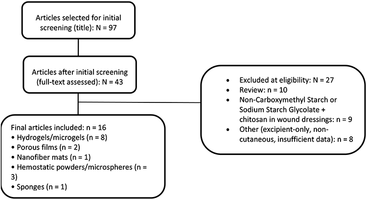 |
Figure 1 Flow diagram of the literature identification, screening, eligibility evaluation, and final study inclusion process conducted in this review. |
The included studies were systematically categorized according to formulation type to enable comparative analysis. Identified categories comprised hydrogels or microgels, porous films, nanofiber mats, hemostatic powders or microspheres, sponges, and polymeric complex or laminate concepts. This classification facilitated evaluation of structure–function relationships across different delivery platforms. Physicochemical properties such as swelling behavior, cross-linking density, mechanical stability, and drug-loading efficiency were extracted and compared. Biological outcomes including antimicrobial activity, antioxidant performance, hemostatic function, and wound-closure rates were also examined. Particular emphasis was placed on the role of chitosan as a bioactive and adhesive polymer matrix. The contribution of sago-derived sodium starch glycolate to hydration dynamics and moisture regulation was analyzed in parallel. Strategies for α-mangostin nanoformulation were evaluated in terms of encapsulation methods and release kinetics. Release mechanisms within the wound microenvironment were discussed in relation to polymer swelling and degradation behavior. Preclinical evidence from infected and diabetic wound models was integrated where available. Translational considerations, including safety, scalability, and regulatory feasibility, were critically appraised. Sustainability aspects were assessed with regard to agro-derived feedstock utilization and green material development. This integrative analysis bridges materials science with therapeutic performance. The synthesis therefore provides a coherent framework linking carrier design, nanoformulation strategy, and biological efficacy. Thus, this methodology defines the analytical scope and conceptual foundation of the present review.
Integrated Physicochemical and Biological Determinants of Chitosan and Sago-Derived Sodium Starch Glycolate at the Wound Interface
Chitosan’s Cationic Interface for Tissue Adhesion, Rapid Hemostasis, and Antibiofilm Control
Chitosan is a cationic aminopolysaccharide obtained through the deacetylation of chitin, and its protonated primary amino groups generate positive charge density in mildly acidic wound environments.33,34 This positive charge enables strong electrostatic interactions with negatively charged cellular membranes and extracellular matrix components. The apparent acid dissociation constant of chitosan lies near physiological acidity, typically around pH 6–6.5.35 As wound pH decreases below this range, protonation increases and electrostatically driven adhesion is enhanced. The degree of deacetylation directly determines the density of available amine groups.33 Molecular weight influences viscosity, mechanical strength, and film-forming capacity.33 Together, these parameters govern nanoparticle stability and dressing performance. Higher charge density generally correlates with stronger antimicrobial and adhesive properties. In chronic wounds where pH approaches neutrality, maintaining cationic activity becomes challenging. Quaternized derivatives have therefore been developed to retain permanent positive charge.36 N-(2-hydroxy)propyl-3-trimethylammonium chitosan chloride is one such example.36 These derivatives extend antibacterial efficacy across a broader pH range. Modified chitosans demonstrate improved stability and functional consistency.37–39 The interplay between protonation behavior and polymer architecture ultimately dictates biological performance. These physicochemical characteristics form the basis for chitosan’s multifunctional activity at the wound interface.
Tissue Adhesion Including Mucoadhesion
Mucoadhesion arises primarily from electrostatic interactions between protonated –NH3⁺ groups on chitosan and negatively charged sialic acid or sulfate residues on mucins.40 Hydrogen bonding and hydrophobic interactions further stabilize adhesion under hydrated conditions.40 Comparative studies demonstrate stronger adhesion of chitosan to gastric and salivary mucins than polyethylene oxide. This multimodal adhesion supports its function as a wet-surface scaffold.41 Adhesion strength depends on molecular weight and degree of deacetylation.40 Surface hydration also modulates interfacial bonding dynamics. Chemical modification can enhance adhesive performance in highly exudative environments. Thiolated chitosan enables disulfide exchange with cysteine-rich mucin domains.42 Catechol-functionalized derivatives provide mussel-inspired wet adhesion. Schiff-base chemistry further strengthens interfacial bonding without compromising compatibility.42 These modifications increase retention on bleeding tissues. Formulation architecture influences hydration and residence time. Thin films promote close surface contact. Hydrogels enable moisture retention and conformability. Electrospun fibers provide high surface area for interfacial interaction.43 Such tunable adhesion improves localized therapeutic delivery within wound beds.
Hemostasis: Rapid Clotting via Cellular and Architectural Effects
Chitosan promotes rapid hemostasis through both physical and biological mechanisms. The polymer concentrates erythrocytes at bleeding sites through electrostatic attraction. Platelet adhesion is enhanced at positively charged interfaces. Rapid fluid absorption within porous matrices accelerates clot formation.33 Physical adsorption contributes to thrombus stabilization. Molecular studies show that chitosan activates platelets via Toll-like receptor-2 signaling.44 This signaling pathway complements its structural hemostatic role.44 Architectural design strongly influences performance. Porous sponges maximize contact area with blood components. Aerogels enhance capillary-driven fluid uptake. High-surface-area fibers further accelerate clotting kinetics.45 Hemostatic efficiency scales with charge density and degree of deacetylation. Enhanced red-cell trapping improves coagulation stability.46 Clinical and preclinical evaluations consistently report shortened bleeding times with chitosan-based dressings.47 The integration of charge-mediated activation and optimized architecture positions chitosan as an effective hemostatic biomaterial.
Antibacterial and Antibiofilm Activity
Chitosan exhibits broad-spectrum antibacterial activity through membrane destabilization mechanisms. Electrostatic interactions disrupt Gram-negative lipopolysaccharide-rich membranes. Gram-positive teichoic-acid-rich envelopes are similarly affected. Increased permeability leads to leakage of intracellular components.36 These effects result in microbial growth inhibition. At the biofilm level, chitosan interferes with extracellular polymeric substance matrices. This disruption reduces bacterial attachment to moist wound surfaces. Biofilm maturation is consequently suppressed.48 Chronic wound environments particularly benefit from antibiofilm strategies. Quaternized derivatives maintain antimicrobial activity near neutral pH.37–39 Sustained cationic charge enhances long-term efficacy.36 Chitosan nanoparticles enable co-delivery of small molecules. These hybrid systems further reduce biofilm biomass and virulence expression. Synergistic formulations amplify antimicrobial performance.49 Together, membrane disruption and matrix interference define chitosan’s antibiofilm capacity.
Design Implications for Nanoparticle Platforms and Dressings
Rational design of chitosan-based systems requires alignment between physicochemical properties and therapeutic objectives. High degrees of deacetylation enhance electrostatic adhesion to tissues.33 Hydrated hydrogel architectures maximize hydrogen bonding interactions.40 For prolonged mucosal residence, thin films and hydrogels are preferred.43 Rapid hemostasis benefits from porous, high-surface-area constructs.33 Aerogel and sponge formats enhance capillary-driven fluid uptake.45 Platelet activation is supported by accessible positive charge.44 For chronic wound infection control, quaternized derivatives sustain activity near neutral pH.36–39 Nanoparticle formulations improve dispersion of hydrophobic therapeutics. Co-encapsulation strategies enable quorum-sensing inhibition.49 Matrix-targeting approaches enhance biofilm disruption.48 Structural stability must be balanced with biodegradability.17 Mechanical integrity influences dressing durability.33 Scalable fabrication methods support translational feasibility.17 Integrating adhesion, hemostasis, and antibiofilm control within a single platform enhances multifunctionality. These design principles collectively inform development of advanced chitosan-based nanoparticle carriers and wound dressings.
Sago-Derived Sodium Starch Glycolate (Crosslinked Carboxymethyl Starch): A “Hydration Engine” for Moisture Control
Sago-derived sodium starch glycolate (SSG) is a crosslinked sodium carboxymethyl starch synthesized from sago starch, in which anionic carboxymethyl groups are introduced into the polysaccharide backbone.18 The presence of these negatively charged carboxylate groups, combined with intragranular covalent crosslinks, underpins its high fluid-uptake capacity and rapid wicking behavior in aqueous environments.18 The sago origin provides a renewable and locally abundant feedstock with tunable substitution chemistry relevant to wound-care biomaterials.50 Measured substitution levels for sago-derived SSG typically fall within a degree of substitution (DS) of approximately 0.23–0.32 under optimized synthesis conditions.20 This range establishes sufficient charge density for rapid hydration while preventing complete dissolution of the polymer network.18 Crosslink strength functions as an additional material parameter that inversely modulates swelling kinetics. Stronger crosslinking restricts polymer chain mobility and reduces free swelling capacity.51 Conversely, lower crosslink density permits greater expansion of the starch network upon fluid contact.52 Classical excipient studies demonstrated that higher carboxymethylation combined with reduced crosslink density enhances water penetration and volumetric expansion.52 Contemporary analyses formalize this relationship by treating crosslink strength as an independent design variable alongside DS. These interdependent chemical parameters directly determine hydration rate and gel integrity. In wound applications, predictable swelling behavior is critical for maintaining structural stability under exudative stress.51 Thus, SSG functions not merely as a disintegrant but as a tunable hydration platform. The chemical architecture of sago-derived SSG therefore defines its capacity to regulate moisture at the wound interface.
As a hydration engine, SSG integrates two complementary water-management mechanisms: bulk swelling and capillary wicking. Bulk swelling enables fluid absorption into the polymer matrix, while capillary pathways facilitate lateral transport of exudate.19 This dual mechanism ensures both retention and redistribution of wound fluid. The anionic polymer network remains partially insoluble due to crosslinking, thereby preserving structural integrity during prolonged exposure to moisture.51 In the wound microenvironment, maintaining a moist yet breathable interface accelerates re-epithelialization and granulation tissue formation. Effective moisture management reduces desiccation without promoting maceration.53 Water-vapor transmission rate (WVTR) and absorption capacity are therefore critical performance indicators. Low-exudate wounds may tolerate lower WVTR values similar to catheter-site films. In contrast, highly exuding wounds require higher vapor transmission to prevent fluid accumulation.54 Carboxymethyl starch systems, including crosslinked nanofiber constructs, demonstrate rapid absorption and evaporation while preserving barrier function.55 These characteristics reinforce their suitability for moisture-balanced wound care. Control of both swelling magnitude and evaporation rate enables adaptation to diverse exudate loads.55 The hydration–evaporation equilibrium ultimately governs wound-bed homeostasis. Therefore, SSG’s water-management properties are central to its functional relevance in topical dressings.
Hydration behavior of sodium carboxymethyl starch networks is influenced by ionic strength within the surrounding medium.56 Counter-ion screening reduces osmotic swelling pressure and decreases equilibrium uptake in saline environments. Multivalent ions may introduce additional physical crosslinks that stiffen the gel matrix.56 Such ionic effects can slow wicking under highly exudative conditions. Formulation studies on sago-derived SSG confirm that rational control of DS and crosslink density produces predictable changes in water uptake and flow-through behavior. This predictability allows translation of tablet-disintegration principles into moist-wound fluid management strategies.18 Moisture-vapor transmission is additionally governed by macro-architecture.57 Porosity, thickness, and capillary connectivity significantly influence WVTR values.58 Foam, fiber, film, and hydrogel formats each present distinct diffusion pathways. Processing methods such as freeze-drying and electrospinning further modulate internal pore structure.57 Microstructure-dependent fluid handling has been confirmed in multiple biopolymer dressing analyses.58,59 Selection of simulated wound fluid composition also alters measured performance metrics.11 Thus, both chemistry and architecture must be co-optimized to achieve targeted hydration behavior. Integrated control of DS, crosslink density, and structural porosity enables precise tuning of wound-fluid dynamics.
Biocompatibility considerations favor starch-based matrices for cutaneous applications.56 Modern reviews highlight the benign degradation profile of carboxymethyl starch networks when residual reagents are carefully controlled.56 Cytocompatibility has been demonstrated in bioactive carboxymethyl starch hydrogels loaded with antimicrobial or anti-inflammatory agents.60 Such systems support cellular proliferation and wound-healing responses.60 From a sustainability perspective, sago-derived SSG offers a renewable polysaccharide backbone with scalable synthesis routes. The material can be formulated as a standalone absorbent or blended into composite hydrogels and fibrous dressings.50 For highly exuding wounds, higher WVTR and greater free-swelling capacity are desirable. This can be achieved by selecting moderate DS values combined with lower crosslink strength.51 For low-to-moderate exudate, denser networks and thicker constructs help maintain a humid microclimate without causing maceration.57 When combined with cationic polymers such as chitosan, anionic carboxylate groups in carboxymethyl starch form polyelectrolyte complexes. These complexes enhance mechanical robustness and modulate diffusion pathways.61 Such interactions balance hydration control with barrier stability in composite dressings.61 (Figure 2)
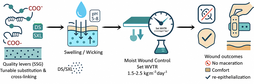 |
Figure 2 Conceptual framework linking degree of substitution (DS) and swelling/crosslink-related index (SXL) to swelling–wicking dynamics, WVTR and absorption control, and resultant wound-healing performance. Notes: The downward arrows indicate the penetration of water/exudate into the SSG matrix. The upward arrow shows the redistribution of fluid due to swelling and the release of vapor/moisture. The droplet symbol is for wound fluid or exposure to moisture. The pH 5–8 symbol is for the relevant testing range for wounds. The WVTR gauge is for the desired moisture-vapor transmission window. Examination marks mean good dressing results with controlled absorption, less risk of maceration, more comfort, and better re-epithelialization. |
Synergy of Chitosan and Sago-Derived Sodium Starch Glycolate at the Wound Interface: Programmed Complementary Functions
At the construct level, combining a cationic, tissue-interactive chitosan layer with an anionic, crosslinked hydration engine based on sago-derived sodium starch glycolate enables a programmed division of labor at the wound interface.16 In this configuration, interfacial adhesion and bioburden control are primarily knowing as chitosan-driven functions.16 In parallel, exudate handling and moisture redistribution are predominantly mediated by the SSG domain. This separation avoids redundancy commonly observed in single-polymer dressings.62 Instead of overlapping roles, each polymer contributes distinct yet complementary performance attributes. Ionic coupling between protonated amine groups of chitosan and carboxymethyl groups of carboxymethyl starch produces a cohesive polyelectrolyte complex network.61 This ionically crosslinked architecture enhances wet mechanical integrity without requiring chemical crosslinkers.61 Charge ratio and ionic strength become tunable design parameters rather than necessitating re-engineering of base polymers.63 Contemporary polyelectrolyte complex literature demonstrates that slightly polycation-leaning stoichiometry preserves excess cationic sites for antimicrobial contact. At the same time, near-electroneutral conditions maximize interpolymer cohesion.63 This balance enables antimicrobial surface activity alongside structural robustness. The resulting laminate or interpenetrated configuration supports spatial specialization across the moisture–infection design space.62 Such architecture reflects intentional programming rather than additive blending. By assigning non-overlapping roles to each domain, functional synergy emerges at both molecular and macroscopic scales. This programmed complementarity defines the mechanistic foundation of the chitosan–SSG hybrid platform.
Transport phenomena represent the shared constraint governing healing efficiency and infection control.64 The hybrid assembly can therefore be conceptualized as a two-channel transport system. The chitosan-rich contact layer primarily regulates diffusive permeability for small molecules and signaling mediators through the mesh characteristics of the polyelectrolyte complex.63 The SSG-rich backing predominantly supports convective exudate transport via swelling and capillary wicking pathways.19 This separation of diffusion and convection reduces wash-off of bioactive compounds compared with homogeneous hydrogel systems.63 Moisture-vapor transmission rate becomes a central performance indicator in this context.65 For moderate-exudate wounds, targeting WVTR values within approximately 2000–2500 g·m−2·24 h minimizes maceration while preventing desiccation.65 Such WVTR windows are consistently reported across modern foam and composite dressing studies.66 Achieving this balance requires coordinated adjustment of front-layer porosity and thickness.65 Simultaneously, crosslink density and degree of substitution in the SSG layer modulate absorption capacity.51 The interplay between architectural parameters and polymer chemistry determines overall vapor flux.58,59 Structural microarchitecture influences both air permeability and fluid retention.57 Selection of simulated wound fluid further affects measured transport behavior.11 Through integrated tuning of these parameters, the hybrid laminate achieves moisture regulation without sacrificing antimicrobial contact or mechanical stability.
Charge programming serves as the central design lever underlying this synergy.61 By selecting a slightly polycation-leaning PEC ratio, residual cationic sites remain available at the tissue interface.63 These free amine groups maintain contact-active antibiofilm pressure.16 Concurrently, the anionic SSG domain rapidly absorbs serum and redistributes fluid away from the microinterface.19 This “adhere-and-clear” function stabilizes early clots and reduces dilution of antimicrobial activity.16 Unlike purely absorbent matrices, the hybrid design preserves programmable cationic contact sites. Drug release kinetics are also influenced by mesh size within the PEC network.63 Reduced free volume slows α-mangostin diffusion without the need for covalent crosslinking.63 This strategy maintains cytocompatibility while achieving controlled release.61 Ionic-strength effects within wound exudate may compress swelling of the PEC layer.56 Therefore, SSG crosslink strength and degree of substitution can be adjusted upward for highly exuding environments.51 For lower-exudate wounds, moderate DS and lower crosslink density support extended residence time and comfort.18 These programmable chemical levers allow adaptation across diverse wound conditions.
Mechanical synergy further reinforces the therapeutic potential of the hybrid construct. Formation of the polyelectrolyte complex increases wet tensile strength compared with either polymer alone.61 Enhanced resistance to cold flow improves dressing durability under physiological stress. Multilayer or graded architectures with a chitosan-rich adhesive face and an SSG-rich absorbent backing demonstrate clinically relevant WVTR and accelerated repair in analogous composite systems.64 Sequential functionality characterizes the hemostatic mechanism of the hybrid.16 The chitosan contact layer initiates platelet engagement and erythrocyte capture at the bleeding interface.33 Subsequently, the SSG domain removes serum to prevent clot washout and sustain thrombus stability.16 This coordinated “capture-then-clear” sequence cannot be replicated by single hydrophilic absorbers.63 Infection control follows a similar hierarchical logic. The contact-active chitosan surface maintains antibiofilm pressure, while the SSG layer acts as a breathable reservoir for sustained cargo release.62,63 (Figure 3)
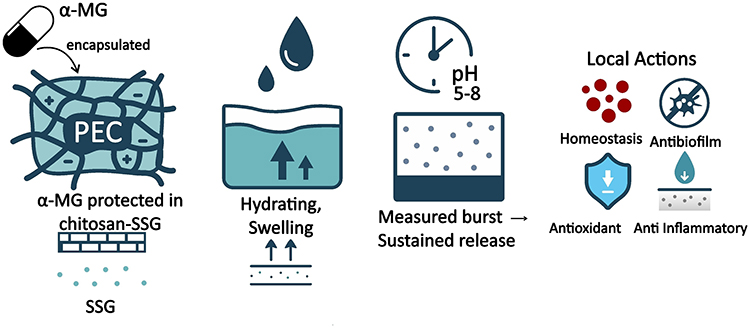 |
Figure 3 Schematic representation of the chitosan–sodium starch glycolate (SSG) hybrid polyelectrolyte complex (PEC) system for α-mangostin delivery and moisture-regulated wound therapy. Notes: The + and − signs represent electrostatic interactions between cationic chitosan amine groups and anionic SSG carboxylate groups. Upward arrows represent hydration-induced swelling, α-mangostin release, and moisture transport, and the clock symbol represents time-dependent biphasic release. The red dots indicate microbial burden or inflammatory/oxidative signals; the shield indicates antimicrobial or protective activity; and the downward arrows indicate reduction of infection, inflammation, oxidative stress, or excess exudate. The Quality by Design (QbD) parameters controlling the performance of the formulation are CS:TPP ratio, pH, ionic strength, CS:SSG ratio, mixing/shear conditions, particle size/polydispersity index (PDI), zeta potential, encapsulation efficiency/drug loading (EE%/DL%), viscoelastic properties (G′/G″), swelling behavior, water vapor transmission rate (WVTR) and stability. |
Evidence-Anchored Recommendations: A Quality by Design Framework
Studies involving wound-facing biomaterials should adopt a minimum-information reporting package encompassing material identity, physicochemical characterization, biological evaluation, and complete procedural transparency.67 Reporting should align with the Minimum Information Reporting in Bio–Nano Experimental Literature framework to enhance reproducibility and cross-laboratory comparability.67 Detailed disclosure of polymer source, degree of substitution, molecular weight, crosslinking chemistry, and processing parameters is essential for data interoperability. Experimental conditions governing swelling behavior, WVTR, mechanical performance, and degradation kinetics must be clearly described. Biological assays should report cell models, culture conditions, exposure times, and statistical methods to enable rigorous interpretation. For in vivo investigations, compliance with ARRIVE 2.0 guidelines is recommended to strengthen internal validity. Randomization, blinding, predefined outcomes, and justified sample sizes reduce bias and improve transparency.68 Such methodological rigor facilitates cross-study comparison and meta-analytic integration. For clinical translation, trial manuscripts should be structured in accordance with the CONSORT checklist and flow diagram, including relevant extensions for device-based interventions. CONSORT remains the prevailing standard for randomized controlled trials.69 Advanced wound dressings are commonly regulated as medical devices, necessitating alignment with applicable regulatory frameworks. Biocompatibility assessment should follow ISO 10993–1 using a risk-based biological evaluation strategy. Documentation should reflect current FDA guidance to support regulatory submissions.70 Physical performance testing must reference EN 13726:2023 for absorption, moisture-vapor transmission, waterproofness, and extensibility, as this revision supersedes earlier European standards. When ASTM E96/E96M is employed for WVTR assessment, the specific method used (desiccant “dry-cup” or water “wet-cup”) and test conditions must be reported, since results from different methods are not directly comparable.
From a Quality by Design (QbD) perspective, development of chitosan–SSG systems should follow a structured hierarchy beginning with definition of a Target Product Profile (TPP). Identification of Critical Quality Attributes (CQAs) relevant to wound performance should follow. Critical Material Attributes (CMAs) and Critical Process Parameters (CPPs) must then be systematically mapped. This framework aligns with ICH Q8(R2) and ICH Q9(R1) guidance on pharmaceutical development and quality risk management. Design-of-experiments methodologies can be applied to delineate a multidimensional design space. In chitosan–SSG hybrids, the polyelectrolyte charge ratio (N⁺/COO−) functions as a key CMA influencing cohesion and antimicrobial contact.63 Degree of deacetylation and molecular weight of chitosan affect adhesion strength and mechanical robustness.33 Degree of substitution and crosslink strength of carboxymethyl starch modulate swelling kinetics and WVTR behavior.51 Laminate porosity and thickness influence vapor flux and exudate absorption capacity.57 Ionic strength impacts polyelectrolyte complex swelling and drug-release profiles under physiological conditions.56 These interrelated parameters determine adhesion under wet conditions, hemostasis efficiency, antibiofilm performance, WVTR control, and drug-release half-times.63 Risk assessment tools such as failure mode and effects analysis may assist in identifying high-impact variables. Establishment of a clear control strategy supports batch-to-batch consistency and scalability. Definition of an explicit design space enhances regulatory confidence and manufacturing robustness. Integration of QbD principles into chitosan–SSG hybrid development strengthens translational readiness and clinical credibility.
Material Selection Framework for Chitosan, Sago-Derived Sodium Starch Glycolate, and Hybrid Systems
Material selection for wound-facing biomaterials should be guided by clearly defined functional priorities rather than polymer availability alone. When wet-surface retention and contact-active antibiofilm control are the dominant objectives, chitosan is best positioned as the wound-contact layer due to its cationic interfacial activity. Its degree of deacetylation, molecular weight, and zeta potential in wound-relevant buffer systems must be explicitly reported, as these parameters directly govern electrostatic adhesion and antimicrobial performance. For gel or film formulations, oscillatory rheology data (G′/G″) together with defined frequency and strain windows are essential to ensure reproducibility and meaningful cross-study comparison. Moderately positive zeta potentials are generally preferable to balance antibacterial efficacy with cytocompatibility, particularly in modified or quaternized systems.71 In bleeding wounds, porous chitosan constructs such as foams or fibrous matrices provide enhanced platelet engagement and erythrocyte trapping. Structural characteristics including pore architecture and thickness should therefore be quantitatively linked to time-to-hemostasis outcomes, and hemostatic evaluation should follow validated bleeding models with reporting aligned to ARRIVE 2.0 guidelines.68 These decision points, associated reporting requirements, and corresponding testing standards are consolidated in Table 1 to provide a structured reference for material deployment. By aligning functional intent with measurable parameters, the framework supports reproducibility and translational clarity.
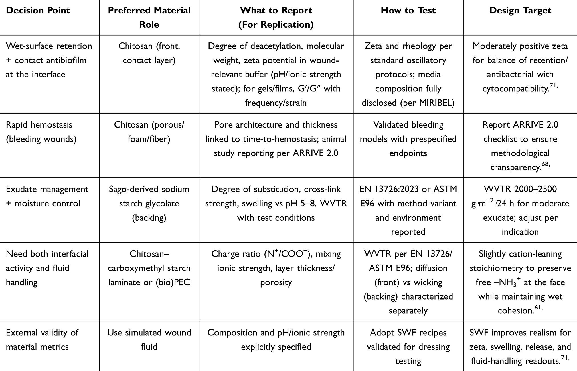 |
Table 1 Evidence-Guided Design and Evaluation Framework for Wound-Facing Biomaterials |
When exudate management and moisture regulation are prioritized, sago-derived sodium starch glycolate functions as a dedicated backing or absorber layer. Reporting should include degree of substitution, quantified crosslink strength, swelling profiles across pH 5–8, and WVTR values with clearly specified environmental conditions. These attributes collectively determine wicking kinetics, structural stability, and vapor permeability. Physical testing should reference EN 13726:2023 or ASTM E96, with explicit disclosure of the selected method variant and test setup. For moderate-exudate wounds, WVTR values in the range of approximately 2000–2500 g·m−2·24 h are frequently associated with maintenance of a moist yet breathable microenvironment, although final tuning remains indication-specific. In clinical contexts requiring both interfacial bioactivity and fluid handling, laminate or polyelectrolyte complex constructs integrating chitosan and carboxymethyl starch are indicated.61 Complexation stoichiometry (N⁺/COO−), mixing ionic strength, and layer thickness or porosity must be transparently reported to ensure mechanistic interpretability. Slightly cation-leaning stoichiometry preserves free –NH3⁺ groups for contact-active antimicrobial function while maintaining cohesive wet integrity.61 Diffusive transport at the contact layer and capillary-driven wicking in the backing layer should be characterized independently to clarify functional separation. Use of simulated wound fluid with explicitly stated composition strengthens external validity of swelling, release, and WVTR measurements.71 Therefore, this evidence-anchored framework integrates material selection, reporting transparency, and performance targets within a Quality by Design mindset.
Formulation Strategies for Alpha-Mangostin Nanoparticles and Mechanisms in the Wound Microenvironment
Alpha-mangostin (α-MG) is a prenylated xanthone with broad-spectrum antimicrobial, anti-inflammatory, and antioxidant activity.21 Despite its pharmacological potential, its poor aqueous solubility and limited physicochemical stability restrict effective exposure at the wound interface. Nanoformulation strategies are therefore required to enhance dispersion, protect the active compound, and prolong local bioavailability. Controlled delivery becomes particularly important in the dynamic wound microenvironment. Early wound phases are often characterized by high microbial burden and elevated reactive oxygen species.72 Subsequent phases involve persistent inflammation and extracellular matrix remodeling.72 An optimal delivery platform should therefore provide an initial antimicrobial pulse followed by sustained anti-inflammatory and antioxidant support.72 Chronic wounds frequently exhibit alkaline pH and altered ionic strength.73 These physicochemical conditions influence drug ionization, polymer charge density, and biofilm resilience.73 Release testing should reproduce these environmental parameters to ensure translational relevance.73 In addition, laboratory characterization should align with updated evaluation guidance for wound dressings.74 Absorption, moisture-vapor transmission, and retention metrics must be interpreted in relation to clinical moisture requirements.74 Together, these considerations define the performance criteria for α-MG nanoparticle delivery systems in wound therapy.
Diverse formulation architectures can accommodate polymer chain–SSG nanoparticle systems to meet these criteria. Figure 4 illustrates representative delivery platforms, including hydrogels, porous films, sponges, microspheres, and nanofiber mats. Hydrogels and microgels provide a hydrated three-dimensional network that supports controlled swelling and sustained release. Porous films offer conformability and barrier function while permitting regulated vapor transmission. Sponges combine high absorption capacity with structural integrity under compressive load. Microspheres enable discrete encapsulation and tunable burst–sustained release profiles. Nanofiber mats provide high surface area and structural mimicry of extracellular matrix architecture.75 Such fibrous scaffolds have been associated with accelerated healing and reduced scar formation.75 Integration of SSG nanoparticles within these platforms enhances hydration-driven wicking and moisture regulation. Concurrently, chitosan-rich domains support tissue adhesion and contact-active antimicrobial effects. The modular nature of these architectures allows tailoring of release kinetics, WVTR, and mechanical robustness. Platform selection should therefore be guided by wound exudate level, infection risk, and required wear time. Structural parameters such as porosity, thickness, and fiber diameter further modulate transport behavior. By combining α-MG nanoencapsulation with programmable polymer–SSG matrices, these delivery systems enable synchronized antimicrobial control, moisture balance, and tissue regeneration support within the wound microenvironment.
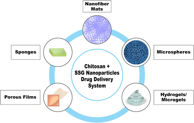 |
Figure 4 Formulation architectures for polymer chain–SSG nanoparticle drug delivery systems across diverse wound-dressing platforms. |
Polymeric Platform: Chitosan-Based Nanoparticles (Ionic Gelation and Polyelectrolyte Complexes)
Polymeric nanoparticles prepared through ionotropic gelation of chitosan with sodium tripolyphosphate (TPP) or via polyelectrolyte complexation with anionic biopolymers provide a mild, water-based encapsulation strategy compatible with heat-sensitive xanthones such as α-mangostin.76 These fabrication approaches avoid organic solvents and preserve molecular integrity of the active compound. The resulting nanoparticles exhibit cationic surface charge that enhances adhesion to negatively charged wound tissues.76 Encapsulation of α-mangostin within chitosan–alginate nanoparticles significantly improves entrapment efficiency and bioactivity compared with the free compound.14 Such polymeric systems enhance apparent solubility and local retention at the wound interface. The positive surface charge also contributes to contact-active antimicrobial effects during early wound phases.76 Residence time is prolonged through mucoadhesive interactions, supporting dose-sparing strategies when fluid flux is elevated.12 Polymeric carriers consistently demonstrate biphasic release kinetics characterized by an initial burst followed by sustained diffusion. This release behavior can be modulated through adjustment of polymer ratio, crosslinking density, and particle size distribution.77 Early burst release facilitates rapid antimicrobial action against high bioburden. Sustained diffusion over subsequent days supports anti-inflammatory and antioxidant modulation.77 Chitosan–TPP and chitosan–alginate complexes are therefore particularly suited when adhesion and controlled multi-day exposure are primary design goals.76 Water-based encapsulation combined with tunable release kinetics reinforces their translational feasibility.12 These characteristics position chitosan-based nanoparticles as front-line platforms for α-mangostin delivery in wound applications.
Lipidic Platform: Nanostructured Lipid Carriers (Optionally Chitosan-Coated)
Nanostructured lipid carriers (NLCs) incorporate α-mangostin within a mixed solid–liquid lipid matrix to enhance solubilization and dermal penetration.26 The lipid core protects hydrophobic molecules from premature degradation while allowing high local drug loading. In vivo studies demonstrate superior wound-healing efficacy of α-mangostin-loaded NLCs compared with the free compound, including in diabetic wound models.26 Lipid matrices also facilitate rapid release in early inflammatory phases.26 Co-formulation with ancillary bioactives such as clove-oil terpenes enhances antimicrobial activity and physical stability.78 These findings underscore the adaptability of lipidic systems for topical therapy. Surface modification with chitosan further improves tissue adhesion and retention.79 Chitosan-coated NLCs exhibit enhanced topical deposition for lipophilic antioxidants.80 This hybrid approach integrates solubilization efficiency with cationic interfacial bioactivity.79 Enhanced contact time supports antimicrobial performance where wash-off is a concern.81 Lipidic systems are particularly advantageous when rapid antimicrobial release and high superficial tissue concentration are desired.81 Particle size and lipid composition can be tuned to optimize penetration depth.26 Controlled release from NLCs complements moisture-regulated dressing platforms. Taken together, lipid-based carriers represent effective options when solubilization and early-phase antimicrobial exposure are prioritized.
However, the benefits of NLCs have to be balanced against formulation-specific stability constraints. During storage NLCs can be physically unstable, as for example particle aggregation, increased particle size, change in polydispersity index, gelation, drug expulsion and reduced encapsulation efficiency. Such changes may be due to lipid crystallization, polymorphic transition, surfactant incompatibility or change in the solid to liquid lipid ratio.81 Lipid oxidation is also a major problem for lipid nanocarriers, particularly when unsaturated oils or oxidation sensitive lipid matrices are used. Oxidative degradation may alter the lipid structure, lower the drug loading stability, affect the drug release behavior and biological performance. Therefore, α-MG-loaded NLCs should be characterized not only in terms of particle size, zeta potential, encapsulation efficiency and wound healing activity, but also in terms of storage stability, oxidative stability, lipid composition, antioxidant protection and performance after sterilization before translation to wound-dressing systems.81
Hydrogel and Film Platforms: Depot Dressings for Sustained Exposure
Hydrogel membranes and film-based dressings provide breathable depot systems capable of stabilizing α-mangostin while maintaining a moist microenvironment.82 Such platforms support re-epithelialization and granulation during inflammatory and proliferative phases.82 Incorporation of α-mangostin into polysaccharide matrices enhances dispersion and reduces crystallization. A registered clinical protocol evaluating an α-mangostin hydrogel film based on a chitosan–alginate matrix reflects increasing translational interest.83 Depot systems enable prolonged local exposure and reduced dosing frequency. Sustained release from hydrogels mitigates inflammation and oxidative stress over multiple days.8 Preclinical formulations co-delivering α-mangostin with complementary phytochemicals demonstrate non-cytotoxicity and accelerated closure.8 Barrier performance and pharmacologic action must be evaluated in parallel.74 WVTR and fluid-handling characteristics influence overall clinical suitability.74 Composite CS/CMC/tannic-acid/beeswax films illustrate multifunctional integration of adhesion, barrier function, and antimicrobial activity.84 Hydrogel elasticity and conformability enhance patient comfort and reduce trauma during dressing changes.82 Structural parameters such as thickness and porosity modulate vapor permeability and drug diffusion.57 Depot dressings are particularly suitable when sustained modulation rather than rapid burst release is required.8 These systems therefore align with later-phase wound healing objectives requiring prolonged signaling regulation.
Mechanistic Alignment with the Wound Microenvironment
α-Mangostin exhibits direct antibacterial activity against Gram-positive organisms, particularly Staphylococcus species prevalent in cutaneous infections.85 It can synergize with β-lactam antibiotics through inhibition of penicillin-binding proteins, reinforcing its relevance during early antimicrobial intervention.85 Beyond planktonic killing, α-mangostin contributes to biofilm suppression and restoration of epithelial barrier integrity.85 Reduced expression of pro-inflammatory cytokines has been documented in preclinical models.85 Chronic wounds are characterized by persistent oxidative stress and dysregulated inflammation.72 The polyphenolic structure of α-mangostin confers antioxidant capacity capable of scavenging reactive oxygen species.72 Such redox modulation supports tissue repair in later wound stages.72 Delivery systems must therefore accommodate temporal shifts in wound physiology. Early antimicrobial pulse followed by sustained anti-inflammatory release is mechanistically advantageous.77 Wound pH and ionic strength influence drug ionization and polymer charge density.73 Release testing should be conducted in simulated wound fluids reflecting relevant salt and protein content.73 Zeta potential characterization under these conditions enhances predictive validity.73 Alignment of carrier design with wound biochemical conditions improves therapeutic synchronization. Integration of antimicrobial, antioxidant, and moisture-regulating mechanisms ultimately supports accelerated and stable re-epithelialization.
Practical Selection Guide: Phase-Matched and Testable Framework
Carrier selection for α-mangostin should be guided by the physiological phase of wound healing rather than by formulation preference alone. Early-stage wounds within the first one to three days typically present high exudate volume, elevated microbial burden, and a microenvironment that may shift toward neutral or alkaline pH. Under these conditions, rapid antimicrobial pressure is essential to prevent biofilm establishment. Lipid-based systems such as nanostructured lipid carriers provide enhanced solubilization and prompt topical drug availability.26 These carriers facilitate high local loading of hydrophobic α-mangostin within superficial tissue layers. Chitosan-coated variants further improve adhesion to wet wound surfaces and extend contact time.79 α-Mangostin exhibits strong anti-staphylococcal activity and can synergize with β-lactam antibiotics, supporting its relevance during the early antimicrobial window.85,86 An intentional early burst release within the first hours may therefore be advantageous in high-burden contexts. As the wound transitions toward the proliferative phase, exudate levels typically moderate and inflammatory signaling becomes the dominant driver of delayed healing. Sustained anti-inflammatory and antioxidant support becomes more critical than rapid antimicrobial pulse. Polymeric chitosan-based nanoparticles enable tunable biphasic release aligned with this need.77 Breathable hydrogel or film depots further extend exposure while preserving a moist interface.82 Cyclodextrin complexation may enhance loading capacity and smooth the release profile in hydrated systems. Therefore, phase-responsive modulation of release kinetics improves therapeutic synchronization with wound biology.
Moisture control remains a central determinant of clinical performance across all wound phases. WVTR targets should be selected according to exudate class, with higher values favored in heavily exuding wounds and mid-range values appropriate during proliferation.74 The measurement method must be explicitly declared because EN 13726 and ASTM E96 variants yield non-interchangeable results. Release kinetics, swelling behavior, and zeta potential should be characterized in simulated wound fluid containing physiologically relevant salts and proteins.73 Such testing conditions capture ionic screening and protein adsorption effects that alter both carrier charge and drug diffusion. In chronic non-healing wounds, including diabetic or venous ulcers, persistent inflammation and oxidative stress require prolonged local exposure rather than repeated burst dosing.72 Depot-oriented hydrogel or polymeric nanoparticle systems with tunable mesh size and porosity are therefore preferable in these contexts. Sustained release with minimal burst can stabilize antioxidant and anti-inflammatory signaling over several days. Moisture regulation must remain balanced to prevent maceration while avoiding desiccation. Breathable laminates integrating absorbent backing layers can support this requirement. Structural parameters such as thickness and porosity should be co-optimized with chemical composition. External validity improves when all assays are conducted under wound-relevant pH and ionic conditions.73 Integration of antimicrobial, antioxidant, and moisture-management performance metrics strengthens translational interpretability. The phase-aligned selection logic, release programming targets, WVTR ranges, and reporting requirements are synthesized in Table 2 to provide a structured and testable decision matrix for α-mangostin delivery platforms.
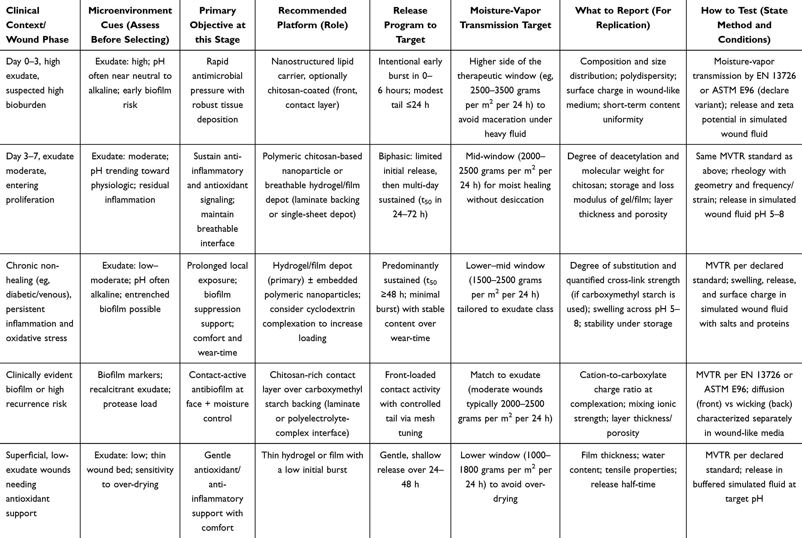 |
Table 2 Phase-Oriented Selection Framework for α-Mangostin Delivery Platforms in Wound Therapy |
Critical Formulation Challenges and Unresolved Release Behavior
A critical evaluation is necessary because the proposed chitosan–SSG–α-MG platform remains formulation-dependent rather than intrinsically superior. Although SSG is attractive because of its rapid liquid uptake and swelling capacity, its role in drug release is not unidirectional. As a superdisintegrant, SSG can accelerate hydration, wicking, and disintegration; however, in a polymeric coating model, the incorporation of SSG produced delayed release followed by burst release, with release behavior depending on SSG concentration, water diffusion, coating permeability, swelling, deformation, and eventual rupture.18,19,51,52,87
SSG functionality is also influenced by the material source, degree of substitution, degree of crosslinking, particle size, viscosity, liquid uptake, and crosslinking strength, indicating that different SSG grades or synthesis conditions may produce different release profiles and even dissolution failure.19,51,52 When SSG or carboxymethyl starch is incorporated into hydrogels, films, sponges, or polyelectrolyte complexes, the release behavior may be further modified by matrix permeability, polyelectrolyte-complex structure, ionic strength, protein adsorption, swelling pressure, and structural relaxation.61,63,73 Therefore, direct α-MG release from sago-derived SSG matrices remains an unresolved issue that can not be assumed from SSG swelling behavior alone.
Existing α-MG delivery studies have shown that the release behaviour varies across nanostructured lipid carriers, cyclodextrin-complexed hydrogels, chitosan–alginate hydrogel films, gellan-gum membranes, hydrogel film-forming sprays, self-healing hydrogels, and chitosan/collagen nanoparticle-loaded hydrogels, indicating that solubilization strategy and carrier architecture strongly affect release kinetics.8,13,26–29,82 Chitosan-based systems also have important limitations, including dependence on molecular weight, degree of deacetylation, protonation state, charge density, and formulation architecture, as well as limited solubility under neutral or physiological conditions, poor mechanical strength in some physical hydrogel formats, and possible wound adhesion during dressing removal.71,88,89 These constraints often require blending, crosslinking, quaternization, polyelectrolyte complexation, or multilayer construction to achieve adequate wet stability and biological performance.61,63,71,88,89 Consequently, the future development of chitosan–sago-derived SSG carriers for α-MG nanoparticles should be framed as a testable formulation strategy requiring optimization of swelling, wet mechanical integrity, exudate handling, α-MG loading, release kinetics, sterilization stability, cytocompatibility, and in vivo wound-healing efficacy rather than as a universally effective platform.
In addition to unresolved release behavior, several formulation-related and translational limitations should be considered. Chitosan-based wound-dressing systems are strongly influenced by polymer properties, particularly molecular weight, degree of deacetylation, modification strategy, and hydrogel or dressing architecture, which may affect adhesion, antimicrobial activity, mechanical strength, and wound compatibility.12,17,88,89 SSG and carboxymethyl starch also show material-dependent performance because their hydration, swelling, liquid uptake, and disintegration- or release-related behavior are affected by the degree of substitution, crosslinking strength, molecular structure, particle-related characteristics, and functional-related material attributes.18–20,51,52
Conflicting release behavior should also be interpreted cautiously. Although SSG can promote hydration, wicking, and swelling, the incorporation of superdisintegrants into polymeric matrices has also been associated with delayed release followed by burst release, depending on matrix composition, water penetration, swelling, deformation, and rupture mechanisms.19,51 In chitosan–carboxymethyl starch systems, drug transport may be further affected by polyelectrolyte-complex formation, polymer ratio, degree of substitution, pH, and ionic conditions.61,63 Therefore, swelling behavior alone cannot be used to predict α-mangostin release from chitosan–SSG hybrid systems.
Clinical translation also remains limited. Most α-mangostin wound-healing formulations reported to date are still based on in vitro experiments, animal models, or formulation-development studies, including nanostructured lipid carriers, cyclodextrin-complexed hydrogels, chitosan–alginate hydrogel films, gellan-gum membranes, and chitosan/collagen nanoparticle-loaded hydrogels.8,13,26–29,82 Although one α-mangostin chitosan–alginate hydrogel film has progressed to a randomized clinical trial protocol, this remains a protocol rather than completed clinical efficacy evidence.83 Consequently, the proposed chitosan–sago-derived SSG platform should be regarded as a promising but formulation-dependent strategy that still requires standardized physicochemical characterization, wound-relevant release testing, reproducibility assessment, and clinically relevant validation before translational superiority can be claimed.67–69,73,74
Discussion – Safety and Biocompatibility: From Regulatory Frameworks to Real Wound Context
Biocompatibility evaluation should be positioned as a risk-managed pathway conducted under ISO 10993–1 and applied to the finished device rather than to individual raw materials alone. Regulatory authorities assess the device as a whole, including sterilization processes, residual reagents, and degradation products. The FDA’s 2023 guidance operationalizes this approach by defining how risks are mapped to biological endpoints and when scientific justification may replace default testing requirements.70 This guidance also clarifies documentation expectations across 510(k), De Novo, IDE, and PMA regulatory pathways.70 Endpoint selection should align with the FDA Biocompatibility Endpoints Tables, which stratify testing according to contact category and duration. Biological risks differ substantially between intact skin exposure and contact with breached or compromised tissue. Limited, prolonged, and long-term contact classifications each carry distinct evidentiary expectations. Transparent study design is essential for reproducibility and regulatory acceptance. In vivo investigations should adhere to ARRIVE 2.0 guidelines, including the Essential 10 and Recommended Set.68 These elements strengthen reporting of randomization, blinding, predefined endpoints, and statistical justification.68 At the clinical stage, trial manuscripts should be structured according to CONSORT 2010, incorporating the 25-item checklist and flow diagram.69 CONSORT compliance enhances clarity, bias control, and interpretability in randomized studies.69 Alignment with these regulatory and reporting frameworks reduces translational uncertainty. Such harmonization ensures that safety evidence remains auditable and reproducible. Integrating standardized reporting with risk-based biological evaluation establishes a defensible pathway from laboratory development to clinical deployment.
Dermal irritation testing should follow ISO 10993–23:2021, which prioritizes reconstructed human epidermis (RhE) assays for device extracts. Independent evaluations demonstrate that RhE-based methods reliably detect even mild irritants when extracts are prepared in accordance with the standard. Chemical characterization must precede biological testing under ISO 10993–18:2020 to identify potential constituents, degradation products, and extractables.90 This approach enables toxicological risk assessment prior to in vivo confirmation. Sample preparation and extraction conditions must comply with ISO 10993–12:2021 and its 2025 Amendment. These procedural variables influence which compounds are mobilized during testing and therefore must be fully disclosed in methodological reporting. Extraction solvent selection, temperature, surface-area-to-volume ratio, and duration affect analytical outcomes. Extractables and leachables testing may be required when crosslinked or chemically modified polymers are employed. Risk-based justification can sometimes replace default endpoint testing when supported by chemical evidence.90 However, such justification must be transparently documented. Chemical assessment and irritation testing are complementary rather than interchangeable steps. Sequential evaluation strengthens confidence in safety conclusions. Devices incorporating multilayer laminates or polyelectrolyte complexes require assessment of potential interfacial reaction products. Comprehensive documentation ensures that biological interpretation reflects real device chemistry rather than isolated component behavior.
Safety interpretation must be integrated with performance metrics to reflect real wound conditions. Laboratory dressing evaluation should reference EN 13726:2023 for absorption, moisture-vapor transmission, waterproofness, and extensibility testing. Results should explicitly state the selected moisture-vapor transmission method, including annex conditions under EN 13726 or the ASTM E96/E96M variant used. Different MVTR methods, such as desiccant “dry-cup” and water “wet-cup”, generate non-comparable vapor gradients and therefore require contextual reporting. Performance data must be interpreted in conjunction with cytocompatibility and irritation findings. At the materials level, chitosan has demonstrated a favorable safety record and clinically relevant hemostatic performance in contemporary formulations.33 Nonetheless, surface charge density and test media composition must be controlled and reported because excessive cationic charge may influence cytotoxicity outcomes.33 Carboxymethyl starch and sodium starch glycolate, including sago-derived variants, exhibit biodegradability and good tissue compatibility in hydrogel and particulate formats.18 However, crosslinked or chemically modified forms may introduce additional chemical species that require evaluation.18 ISO 10993–23 irritation testing and ISO 10993–18 chemical characterization are therefore recommended for modified systems.90 Risk assessment should consider both polymer backbone stability and processing residues. Performance–safety alignment is particularly critical in chronic wounds where prolonged exposure occurs. Ultimately, translating laboratory promise into clinical reliability requires simultaneous attention to chemical characterization, biological endpoints, and standardized performance metrics under realistic wound conditions.
Efficacy: Bridging the Early Antimicrobial Pulse to Sustained Anti-Inflammatory and Antioxidant Support
An effective α-mangostin delivery strategy can be conceptualized as a phase-matched efficacy model aligned with wound-healing biology. During the first 24–72 hours, wounds are frequently characterized by high exudate volume, elevated microbial burden, and oxidative stress.81 In this early window, rapid antimicrobial exposure is essential to suppress pathogen proliferation and prevent biofilm maturation. Nanostructured lipid carriers loaded with α-mangostin have demonstrated superior in vivo healing outcomes in diabetic wound models compared with free drug controls.26 These findings confirm the suitability of lipid-based systems when rapid tissue availability and enhanced topical deposition are required.26 Surface modification with chitosan or other cationic coatings further increases wet-surface adhesion and retention.84 Such mucoadhesive enhancement is particularly relevant in exudative environments where wash-off limits contact time. Beyond planktonic killing, α-mangostin exhibits broad antibacterial activity against Gram-positive pathogens commonly implicated in cutaneous infections.21 This activity supports its role in establishing early antimicrobial pressure. The combination of rapid release and strong surface retention defines the “antimicrobial pulse” phase. This phase aims to reduce microbial load before transition to proliferative repair. Early bacterial suppression also mitigates inflammatory amplification. Effective front-loaded delivery therefore establishes a biochemical environment conducive to subsequent tissue regeneration. Alignment of platform choice with this temporal requirement improves therapeutic coherence.
As microbial burden subsides and the wound transitions toward proliferation, sustained modulation of inflammation and oxidative stress becomes more critical than rapid burst dosing.81 Polymeric chitosan-based nanoparticles and breathable hydrogel or film depots are well suited for this maintenance phase. These systems provide controlled multi-day release while preserving a moist and breathable interface.82 Gellan-gum hydrogel membranes loaded with α-mangostin have demonstrated enhanced cutaneous healing markers in preclinical models.82 Clinical translation is further suggested by the registration of a chitosan–alginate α-mangostin hydrogel film protocol, indicating practical feasibility of sheet-based depots.83 Where solubility constrains drug loading, cyclodextrin complexation such as 2-hydroxypropyl-β-cyclodextrin or γ-cyclodextrin increases aqueous compatibility and supports sustained release within polysaccharide matrices.27 This strategy provides an orthogonal formulation lever without altering base polymer chemistry. Sustained antioxidant signaling aligns with the polyphenolic redox activity of α-mangostin in proliferative phases.72 For translational validity, release kinetics, surface charge, and fluid-handling properties should be evaluated in simulated wound fluid containing physiologically relevant salts and proteins.73 Such testing conditions better reflect ionic screening and protein adsorption effects than simple aqueous buffers.73 Integrating antimicrobial pulse delivery with depot-based sustained support creates a two-stage therapeutic logic. This structured regimen links pharmacologic activity to wound-phase physiology. Reporting pharmacologic endpoints alongside realistic device performance metrics strengthens interpretability and reproducibility in wound-care research.
Auditable Quality by Design: Platform-Specific CQA/CPP and Method-Faithfulness Audit for Dressings
An auditable Quality by Design framework should be applied to wound-topical nanoplatforms to ensure reproducibility and lifecycle control. The structured stack of Target Product Profile, Critical Quality Attributes, Critical Material Attributes or Critical Process Parameters, and control strategy should be explicitly aligned with ICH Q8(R2) and ICH Q9(R1). This alignment embeds risk-based thinking into formulation development and reporting. For wound dressings incorporating α-mangostin, the Target Product Profile must integrate both pharmacologic and barrier-performance objectives. Critical Quality Attributes for hydrogel and film platforms include moisture-vapor transmission rate, viscoelastic properties, swelling behavior across pH 5–8, and wet-surface adhesion.91 These attributes directly influence clinical performance in exudative and proliferative wound phases. Oscillatory rheology should be reported with full disclosure of geometry, strain range, and frequency window to permit cross-study comparison.91 Swelling studies should reflect physiologically relevant ionic conditions. Adhesion testing should specify substrate type and hydration status. Critical Material Attributes such as polymer molecular weight, degree of substitution, crosslink density, and charge ratio must be linked quantitatively to CQAs. Critical Process Parameters including mixing order, ionic strength during complexation, drying method, and sterilization route should also be documented. A defined control strategy ensures consistency across production batches. Explicit linkage between material parameters and clinical-relevant CQAs strengthens translational predictability. Such integration transforms empirical formulation into lifecycle-managed design. Transparent QbD documentation enables independent auditing and regulatory alignment.
Moisture-vapor transmission testing requires explicit declaration of the applied standard and method variant. EN 13726:2023 unifies absorbency, WVTR, watertightness, and extensibility test methods and should be referenced when used. ASTM E96/E96M variants, including desiccant “dry-cup” and water “wet-cup” approaches, generate different vapor gradients and are not directly comparable. Selection of method should reflect the intended clinical moisture scenario. Reporting must include temperature, relative humidity, and vapor pressure gradient conditions. Without such disclosure, WVTR values lack interpretive validity. A method-faithfulness audit is therefore proposed as a structured checklist accompanying performance data. This audit evaluates whether simulated wound fluid composition, ionic strength, protein content, pH, and testing temperature approximate realistic wound conditions. Simulated wound fluid should be clearly specified rather than replaced by distilled water or simple phosphate buffer.73 Release kinetics and retention behavior may differ substantially under protein-containing media due to charge screening and adsorption effects.73 Fluid-handling capacity should also be evaluated under clinically relevant load conditions. Alignment of pharmacologic release profiles with standardized dressing metrics prevents overestimation of therapeutic benefit. Method-faithful reporting narrows the gap between benchtop characterization and bedside performance. Integration of QbD structure with realistic test environments strengthens confidence in the reproducibility and regulatory acceptability of wound nanoplatforms.
Sustainability: Sago Feedstock, Clean Processing, and Scale-Relevant Translation
Sago palm (Metroxylon sagu) represents a peat-adapted starch source compatible with paludiculture systems that avoid drainage-intensive agriculture.92 Cultivation in rewetted peatlands reduces fire risk and supports ecological restoration.92 A case study from Tebing Tinggi, Riau, demonstrated recovery of sago productivity following peat rewetting, reinforcing its viability as a sustainable local starch supply.93 This ecological compatibility strengthens the argument for sago-derived excipients in wound-care materials. Conversion of sago starch into sodium starch glycolate or carboxymethyl starch has been achieved through optimized aqueous processes yielding reproducible degrees of substitution in the range of approximately 0.23–0.32.35,20 Such controlled substitution establishes a consistent functional baseline for absorber or backing layers in wound dressings.18 Recent analytical methods for quantifying crosslink strength further enable objective specification of critical quality attributes in sago-based SSG grades.51 These advances elevate sago-derived SSG from commodity excipient to performance-defined biomaterial. Evidence compiled across hydrogel sheets, porous films, nanofiber mats, microparticles, and hemostatic powders demonstrates architectural versatility. The systems summarized in Table 3 span radiation-processed hydrogels, polyelectrolyte complexes, bioactive nanocomposites, oxygen-releasing sponges, and porous microspheres. Multiple CMS- and SSG-based constructs report accelerated wound closure, antibacterial activity, antioxidant effects, or improved wet stability in vitro and preclinical models.60,94–104 However, method-faithfulness reporting remains inconsistent, with simulated wound fluid and explicit WVTR variants often unreported. This gap underscores the need to align sustainability-driven material innovation with standardized performance validation. Thus, the data support characterization of sago-derived SSG as a scalable “hydration and absorption engine” adaptable across dressing architectures.
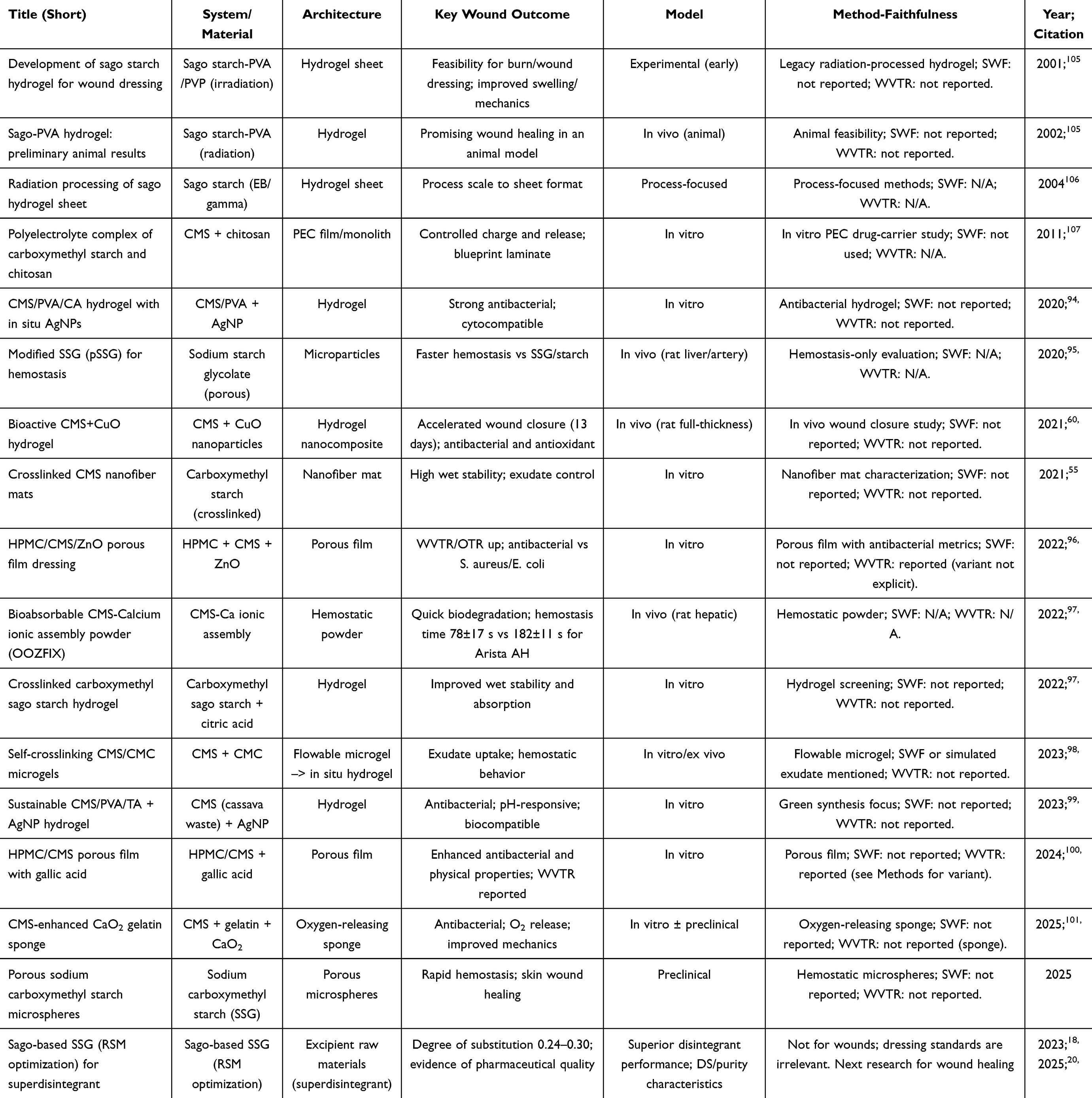 |
Table 3 Design and Performance of Carboxymethyl Starch and Sodium Starch Glycolate Dressings: Functional Outcomes and the Sago-Based Advantage |
Downstream processing strategies further reinforce sustainability when solvent-free or aqueous fabrication routes are employed. Chitosan-based contact layers can be assembled via ionotropic gelation or polyelectrolyte complexation under mild aqueous conditions, eliminating reliance on organic solvents.76 Nanostructured lipid carriers intended for early antimicrobial delivery can be produced by high-pressure homogenization, a scale-ready and largely solvent-minimized process.81 Such manufacturing compatibility aligns environmental considerations with industrial feasibility. Integration of sago-derived SSG as an absorber backing layer complements these clean fabrication approaches. Composite laminates combining chitosan contact surfaces with SSG absorbent backings offer a structurally rational configuration grounded in existing CMS/SSG evidence. Sustainability claims, however, must be accompanied by method-faithful characterization. Uptake, release, and surface-charge measurements should be conducted in simulated wound fluid with clearly stated salt, protein, and osmolality composition. Variability in test fluid formulation can substantially alter fluid-handling and release outcomes. Moisture-vapor transmission testing must also specify the applied standard and orientation, including EN 13726 variants or ASTM E96 dry-cup versus wet-cup conditions. Opposing vapor gradients generated by different methods produce non-comparable values.73 Transparent disclosure of these parameters ensures cross-laboratory reproducibility. Alignment of ecological feedstock sourcing, clean processing, and standardized evaluation strengthens translational credibility. The structured integration of sago-derived SSG absorber layers with chitosan contact interfaces and, where indicated, lipid-based antimicrobial pulse systems represent a scalable and sustainability-aligned dressing design pathway.
Authors’ Perspective: Translational Integration and in vivo Validation
Wound care requires a phase-aligned and specification-driven design that integrates interfacial bioactivity with controlled moisture management. A chitosan-based contact layer provides wet-tissue adhesion through electrostatic interactions with negatively charged wound substrates. Its cationic surface contributes to rapid hemostasis via erythrocyte aggregation and platelet activation. Contact-active antibiofilm pressure further supports early microbial control at the wound surface. In parallel, sago-derived sodium starch glycolate functions as a programmable hydration and absorption engine. Through defined degree of substitution and crosslink strength, SSG regulates swelling, capillary wicking, and moisture-vapor transmission. This dual-layer configuration enables decoupling of antimicrobial interface control from exudate handling. The system is designed to maintain a therapeutic micro-moist environment that avoids both desiccation and maceration. Integration of locally sourced sago feedstock reinforces supply resilience and sustainability. Aqueous conversion of sago starch to SSG with quantifiable substitution and crosslink parameters supports reproducible material specifications. The laminate architecture accommodates a two-stage therapeutic logic. An early antimicrobial pulse can be delivered, for example, via chitosan-coated nanostructured lipid carriers incorporated at the contact interface. Subsequent sustained anti-inflammatory and antioxidant exposure may be achieved using polymeric nanoparticle or hydrogel depots. Such temporal programming aligns drug delivery with wound-phase physiology. The construct therefore integrates material chemistry, device architecture, and pharmacologic scheduling within a unified design strategy.
Translation of this concept into preclinical validation requires methodologically sound in vivo assessment. A representative rodent wound-healing evaluation model is illustrated in Figure 5, depicting self-assembly of chitosan–SSG nanoparticles followed by topical treatment and monitoring of wound closure progression. The model enables assessment of hemostatic response, infection control, and tissue regeneration outcomes under controlled experimental conditions. Quantitative endpoints may include time to hemostasis, reduction in bacterial burden, and percentage wound closure over time. Histological evaluation can further characterize re-epithelialization and granulation tissue formation. Such in vivo validation supports mechanistic interpretation of interfacial and hydration-driven effects. Embedding the platform within a Quality-by-Design framework strengthens translational rigor. The workflow links Target Product Profile to Critical Quality Attributes and Critical Material or Process Parameters, culminating in a defined control strategy. Method-faithful reporting requires evaluation of release kinetics, surface charge, swelling, and fluid handling in simulated wound fluid of specified composition. Explicit declaration of the applied moisture-vapor transmission standard and variant ensures cross-study comparability. Alignment between pharmacologic endpoints and standardized dressing metrics prevents overestimation of therapeutic benefit. Integration of sustainability, performance, and regulatory traceability enhances platform credibility. In this framework, sago-derived SSG functions not merely as a starch derivative but as a specification-defined biomaterial. The combined chitosan–SSG–α-mangostin system may provide a translationally coherent design framework, but its therapeutic value remains dependent on formulation-specific validation of release behavior, wet mechanical stability, cytocompatibility, sterilization tolerance, and in vivo wound-healing performance.
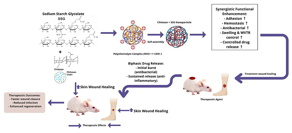 |
Figure 5 Schematic representation of in vivo rodent wound-healing evaluation following treatment with chitosan–SSG nanoparticles. Notes: The horizontal arrows represent the sequential workflow from formulation to application, starting with SSG and chitosan complexation, followed by nanoparticle self-assembly, topical application, and wound-healing evaluation. The + signs indicate the cationic chitosan-rich interactions involved in the formation of the PEC. The arrow upward shows the functional improvement, such as adhesion, hemostasis, antibacterial ability, swelling/water vapor transmission rate, controlled drug release, and wound healing response. The leftward arrows indicate the therapeutic effects and outcomes, like quick wound closure, reduced infection, and enhanced regeneration. |
Conclusion
The article provides four important points about the development of chitosan-sago-derived sodium starch glycolate (SSG) systems for α-mangostin wound administration. Firstly, chitosan and sago-derived SSG complement each other, with chitosan responsible for wound-surface adhesion, hemostasis, and antibacterial activity, while SSG is responsible for hydration control, swelling-driven fluid absorption, and moisture control. Second, α-mangostin nanoformulation is a sensible technique for the enhancement of local distribution of a poorly water-soluble bioactive molecule with antibacterial, antioxidant, and anti-inflammatory potential. Third, the suggested chitosan–SSG platform should be created as specification-driven systems in which essential parameters such as degree of substitution, crosslinking strength, particle size, swelling capacity, WVTR, release kinetics, and wet mechanical stability are explicitly defined. Fourth, while mechanistic promise exists, the platform is formulation-dependent, and the need for standardized physicochemical testing, simulated wound fluid evaluation, stability assessment, sterilization compatibility, and in vivo validation must be addressed before clinical translation. In conclusion, chitosan–sago-derived SSG is a durable and customizable functional carrier platform, whereas its therapeutic effectiveness depends on the repeatable formulation design and clinically relevant validation.
Acknowledgments
This publication charge is funded by Unpad through the Indonesian Endowment Fund for Education (LPDP) on behalf of the Indonesian Ministry of Higher Education, Science and Technology and managed under the EQUITY Program (Contract No. 4303/ B3/DT.03.08/2025 and 3927/UN6. RKT/HK.07.00/2025).
Author Contributions
All authors made a significant contribution to the work reported, whether that is in the conception, study design, execution, acquisition of data, analysis and interpretation, or in all these areas; took part in drafting, revising or critically reviewing the article; gave final approval of the version to be published; have agreed on the journal to which the article has been submitted; and agree to be accountable for all aspects of the work.
Funding
The authors gratefully acknowledge financial support from the Indonesia Endowment Fund for Education (LPDP), Ministry of Finance of the Republic of Indonesia. Institutional support from the Faculty of Pharmacy, Universitas Padjadjaran, and the National Research and Innovation Agency (BRIN) is also gratefully acknowledged.
Disclosure
The authors declare that there are no conflicts of interest in this work.
References
1. Falanga V, Isseroff RR, Soulika AM, et al. Chronic wounds. Nat Rev Dis Prim. 2022;8(1).
2. Chen X, Shi X, Xiao H, et al. Research hotspot and trend of chronic wounds: a bibliometric analysis from 2013 to 2022. Wound Repair Regener. 2023;31(5):597–28. doi:10.1111/wrr.13117
3. Goswami AG, Basu S, Banerjee T, et al. Biofilm and wound healing: from bench to bedside. Eur J Med Res. 2023;28(1):157.
4. Nasra S, Pramanik S, Oza V, et al. Advancements in wound management: integrating nanotechnology and smart materials for enhanced therapeutic interventions. Discover Nano. 2024;19(1):159.
5. Hu Y, Yu L, Dai Q, et al. Multifunctional antibacterial hydrogels for chronic wound management. Biomater Sci. 2024;12:2460–2479.
6. Sangnim T, Puri V, Dheer D, et al. Nanomaterials in the wound healing process: new insights and advancements. Pharmaceutics. 2024;16(3):300. doi:10.3390/pharmaceutics16030300
7. Afshar M, Rezaei A, Eghbali S, et al. Nanomaterial strategies in wound healing: a comprehensive review of nanoparticles, nanofibres and nanosheets. Int Wound J. 2024;21(7). doi:10.1111/iwj.14953
8. Chanmontri M, Kiti K, Surassmo S, et al. Effect of incorporating α-mangostin or curcumin-β-cyclodextrin inclusion complexes on biological activities of injectable self-healing quaternized chitosan/oxidized pectin hydrogels for wound dressing applications. Polymer. 2025;334:128721. doi:10.1016/j.polymer.2025.128721
9. Zhang W, Liu L, Cheng H, et al. Hydrogel-based dressings designed to facilitate wound healing. Mater Adv. 2023;5:1364–1394.
10. Koshenaj K, Ferrari G. A comprehensive review on starch-based hydrogels: from tradition to innovation, opportunities, and drawbacks. Polymers. 2024;16(14):1991.
11. Minsart M, Van Vlierberghe S, Dubruel P, et al. Commercial wound dressings for the treatment of exuding wounds: an in-depth physico-chemical comparative study. Burns Trauma. 2022;10. doi:10.1093/burnst/tkac024
12. Naveedunissa S, Meenalotchani R, Manisha M, et al. Advances in chitosan based nanocarriers for targetted wound healing therapies: a review. Carbohydr Polym Technol Appl. 2025;11:100891.
13. Kusnadi, Herdiana Y, Rochima E, et al. The potential of α-mangostin-loaded chitosan/collagen nanoparticles in hydrogel formulation for enhanced wound healing. Nanotechnol Sci Appl. 2025;19:1–24. doi:10.2147/NSA.S563394
14. Muchtaridi M, Suryani AI, Wathoni N, et al. Chitosan/alginate polymeric nanoparticle-loaded α-mangostin: characterization, cytotoxicity, and in vivo evaluation against breast cancer cells. Polymers. 2023;15(18):3658. doi:10.3390/polym15183658
15. Kusnadi K, Herdiana Y, Rochima E, et al. Collagen-based nanoparticles as drug delivery system in wound healing applications. Int J Nanomed. 2024;19:11321–11341.
16. Rajkumar DS, Keerthika K, Vijayaragavan V, et al. Chitosan-based biomaterial in wound healing: a review. Cureus. 2024;16(2). doi:10.7759/cureus.55193
17. Su J, Liu C, Sun A, et al. Hemostatic and antimicrobial properties of chitosan-based wound healing dressings: a review. Int J Biol Macromol. 2025;306:141570.
18. Putra ON, Musfiroh I, Elisa S, et al. Sodium starch glycolate (SSG) from sago starch (Metroxylon sago) as a superdisintegrant: synthesis and characterization. Molecules. 2024;29(1):151.
19. Berardi A, Janssen PHM, Dickhoff BHJ. Technical insight into potential functional-related characteristics (FRCs) of sodium starch glycolate, croscarmellose sodium and crospovidone. J Drug Delivery Sci Technol. 2022;70:103261.
20. Putra ON, Musfiroh I, Paramitasari D, et al. Sago-starch-derived sodium starch glycolate: an effective superdisintegrant to enhance formulation performance. Polymers. 2025;17(9):1–24. doi:10.3390/polym17091208
21. Górecka H, Gu´zniczak MGZ, Buzalewicz I, et al. Alpha-mangostin: a review of current research on its potential as a novel antimicrobial and anti-biofilm agent. 2025.
22. Nurhidayah W, Widyasari EM, Daruwati I, et al. Radiosynthesis, stability, lipophilicity, and cellular uptake evaluations of [(131)I]Iodine-α-mangostin for breast cancer diagnosis and therapy. Int J Mol Sci. 2023;24(10):8678. doi:10.3390/ijms24108678
23. Mardianingrum R, Hariono M, Ruswanto R, et al. Synthesis, anticancer activity, structure–activity relationship, and molecular modeling studies of α-mangostin derivatives as hERα inhibitor. J Chem Inf Model. 2022;62(21):5305–5316. doi:10.1021/acs.jcim.1c00926
24. Amalia R, Dewi C, Fristiohady A, et al. Unraveling the influence of α-mangostin on MDA-MB-231 cell line via WNT/β-catenin signaling pathway: in silico and in vitro approaches. Front Pharmacol. 2025;16:1600281. doi:10.3389/fphar.2025.1600281
25. Budiman A, Nurani NV, Laelasari E, et al. Effect of drug–polymer interaction in amorphous solid dispersion on the physical stability and dissolution of drugs: the case of alpha-mangostin. Polymers. 2023;15(14):3034. doi:10.3390/polym15143034
26. Suhandi C, Wilar G, Mohammed AFA, et al. Propolis-based nanostructured lipid carrier of α-mangostin for promoting diabetic wound healing in alloxan-induced mice. J Inflamm Res. 2025;18:7443–7457. doi:10.2147/JIR.S525243
27. Wathoni N, Sari DP, Suharyani I, et al. Enhancement of α-mangostin wound healing ability by complexation with 2-hydroxypropyl-β-cyclodextrin in hydrogel formulation. Pharmaceuticals. 2020;13(10):1–16. doi:10.3390/ph13100290
28. Milanda T, Cindana Mo’o FR, Mohammed AFA, et al. Alginate/chitosan-based hydrogel film containing α-mangostin for recurrent aphthous stomatitis therapy in rats. Pharmaceutics. 2022;14(8):1709. doi:10.3390/pharmaceutics14081709
29. Suhandi C, Wilar G, Mohammed AFA, et al. Hydrogel film-forming spray formulation containing propolis-based nanostructured lipid carriers of α-mangostin for diabetic wound repair. OpenNano. 2026;27:100275. doi:10.1016/j.onano.2025.100275
30. Baniasadi H. State-of-the-art in natural hydrogel-based wound dressings: design, functionalization, and fabrication approaches. Adv Colloid Interface Sci. 2025;342:103527. doi:10.1016/j.cis.2025.103527
31. Alberts A, Tudorache DI, Niculescu AG, et al. Advancements in wound dressing materials: highlighting recent progress in hydrogels, foams, and antimicrobial dressings. Gels. 2025;11(2):123. doi:10.3390/gels11020123
32. Lin X, Zhang X, Wang Y, et al. Hydrogels and hydrogel-based drug delivery systems for promoting refractory wound healing: applications and prospects. Int J Biol Macromol. 2025;285:138098.
33. Cassano R, Perri P, Scarcello E, et al. Chitosan hemostatic dressings: properties and surgical applications. Polymers. 2024;16(13):1770. doi:10.3390/polym16131770
34. Chicea D, Nicolae-Maranciuc A. A review of chitosan-based materials for biomedical, food, and water treatment applications. Materials. 2024;17(23):5770. doi:10.3390/ma17235770
35. Das A, Ghosh S, Pramanik N. Chitosan biopolymer and its composites: processing, properties and applications- A comprehensive review. Hybrid Adv. 2024;6:100265. doi:10.1016/j.hybadv.2024.100265
36. Tan H, Ma R, Lin C, et al. Quaternized chitosan as an antimicrobial agent: antimicrobial activity, mechanism of action and biomedical applications in orthopedics. Int J Mol Sci. 2013;14(1):1854–1869. doi:10.3390/ijms14011854
37. Chiu PH, Wu ZY, Hsu CC, et al. Enhancement of antibacterial activity in electrospun fibrous membranes based on quaternized chitosan with caffeic acid and berberine chloride for wound dressing applications. RSC Adv. 2024;14(47):34756–34768. doi:10.1039/D4RA05114A
38. Miranda A, Brandquist ND, Johnson K, et al. Quaternized chitosan derivatives inhibit growth and affect biofilm formation of Staphylococcus aureus. Sci Rep. 2025;15(1). doi:10.1038/s41598-025-11891-1
39. Cibotaru S, Anisiei A, Platon VM, et al. Imino-quaternized chitosan/chitosan nanofibers loaded with norfloxacin as potential bandages for wound healing. Int J Biol Macromol. 2025;314:144304. doi:10.1016/j.ijbiomac.2025.144304
40. Collado-González M, Espinosa YG, Goycoolea FM. Interaction between chitosan and mucin: fundamentals and applications. Biomimetics. 2019;4(2):32. doi:10.3390/biomimetics4020032
41. Stie MB, Cunha C, Huang Z, et al. A head-to-head comparison of polymer interaction with mucin from porcine stomach and bovine submaxillary glands. Sci Rep. 2024;14(1). doi:10.1038/s41598-024-72233-1
42. Jawadi Z, Yang C, Haidar ZS, et al. Bio-inspired muco-adhesive polymers for drug delivery applications. Polymers. 2022;14(24):5459. doi:10.3390/polym14245459
43. Ahmad K, Zhang Y, Chen P, et al. Chitosan interaction with stomach mucin layer to enhances gastric retention and mucoadhesive properties. Carbohydr Polym. 2024;333:121926. doi:10.1016/j.carbpol.2024.121926
44. Huang Z, Zhang D, Tong L, et al. Protonated-chitosan sponge with procoagulation activity for hemostasis in coagulopathy. Bioact Mater. 2024;41:174–192. doi:10.1016/j.bioactmat.2024.07.012
45. Du X, Zhang T, Liu Y, et al. A self-elastic chitosan sponge integrating active and passive hemostatic mechanisms for effectively managing uncontrolled coagulopathic hemorrhage. Mater Today Bio. 2024;26:101031. doi:10.1016/j.mtbio.2024.101031
46. Gheorghiță D, Moldovan H, Robu A, et al. Chitosan-based biomaterials for hemostatic applications: a review of recent advances. Int J Mol Sci. 2023;24(13):10540. doi:10.3390/ijms241310540
47. Perri P, Curcio F, De Luca M, et al. Evaluation of chitosan-based axiostat as hemostatic dressing for endovascular procedures in patients with leriche syndrome on anticoagulant therapy. Pharmaceuticals. 2025;18(4):584. doi:10.3390/ph18040584
48. Nag M, Lahiri D, Mukherjee D, et al. Functionalized chitosan nanomaterials: a jammer for quorum sensing. Polymers. 2021;13(15):2533. doi:10.3390/polym13152533
49. Badawy MSEM, Riad OKM, Taher FA, et al. Chitosan and chitosan-zinc oxide nanocomposite inhibit expression of LasI and RhlI genes and quorum sensing dependent virulence factors of Pseudomonas aeruginosa. Int J Biol Macromol. 2020;149:1109–1117. doi:10.1016/j.ijbiomac.2020.02.019
50. Tuan Mohamood NFAZ, Zainuddin N, Ahmad Ayob M, et al. Preparation, optimization and swelling study of carboxymethyl sago starch (CMSS)-acid hydrogel. Chem Cent J. 2018;12(1). doi:10.1186/s13065-018-0500-8
51. Janssen PHM, Rijpkema W, Stout K, et al. Quantifying cross-linking strength in sodium starch glycolate and its impact on tablet disintegration and dissolution. Eur J Pharm Biopharm. 2025;214:114803. doi:10.1016/j.ejpb.2025.114803
52. Rudnic EM, Kanig JL, Rhodes CT. Effect of molecular structure variation on the disintegrant action of sodium starch glycolate. J Pharmaceut Sci. 1985;74(6):647–650. doi:10.1002/jps.2600740613
53. Wong CCQ, Tomura K, Yamamoto O. Wound healing performance in a moist environment of crystalline glucose/mannose film as a new dressing material using a rat model: comparing with medical-grade wound dressing and alginate. Pharmaceuticals. 2023;16(11):1532. doi:10.3390/ph16111532
54. Bainbridge P, Browning P, Bernatchez SF, et al. Comparing test methods for moisture-vapor transmission rate (MVTR) for vascular access transparent semipermeable dressings. J Vasc Access. 2023;24(5):1000–1007. doi:10.1177/11297298211050485
55. Luan Z, Zhang H, Hu J, et al. Crosslinked carboxymethyl starch nanofiber mats: preparation, water resistance and exudates control ability. Eur Polym J. 2021;154:110568. doi:10.1016/j.eurpolymj.2021.110568
56. Lee CS, Hwang HS. Starch-based hydrogels as a drug delivery system in biomedical applications. Gels. 2023;9(12):951. doi:10.3390/gels9120951
57. Zehrer CL, Holm D, Solfest SE, et al. A comparison of the in vitro moisture vapour transmission rate and in vivo fluid-handling capacity of six adhesive foam dressings to a newly reformulated adhesive foam dressing. Int Wound J. 2014;11(6):681–690. doi:10.1111/iwj.12030
58. Gefen A, Alves P, Beeckman D, et al. Fluid handling by foam wound dressings: from engineering theory to advanced laboratory performance evaluations. Int Wound J. 2024;21(2). doi:10.1111/iwj.14674
59. Palácio SB, Penello SO, Hodel KVS, et al. Commercial, non-commercial and experimental wound dressings based on bacterial cellulose: an in-depth comparative study of physicochemical properties. Fibers. 2025;13(9):127. doi:10.3390/fib13090127
60. Abdollahi Z, Zare EN, Salimi F, et al. Bioactive carboxymethyl starch-based hydrogels decorated with cuo nanoparticles: antioxidant and antimicrobial properties and accelerated wound healing in vivo. Int J Mol Sci. 2021;22(5):1–18. doi:10.3390/ijms22052531
61. Castro D, Brovina V, Litvinov M, et al. Effect of degree of substitution and polymer ratio on the structure of chitosan: carboxymethyl starch (Bio)polyelectrolyte complexes. Polymers. 2024;16(24):3539. doi:10.3390/polym16243539
62. Gou Y, Hu L, Liao X, et al. Advances of antimicrobial dressings loaded with antimicrobial agents in infected wounds. Front Bioeng Biotechnol. 2024;12. doi:10.3389/fbioe.2024.1431949
63. Olea AF, Carrasco H, Santana F, et al. Formation and characterization of chitosan-based polyelectrolyte complex containing antifungal phenylpropanoids. Polymers. 2024;16(23):3348. doi:10.3390/polym16233348
64. Lin T, Chen D, Geng Y, et al. Carboxymethyl chitosan/sodium alginate/chitosan quaternary ammonium salt composite hydrogel supported 3J for the treatment of oral ulcer. Gels. 2023;9(8):659. doi:10.3390/gels9080659
65. Nuutila K, Eriksson E. Moist wound healing with commonly available dressings. Adv Wound Care. 2021;10(12):685–698. doi:10.1089/wound.2020.1232
66. Lan Z, Kar R, Chwatko M, et al. High porosity PEG-based PEG -based hydrogel foams with self-tuning moisture balance as chronic wound dressings. J Biomed Mat Res. 2023;111(4):465–477. doi:10.1002/jbm.a.37498
67. Faria M, Björnmalm M, Thurecht KJ, et al. Minimum information reporting in bio–nano experimental literature. Nature Nanotechnol. 2018;13(9):777–785. doi:10.1038/s41565-018-0246-4
68. du Sert NP, Hurst V, Ahluwalia A, et al. The arrive guidelines 2.0: updated guidelines for reporting animal research. PLoS Biol. 2020;18(7).
69. Moher D, Hopewell S, Schulz KF, et al. CONSORT 2010 explanation and elaboration: updated guidelines for reporting parallel group randomised trials. BMJ. 2010;340(mar23 1):c869–c869. doi:10.1136/bmj.c869
70. Health USDo, Human Services F, Drug A. Use of International Standard ISO 10993-1, “Biological evaluation of medical devices-Part 1: evaluation and testing within a risk management process. Guid Industry Food Drug Administr Staff Preface Public Comment. 2023.
71. Hong F, Qiu P, Wang Y, et al. Chitosan-based hydrogels: from preparation to applications, a review. Food Chemistry. 2024;21. doi:10.1016/j.fochx.2023.101095
72. Dong Y, Wang Z. ROS-scavenging materials for skin wound healing: advancements and applications. Front Bioeng Biotechnol. 2023;11. doi:10.3389/fbioe.2023.1304835
73. Svensby AU, Nygren E, Gefen A, et al. The importance of the simulated wound fluid composition and properties in the determination of the fluid handling performance of wound dressings. Int Wound J. 2024;21(5). doi:10.1111/iwj.14861
74. Nygren E, Malone M, Gefen A. Laboratory evaluations of wound dressings: key advances to reflect clinical reality. Int Wound J. 2025;22(S1). doi:10.1111/iwj.70333
75. Jonidi Shariatzadeh F, Currie S, Logsetty S, et al. Enhancing wound healing and minimizing scarring: a comprehensive review of nanofiber technology in wound dressings. Pro Mater Sci. 2025;147:101350. doi:10.1016/j.pmatsci.2024.101350
76. Rohtagi P, Garg U, Jain N, et al. Chitosan and hyaluronic acid-based nanocarriers for advanced cancer therapy and intervention. Biomaterials Adv. 2024;157:213733. doi:10.1016/j.bioadv.2023.213733
77. Wathoni N, Rusdin A, Motoyama K, et al. Nanoparticle drug delivery systems for α-mangostin. Nanotechnol Sci Appl. 2020;Volume 13:23–36. doi:10.2147/NSA.S243017
78. Sawatphakdee G, Yostawonkul J, Oontawee S, et al. Feasibility of nanostructured lipid carrier loaded with alpha-mangostin and clove oil for canine periodontal therapy. Animals. 2024;14(14):2084. doi:10.3390/ani14142084
79. Burki FA, Shah KU, Razaque G, et al. Optimization of chitosan-decorated solid lipid nanoparticles for improved flurbiprofen transdermal delivery. ACS Omega. 2023;8(22):19302–19310. doi:10.1021/acsomega.2c08135
80. Barari F, Maghsoudian S, Alinezhad V, et al. Effective topical delivery of astaxanthin via optimized chitosan-coated nanostructured lipid carriers: a promising strategy for enhanced wound healing and tissue regeneration. J Drug Delivery Sci Technol. 2025;110:107052. doi:10.1016/j.jddst.2025.107052
81. Wathoni N, Suhandi C, Elamin KM, et al. Advancements and challenges of nanostructured lipid carriers for wound healing applications. Int J Nanomed. 2024;Volume 19:8091–8113. doi:10.2147/IJN.S478964
82. Shahzad S, Khan IU, Khalid I. α-Mangostin encapsulated gellan gum membranes for enhanced antibacterial, anti-inflammatory, antioxidant and wound healing activity. Int J Biol Macromol. 2025;308:142493. doi:10.1016/j.ijbiomac.2025.142493
83. Vitamia C, Iftinan GN, Latarissa IR, et al. α-Mangostin hydrogel film with chitosan alginate base for recurrent aphthous stomatitis (RAS) treatment: study protocol for double-blind randomized controlled trial. Front Pharmacol. 2024;15. doi:10.3389/fphar.2024.1353503
84. Ali M, Ullah S, Ullah S, et al. Innovative biopolymers composite based thin film for wound healing applications. Sci Rep. 2024;14(1):27415.
85. Park SY, Lee JH, Ko SY, et al. Antimicrobial activity of α-mangostin against Staphylococcus species from companion animals in vitro and therapeutic potential of α-mangostin in skin diseases caused by S. pseudintermedius. Front Cell Infect Microbiol. 2023;13. doi:10.3389/fcimb.2023.1203663
86. Ge R, Zhao H, Tang Q, et al. A novel α-mangostin derivative synergistic to antibiotics against MRSA with unique mechanisms. Microbiol Spectr. 2024;12(12). doi:10.1128/spectrum.01631-24
87. Abuzeineh H, Abdel Rahim S, Cespi M, et al. Time-controlled release by the incorporation of superdisintegrants within the coat of zein dry coated tablets. J Drug Delivery Sci Technol. 2021;65:102716. doi:10.1016/j.jddst.2021.102716
88. Ji M, Li J, Wang Y, et al. Advances in chitosan-based wound dressings: modifications, fabrications, applications and prospects. Carbohydr Polym. 2022;297:120058. doi:10.1016/j.carbpol.2022.120058
89. Luo Y, Cui L, Zou L, et al. Mechanically strong and on-demand dissoluble chitosan hydrogels for wound dressing applications. Carbohydr Polym. 2022;294:119774. doi:10.1016/j.carbpol.2022.119774
90. Pellevoisin C, Coleman KP, Hoffmann S. ISO 10993-23 In vitro irritation testing for medical devices: substantiating applicability to mild irritants and non-extractables. Toxicol In Vitro. 2022;82:105371. doi:10.1016/j.tiv.2022.105371
91. Demeter M, Scărișoreanu A, Călina I. State of the art of hydrogel wound dressings developed by ionizing radiation. Gels. 2023;9(1):55. doi:10.3390/gels9010055
92. Temmink RJM, Robroek BJM, van Dijk G, et al. Wetscapes: restoring and maintaining peatland landscapes for sustainable futures. Ambio. 2023;52(9):1519–1528. doi:10.1007/s13280-023-01875-8
93. Monda Y, Kuwahara S, Aoki Y, et al. Productivity of sago palms on smallholder plantations after rewetting in previously drained peatland: a case study on Tebing Tinggi Island, Riau Province, Indonesia. Tropics. 2022;31(1):11–32. doi:10.3759/tropics.MS21-15
94. Ounkaew A, Kasemsiri P, Jetsrisuparb K, et al. Synthesis of nanocomposite hydrogel based carboxymethyl starch/polyvinyl alcohol/nanosilver for biomedical materials. Carbohydr Polym. 2020;248:116767. doi:10.1016/j.carbpol.2020.116767
95. Panwar V, Thomas J, Sharma A, et al. In-vitro and in-vivo evaluation of modified sodium starch glycolate for exploring its haemostatic potential. Carbohydr Polym. 2020;235:115975. doi:10.1016/j.carbpol.2020.115975
96. Pitpisutkul V, Prachayawarakorn J. Hydroxypropyl methylcellulose/carboxymethyl starch/zinc oxide porous nanocomposite films for wound dressing application. Carbohydr Polym. 2022;298:120082. doi:10.1016/j.carbpol.2022.120082
97. Ko YG, Kim BN, Kim EJ, et al. Bioabsorbable carboxymethyl starch–calcium ionic assembly powder as a hemostatic agent. Polymers. 2022;14(18):3909. doi:10.3390/polym14183909
98. Panwar V, Sharma A, Murugesan P, et al. Free-flowing, self-crosslinking, carboxymethyl starch and carboxymethyl cellulose microgels, as smart hydrogel dressings for wound repair. Int J Biol Macromol. 2023;246:125735. doi:10.1016/j.ijbiomac.2023.125735
99. Srikhao N, Theerakulpisut S, Chindaprasirt P, et al. Green synthesis of nano silver-embedded carboxymethyl starch waste/poly vinyl alcohol hydrogel with photothermal sterilization and pH-responsive behavior. Int J Biol Macromol. 2023;242:125118. doi:10.1016/j.ijbiomac.2023.125118
100. Pitpisutkul V, Prachayawarakorn J. Porous antimicrobial crosslinked film of hydroxypropyl methylcellulose/carboxymethyl starch incorporating gallic acid for wound dressing application. Int J Biol Macromol. 2024;256:128231. doi:10.1016/j.ijbiomac.2023.128231
101. Andam S, Akbari B, Nourmohammadi J, et al. Physicochemical, biological, and oxygen-releasing properties of carboxymethyl starch added to calcium peroxide-containing gelatin sponge dressings. Mater Today Chem. 2025;47:102798. doi:10.1016/j.mtchem.2025.102798
102. Jolly SS, Rattan V. Is chitosan-based dressing more effective than gauze pressure in achieving early hemostasis after dental extractions in patients with deranged coagulation profiles? Archiv Craniofacial Surg. 2025;26(2):65–69. doi:10.7181/acfs.2024.0082
103. Strużyna J, Korzeniowski T, Tobiasz M, et al. Clinical evaluation of the efficiency and safety of the Tromboguard® hemostatic dressing for donor sites of split-thickness skin graft: a multicenter study. Burns Open. 2024;8(3):197–203. doi:10.1016/j.burnso.2024.05.001
104. Ling Tan JS, Roberts CJ, Billa N. Mucoadhesive chitosan-coated nanostructured lipid carriers for oral delivery of amphotericin B. Pharmaceut Develop Technol. 2019;24(4):504–512. doi:10.1080/10837450.2018.1515225
105. Hashim K, Zaman K, Mohd Dahlan HJ, et al. Development of sago starch hydrogel for wound dressing. 2001.
106. Hashim K, Yacob N 2 radiation processing of sago hydrogel thin film. 2004.
107. Assaad E, Wang YJ, Zhu XX
 © 2026 The Author(s). This work is published and licensed by Dove Medical Press Limited. The
full terms of this license are available at https://www.dovepress.com/terms
and incorporate the Creative Commons Attribution
- Non Commercial (unported, 4.0) License.
By accessing the work you hereby accept the Terms. Non-commercial uses of the work are permitted
without any further permission from Dove Medical Press Limited, provided the work is properly
attributed. For permission for commercial use of this work, please see paragraphs 4.2 and 5 of our Terms.
© 2026 The Author(s). This work is published and licensed by Dove Medical Press Limited. The
full terms of this license are available at https://www.dovepress.com/terms
and incorporate the Creative Commons Attribution
- Non Commercial (unported, 4.0) License.
By accessing the work you hereby accept the Terms. Non-commercial uses of the work are permitted
without any further permission from Dove Medical Press Limited, provided the work is properly
attributed. For permission for commercial use of this work, please see paragraphs 4.2 and 5 of our Terms.