Back to Journals » Clinical Ophthalmology » Volume 18
Ray-Tracing Customization in Myopic and Myopic Astigmatism LASIK Treatments for Low and High Order Aberrations Treatment: 2-Year Visual Function and Psychometric Value Outcomes of a Consecutive Case Series
Authors Kanellopoulos AJ
Received 12 November 2023
Accepted for publication 25 January 2024
Published 22 February 2024 Volume 2024:18 Pages 565—574
DOI https://doi.org/10.2147/OPTH.S444174
Checked for plagiarism Yes
Review by Single anonymous peer review
Peer reviewer comments 2
Editor who approved publication: Dr Scott Fraser
Video abstract presented by Kanellopoulos.
Views: 77
Anastasios John Kanellopoulos1,2
1Medical Director: The Laservision Clinical and Research Institute, Athens, Greece; 2Clinical Professor, Department of Ophthalmology, NYU Grossman Medical School, Department of Ophthalmology, New York City, NY, USA
Correspondence: Anastasios John Kanellopoulos, Clinical Professor of Ophthalmology, NYU Medical School, Medical Director: The Laservision Clinical and Research Institute, 17 Tsocha Street, Athens, 115 21, Greece, Tel + 30 210 7472777, Fax + 30 210 7472789, Email [email protected]
Purpose: The safety and long-term efficacy of automated ray-tracing customized myopic and myopic astigmatic femtosecond laser-assisted LASIK.
Methods: This consecutive case series retrospective analysis, of 20 subjects (40 eyes) treated with automated raytracing named Wavelight Plus, to include low and high order aberrations based on a three-dimensional custom virtual eye for each case-calculated from interferometry data-obtained from a single diagnostic device that also provides Hartman–Shack Wavefront and Scheimpflug tomography data. We evaluated before and after the customized LASIK procedure: visual acuity, refractive error, high order aberrations, contrast sensitivity, and psychometric post-operative visual function data.
Results: At 24 months, the comparison of the pre-operative to the post-operative refractive and visual function value changes in average were: subjective manifest refraction from − 4.38 ± 2.54 diopters (D) (range − 9.75 to − 1.25 D) to +0.11 ± 0.19 D; subjective manifest refractive astigmatism from − 0.76 ± 0.91 D (range − 2.75 to 0 D) to − 0.13 ± 0.16 D, corneal astigmatism from − 1.16 ± 0.64 D (range − 0.2 to − 2.8) to − 0.47 ± 0.11 D. 65% of the eyes studied demonstrated an increase of at least one line of vision, while from the same group 38% demonstrated 2 lines of increase. High order aberrations, contrast sensitivity as well as the subjective psychometric input based on the VFQ-25 questionnaire demonstrated actual improvement.
Conclusion: This longer-term follow-up, single-arm retrospective consecutive case series documents LASIK treatment customization that appears to be safe and effective in the correction of myopia and myopic astigmatism. Markedly improved objective and subjective visual function post-operatively, underlying the potential importance of simultaneously attempting to correct high order aberrations and improving the spatial alignment of total, measured human eye optics.
Keywords: raytracing excimer customization of myopic corrections, topography-guided, wavefront-guided, femtosecond-laser assisted myopic LASIK, customized excimer laser ablation: human eye optical tilt between the cornea and the crystalline lens
Introduction
Cornea-based refractive surgery, and laser vision correction specifically, is based on the treatment of the subjective manifest refraction, as a globally accepted rule universally, regarding technology and technique: safety and efficacy in the percentage of post-operative spectacle independence. This refraction is usually the subjective manifest refraction established by the “dry” (without prior pupil dilatation and/or cycloplegia) manifest or with consideration, or even for some clinicians’ actual adaptation of cycloplegia (usually named “wet”) manifest refraction instead.1,2
There have been several customization methods introduced, in order, to enhance the target for myopia and/or myopic-astigmatic correction (low order aberrations) as well as high order aberrations: coma, corneal asphericity and trefoil. Additionally, excimer laser tracking has been customized to center on the corneal vertex instead of the pupillary center, as it represents the point on the cornea surface, theoretically incident to the line of sight. Cyclorotation adjustment has been an additional customization tool aiming to optimize the accuracy of excimer laser cylindrical correction and the accurate delivery of correction for possible higher order aberrations inclusive in the corrective delivery to the cornea stroma. Wavefront-guided treatments, topography-guided treatments have for years addressed some of these customization approaches with enhanced clinical outcomes reported.3–28
The evaluation the low and high order aberrations of the human eye via ray-tracing has been studied in the past manually and respective customization was clinically applied in myopic LASIK procedures through manual calculations.29,30 The purpose was to explore more accurate corrections regarding effective refractive error and potentially improve further visual function.
Wavefront analysis, tomographic corneal and anterior chamber Scheimpflug tomography analysis, axial length measurements, expected wound and epithelial remodeling healing patterns and potential biomechanical changes in respect to the amount of tissue removed were pivotal input points for these calculations and outcome projections. The initial clinical outcomes of this work were reported as early as 2008.29–31 Subsequent research and development to include the configuration and clinical use a combination-diagnostic device, in conjunction with software development enabling standardized automated calculations, by the Wavelight/Alcon team, came to fruition in 2019 as automated ray-tracing with data provided by a single device, the Sitemap, received the Conformité Européene (CE) approval mark for clinical application as a novel refractive surgery product named at the time Innoveyes. Sitemap is the trade name of the specific diagnostic device that was developed and approved, able to offer the appropriate validated data. These data were used initially by the Innoveyes as called earlier, and Wavelight Plus currently, automated software for ray-tracing calculations that produce the customized ablation algorithm and pattern for treatment of the low and high order aberrations by to be the EX500 excimer laser for myopia and myopic astigmatism LASIK correction as reported first by our team.32–34
Methods
The primary hypothesis in this study was: Ray-Tracing customization LASIK cases outcomes regarding lines of vision gained (comparing post-operative uncorrected distance visual accuity [UDVA], to pre-operative corrected vidual accuity [CDVA]). 20 consecutive cases undergoing bilateral myopic LASIK were included in the study. Ethics Committee approval was obtained from the Laservision Ambulatory Surgical Unit Ethics Committee and adhered to the tenets of the Declaration of Helsinki. All cases following extensive verbal consent submitted written informed consent as well. The study received approval and registration with the Greek Authority of Pharmaco-technological medical studies (EOF Dec 2020) and registered for within the EU with the German Clinical Trials Register (www.drks.de/drks_web/; ID: DRKS00020388).
Study Objectives in Summary
- 1 – The Comparison of pre-operative CDVA to 24-month post-operative UDVA (lines of visual acuity gained)
- 2 – Secondary Study Endpoints:
Subjective post-operative visual function feedback from the patients based on the National Eye Institute 25-Item Visual Function Questionnaire (VFQ-25) interviewer-administered format developed by RAND as evaluated on post-operative month 24.35
Attempted vs. achieved subjective manifest refractive error correction at 24 months.
Comparison of pre-operative to post-operative total corneal astigmatism as documented by Scheimpflug tomography (Pentacam) inclusive in the sitemap measurement prior and at 24 months after LASIK treatment.
Assessment of low contrast sensitivity measured pre-operatively and at 24 months post-operative utilizing the functional vision analyzer (Stereo Optical, Chicago, IL, USA).
The methodology also included evaluation of the type, severity, duration and frequency of all and any, potential adverse events to be encountered during the 24-month span of this study.
Inclusion/Exclusion
Inclusion Criteria
Successful completion of primary ray-tracing customized LASIK cases using the Alcon/Wavelight refractive suite and the Wavelight Plus automated customization software, as described above. Subjects between 18 to 65 years of age. Pre-operative myopia up to −10 D of myopia and up to −4 D of astigmatism. Pre-operative central corneal thickness of at or over 500 μm, confirmed by both corneal tomography and anterior segment OCT (Avanti-Angioflow OCT, Optovue, CA, USA). A negative pregnancy test given just prior to the procedure for all female subjects.
Exclusion Criteria
Previous ocular surgery, clinically significant corneal abnormalities including scar in the visual axis, anterior basement membrane dystrophy, clinical signs of dry eye and/or blepharitis, to include significant superficial punctuate keratitis and/or significant epithelial irregularity in the anterior segment OCT epithelial mapping (differences of over 5um, and or average under 49um and over 55um). Tomographic evidence of Keratoconus and even keratoconus suspect, as defined by corneal tomography using the Amsler-Krumeich criteria.
The Diagnostic Device
The diagnostic device used to provide the imaging data was the Sitemap (Alcon/Wavelight, ft. Worth, TX, USA) based on the Pentacam®AXL Wave device (Oculus, Germany) that has been modified, in order to provide the proprietary diagnostic imaging for the ray-tracing treatment, automated calculations by the Wavelight Plus software applied by the EX500 excimer laser ablations.
The modification changes have been made to adapt the diagnostic information captured on each patient by the Sitemap device, subsequently transfer them as data to the EX500 excimer platform Wavelight plus software through an independent and dedicated to the Alcon/Wavelight refractive suite (the FS200 femtosecond laser and the EX500 excimer laser).
Laser Spot Profile
The excimer laser spot profile has been renovated so its treatment delivery when using the raytracing, Wavelight-Plus software differs from the up-to-now Wavefront-optimized profile, to improve disadvantages associated with previous profile patterns.
The EX500 excimer when executing a raytracing Wavelight-Plus customized treatment actually delivers throughout its span, a combined sphere and cylinder correction, along with, high-order aberrations-in contrast to wavefront optimized treatments by the same platform that treat sphere first and cylinder last, The Wavelight Plus software has been designed to take into account data studied previously on expected epithelial remodeling relevant to the amount and surface (optical zone) of tissue removed as well as potential biomechanical shifts anticipated relevant to the amount of tissue removed. It additionally automatically adjusts for nomogram anticipated deviations, pre-emptively in the final ablation profile calculated.
Myopic and myopic-astigmatic excimer treatment transition zone: There is additional change in the traditional for this manufacturer excimer laser delivered transition zone in the Wavelight Plus software calculations and treatment-parameter, delivery by the EX500 excimer laser. In previous treatment profiles offered by the Alcon/Wavelight Refractive Suite (Wavefront optimized, Asphericity-adjusted, Wavefront-guided and topography-guided) using optical zone of 6.5 mm for myopia, a transitional zone of 0.75mm was added, by default, by the software, while in myopic astigmatism: a 1.5mm transition zone respectively. The Wavelight-Plus ray-tracing transition zone is uniformly set at 2.5mm by default, a marked difference from previous methodology.
Automatic Nomogram Adjustment
The amount of myopia to be removed is still calculated based on the Munnerlyn formula and as mentioned before, with added adjustment for the expected epithelial changes, biomechanical changes, and the actual flying spot laser beam incidence: meaning the spot incidence on the peripheral part of the ablation is boosted with an extra spot in order to compensate for that loss of energy delivered at that specific spot.
Ray tracing adjustment: the axial length measurements give precise measurements of anterior chamber depth as well as total eye axial length, measurements integral to the automated calculation algorithm of the Wavelight-Plus ray-tracing customization in-order-to optimize the excimer laser ablation profile to the refractive error and higher order aberrations attempted to be corrected. The Wavelight Plus ray-tracing software uses the Sitemap data in several steps:
- 1 – The corneal tomography measurements are used to calculate 2000 rays traced virtually by this software up-to the anterior surface of the lens.
- 2 – The Hartman–Shack Wavefront data also captured by the Sitemap are utilized by this software to virtually trace 2000 rays from the retina surface to the anterior lens surface in a retrograde fashion.
- 3 – The potential optical tilt that theoretically derives from the ray-tracing automated calculation, when defining the spatial orientation between the cornea and the lens – both viewed virtually as three-dimensional refractive units – is also calculated and included in the ablation calculation as “tilt correction”, in order to make them theoretically function post-operatively in parallel fashion and thus optically optimal.
All the Sitemap measurements noted above: Scheimpflug tomography, Hartman–Shack wavefront analysis and interferometry are internally validated by the Sitemap device for consistency as several are required for accuracy assessment that takes place by the device automatically.
The sequence of imaging by the Sitemap that we used uniformly was for each eye Wavefront measurements, followed by Scheimpflug tomography and completing with interferometry.
Following the Wavelight-Plus automated ray-tracing calculation, the default treatment presented by the software is the calculated one and not the subjective manifest refraction entry, as was usual in all predecessors customized software by the same manufacturer noted above – yet another difference to previous customized versions used by the same excimer laser.
This actual ray-tracing customized treatment can be adjusted in a final step by the surgeon prior to ablation for any of the low order aberration parameters: sphere, cylinder and axis, but cannot be adjusted in regard to the higher order aberrations calculated by the ray-tracing analysis and planned to be corrected – these are not the high order aberrations calculated by the Hartman–Shack Wavefront captured by the Sitemap, but those by the automated ray-tracing Wavelight Plus software.
The calculated tilt correction by the ray-tracing software, also cannot be adjusted by the surgeon.
In summary refraction and visual function data evaluated in all cases were:
- 1 – Uncorrected distance visual acuity: UDVA
- 2 – Corrected distance visual acuity; CDVA with the subjective manifest clinical refraction:
- 3 – low contrast sensitivity.
- 4 – Sitemap measurements and refraction calculation pre-operative and at 24 months post-operative.
- 5 – Subjective post-operative visual function feedback from the patients based on the National Eye Institute 25-Item Visual Function Questionnaire (VFQ-25) modified by RAND34 interviewer-administered format (Appendix II) and evaluated on post-operative month 24.
Results
We treated a total of 21 consecutive patients, 20 were followed-up for the 24th post-operative month, one patient a female had to relocate outside Greece, by month 1 following her LASIK treatment and was excluded as we did not have 3 nor 24-month data. Of the cases presented herein there were 12 female and 8 males. The patient demographic and pre-operative refraction data are summarized in Table 1.
 |
Table 1 Patient Demographic and Pre-operative Refraction Data |
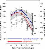
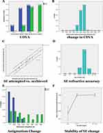
The average subjective manifest refractive error changed from −4.38 ± 2.54 diopters (D) pre-operative to +0.11 ± 0.19 D 24 months post-operative; Subjective manifest refractive astigmatism was measured from −0.76 ± 0.91 D (range −2.75 to 0 D) pre-operative to −0.13 ± 0.16 D 24 months post-operative, tomographic astigmatism changed from −1.16 ± 0.64 D (range −0.2 to −2.8) pre-operative to −0.47 ± 0.11 D post-operative, all at 24 months follow-up. Of all cases 65% appeared to improve by at least one line of vision, while 38% appeared to improve by 2 lines of post-operative UDVA when compared to pre-operative CDVA. All the visual function and refraction data noted above are summarized in Figure 1. Pre-to post-operative total high order aberrations measured by the RMSh metric (total high order aberrations) by the Sitemap device, changed from 0.25 um pre-op to 0.35 um 24 months post-op. Low contrast sensitivity appeared better 24 months post-operatively, when compared to pre-op, in all frequencies studied as noted in Figure 2.
 |
Figure 2 Low contrast sensitivity data comparison: pre-operative in red, compared to 24 months post-operative in blue using the Stereo Vision device. |
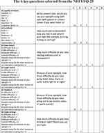
Regarding the NEI VFQ-25 questionnaire administered at 24 months we chose 6 key questions from the large number of questions evaluated,35 relating to post-operative quality of visual function:
- 1 – subjective quality of vision,
- 2 – subjective patient comfort with their eyes,
- 3 – near vision subjective function,
- 4 – night vision subjective function,
- 5 – subjective comfort while performing weekly hobbies,
- 6 – and last subjective quality of vision in night driving.
All summarized in Figure 3.
 |
Figure 3 NEI VFQ-25 questionnaire, 6 selected key question responses. Credits: The VFQ-25 questionnaire used in this study was developed at RAND under the sponsorship of the National Eye Institute.35 |
Discussion
Clinical evaluation of wavefront-guided customized treatment of high order aberrations: coma, trefoil and spherical aberration has been shown to improve post-myopic LASIK visual function outcomes.2,7,11,12 Customization of myopic LASIK treatments based on topography, has also demonstrated clinically visual function advantages.13,14,19–28 The subjective manifest refraction measurements present usually less cylindrical power and often of different axis to that measured by tomography, studied in the FDA study by ALCON (WaveLight ALLEGRETTO WAVE® Eye-Q Excimer Laser - P020050/S012, Summary of Safety and Effectiveness Data, page 31: (assessed on March 1st, 2020 at: https://www.accessdata.fda.gov/cdrh_docs/pdf2/P020050S012b.pdf27 demonstrating improved visual outcomes regarding lines of visual acuity gained post-operative. We introduced and reported the principle of topography-modified refraction (TMR) in a contralateral eye study demonstrating that this treatment refraction modification in myopic LASIK may offer improved visual outcomes.28
The customization outcomes described herein, based on the study of myopic and myopic astigmatic LASIK treatments using the Wavelight-Plus ray-tracing automated customization platform appear to offer the same level, if not better visual outcomes previously reported with topography-guided customization and manual adjustment of the low order aberrations with TMR,28 with automated calculations instead, validating the clinical significance of the previous work we reported above and the steps taken to calculate low and high order aberrations.
Conclusion
This small single-arm observational consecutive case series appears to offer effective and safe long-term correction of myopia and myopic astigmatism, by employing the FS200 femtosecond laser and the EX500 Excimer laser and the customization platform of Wavelight-Plus with automated ray-tracing. The visual function data post-operative as well as the positive subjective feedback documented by the subjects underline the potential importance of correcting higher aberrations and improving human eye optics that as a result may offer higher visual function performance post-operative when compared to pre-operative, for most patients treated.
Credits
The VFQ-25 questionnaire used in this study was developed at RAND under the sponsorship of the National Eye Institute.
Study Data Sharing
The authors have posted individual deidentified participant data to be asseccible on the registry link below. The specific data are noted on a spreadheet and include patient sex, age, preoperative refraction, keratometry, UDVA and CDVA, as well as the 1 month 3 month and 24 month data. They will be accessible indefinitely as noted in the statement below: All individual deidentified participant data will be available at the ISRCTN registry as: ISRCTN73528436. Registry access link: https://doi.org/10.1186/ISRCTN73528436.
Funding
This study # 59917561, was funded by a non-restricted investigator-initiated study, by Alcon Hellas. Presented in part as a poster at the winter ESCRS meeting in Portugal, February 2022 and the ASCRS meeting in Washington, DC April 2022.
Disclosure
The author (AJK) serves among other commercial entities as a scientific consultant for Alcon/Wavelight (Erlangen, Germany).
References
1. Rapuano CJ, Boxer-Wachler BS, Davis EA, et al. Authors of the 2013–2014 edition of: refractive surgery, chapter 2: patient evaluation from the basic and clinical science course, section 13, AAO, page 33.
2. Doane JF, Slade SG. An introduction to wavefront-guided refractive surgery. Int Ophthalmol Clin. 2003;43(2):101–117. doi:10.1097/00004397-200343020-00011
3. Wen D, McAlinden C, Flitcroft I, et al. Postoperative efficacy, predictability, safety, and visual quality of laser corneal refractive surgery: a network meta-analysis. Am J Ophthalmol. 2017;178:65–78. doi:10.1016/j.ajo.2017.03.013
4. Lukenda A, Martinović ZK, Kalauz M. Excimer laser correction of hyperopia, hyperopic and mixed astigmatism: past, present, and future. Acta Clin Croat. 2012;51(2):299–304.
5. Reggiani-Mello G, Krueger RR. Comparison of commercially available femtosecond lasers in refractive surgery. Expert Rev Opthalmol. 2011;6(1):55–56. doi:10.1586/eop.10.80
6. Salomão MQ, Wilson SE. Femtosecond laser in laser in situ keratomileusis. J Cataract Refract Surg. 2010;36(6):1024–1032. doi:10.1016/j.jcrs.2010.03.025
7. Vega-Estrada A, Alió JL, Arba Mosquera S, Moreno LJ. Corneal higher order aberrations after LASIK for high myopia with a fast repetition rate excimer laser, optimized ablation profile, and femtosecond laser-assisted flap. J Refract Surg. 2012;28(10):689–696. doi:10.3928/1081597X-20120921-03
8. Winkler von Mohrenfels C, Khoramnia R, Lohmann CP. Comparison of different excimer laser ablation frequencies (50, 200, and 500 Hz). Graefes Arch Clin Exp Ophthalmol. 2009;247(11):1539–1545. doi:10.1007/s00417-009-1102-x
9. Iseli HP, Mrochen M, Hafezi F, Seller T. Clinical photoablation with a 500-Hz scanning spot excimer laser. J Refract Surg. 2004;20(6):831–834. doi:10.3928/1081-597X-20041101-12
10. de Ortueta D, Magnago T, Triefenbach N, Arba Mosquera S, Sauer U, Brunsmann U. In vivo measurements of thermal load during ablation in high-speed laser corneal refractive surgery. J Refract Surg. 2012;28(1):53–58. doi:10.3928/1081597X-20110906-01
11. Kanellopoulos AJ, Pe LH. Wavefront-guided enhancements using the Wavelight excimer laser in symptomatic eyes previously treated with LASIK. J Refract Surg. 2006;22(4):345–349. doi:10.3928/1081-597X-20060401-08
12. Smadja D, Reggiani-Mello G, Santhiago MR, Krueger RR. Wavefront ablation profiles in refractive surgery: description, results, and limitations. J Refract Surg. 2012;28(3):224–232. doi:10.3928/1081597X-20120217-01
13. Kanellopoulos AJ. Topography-guided custom retreatments in 27 symptomatic eyes. J Refract Surg. 2005;21(5):S513–8. doi:10.3928/1081-597X-20050901-19
14. Kanellopoulos AJ. Topography-guided hyperopic and hyperopic astigmatism femtosecond laser-assisted LASIK: long-term experience with the 400 Hz eye-Q excimer platform. Clin Ophthalmol. 2012;6:895–901. doi:10.2147/OPTH.S23573
15. Zheng H, Song LW. Visual Quality of Q-value-guided LASIK in the Treatment of High Myopia. Yan Ke Xue Bao. 2011;26(4):208–210. doi:10.3969/j.issn.1000-4432.2011.04.005
16. Alio JL, Vega-Estrada A, Piñero DP. Laser-assisted in situ keratomileusis in high levels of myopia with the Amaris excimer laser using optimized aspherical profiles. Am J Ophthalmol. 2011;152(6):954–963. doi:10.1016/j.ajo.2011.05.009
17. El Awady HE, Ghanem AA, Saleh SM. Wavefront-optimized ablation versus topography-guided customized ablation in myopic LASIK: comparative study of higher order aberrations. Ophthalmic Surg Lasers Imaging. 2011;42(4):314–320. doi:10.3928/15428877-20110421-01
18. Kanellopoulos AJ. Reporting acuity outcomes and refractive accuracy after LASIK. J Refract Surg. 2014;30(12):798–799. doi:10.3928/1081597X-20141113-01
19. Gobbi PG, Carones F, Brancato R. Keratometric index, videokeratography, and refractive surgery. J Cataract Refract Surg. 1998;24(2):202–211. (). doi:10.1016/S0886-3350(98)80201-0
20. Kanellopoulos AJ, Asimellis G. In vivo three-dimensional corneal epithelium imaging in normal eyes by anterior-segment optical coherence tomography: a clinical reference study. Cornea. 2013;32(11):1493–1498. doi:10.1097/ICO.0b013e3182a15cee
21. Kanellopoulos AJ, Asimellis G. In vivo 3-dimensional corneal epithelial thickness mapping as an indicator of dry eye: preliminary clinical assessment. Am J Ophthalmol. 2014;157(1):63–68. doi:10.1016/j.ajo.2013.08.025
22. Kanellopoulos AJ, Aslanides IM, Asimellis G. Correlation between epithelial thickness in normal corneas, untreated ectatic corneas, and ectatic corneas previously treated with CXL; is overall epithelial thickness a very early ectasia prognostic factor? Clin Ophthalmol. 2012;6:789–800. doi:10.2147/OPTH.S31524
23. Hwang ES, Schallhorn JM, Randleman JB. Utility of regional epithelial thickness measurements in corneal evaluations. Surv Ophthalmol. 2020;65(2):187–204. doi:10.1016/j.survophthal.2019.09.003
24. Salomão MQ, Hofling-Lima AL, Lopes BT, et al. Role of the corneal epithelium measurements in keratorefractive surgery. Curr Opin Ophthalmol. 2017;28(4):326–336. doi:10.1097/ICU.0000000000000379
25. Reinstein DZ, Archer TJ, Gobbe M. Refractive and topographic errors in topography-guided ablation produced by epithelial compensation predicted by 3D Artemis VHF digital ultrasound stromal and epithelial thickness mapping. JRefract Surg. 2012;28(9):657–663. doi:10.3928/1081597X-20120815-02
26. Kanellopoulos AJ, Asimellis G. Comparison of Placido disc and Scheimpflug image-derived topography-guided excimer laser surface normalization combined with higher fluence CXL: the Athens Protocol, in progressive keratoconus. Clin Ophthalmol. 2013;7:1385–1396. doi:10.2147/OPTH.S44745
27. WaveLight ALLEGRETTO WAVE® Eye-Q Excimer Laser - P020050/S012, Summary of Safety and Effectiveness Data, page 31. Available from: https://www.accessdata.fda.gov/cdrh_docs/pdf2/P020050S012b.pdf.
28. Kanellopoulos AJ. Topography-modified refraction (TMR): adjustment of treated cylinder amount and axis to the topography versus standard clinical refraction in myopic topo-guided LASIK. Clin Ophthalmol. 2016;3(10):2213–2221. doi:10.2147/OPTH.S122345
29. Mrochen M, Bueeler M, Donitzky C, et al. Optical Ray Tracing for the calculation of optimized corneal ablation profiles in refractive surgery planning. J Refract Surg. 2008;24(4):S446–S451. doi:10.3928/1081597X-20080401-23
30. Schumacher S, Seiler T, Cummings A, Maus M, Mrochen M. Optical ray tracing-guided laser in situ keratomileusis for moderate to high myopic astigmatism. J Cataract Refract Surg. 2012;38(1):28–34. doi:10.1016/j.jcrs.2011.06.032
31. Cummings AB, Kelly GE. Optical ray tracing-guided myopic laser in situ keratomileusis: 1-year clinical outcomes. Clin Ophthalmol. 2013;7:1181–1191. doi:10.2147/OPTH.S44720
32. Kanellopoulos AJ. Initial outcomes with customized myopic LASIK, guided by automated ray tracing optimization: a novel technique. Clin Ophthalmol. 2020;17(14):3955–3963. doi:10.2147/OPTH.S280560
33. Kanellopoulos AJ. Keratoconus management with customized photorefractive keratectomy by artificial intelligence ray-tracing optimization combined with higher fluence corneal crosslinking: the ray-tracing Athens protocol. Cornea. 2021;40(9):1181–1187. doi:10.1097/ICO.0000000000002739
34. Kanellopoulos AJ. Combined photorefractive keratectomy and corneal cross-linking for keratoconus and ectasia: the Athens protocol. Cornea. 2023;42(10):1199–1205. doi:10.1097/ICO.0000000000003320
35. Mangione CM, Lee PP, Gutierrez PR, Spritzer K, Berry S, Hays RD. National eye institute visual function questionnaire field test investigators development of the 25-item national eye institute visual function questionnaire. Arch Ophthalmol. 2001;119(7):1050–1058.
 © 2024 The Author(s). This work is published and licensed by Dove Medical Press Limited. The full terms of this license are available at https://www.dovepress.com/terms.php and incorporate the Creative Commons Attribution - Non Commercial (unported, v3.0) License.
By accessing the work you hereby accept the Terms. Non-commercial uses of the work are permitted without any further permission from Dove Medical Press Limited, provided the work is properly attributed. For permission for commercial use of this work, please see paragraphs 4.2 and 5 of our Terms.

