")
Back to Journals » International Medical Case Reports Journal » Volume 14
Rapid Morphological Restoration of Normal Foveal Contour After Spontaneous Epiretinal Membrane Separation from Retina in Eye with Macular Pseudohole: A Case Report
Received 9 January 2021
Accepted for publication 3 March 2021
Published 31 March 2021 Volume 2021:14 Pages 211—214
DOI https://doi.org/10.2147/IMCRJ.S301252
Checked for plagiarism Yes
Review by Single anonymous peer review
Peer reviewer comments 3
Editor who approved publication: Dr Scott Fraser
Ryoma Kamada, Takeshi Iwase
Department of Ophthalmology, Akita University Graduate School of Medicine, Akita, Japan
Correspondence: Takeshi Iwase
Department of Ophthalmology, Akita University Graduate School of Medicine, 1-1-1, Hondou, Akita, 010-8536, Japan
Tel +81-18-884-6167
Fax +81-18-836-2621
Email [email protected]
Purpose: This study aims to report a case of idiopathic epiretinal membrane (ERM) spontaneously separated from the retinal surface even in eyes with a macular pseudohole, which was immediately followed by resolution of visual deterioration and morphological findings.
Case Report: A 66-year-old man presented with visual deterioration and metamorphopsia in the right eye. An ERM with a macular pseudohole in the right eye was shown by fundus examinations and optical coherence tomography (OCT) images. We intended to perform vitrectomy to remove the ERM, but within 2 months after the initial visit, the ERM spontaneously separated from the retina. Fundus photograph showed that the ERM and the macular pseudohole were absent and the fundus looked almost normal, and OCT image showed no ERM and an almost normal appearance of the retina without remaining undulations. After the ERM separation, his vision improved to 20/15.
Conclusion: Cases with an ERM-associated macular pseudohole should be carefully monitored for the possibility of a spontaneous separation of the ERM from the retina.
Keywords: epiretinal membrane, macular pseudohole, spectral domain optical coherence tomography, spontaneous separation
Introduction
The epiretinal membrane (ERM) is characterized by epiretinal fibrocellular proliferation,1 usually occurring in elderly patients, and is usually associated with posterior vitreous detachment (PVD).2,3 In general, ERM slowly advances and can cause a disruption of the foveal structure,4 leading to visual dysfunction and metamorphopsia.5 To release traction, vitrectomy and ERM peeling are used and are effective in improving the visual dysfunction, metamorphopsia, and visual acuity.6–8
A spontaneous separation of the ERM from the retina is a relatively rare occurrence.9 It has been reported that spontaneous separation may occur in eyes with secondary ERM, particularly in young patients.10,11 Self-separation of an idiopathic ERM, however, is exceedingly rare in eyes with a macular pseudohole.12,13
In this study, we present a case of idiopathic ERM that spontaneously separated from the retinal surface of the eye with a macular pseudohole, immediately following resolution of visual deterioration and morphological findings.
Case Description
This study was carried out at the Akita University Hospital. The procedures conformed to the principles of the Declaration of Helsinki, and an informed written consent was obtained for the treatment and publication of this case report and accompanying images, following an explanation of the procedures to be used and the possible complications.
A 66-year-old man who had metamorphopsia due to an ERM in the right eye was referred to our hospital for treatment (Figure 1). Our examination showed that he had an ERM with a macular pseudohole with a visual acuity of 20/20 in the right eye. The patient had not retinal breaks and history of any ocular and systemic disorders, indicating that the ERM was primary and not secondary to trauma or inflammatory diseases. Fundus photograph taken at the initial examination in our hospital, showing an ERM with a round hole, centered on the fovea giving the appearance of a macular pseudohole. Spectral domain optical coherence tomography (SD-OCT) image shows the presence of PVD, retinal folds with a contraction of the ERM in the macular region, and partial separation of the ERM from the sensory retina in the inferior area. Moreover, without vitreous traction, the ERM appeared to be more contracted compared to that in an OCT image taken at the referral hospital 3 months earlier.
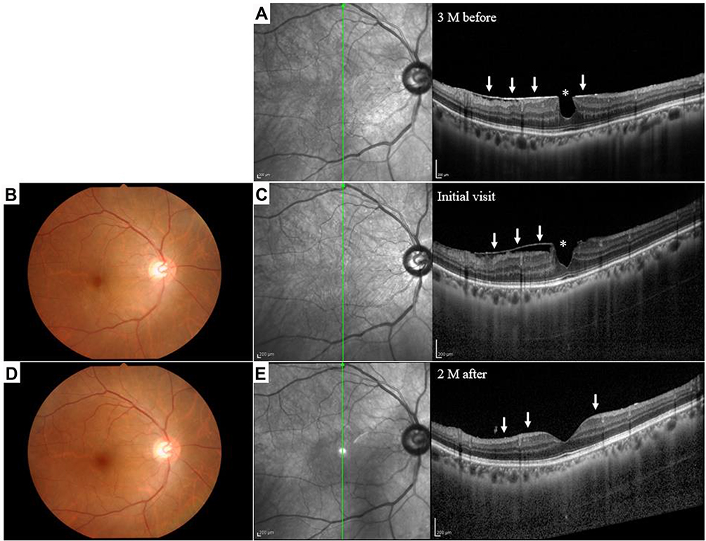 |
Figure 1 Fundus photographs and spectral domain optical coherence tomographic (SD-OCT) images of a 66-year-old man with metamorphopsia caused by an epiretinal membrane (ERM). A macular pseudohole was also present. (A) An SD-OCT image showing an ERM which is seen as a hyperreflective layer (white arrows) on the retina. A macular pseudohole (asterisk) can also be seen in the SD-OCT image. These images were taken at the referral hospital 3 months before the initial examination in our hospital. (B) Fundus photograph taken at the initial examination in our hospital showing an ERM with a round hole centered on the fovea giving the appearance of a macular pseudohole. (C) SD-OCT image shows the macular pseudohole (asterisk) and retinal folds with a contraction of the ERM in the macular area and a partially separation of the ERM from the sensory retina in the inferior area at the initial examination (white arrows). (D) Fundus photograph taken 2 months later. The ERM and the macular pseudohole are absent and the fundus has an almost normal appearance. (E) SD-OCT image shows no ERM and almost normal appearance of the retina without remaining undulations 2 months after the initial visit (white arrows). |
We intended to perform vitrectomy to remove the ERM, but within 2 months after the initial visit, the ERM spontaneously separated from the retina. Fundus photograph showed that the ERM and the macular pseudohole are absent and the fundus has an almost normal appearance, and without remaining undulations, SD-OCT image showed no ERM and almost normal retina appearance. His vision improved to 20/15 after the ERM separation.
Discussion
Several cases of spontaneous ERM separation in eyes with a macular pseudohole have been reported, and the time of separation was distinct and depended on the patient’s age.14–18 However, the separation can occur as long as 5 years after the detection of the ERM. Our case showed a rapid progression of the ERM contraction resulting in spontaneous separation from the retina within a total of 5 months (3 months before and 2 months after the initial visit) after it was first detected. Our case also had a rapid recovery of the foveal structure and improvement of vision.
The pathophysiology of spontaneous ERM separation is multifactorial and several mechanisms are being considered.17 The first one is PVD with pulling of ERM by detaching vitreous. Spontaneous ERM separation is more common in eyes with PVD.9 Second, the immature ERM’s contracting forces become stronger than its adhesions to the retina, resulting in slow tangential traction on the ERM’s edges and gradual separation from the edges toward the center. The third one is acute tearing of ERM at its weakest central point and retraction of a part of the membrane toward the epicenter, including increased stress on the ERM like Valsalva.17 The exact cause of spontaneous ERM separation is not easy to detect in each case. However, the second and third mechanisms should be related with the spontaneous ERM separation in the current case since the case had PVD at the initial visit.
It has been reported that eyes with a macular pseudohole associated with an ERM have a relatively well-preserved foveal function.19 This is probably because this type of ERM does not cover the foveal area and has a limited impact on the function of the foveal area. Furthermore, the structural characteristic would be related to the faster restoration of the foveal structure in comparison to the normal idiopathic ERM, which is typically associated with a shift in the foveal morphology as seen by the reduction of foveal concavity.19
Conclusion
Therefore, we suggest that cases with an ERM-associated macular pseudohole should be closely monitored for the possibility of a spontaneous separation of the ERM from the retina.
Ethics Approval and Informed Consent
Written informed consent was obtained from the patient for publication of this case report including any accompanying images. The Institutional Review Board was informed, and approval was not required for reporting the case.
Funding
The authors received no financial support for the research, authorship, or publication of this article.
Disclosure
The authors report no conflicts of interest in this work.
References
1. Hiscott PS, Grierson I, McLeod D. Natural history of fibrocellular epiretinal membranes: a quantitative, autoradiographic, and immunohistochemical study. Br J Ophthalmol. 1985;69:810–823. doi:10.1136/bjo.69.11.810
2. Fraser-Bell S, Guzowski M, Rochtchina E, Wang JJ, Mitchell P. Five-year cumulative incidence and progression of epiretinal membranes: the Blue Mountains Eye Study. Ophthalmology. 2003;110(1):34–40. doi:10.1016/S0161-6420(02)01443-4
3. Fraser-Bell S, Ying-Lai M, Klein R, Varma R. Prevalence and associations of epiretinal membranes in latinos: the Los Angeles Latino Eye Study. Invest Ophthalmol Vis Sci. 2004;45(6):1732–1736. doi:10.1167/iovs.03-1295
4. Ichikawa Y, Imamura Y, Ishida M. Inner nuclear layer thickness, a biomarker of metamorphopsia in epiretinal membrane, correlates with tangential retinal displacement. Am J Ophthalmol. 2018;193:20–27. doi:10.1016/j.ajo.2018.06.001
5. Appiah AP, Hirose T, Kado M. A review of 324 cases of idiopathic premacular gliosis. Am J Ophthalmol. 1988;106(5):533–535. doi:10.1016/0002-9394(88)90581-8
6. Sivalingam A, Eagle RC
7. Pesin SR, Olk RJ, Grand MG, et al. Vitrectomy for premacular fibroplasia. Prognostic factors, long-term follow-up, and time course of visual improvement. Ophthalmology. 1991;98:1109–1114. doi:10.1016/S0161-6420(91)32169-9
8. Park DW, Dugel PU, Garda J, et al. Macular pucker removal with and without internal limiting membrane peeling: pilot study. Ophthalmology. 2003;110(1):62–64. doi:10.1016/S0161-6420(02)01440-9
9. Yang HS, Hong JW, Kim YJ, Kim JG, Joe SG. Characteristics of spontaneous idiopathic epiretinal membrane separation in spectral domain optical coherence tomography. Retina. 2014;34:2079–2087. doi:10.1097/IAE.0000000000000199
10. Meyer CH, Rodrigues EB, Mennel S, Schmidt JC, Kroll P. Spontaneous separation of epiretinal membrane in young subjects: personal observations and review of the literature. Graefes Arch Clin Exp Ophthalmol. 2004;242(12):977–985. doi:10.1007/s00417-004-0934-7
11. Andreev AN, Bushuev AV, Svetozarskiy SN. A case of secondary epiretinal membrane spontaneous release. Case Rep Ophthalmol Med. 2016;2016:4925763. doi:10.1155/2016/4925763
12. Purtskhvanidze K, Balken L, Hamann T, et al. Long-term follow-up of lamellar macular holes and pseudoholes over at least 5 years. Graefes Arch Clin Exp Ophthalmol. 2018;256(6):1067–1078. doi:10.1007/s00417-018-3972-2
13. Kida T, Morishita S, Fukumoto M, Sato T, Oku H, Ikeda T. Long-term evaluation of spontaneous release of epiretinal membrane and its possible pathogenesis. Clin Ophthalmol. 2017;11:1607–1610. doi:10.2147/OPTH.S146692
14. Oono Y, Nakamura S, Yoshimura K, Yamakawa R, Takayama T. Recurrence after spontaneous resolution of an idiopathic epiretinal membrane. Case Rep Ophthalmol. 2011;2:55–58. doi:10.1159/000324462
15. Menteş J, Nalçacı S. An unusual case: self-separation of an idiopathic epiretinal membrane. Turk J Ophthalmol. 2020;50(1):56–58. doi:10.4274/tjo.galenos.2019.62372
16. Kim JH, Kim JW, Kim CG, Lee DW. Long-term natural history of the idiopathic epiretinal membrane in children and young adults. Graefes Arch Clin Exp Ophthalmol. 2020;258(10):2141–2150. doi:10.1007/s00417-020-04787-5
17. Mansour AM, Mansour HA, Arevalo JF. Spontaneous release of epiretinal membrane in a young weight-lifting athlete by presumed central rupture and centrifugal pull. Clin Ophthalmol. 2014;8:2243–2250. doi:10.2147/OPTH.S74163
18. Nomoto H, Matsumoto C, Arimura E, et al. Quantification of changes in metamorphopsia and retinal contraction in eyes with spontaneous separation of idiopathic epiretinal membrane. Eye (Lond). 2013;27(8):924–930. doi:10.1038/eye.2013.108
19. Hwang JU, Sohn J, Moon BG, et al. Assessment of macular function for idiopathic epiretinal membranes classified by spectral-domain optical coherence tomography. Invest Ophthalmol Vis Sci. 2012;53:3562–3569. doi:10.1167/iovs.12-9762
 © 2021 The Author(s). This work is published and licensed by Dove Medical Press Limited. The full terms of this license are available at https://www.dovepress.com/terms.php and incorporate the Creative Commons Attribution - Non Commercial (unported, v3.0) License.
By accessing the work you hereby accept the Terms. Non-commercial uses of the work are permitted without any further permission from Dove Medical Press Limited, provided the work is properly attributed. For permission for commercial use of this work, please see paragraphs 4.2 and 5 of our Terms.
© 2021 The Author(s). This work is published and licensed by Dove Medical Press Limited. The full terms of this license are available at https://www.dovepress.com/terms.php and incorporate the Creative Commons Attribution - Non Commercial (unported, v3.0) License.
By accessing the work you hereby accept the Terms. Non-commercial uses of the work are permitted without any further permission from Dove Medical Press Limited, provided the work is properly attributed. For permission for commercial use of this work, please see paragraphs 4.2 and 5 of our Terms.