Back to Journals » Clinical, Cosmetic and Investigational Dermatology » Volume 15
Quantitative Evaluation of the Effectiveness of Chemical Peelings in Reducing Acne Lesions Based on Gray-Level Co-Occurrence Matrix (GLCM)
Authors Odrzywołek W ![]() , Deda A
, Deda A ![]() , Zdrada J, Wilczyński S
, Zdrada J, Wilczyński S ![]() , Błońska-Fajfrowska B, Lipka-Trawińska A
, Błońska-Fajfrowska B, Lipka-Trawińska A
Received 17 May 2022
Accepted for publication 18 August 2022
Published 12 September 2022 Volume 2022:15 Pages 1873—1882
DOI https://doi.org/10.2147/CCID.S375131
Checked for plagiarism Yes
Review by Single anonymous peer review
Peer reviewer comments 3
Editor who approved publication: Dr Jeffrey Weinberg
Wiktoria Odrzywołek,1 Anna Deda,2 Julita Zdrada,1 Sławomir Wilczyński,1 Barbara Błońska-Fajfrowska,1 Aleksandra Lipka-Trawińska1
1Department of Basic Biomedical Science, Faculty of Pharmaceutical Sciences in Sosnowiec, Medical University of Silesia, Katowice, Poland; 2Department of Cosmetology, Faculty of Pharmaceutical Sciences in Sosnowiec, Medical University of Silesia, Katowice, Poland
Correspondence: Wiktoria Odrzywołek, Department of Basic Biomedical Science, Faculty of Pharmaceutical Sciences in Sosnowiec, Medical University of Silesia, 41-200 Sosnowiec, ul. Kasztanowa 3, Katowice, Poland, Tel +32269 98 30, Email [email protected]
Purpose: Acne vulgaris is a chronic, inflammatory disease accompanied by lesions affecting the structure of the skin. Chemical peels are one of the methods of reducing acne vulgaris. There is still a lack of quantitative methods of assessing impact of cosmetic procedure on the skin. Skin condition depends on skin texture characterization; therefore, the analysis that provides data about the textures can be helpful in assessing the effectiveness of cosmetic treatments.
Patients and Methods: The study involved 24 volunteers with acne lesions. Each participant underwent 4 treatments using chemical peels at two-week intervals. Before, during and after procedure clinical photography were made. To assess effectiveness of chemical peeling in acne lesion reduction, we were used gray-level co-occurrence matrix (GLCM) analysis. Qualitative assessment of acne severity was made by 12 experts in dermatology.
Results: After a series of treatments, the GLCM contrast value decreased in each area of the face, and the GLCM homogeneity value increased, which means that the number of acne lesions was reduced. Expert assessment according to the IGA scale confirms the effectiveness of therapy with both salicylic and glycolic acid and pyruvic acid.
Conclusion: The results of this study prove that GLCM analysis is a useful tool for assessing the effectiveness of chemical peel treatments. It can also be used for quantitative assessment of skin texture.
Keywords: gray-level co-occurrence matrix, acne, chemical peelings, acne treatment, skin texture
Introduction
Acne vulgaris is a chronic, inflammatory disease of the pilosebaceous unit. It is mainly caused by increased sebum production, hyperkeratinization of the hair follicles, bacterial colonization and inflammation. One component of acne pathogenesis is inflammation, which develops as a result of the activation of multiple inflammatory pathways. Inflammation occurs in all acne lesions, including micro-blackheads. Immunohistochemical studies show a higher level of pro-inflammatory factors (CD4 cells, macrophages and interleukin (IL) −1-alpha) in the skin of acne patients compared to the skin of non-acne patients. The inflammation associated with acne stimulates melanogenesis and pigment deposition.1–4
Chemical peels are one of the methods of reducing acne vulgaris. The high effectiveness of peels is mainly due to the wide possibility of adjusting the treatment to the patient. Various substances can be used, with different combinations, different concentrations and different duration of action on the skin. Currently, the most commonly used surface peels are: glycolic acid, trichloroacetic acid, salicylic acid, pyruvic acid and resorcinol. A medium-strength organic acid (AHA, a-keto, etc.) usually dissociates in water. When the acid molecule is transferred to the liquid crystal structure of the intercellular cement, it triggers a chemo-exfoliating effect. The result of this condition is the acceleration of exfoliation of the epidermis, smoothing out the skin tone, reducing the secretion of sebum and the amount of acne lesions.5–8
Despite the high effectiveness of peels, there is still a lack of quantitative methods of assessing their impact on the skin. The most commonly used instrumentation to evaluate the properties of skin involves single point measurements. This methods measure the biomechanical parameters of the skin, such as the level of sebum secretion and the content of chromophores: melanin and hemoglobin, which provide information on the effect of the therapy on the activity of the sebaceous glands, as well as discoloration and inflammation. However, they cannot assess the reduction of acne lesions. Polarized light photography is the method of assessing inflammatory changes occurring in acne. The use of cross-polarization filters enables the visualization of the subsurface features.9 The development non-invasive methods would allow not only to assess the effectiveness and safety of these popular aesthetic medicine procedures, but also to optimize the treatment parameters in relation to the baseline skin features.
This paper proposes an innovative method based on the use of dedicated image analysis and processing algorithms that quantify the effectiveness of peels in reducing acne lesions. In addition, a qualitative analysis was performed to assess the severity of acne lesions before and after a series of treatments using the IGA scale by experts. The results of the qualitative analysis were compared and correlated with each other.
Materials and Methods
Patients
Treatment Procedure
The study involved 24 volunteers with acne lesions (Table 1). Each participant underwent 4 treatments using chemical peels at two-week intervals. The quantitative analysis of the advancement of acne lesions was performed at 3 time points t0 - before the series of treatments, t1 - during (after 2 treatments) and t2 - after a series of 4 treatments. The analysis was based on the photographic documentation made using the Fotomedicus system, Elfo, Poland. The photos were taken with a cross-polarization filter, which avoids the effect of light reflection from the skin surface and provides a more objective visualization of inflammatory lesions and acne discoloration. The treatment area covers the facial skin, divided into two parts: right and left. Pre Peeling Cleanser (Mene & Moy System, Miami, USA) was applied to the entire face and was washed off after 5 minutes. Then, on the right side of the face, Alfa Beta Complex Gel (Mene & Moy, Miami, USA) containing salicylic and glycolic acid was applied, and on the left side, a preparation Perfarma Pyruvic Peeling containing 50% pyruvic acid (Perfarma P.D, Roma, Italy) was applied. The preparations were washed off after the appearance of the erythematous reaction.10
 |
Table 1 Patient Characteristics |
The inclusion criteria for the research were:
- acne vulgaris,
- age over 18,
- informed consent to participate in the research.
The exclusion criteria from the study were:
- use of anti-acne agents for a minimum period of 3 months from the end of treatment,
- pregnancy and breastfeeding,
- age under 18,
- taking photosensitizing drugs,
- tan.
Ethics Statement
The authors confirm that the ethical policies of the journal, as noted on the journal’s author guidelines page, have been adhered to and the appropriate ethical review committee approval has been received. The study was conducted in accordance with the Declaration of Helsinki. The research obtained a positive opinion of the Ethics Committee of the SUM No. PCN/0022/KB1/12/I/20 on 19 May 2020. All volunteers signed a voluntary consent to participate in the study.
The Course of the Study
Image Analysis
Qualitative Assessment of Acne Severity
Clinical photographs of patients taken in the cross-polarized light at an angle of 45 ° (right and left half-profile) were juxtaposed and presented to a group of experts in dermatology - The photos were evaluated by 12 experts. Experts assessed the severity of acne lesions according to the IGA scale, based on the criteria given in the table (Table 2) before the series of treatments and after the series of exfoliation treatments. The analysis was in the form of a blinded sample - The experts were not informed about the procedure performed. Their task was to analyze the condition of the skin.
 |
Table 2 Investigator’s Global Assessment (IGA) of Acne Severity |
GLCM Matrix
The assessment of acne lesions is currently based primarily on qualitative methods. Visually, the number and advancement of changes are assessed. Acne lesions are more intense when the contrast between them and unchanged skin is greatest. In view of the above, an algorithm for quantification was proposed, which takes into account this dependence: the gray-level co-occurrence matrix - GLCM. GLCM analysis enables data to be obtained from parameters such as contrast and homogeneity.
Gray-level co-occurrence matrix analysis determines the spatial relationship between pixels by identifying quantitatively the difference in intensity between the examined pixel and its adjacent pixels. Thus, the GLCM informs about how often the pixels of a given brightness are located at a certain distance from each other. Most often analyzed are pixels that are adjacent to each other. The GLCM matrix is described by parameters such as distance and angle, for which pairs of pixels are tested.11–14 The analysis can be performed at 0 °, 45 °, 90 ° and 135 °. Texture information can be obtained by statistically analyzing two-dimensional images.15
Contrast is the difference between the highest and lowest values of adjacent sets of pixels. High contrast is usually equated with hard textures, while low contrast is equated with smooth. For homogeneous images, the contrast value is 0 or close to 0. Homogeneity is a measure of the similarity of the image, it takes values between 0–1. Higher homogeneity values show less gray differences (if we reduce the images to gray levels) among the pixel set. Homogeneity is maximal when all elements in the image are the same (if there is no variation among pixels, then homogeneity is 1).15 Homogeneity (formula 1) and contrast (formula 2) and GLCM are inversely correlated in terms of an equivalent distribution in the population pairs of pixels. This means that the homogeneity decreases as the contrast increases, and vice versa.16,17
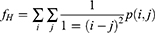
I, j - coordinates of the co-occurrence matrix space
p(i, j) - element in the co-occurrence matrix at the coordinates i and j.
The ROI areas were arbitrarily distinguished from clinical photos: forehead right side, cheek right side, nose right side, forehead left side, forehead right side, nose left side. The Gray-Level Co-occurrence Matrix- GLCM was used to analyze ROI. The photos were converted to grayscale, obtaining monochrome images with 256 gray levels. The pixels that are adjacent to each other were analyzed. In this study, pixels situated at an angle of 0 ° in the immediate vicinity were analyzed. The analysis was performed in Matlab Version 7.11.0.584 (R2010b).
Statistical Analysis
Statistical analysis was performed in the Statistica 13.3 software (TIBCO Software, Palo Alto, CA, USA). The normality of the distribution was assessed using the Shapiro–Wilk W-test. Normal distributions were not obtained in the results of contrast, homogeneity and expert assessment. For the analysis of contrast and homogeneity of the image, the Friedman ANOVA test was used. Expert ratings were analyzed using the Wilcoxon test. The level of statistical significance was assumed to be α = 0.05.
Results
Figure 1 show an exemplary photo visualizing the obtained effects. Not only a smaller number of inflammatory changes can be observed after the series of procedures used, but also a change in the overall color of the skin.
 |
Figure 1 Clinical photographs of the cheeks area of volunteer No. 1 before the application of a preparation containing 50% pyruvic acid - t0 (A and E) and after a series of treatments (B and F) - t2 and before the application of Alfa Beta Complex Gel (Mene & Moy System) containing salicylic and glycolic acid (C and G) - t0 and after a series of treatments (D and H) - t2. |
All volunteers felt temporary burning and itching sensations after the application of pyruvic acid, they observed a few days of exfoliation on both sides of the face on the 3rd day after the procedure.
GLCM Contrast Analysis
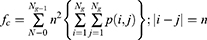
As a result of the use of a mixture of glycolic acid and salicylic acid, the GLCM contrast of the left side of the forehead (p <0.05), cheek (p <0.01) and nose (p <0.01) decreased in a statistically significant manner (Figure 2). Before performing a series of acne treatments, the highest contrast was observed in the nose area and the lowest in the cheek area. After a series of procedures, this tendency did not change, the nose still remained the most contrasting ROI and the cheek the least contrasting. Moreover, it should be noted that for all three examined areas, ie forehead, cheeks, nose, after a series of treatments, the standard deviation of the mean contrast decreased, which also indicates a skin tone alignment and skin texture.
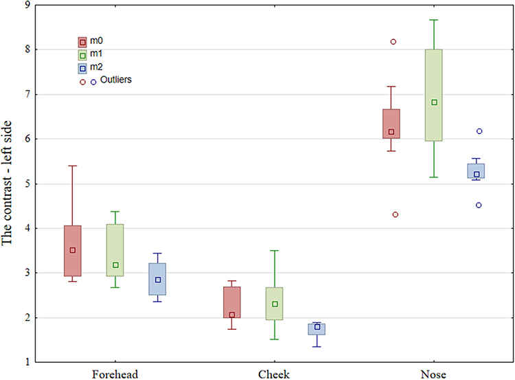 |
Figure 2 Contrast of ROI areas of the left side of the face obtained from photos taken under cross-polarized light. Median, interquartile range, min-max. |
As a result of procedures using pyruvic acid peeling, the contrast of the right side of the nose (p <0.01) decreased in a statistically significant manner (Figure 3). On the forehead (p = 0.065) and on the cheek (p = 0.053), a tendency was shown to fall below the adopted threshold of statistical significance (p <0.05). As on the left side, the highest initial contrast was observed on the nose and the lower values on the forehead and cheeks, respectively. For all three examined areas, ie forehead, cheeks, nose, after a series of treatments, the standard deviation of the mean contrast decreased, which also suggests a reduction of acne lesions.
 |
Figure 3 Contrast of ROI areas of the right side of the face obtained from photos taken in cross polarized light. Median, interquartile range, min-max. |
GLCM Homogeneity Analysis
As a result of the use of a mixture of glycolic acid and salicylic acid, the homogeneity of the left side of the forehead (p <0.001), cheek (p <0.01) and nose (p <0.05) increased in a statistically significant manner (Figure 4). As expected, the initial homogeneity was highest for the cheeks and lower for the forehead and nose, respectively, because according to the proposed GLCM algorithm, homogeneity is inversely proportional to contrast.
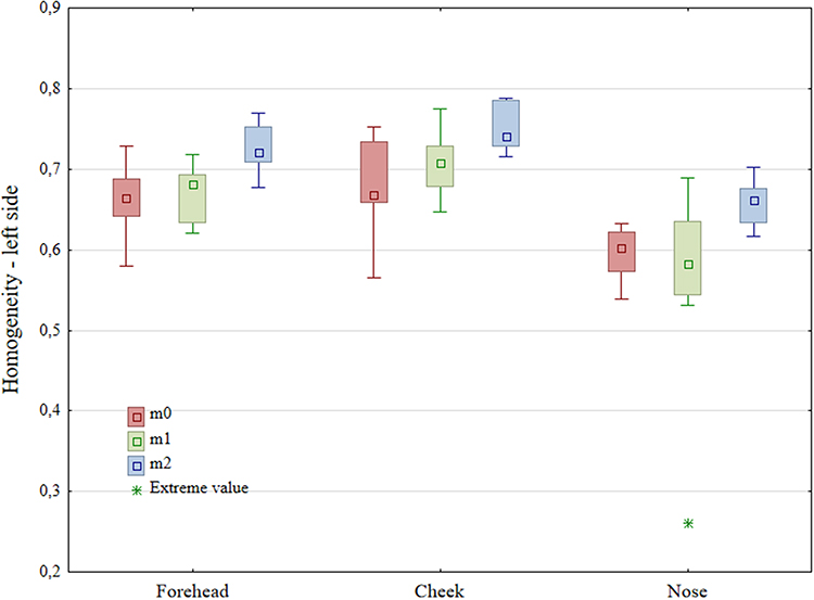 |
Figure 4 Homogeneity of ROI areas of the left side of the face, obtained from photos taken in cross-polarized light. Median, interquartile range, min-max. (*-extreme value). |
Similar results as for the left side of the face were recorded for the right side (Figure 5), where the application of pyruvic acid peeling resulted in a statistically significant increase in homogeneity after a series of treatments: forehead (p <0.01), cheek (p <0.01) and nose (p < 0.001). Contrary to the contrast value, the highest homogeneity was observed on the cheek and the lowest on the nose. After a series of anti-acne procedures, this trend has not changed, the cheek is still the most homogeneous ROI and the nose the least homogeneous.
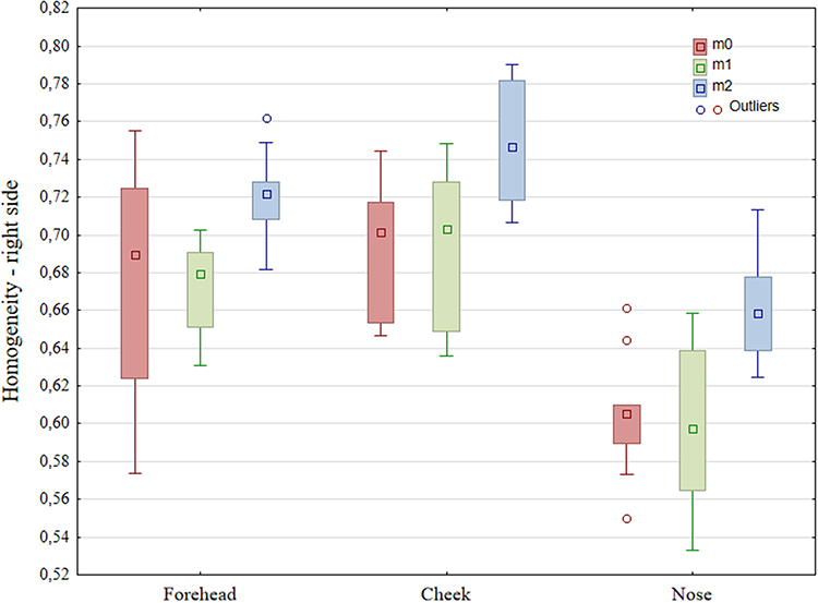 |
Figure 5 Homogeneity of ROI areas of the right side of the face, obtained from photos taken in cross-polarized light. Median, interquartile range, min-max. |
Qualitative Analysis
According to experts, there was an improvement in the skin’s condition and reduction of acne lesions, after applying a series of treatments using pyruvic acid and after using a mixture of glycolic and salicylic acids (Figure 6). Experts assessed the condition of the patients’ skin before the treatments as “mild”, and as “almost clear” after the treatments. This indicates an improvement in the condition of the skin and a significant reduction in the number of papules and pustules in the examined patients after a series of chemical peels. The IGA classification is a qualitative and subjective scale. Each of the experts assessing the severity of acne lesions is guided by their own experience and may interpret acne changes differently. In some cases, it may be difficult to classify acne to a certain severity. This translates into a lack of consistency in the experts’ assessment.
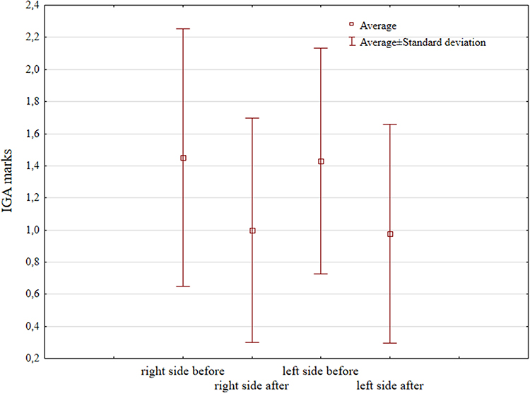 |
Figure 6 Average marks according to the IGA scale used by the experts. |
Discussion
Most of the studies to date comparing the effects of chemical peels are based on subjective evaluation or on developed scales also based on subjective evaluation.18,19 Due to the limited possibilities of an objective, repeatable and quantitative method of skin assessment during the course of acne therapy, it was decided to use methods of image analysis and processing that had not been used for this purpose so far. The impact of pyruvic acid and a mixture of salicylic and glycolic acids on the facial skin of patients with acne vulgaris was quantified by using grey level co-occurrence matrix (GLCM).
Each digital photograph has distinctive features related to color, contrast, size and shape. One of the methods of image analysis that allows to describe these features is the GLCM matrix. However, there are no scientific reports indicating the use of the GLCM matrix to quantify the reduction of acne lesions. The GLCM matrix has been a method used so far to analyze computed tomography images, determine the density of breast tissue, bone quality, and assess cellulite. It is an increasingly popular method of image analysis due to the high potential of medical imaging research.20–22
Mukul Shirvaikar,13 using the GLCM matrix, estimated the roughness of the surface of the articular cartilage in the case of early signs of degeneration in the range from 0.09 µm to 2.51 µm with an accuracy of 0.03 µm. Its results indicate high precision and the possibility of obtaining accurate data from the analysis GLCM. Xiang Ou12 used the GLCM matrix in his study to analyze skin capacitive images measured with a fingerprint reader, as well as to analyze the permeation of solvents through the skin. The results show that GLCM analysis is an effective way to extract and analyze skin texture information, which can be a valuable source of data to evaluate the effects of medical and cosmetic treatments. The GLCM matrix could also be a valuable benchmark for skin aging studies and provide data to evaluate the efficacy of skin permeation of solvents. Fabiane Leonel Utino11 in the study used the GLCM matrix to distinguish sarcoidosis from tuberculoid leprosy. Among the GLCM parameters, the entropy and SAM values were similar, while the contrast was significantly higher in sarcoidosis (p = 0.02 p = 0.02; 4908.31 vs.2822).
GLCM is one of the effective quantitative analysis methods for assessing skin texture. The textures often refers to the distribution of colors on the surface, thus providing information about the spatial distribution of colors in the examined image. They are characterized by variations in intensity or color that usually result from the roughness of the object’s surface. Contrast and homogeneity show the presence of a particular distribution textural pattern within the picture.23,24 Acne lesions change the texture of the skin, papules and pustules are lofty structures, and open blackheads and scars are concave. The contrast value reflects the sharpness and density of the texture as well as the range of the groove depth. The lower the contrast, the greater the homogeneity of the image, and thus the even structure of the tested surface. In addition to the anti-acne effectiveness of peels, image analysis using GLCM contrast assesses the surface structure of the skin. Healing of acne lesions may occur with scarring. Despite the healing of inflammatory and non-inflammatory lesions, the skin structure remains irregular. Image analysis makes possible to evaluate the complete treatment process.
Expert assessment according to The IGA scale, like the GLCM method, confirms the effectiveness of therapy with both salicylic and glycolic acid and pyruvic acid. The IGA scale is based on the subjective opinion of the examiner, which contributes to the discrepancy between the opinions of individual experts. Experts had doubts as to the unambiguous classification of 2 patients, which is illustrated by the problem of qualitative scales, where the severity of acne lesions can be interpreted in various ways. The proposed method of using the GLCM matrix to assess the effectiveness of chemical peels is an objective tool that solves the problem of mistakes in the interpretation of experts. The use of the Fotomedicus system makes it possible to obtain repeatable clinical photographs, eliminating the risk of errors in the further analysis of the results.
Limitations
- Only patients with mild acne were included in the study.
- All patients had phototype II according to the Fitzpatrick scale.
- No comparison of the GLCM method with other non-invasive methods of assessing acne lesions.
Conclusion
The effectiveness of chemical peels with the use of glycolic, salicylic and pyruvic acid in reducing acne lesions has been proven in many studies.18,25 In this study, we used the GLCM analysis as a new method for assessing the effectiveness of acne therapy. After a series of treatments, the contrast value decreased in each area of the face, and the homogeneity value increased, which means that the number of acne lesions was reduced. The results of this study prove that GLCM analysis is a useful tool for assessing the effectiveness of chemical peel treatments. It may also be helpful in choosing the right preparation depending on the severity of acne. The effectiveness of the GLCM analysis is confirmed by the opinion of experts on the assessment of the improvement of skin condition and the degree of reduction of acne lesions.
Acknowledgments
This study is supported by the Medical University of Silesia (PCN-1-013/K/0/O, PCN-2-061/N/1/O and PCN-2-027/N/1/O).
Disclosure
The authors report no conflicts of interest in this work.
References
1. Arndt KA, Leboit PE, Wintroub BU, et al. Acne and Rosacea: applying emerging science to improve outcomes. Semin Cutan Med Surg. 2018;37(3):1–20.
2. Gudjonsson JE, Kabashima K, Eyerich K. Mechanisms of skin autoimmunity: cellular and soluble immune components of the skin. J Allergy Clin Immunol. 2020;146(1):8–16. doi:10.1016/j.jaci.2020.05.009
3. Elbuluk N, Grimes P, Chien A, et al. The pathogenesis and management of acne-induced post-inflammatory hyperpigmentation. Am J Clin Dermatol. 2021;22(6):829–836. doi:10.1007/s40257-021-00633-4
4. Chaowattanapanit S, Silpa-Archa N, Kohli I, Lim HW, Hamzavi I. Postinflammatory hyperpigmentation: a comprehensive overview: treatment options and prevention. J Am Acad Dermatol. 2017;77(4):607–621. doi:10.1016/j.jaad.2017.01.036
5. Sofen B, Prado G, Emer J. Melasma and post inflammatory hyperpigmentation: management update and expert opinion. Skin Therapy Lett. 2016;21(1):1–7.
6. Pathak A, Mohan R, Rohrich RJ. Chemical peels: role of chemical peels in facial rejuvenation today. Plast Reconstr Surg. 2020;145(1):58e–66e. doi:10.1097/PRS.0000000000006346
7. Berardesca E, Cameli N, Primavera G, Carrera M. Clinical and instrumental evaluation of skin improvement after treatment with a new 50% pyruvic acid peel. Dermatologic Surg. 2006;32(4):526–531. doi:10.1111/j.1524-4725.2006.32106.x
8. Jaffary F, Faghihi G, Saraeian S, Hosseini SM. Comparison the effectiveness of pyruvic acid 50% and salicylic acid 30% in the treatment of acne. J Res Med Sci. 2016;21:31. doi:10.4103/1735-1995.181991
9. Kollias N, Stamatas GN. Optical non-invasive approaches to diagnosis of skin diseases. J Investig Dermatol Symp Proc. 2002;(1):64–75. doi:10.1046/j.1523-1747.2002.19635.x
10. Zdrada J, Odrzywołek W, Deda A, Wilczyński S, Błońska-Fajfrowska B. Analysis of the effectiveness of chemical peelings in the treatment of acne vulgaris assessed using high-frequency ultrasound—A comparative study. J Cosmet Dermatol. 2021;20(9):2810–2815. doi:10.1111/jocd.13934
11. Utino FL, Garcia M, Velho PENF, et al. Second-harmonic generation imaging analysis can help distinguish sarcoidosis from tuberculoid leprosy. J Biomed Opt. 2018;23(12):1–7. doi:10.1117/1.JBO.23.12.126001
12. Ou X, Pan W, Xiao P. In vivo skin capacitive imaging analysis by using grey level co-occurrence matrix (GLCM). Int J Pharm. 2014;460(1–2):28–32. doi:10.1016/j.ijpharm.2013.10.024
13. Shirvaikar M, Huang N, Dong XN. THE MEASUREMENT OF BONE QUALITY USING GRAY LEVEL CO-OCCURRENCE MATRIX TEXTURAL FEATURES. J Med Imaging Heal Informatics. 2016;6(6):1357–1362. doi:10.1166/jmihi.2016.1812
14. Rebouças Filho PP, Peixoto SA, Medeiros da Nóbrega RV, et al. Automatic histologically-closer classification of skin lesions. Comput Med Imaging Graph. 2018;68:40–54. doi:10.1016/j.compmedimag.2018.05.004
15. Calin MA, Parasca SV, Calin MR, Petrescu E. An analysis of human dorsal hand skin texture using hyperspectral imaging technique for assessing the skin aging process. Appl Spectrosc. 2017;71(3):391–400. doi:10.1177/0003702816659667
16. Saito A, Numata Y, Hamada T, et al. A novel method for morphological pleomorphism and heterogeneity quantitative measurement: named cell feature level co-occurrence matrix. J Pathol Inform. 2016;7:36. doi:10.4103/2153-3539.189699
17. Manivannan K, Aggarwal P, Devabhaktuni V, Kumar A, Nims D, Bhattacharya P. Particulate matter characterization by gray level co-occurrence matrix based support vector machines. J Hazard Mater. 2012;223–224:94–103. doi:10.1016/j.jhazmat.2012.04.056
18. In Jae J, Dong Ju H, Dong Hyun K, Yoon MS, Lee HJ. Comparative study of buffered 50% glycolic acid (pH 3.0) + 0.5% salicylic acid solution vs Jessner’s solution in patients with acne vulgaris. J Cosmet Dermatol. 2018;17(5):797–801. doi:10.1111/jocd.12445
19. Nofal E, Nofal A, Gharib K, Nasr M, Abdelshafy A, Elsaid E. Combination chemical peels are more effective than single chemical peel in treatment of mild-to-moderate acne vulgaris: a split face comparative clinical trial. J Cosmet Dermatol. 2018;17(5):802–810. doi:10.1111/jocd.12763
20. Sethi G, Saini BS. Computer aided diagnosis system for abdomen diseases in computed tomography images. Biocybern Biomed Eng. 2016;36(1):42–55. doi:10.1016/j.bbe.2015.10.008
21. Kumar I, Virmani J, Thakur S. A classification framework for prediction of breast density using an ensemble of neural network classifiers. Biocybern Biomed Eng. 2017;37(1):217–228. doi:10.1016/j.bbe.2017.01.001
22. de la Casa Almeida M, Suárez Serrano C, Jiménez Rejano JJ, Ríos Díaz J, Benitez Lugo ML, Rebollo Roldán JR. Reliability of texture analysis using co-occurrence matrices (glcm) on photographic image in the assessment of cellulite in a Spanish population. J Eur Acad Dermatol Venereol. 2015;29(2):315–324. doi:10.1111/jdv.12534
23. Maktabdar Oghaz M, Maarof MA, Rohani MF, Zainal A, Shaid SZM. An optimized skin texture model using gray-level co-occurrence matrix. Neural Comput Appl. 2019;31(6):1835–1853. doi:10.1007/s00521-017-3164-8
24. Pang H, Chen T, Wang X, Chang Z, Shao S, Zhao J. Quantitative evaluation methods of skin condition based on texture feature parameters. Saudi J Biol Sci. 2017;24(3):514–518. doi:10.1016/j.sjbs.2017.01.021
25. Garg VK, Sinha S, Sarkar R. Glycolic acid peels versus salicylic-mandelic acid peels in active acne vulgaris and post-acne scarring and hyperpigmentation: a comparative study. Dermatol Surg. 2009;35(1):59–65. doi:10.1111/j.1524-4725.2008.34383.x
 © 2022 The Author(s). This work is published and licensed by Dove Medical Press Limited. The
full terms of this license are available at https://www.dovepress.com/terms
and incorporate the Creative Commons Attribution
- Non Commercial (unported, 3.0) License.
By accessing the work you hereby accept the Terms. Non-commercial uses of the work are permitted
without any further permission from Dove Medical Press Limited, provided the work is properly
attributed. For permission for commercial use of this work, please see paragraphs 4.2 and 5 of our Terms.
© 2022 The Author(s). This work is published and licensed by Dove Medical Press Limited. The
full terms of this license are available at https://www.dovepress.com/terms
and incorporate the Creative Commons Attribution
- Non Commercial (unported, 3.0) License.
By accessing the work you hereby accept the Terms. Non-commercial uses of the work are permitted
without any further permission from Dove Medical Press Limited, provided the work is properly
attributed. For permission for commercial use of this work, please see paragraphs 4.2 and 5 of our Terms.