
Back to Journals » Lung Cancer: Targets and Therapy » Volume 16
Pulmonary Nodular Lymphoid Hyperplasia: Reviewing a Lung Cancer Mimicker
Authors Schneider Galvão G  , Fávero Prietto dos Santos J , Hsu YHR , Rogalla P
Received 10 April 2025
Accepted for publication 3 June 2025
Published 10 December 2025 Volume 2025:16 Pages 161—166
DOI https://doi.org/10.2147/LCTT.S527578
Checked for plagiarism Yes
Review by Single anonymous peer review
Peer reviewer comments 2
Editor who approved publication: Dr Sai-Hong Ou
Gabriela Schneider Galvão,1,* Jônatas Fávero Prietto dos Santos,1,* Ying-Han R Hsu,2 Patrik Rogalla1
1Joint Department of Medical Imaging, University of Toronto, Toronto, ON, Canada; 2Laboratory Medicine Program, University Health Network, Toronto, ON, Canada
*These authors contributed equally to this work
Correspondence: Patrik Rogalla, Joint Department of Medical Imaging, University of Toronto, Toronto General Hospital, 1 PMB 291, 585 University Avenue, Toronto, ON, M5G 2N2, Canada, Email [email protected]
Background: Pulmonary nodular lymphoid hyperplasia is a rare benign lymphoproliferative disorder. Patients are commonly asymptomatic. Its radiological presentation is characterized by peripheral single nodular lesion, multiple nodules or mass-like consolidation. Therefore, it can mimic other more frequent pulmonary diseases, such as lung cancer and pulmonary lymphoma. The diagnosis is often late, relying on histopathological findings.
Case Presentation: We report a case of a 66-year-old woman who presented with cough and upper respiratory tract symptoms. Chest CT revealed a mass in the right lower lobe, mediastinal and hilar enlarged lymph nodes and multifocal ground-glass and part solid nodules. PET-CT findings were concerning for primary lung malignancy. She underwent transbronchial needle aspiration biopsy of the enlarged lymph nodes and pulmonary mass. The results were negative for malignancy, but immunophenotyping by flow cytometry was concerning for a lymphoproliferative disorder. For this reason, a percutaneous CT-guided transthoracic core needle biopsy of the mass was performed. Immunophenotypic features of lymphoma were not identified. Overall findings were suggestive of nodular lymphoid hyperplasia. The follow-up chest CT showed near complete resolution of the mass with a residual ground-glass lesion and stability of the enlarged lymph nodes, ground-glass and part solid nodules.
Conclusion: Pulmonary nodular lymphoid hyperplasia is an uncommon lung disorder, usually detected incidentally on chest imaging. Its radiological features can mimic lung cancer and other more prevalent pulmonary diseases. Therefore, a multidisciplinary approach, including histopathological confirmation is essential for an accurate diagnosis. Although rare, it should be considered in the differential diagnosis of pulmonary malignancies.
Keywords: pulmonary nodular lymphoid hyperplasia, lung cancer, differential diagnosis, chest CT
Introduction
Pulmonary nodular lymphoid hyperplasia (PNLH) is a rare benign disease of middle-aged patients with no sex predilection, which was initially known as pseudolymphoma. This entity is characterized by a localized, reactive polyclonal lymphoproliferative lesion.1
Patients are most commonly asymptomatic (70%), but they can present with respiratory and general symptoms, including cough, sputum, fatigue, dyspnea and hemoptysis.1–3 On imaging, the features are similar to other much more common pathologies, such as primary lung cancer and lymphoma. The diagnosis is frequently late, as it is usually found incidentally on imaging exams, and the definite diagnosis relies on histopathological analysis. Histopathological findings can also overlap with other neoplastic and non-neoplastic diseases. Therefore, a clinical and radiological correlation with pathology is important for making the correct diagnosis.4 Resection of these lesions is curative, and the prognosis of these patients is excellent. There are a few case reports suggesting that these lesions can also spontaneously resolve.5
We report a case of a symptomatic patient that presented with a large mass in the Chest CT and with enlarged thoracic lymph nodes. Initially, it was concerning for primary lung cancer and therefore it was biopsied.
Case Presentation
A 66-year-old woman with a past medical history of active tobacco smoking (30 pack/years), hypertension, type 2 diabetes mellitus, coronary artery disease, and congestive heart failure presented to her family physician with cough and upper respiratory tract infection symptoms. She denied associated signs and symptoms, including sputum production, hemoptysis, dyspnea, fever or weight loss. At the time of presentation, she was on bisoprolol and empagliflozin. She retired from office work and denied history of occupational exposures. She reported a second-degree family history of leukemia. Physical examination revealed a frail patient with low body mass index, otherwise unremarkable.
A chest CT was requested, which revealed a large mass in the superior segment of the right lower lobe measuring 4.6×4.3 x 3.7 cm (Figure 1) associated with ipsilateral hilar and interlobar, and bilateral mediastinal enlarged lymph nodes (Figure 2). There were also multifocal heterogenous ground-glass nodules with a macro-reticular pattern, and part solid nodules in both lungs measuring up to 2.0 cm (Figure 3). Incidental bilateral adrenal gland nodules measuring up to 1.5 cm were noted in the upper abdomen. Given the presence of findings concerning for metastatic primary lung cancer, a CT-guided lung biopsy was performed in a community hospital, which was nondiagnostic. A dedicated CT abdomen with adrenal washout protocol was also performed showing that adrenal gland nodules were consistent with adenomas. She ultimately was referred to the division of Thoracic Surgery in a tertiary care center for further workup and management.
 |
Figure 1 Chest CT axial (a) and coronal (b) reconstructions demonstrating a large mass in the superior segment of the right lower lobe (white arrow). The lesion was metabolically active on PET-CT (SUVmax 9.3) (c and d) with a central area of relative photopenia, suggestive of necrosis. |
 |
Figure 2 Chest CT showing enlarged metabolically active subaortic (a and c) and right hilar (b and d) lymph nodes (white arrows). |
 |
Figure 3 Chest CT demonstrating multifocal subsolid lesions (white arrows) in the right upper lobe (a) and left upper lobe (c), with associated increased FDG uptake on PET-CT (b and d). |
The patient underwent a whole body 18F-FDG PET-CT, which showed hypermetabolism in the right lower lobe mass reaching a SUVmax of 9.3, with heterogenous FDG avidity and central photopenia likely related to areas of necrosis, findings highly concerning for primary lung malignancy. Multiple additional ground-glass and part solid nodules were morphologically stable, showing no or mild increased FDG uptake (reaching a SUVmax of 3.8), raising the possibility of adenocarcinoma spectrum lesions. Ipsilateral hilar and bilateral mediastinal nodes were also hypermetabolic, reaching a SUVmax of 4.8 on station 5 (subaortic) measuring 2.0 cm (stable in size). No additional hypermetabolic lesion concerning for metastatic disease was identified. Subsequently, the patient underwent an endobronchial ultrasound (EBUS) guided biopsy using a radial probe with assistance of an on-site cytopathologist. The right lower lobe mass and nodal (stations 11R, 7, 4R, and 4L) transbronchial needle aspiration biopsies were negative for malignancy. There was no evidence of carcinoma or granulomatous inflammation. Immunophenotyping by flow cytometry showed features of a reactive lymphoid population, and the morphologic and immunophenotypic findings were consistent with reactive lymphoid hyperplasia.
Given the suspicion of a lymphoproliferative disorder, a second percutaneous CT-guided transthoracic core needle biopsy of the right lower lobe mass was performed obtaining a 2.0 cm core biopsy using an 18-gauge needle, which revealed lymphoid proliferation including multiple lymphoid follicles (Figure 4). Immunophenotypic features diagnostic of lymphoma were not identified. Overall findings were suggestive of nodular lymphoid hyperplasia. The patient underwent a follow-up CT chest in 3 months, which showed interval near complete spontaneous resolution of the right lower lobe mass, with residual ground-glass opacity at the same level (Figure 5), with similar morphologic features in comparison to the additional pulmonary nodules, which have remained stable in size and in appearance. Enlarged right hilar and bilateral mediastinal lymph nodes were also stable in this follow-up and were deemed to be likely reactive in nature. The patient was referred to a smoking cessation clinic and she will continue under clinical and imaging surveillance.
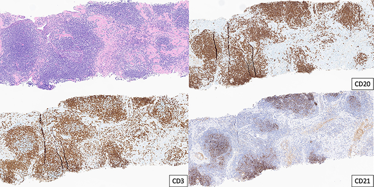 |
Figure 4 H&E section of core biopsy showing reactive lymphoid hyperplasia with frequent lymphoid follicles highlighted by CD3, CD20 and CD21 immunohistochemical studies. |
 |
Figure 5 3-month follow-up chest CT showing significant interval decrease of the right lower lobe mass with a residual groundglass opacity (white arrow on (a), similar to the pre-existent multifocal lesions, and persistent prominent subaortic (b) and right hilar (c) lymph nodes (white arrows on (b and c)). |
Discussion
PNLH is a rare benign lymphoproliferative disorder of unclear etiology. Differently from the spectrum of malignant lymphoma, PNLH is a non-neoplastic reactive and localized process. Other entities characterized by polyclonal lymphoid infiltrates in the lungs include follicular bronchiolitis and lymphoid interstitial pneumonia. However, they present with different distribution patterns, predominantly peribronchovascular and diffuse distribution, respectively.1
Patients with PNLH are most commonly asymptomatic but can also present with respiratory and nonspecific general symptoms.1–3 Radiological manifestations of PNLH have been described as a single nodule, multiple nodular lesions or as consolidation, most often peripheral.5,6 This poses a diagnostic challenge, since it mimics the presentation of other much more common pulmonary pathologies, such as lung cancer, metastatic disease and primary pulmonary lymphoma.6,7 Lymphadenopathy and pleural invasion are not typical findings.6 FDG PET can help differentiate benign from malignant lesions when there is no uptake. However, benign pathologies can also present increased metabolic uptake.5 In this scenario, lung malignancy is the first differential diagnosis. In our case, the mass showed hypermetabolism with heterogeneous FDG avidity and with associated mediastinal and hilar lymphadenopathy, and therefore it was considered highly concerning for primary lung malignancy.
The case we reported illustrates the diagnostic challenge of PNLH. This report reinforces the importance of a multidisciplinary approach to make an accurate diagnosis, combining clinical presentation, radiological findings, and histopathological confirmation. In this case, the final diagnosis of PNLH allowed conservative management of the patient, with spontaneous resolution in the follow-up, avoiding unnecessary surgical procedures. Although rare, PNLH should be considered in the differential diagnosis of malignancy.
Ethics Approval
Institutional authorization is not applicable.
Consent to Participate
The authors declare that written informed consent was obtained from the patient for the publication.
Consent for Publication
Written informed consent was obtained from the patient for publication of this case report and any accompanying images.
Author Contributions
All authors made a significant contribution to the work reported, whether that is in the conception, study design, execution, acquisition of data, analysis and interpretation, or in all these areas; took part in drafting, revising or critically reviewing the article; gave final approval of the version to be published; have agreed on the journal to which the article has been submitted; and agree to be accountable for all aspects of the work.
Disclosure
The authors have nothing to declare.
References
1. Hare SS, Souza CA, Bain G, et al. The radiological spectrum of pulmonary lymphoproliferative disease. Br J Radiol. 2012;85(1015):848–864. doi:10.1259/bjr/16420165
2. Fang L, Xu J, Wang L, He Z, Lv W, Hu J. Pulmonary nodular lymphoid hyperplasia: a rare benign disease with malignant mask. Ann Translat Med. 2019;7(3):43. doi:10.21037/atm.2018.12.59
3. Miyoshi S, Hamada H, Katayama H, et al. A case of pulmonary nodular lymphoid hyperplasia with a resected cavity, followed by spontaneous regression of the remaining lesions. Intern Med. 2010;49(15):1617–1621. doi:10.2169/internalmedicine.49.3487
4. Yell M, Rosado FG. Pulmonary nodular lymphoid hyperplasia. Arch Pathol Lab Med. 2019;143(9):1149–1153. doi:10.5858/arpa.2018-0188-rs
5. Suga K, Yasuhiko K, Hiyama A, Hori K, Tanaka N, Ueda K. F-18 FDG PET/CT findings in a case of multifocal nodular lymphoid hyperplasia of the lung. Clin Nucl Med. 2009;34(6):374–376. PMID: 19487850. doi:10.1097/RLU.0b013e3181a34591
6. Sirajuddin A, Raparia K, Lewis VA, et al. Primary pulmonary lymphoid lesions: radiologic and pathologic findings. RadioGraphics. 2016;36(1):53–70. doi:10.1148/rg.2016140339
7. Kajiwara S, Sakai S, Soeda H, et al. Multifocal nodular lymphoid hyperplasia of the lung. J Thorac Imaging. 2005;20(3):239–241. PMID: 16077344. doi:10.1097/01.rti.0000158404.40711.26
 © 2025 The Author(s). This work is published and licensed by Dove Medical Press Limited. The
full terms of this license are available at https://www.dovepress.com/terms
and incorporate the Creative Commons Attribution
- Non Commercial (unported, 4.0) License.
By accessing the work you hereby accept the Terms. Non-commercial uses of the work are permitted
without any further permission from Dove Medical Press Limited, provided the work is properly
attributed. For permission for commercial use of this work, please see paragraphs 4.2 and 5 of our Terms.
© 2025 The Author(s). This work is published and licensed by Dove Medical Press Limited. The
full terms of this license are available at https://www.dovepress.com/terms
and incorporate the Creative Commons Attribution
- Non Commercial (unported, 4.0) License.
By accessing the work you hereby accept the Terms. Non-commercial uses of the work are permitted
without any further permission from Dove Medical Press Limited, provided the work is properly
attributed. For permission for commercial use of this work, please see paragraphs 4.2 and 5 of our Terms.