Back to Journals » Nature and Science of Sleep » Volume 12
Psychomotor Performance in Patients with Obstructive Sleep Apnea Syndrome
Authors Lusic Kalcina L ![]() , Pavlinac Dodig I
, Pavlinac Dodig I ![]() , Pecotic R
, Pecotic R ![]() , Valic M
, Valic M ![]() , Dogas Z
, Dogas Z ![]()
Received 16 October 2019
Accepted for publication 9 February 2020
Published 9 March 2020 Volume 2020:12 Pages 183—195
DOI https://doi.org/10.2147/NSS.S234310
Checked for plagiarism Yes
Review by Single anonymous peer review
Peer reviewer comments 2
Editor who approved publication: Dr Sutapa Mukherjee
Linda Lusic Kalcina, Ivana Pavlinac Dodig, Renata Pecotic, Maja Valic, Zoran Dogas
Department of Neuroscience, Split Sleep Medicine Center, University of Split School of Medicine, Split, Croatia
Correspondence: Zoran Dogas
Department of Neuroscience, Split Sleep Medicine Center, University of Split School of Medicine, Soltanska 2, Split 21000, Croatia
Tel +38521557905
Fax +38521557955
Email [email protected]
Purpose: Determinants of obstructive sleep apnea (OSA) are hypoxemia and hypercapnia, as well as (micro) arousals from sleep, resulting in chronic sleep fragmentation, sleep deprivation, and excessive daytime sleepiness (EDS). All of the above-mentioned factors might contribute to psychomotor impairment seen in OSA patients. Additionally, this study aimed to assess the contribution of BMI, age, EDS assessed with Epworth sleepiness scale (ESS), and severity of OSA assessed with apnea-hypopnea index (AHI) to the reaction time on chronometric tests in OSA patients and controls. It is hypothesized that moderate and severe OSA have adverse effects on reaction time of perception to visual stimulus, of solving simple arithmetic operations, and of psychomotor limbs coordination assessed by chronometric psychodiagnostic test battery.
Patients and Methods: This study was conducted on 206 male participants; 103 of them had moderate or severe OSA diagnosed by whole-night polysomnography/polygraphy. Control participants (N=103), matched to patients with OSA by age and BMI, had no reported OSA in their medical history, no increased risk for OSA, nor EDS. All participants were assessed with three chronometric psychodiagnostic tests, measuring the reaction time of perception to visual stimulus, of solving simple arithmetic operations, and of psychomotor limbs coordination.
Results: Participants from the OSA group achieved impaired results compared to control participants in minimum single task solving time in speed of solving simple arithmetic operations (3± 0.9 and 2.6± 0.6, P< 0.001), and in minimum solving time of a single task in complex psychomotor limbs coordination (0.69± 0.2 and 0.61± 0.1, P=0.007). Regression analysis revealed no significant contribution of daytime sleepiness to the results achieved in each of the tests.
Conclusion: It is concluded that severe OSA impaired speed of perception, convergent, and operative thinking. Moreover, it is suggested that EDS did not contribute to poor psychomotor outcome in patients with OSA in this study, when age was controlled for.
Keywords: obstructive sleep apnea syndrome, overnight polysomnography, psychomotor performance, psychodiagnostic test, daytime sleepiness
Plain Language Summary
Chronic difficulties with sleep and consequent excessive daytime sleepiness might contribute to psychomotor impairment noted in obstructive sleep apnea (OSA). The use of a chronometric instrument provides information on the extent of such dysfunction, regarding the subjects’ reaction time. Reaction time is measured as time needed for a certain psychological activity. This study was conducted on 103 male subjects with moderate or severe OSA diagnosed by whole-night sleep recordings in the sleep laboratory. Control subjects were matched to OSA subjects by age and body mass index, with no previous diagnosis of OSA or increased risk for OSA, nor excessive daytime sleepiness. Patients with OSA showed a prolonged reaction time of perception to visual stimulus, of solving simple arithmetic operations, and psychomotor limbs coordination, in comparison to controls. When age was accounted for, sleepiness did not contribute to the psychomotor outcome in OSA patients. The novelty of the current study is the evaluation of patients with an instrument assessing reaction time, including an assessment of executive function and processing speed, abilities that have been indicated as important in carrying out complex behaviors associated with safe driving. In addition, the study provided data on impaired stability in reaction time during the process of testing among male OSA patients compared to controls.
Introduction
There is growing interest in obstructive sleep apnea (OSA) and its impact on physiological and psychological performances, due to its high prevalence, particularly in men, and association with adverse medical complications.1 Substantial evidence supports findings that apneic patients have poor outcomes in executive and motor functioning, visuospatial and psychomotor functions, memory, vigilance and attention, concept formation, construction, perception, attention, and alertness.2–5 It has been proposed that intermittent hypoxia per se might contribute to the etiology of such dysfunction in apneic patients,6 as well as (micro) arousals from sleep, which result in chronic sleep fragmentation, sleep deprivation, and consequent excessive daytime sleepiness (EDS).2,6
Most studies of neuropsychological correlates of OSA have focused on impaired psychomotor performance, due to its consequences on OSA patients, such as occupational injuries,7 motor vehicle accidents,8 and overall health-related quality-of-life.9 Neurocognitive processing speed is a measure of such psychomotor function, which has often been reported with tests of 2-hand coordination or reaction time tests.10 When assessing neuropsychological decline in patients with OSA, a recent systematic review defines existing findings as imprecise, having unknown consistency of evidence from studies as well as evidence limited by the risk of bias.11 Although it is well established that psychomotor speed assessed with the Stroop test12,13 is impaired in OSA patients, more complex psychomotor performance measures are still not well established as being deteriorated in OSA. Psychomotor impairment in OSA patients has previously been assessed according to performance on reaction time tests such as the Psychomotor vigilance test (PVT),6,9,14–16 Multiple Unprepared Reaction Time Test (MURT),17 Finger tapping test,18 Trail Making Test,19 and computerized Movement Detection Time Test (MDT).20 Findings of decreased reaction time on PVT tests in OSA patients have been reported on community samples without control groups,21 OSA patients compared to control patients being enrolled in sleep clinics,15 or without control groups.9 Reaction times on MURT tests have also been reported as increased in OSA patients, including a small group of healthy patients with its gender distribution differing from OSA patients.17 In addition, compared with control patients having lower BMI, OSA patients performed worse on the Trail Making Test.19 Contrary to previous findings, reports on the sequential finger tapping task indicated similar performance of OSA patients and age/education matched controls.18
It has been reported that the results from previous studies on psychomotor deficits in OSA patients are diverse due to the fact that different studies, even when they are measuring the same function, employ various neuropsychological instruments.2 Considering reported discrepancies among findings, the current study included chronometric assessment of various aspects of central nervous system activities, assessing psychomotor performance with reaction times in tasks of different complexity and controlling for the factors in the control group that have been reported as lacking in previous research. The psychodiagnostic chronometric instrument, Complex Reactiometer Drenovac test battery (CRD), enabled the measurement of a reaction time to a moving visual stimulus, to a complex visual stimulus demanding the reaction of upper and lower limbs, and to a visual task demanding convergent thinking.22,23 CRD has been defined as one of the few test batteries providing data on the stability of psychomotor functioning during the process of testing, assessed with the ballast measures of reaction time.22 Psychomotor functioning in other clinical populations has already been assessed with the chronometric instrument used in this study,24,25 and it has been proposed that reaction time improves as a consequence of continuous positive airway pressure (CPAP) or mandibular advancement device (MAD) therapy.26,27
This study was performed in order to evaluate the influence of newly-diagnosed OSA on reaction time and information processing in solving simple and complex psychomotor tasks of the CRD-series compared to matched control participants. The novelty of the current study is employment of the ballast outcome measures, quite unique and precise indicators of psychomotor performance stability during the process of testing. Based on previous findings of prolonged reaction time in PVT,5,14–16 MDT,20 Stroop test,12,13 and executive function impairment of OSA patients,6,28,29 we hypothesized that moderate and severe OSA have adverse effects on the reaction time of perception, reaction time of convergent thinking during solving simple arithmetic tasks, and operative thinking during complex psychomotor coordination task assessed by psychodiagnostic chronometric test battery. Additionally, we aimed to assess the contribution of BMI, age, EDS assessed with Epworth sleepiness scale (ESS), and severity of OSA assessed with apnea-hypopnea index (AHI) to the reaction time on chronometric tests in OSA patients and controls.
Methods
Setting
This research was undertaken at the University of Split School of Medicine in Croatia. All sleep analysis data, sleep history data, and neuropsychological testing data were collected from the beginning of 2011 to the end of 2014. All patients were admitted to the Split Sleep Medicine Center, where the diagnostic procedures polysomnography (PSG) or polygraphy (PG) were performed by sleep technologists and an expert certified sleep medicine–physician specialist. The psychomotor testing was performed in a separated and specially equipped neuroscience laboratory that provided isolation from external visual and auditory influences. All OSA patients and control subjects were assessed with psychomotor tests during the morning hours, from 8 am to 11 am. All PSG and PG studies were performed in the time period between 9 pm and 8 am.
Participants
Ethical standards of the institutional research committee and the 1964 Helsinki declaration with its latter amendments were applied to the protocol of the study. All participants were informed about the procedures and the aims of the study, and signed the informed consent forms. Following Ethics Committee for Biomedical research and institutional approvals at the University of Split School of Medicine, a total of 206 male participants was studied. All participants were interviewed about their medical history, their body height and weight were measured, and body mass index (BMI) was calculated. The main inclusion criterion was diagnosis of moderate or severe OSA, whereas exclusion criteria were the following: age younger than 18 years, female gender, previous treatment of OSA, diagnosis of malignant disease, mental disability, or severe physical disability. Table 1 summarizes the demographic data of study subjects.
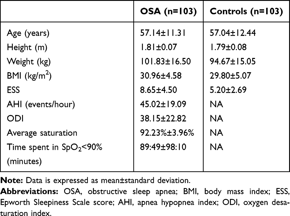 |
Table 1 Demographic Characteristics of Patients with Obstructive Sleep Apnea and Control Participants |
A total of 103 participants were patients diagnosed with moderate or severe OSA in the Split Sleep Medicine Center, following complete all night PSG (Philips Respironics, ALICE 5LE, Eindhoven, the Netherlands, n=32) or PG (Somnocheck, PdX, Embleta, Stardust, n=71). PSG recordings included EEG, EOG, mental and tibial EMG, thoracic and abdominal movements, ECG, pulse oximetry, nasal airflow, and the intensity of snoring. PG recordings included thoracic and abdominal movements, pulse oximetry and nasal airflow. Guidelines of the American Academy of Sleep Medicine (AASM)30 and European Sleep Research Society (ESRS) were used to manually score and evaluate polysomnographic and polygraphic data.31 If an overnight recording lasted less than 6 hours evaluation was not undertaken, and a new PSG/PG was performed. The patients were enrolled in the study if the PSG/PG recording revealed moderate (AHI≥15) or severe (AHI≥30) OSA.
Control subjects (n=103) were drawn from the pool of data obtained from the 10,001 Dalmatian project of The Croatian National Biobank,32 consisting of participants from the general population selected during routine physical examination.
Control participants were matched to patients with OSA by BMI and age. All control subjects had no medical history of sleep disorders and had no increased risk for OSA according to the STOP questionnaire. We considered patients to have increased risk for OSA if they answered positively to two or more questions on the STOP questionnaire. Moreover, control participants did not have EDS as measured by Croatian language validated Epworth sleepiness scale (ESS),33 with a cut-off value of 10, which might indicate the presence of OSA.
A certified physician obtained a detailed medical history during which all self-reported medical conditions were assessed, and all patients provided previous medical examination findings and medical specialists’ reports (Table 2). Data were available for all 103 OSA patients, whereas, in the control group, data on comorbidity and drugs were missing for four respondents’ due to their request of anonymity. In addition, the association with the performance on neuropsychological tasks has been proposed even for disorders treated with antidiabetic medications and antihypertensives (eg, ACE inhibitors, calcium channel blockers, and diuretics), and even for proton-pump inhibitors.34 Taking that association into account, we documented all OSA and control patients’ medications during the medical examination, which are presented in Table 3.
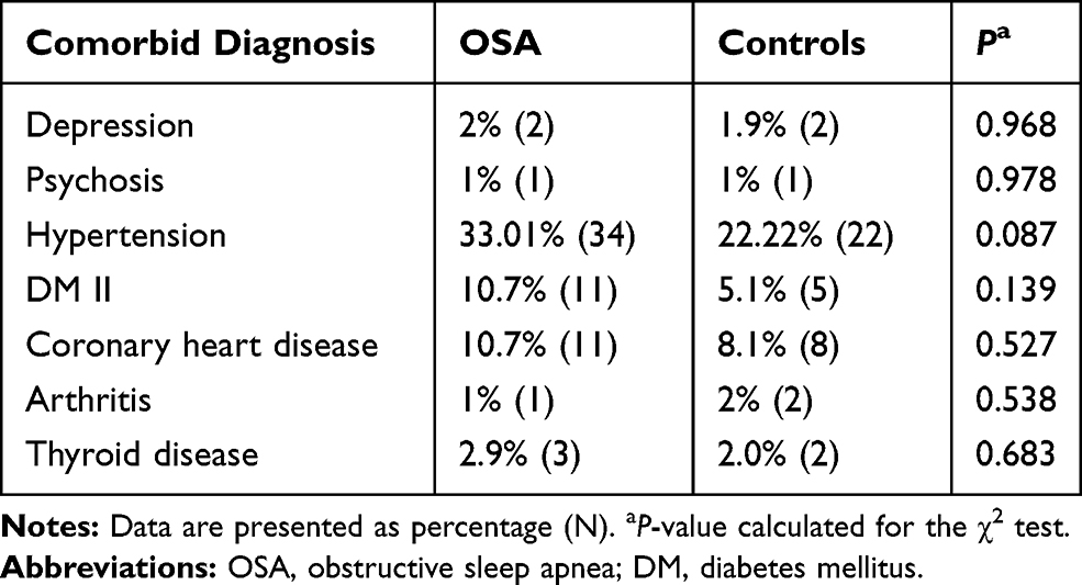 |
Table 2 Frequency of Comorbid Disease in OSA Patients and Controls |
 |
Table 3 Frequency of Prescription Medication in OSA Patients and Controls |
As presented in Tables 2 and 3, there were two OSA patients and two controls diagnosed with depression reporting the use of antidepressant medications, and one patient with psychosis in each group who reported using antipsychotics. All of them were under psychiatric treatment. The similar distribution of patients with psychiatric diagnosis and reports of using antidepressants or antipsychotics indicates that the underlying effect of a psychiatric diagnosis or psychoactive medication was not the reason for the established differences in psychomotor performance.
No differences were noted regarding the average years of education among OSA and control subjects (12.54±2.6 vs 12.85±2.9; respectively). Therefore, level of education should not be considered as a covariate in the association of OSA and psychomotor function reported in this study. Also, among the control participants, 41 subjects reported being retired, whereas 34 OSA patients were retired.
Psychomotor Testing
A battery of computer generated chronometric psychodiagnostic tests have found that CRD is a reliable and objective measure for psychological characteristics sensitive enough to detect minor changes in psychomotor performance.22,23,35 This chronometric instrument can measure memory, perception, psychomotor reaction, and thinking as dynamic functions of the central nervous system and attention and functional disturbances. In accordance with the chronometric approach, time needed to process information provides data on the psychomotor function presented as the reaction time. Tests performed in this study are applicable independently of age, language, and background education.22
In our experiments we used three representative tests from the CRD-series, always applied in the same order, increasing complexity with each following test. The assessment included the CRD 311 (speed of perception to visual stimulus), CRD 411 (speed of complex psychomotor limbs coordination), and CRD 11 (speed of solving simple arithmetic operations) tests. Discrimination of simple light signal position was assessed with the CRD 311 test. Complex psychomotor coordination was assessed with the CRD 411 test, measuring coordinated movements of participants’ upper and lower limbs. Finally, the CRD 11 test measured the speed of simple arithmetic operations of summation and subtraction.
In the CRD 311 test, there were 60 tasks included. The panel had nine small light buttons in one row where one light was appearing in a random sequence, and the respondent was instructed to press the square below the light as fast as possible when the light appears, with his index finger of the dominant hand. After the correct square was pressed, the random new light appeared on the panel, and the respondent had to continue by pressing the square below the new light. The task was finished when all 60 lights appeared.
In the CRD 11 test, the panel included 12 lights in the middle area, and one light was appearing in each task indicating which numbers (horizontal and vertical to the light) to add or subtract. The highest number on the panel was 13. The arithmetic operation was defined depending on the light with a plus (+) or a minus (–) symbol appearing in the left/right corner field of the panel. Participants were required to press the correct answer, by pressing the sum or subtraction of the numbers presented in the bottom row (from 6 to 17). After the correct answer was applied, the following task appeared on the panel. Participants had to solve 35 tasks in order to finish the test.
In the CRD 411 test, the panel included four circles in which four lights might appear during the test (two in the top row, and two in the bottom row). Two lights in the top row (one left and one right) indicated that the respondent should press the button with the corresponding hand. Two lights in the bottom row (one left and one right) indicated that the respondent should press pedals on the floor with the corresponding foot. Among the 35 tasks in the test, there was a possibility that one, two, or three lights will appear. Therefore, the respondent had to press one hand or one foot, both hands or both feet, and all possible combinations of hands and feet (when three lights are appearing) depending on the lights on the panel. If more than one light appeared, the buttons or pedals had to be pressed simultaneously in order to continue the task. The following task was initiated after a correct response to the previous one.
The participants were instructed to solve the tasks accurately and quickly. At each single testing, all the participants were assigned the same variation of each test to avoid the possible influence of the test complexity on the final results. All subjects underwent a trial of all of the tests used, in order to familiarize the participants with the tests. An additional condition for all respondents was waking up ≥1 hour before the testing.
Several parameters were recorded and used for statistical analysis for each test: minimum (best) single task solving time (MinT), median time for task solving (MedT), total test solving time (TTST), total ballast (TB), start ballast (SB), and end ballast (EB). MinT, MedT, and TTST are indicators of speed and accuracy. Ballasts are indicators of stability and represent the wasted time that was not spent on solving the tests. They are computed as a summation of MinT and all other reaction time differences through the test (TB=∑Ti-MinT), where Ti is a single task reaction time at the total test (TB), as well as the first (SB) and last half (EB) of the test.22,23
Statistical Analysis
Statistical analysis of the data was performed in MedCalc for Windows, version 11.5.1.0 (MedCalc Software, Mariakerke, Belgium). Means±standard deviations were descriptors of continuous data. The comparisons of psychomotor performance, as well as daytime sleepiness assessed with ESS, were performed using the t-test for independent samples. In cases with positive or negative asymmetric data following Shapiro Wilks test, a parametric analysis was performed due to the sample size of 206 and homogeneity of variances of the two samples, and following inspection of Q-Q plots and skewness or kurtosis of asymmetrically distributed data. The comparisons between severe and moderate OSA groups and control groups were performed with the Kruskal Wallis test due to large differences in sample size of severe and moderate OSA patients’ groups. Specific differences among groups depending on the severity of OSA were assessed with Mann Whitney U-test for independent samples, and results were presented with median (interquartile range). Correlations between psychomotor outcomes and ESS score, BMI, age, and AHI were calculated using Pearson correlation coefficients. R2 is calculated as multiple regression coefficient, where β values indicate the predictive contribution of each factor in the regression model. Statistical values with a P<0.05 were considered significant.
Results
Demographic data indicate that BMI and age were not significantly different in control and OSA groups (Table 1). Indicators of hypoxemia in OSA patients are shown in Table 1.
Reaction Times and Stability of Performance Across Time
Participants from the OSA group had increased reaction time in comparison to controls in all applied tests (Table 4). The minimal single task solving time was increased by 0.41 seconds in OSA patients when solving simple arithmetic operations tests (P<0.001), and by 0.8 seconds when testing complex psychomotor limbs coordination (P=0.007). The total test solving time was 2.5 seconds longer in OSA patients assessed with the light signal position discrimination test (P=0.013), and 10.9 seconds longer during the assessment of complex psychomotor limbs coordination (P=0.016).
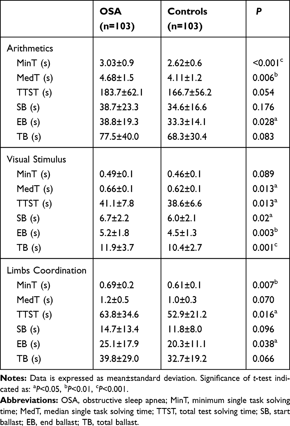 |
Table 4 Psychomotor Test Performance of Patients with Obstructive Sleep Apnea and Control Participants |
In order to assess the stability of performance across time, total ballast measure was expressed as a sum of differences between minimum single task solving time and each of all other single task solving times. This measure was calculated for the whole test, the first half (start ballast) and for the last half (end ballast) in each of the tests applied. Stability in time necessary to solve each task in a specific test was decreased toward the end of the test among OSA patients, as reported in significantly different ballast measures (Table 4). Specifically, control patients exhibited an increased stability of test solving time toward the end of the test in speed of solving simple arithmetic operations (P=0.028) compared to OSA patients. Additionally, the stability of performance was impaired in OSA patients during the first and last half of the test in the speed of perception to visual stimulus (P=0.02 and P=0.003; respectively), compared to controls. A pronounced difference was observed in the stability toward the end of the test assessing complex psychomotor limbs coordination (P=0.038), indicating pronounced deceleration toward the end of the test among OSA patients.
Apnea Severity, Excessive Daytime Sleepiness and Psychomotor Performance
As a measure of apnea severity, AHI positively correlated with the speed of perception to visual stimulus test results in the OSA group (r=0.256, P=0.011 for TTST), as shown in Figure 1 and Table 5. No significant correlation was found between results achieved on the psychomotor limbs coordination tests, speed of solving simple arithmetic operations, and AHI of the apneic patients (Table 5).
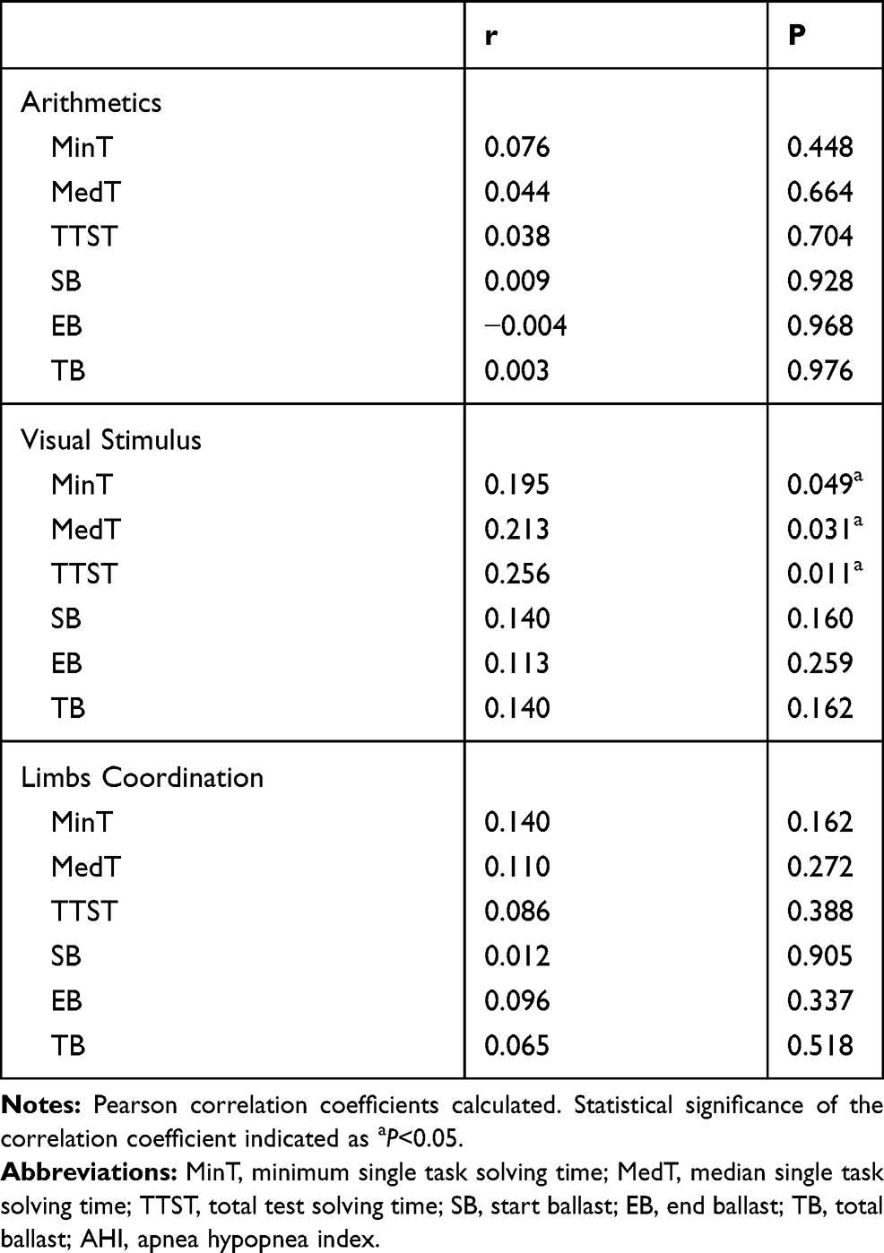 |
Table 5 Pearson Correlation Coefficients Between Psychomotor Tests Results and Severity of Apnea Expressed as AHI in Patients with Obstructive Sleep Apnea |
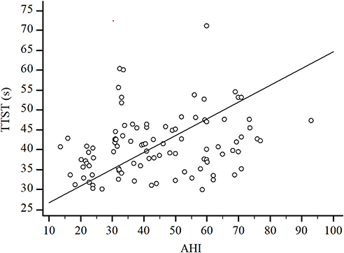 |
Figure 1 Correlation between speed of perception to visual stimulus and AHI index of patients with OSA. Notes: The scatter plot represents the association of a total test solving time needed to perform the perception to visual stimulus test and the severity of apnea expressed as AHI (r=0.256; P=0.011). Abbreviations: CRD 311, test of speed of perception to visual stimulus; AHI, apnea hypopnea index; TTST, total test solving time. |
No significant correlation was found between excessive daytime sleepiness measured by ESS and TTST for simple arithmetic operations in OSA patients (P=0.439) or controls (P=0.912). Likewise, the speed of perception to visual stimulus was not associated with ESS in OSA patients (P=0.534), or controls (P=0.305). However, a negative correlation was found between psychomotor limbs coordination and ESS score in the OSA group (r=−0.195, P=0.048 for TTST), indicating that higher daytime sleepiness of OSA patients was associated with shorter reaction time. In control patients, a positive correlation was found between the minimal single task solving time of limbs coordination psychomotor tests and ESS score (r=0.234, P=0.017), indicating the slower performance of controls with subjective sleepiness.
Regression analysis revealed no significant contribution of daytime sleepiness to the results achieved in each of the tests (Table 6). The regression model including BMI, age, ESS, and AHI yielded only age as a significant predictor of psychomotor tests used on OSA patients in this study, and the regression model including BMI, age, and ESS as predictors in control patients also indicated that only age remained a significant predictor of psychomotor performance (Table 6). As presented in Figure 2, when TTST and MinT of OSA and control participants are shown in different age groups, the averaged reaction time is increased in the OSA group for all three CRD-series tests.
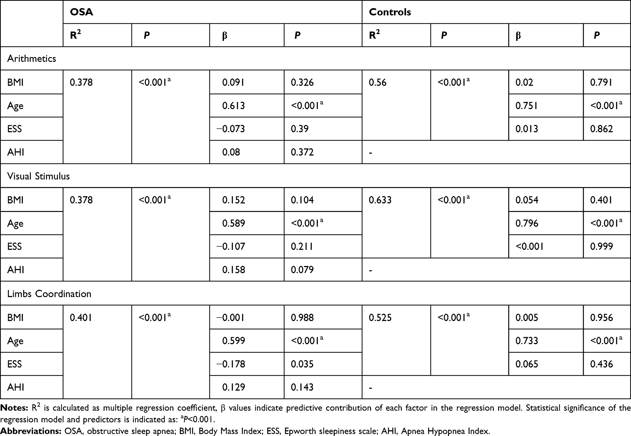 |
Table 6 Regression Analysis of Obesity, Age, Daytime Sleepiness and Severity of Apnea Indicators in the Prediction of Psychomotor Test Performance Among Patients with Obstructive Sleep Apnea |
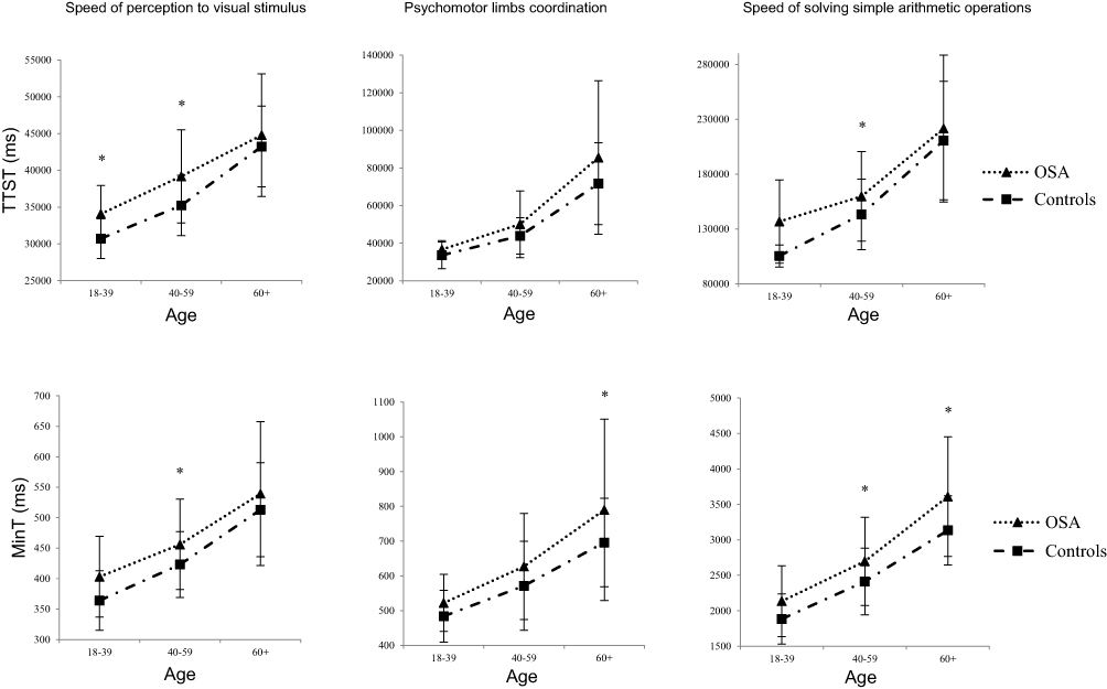 |
Figure 2 Total and minimal psychomotor reaction time of obstructive sleep apnea patients (n=103) and controls (n=103), divided at age groups.Notes: Average minimum solving time of a single task and total test solving time for all three psychomotor tests (test of speed of perception to visual stimulus, test of speed of solving simple arithmetic operations, test of psychomotor limbs coordination) of patients with OSA (n=103) and control (n=103) subjects, divided at age groups. Solid lines are showing the reference range of test results in healthy subjects, dotted lines are showing the average test results for patients with OSA and dashed lines are showing the average test results for control subjects. Asterisk (*) indicates a significant difference with P<0.05 among OSA patients and controls of a single age group in a specific chronometric measure following t-test for independent samples.Abbreviations: TTST, total test solving time; MinT, minimum solving time of a single task. |
When assessed in specific OSA severity groups, findings reveal that significant differences were found among severe OSA compared to healthy controls, whereas moderate OSA showed no significant differences in psychomotor performance compared to controls (Table 7).
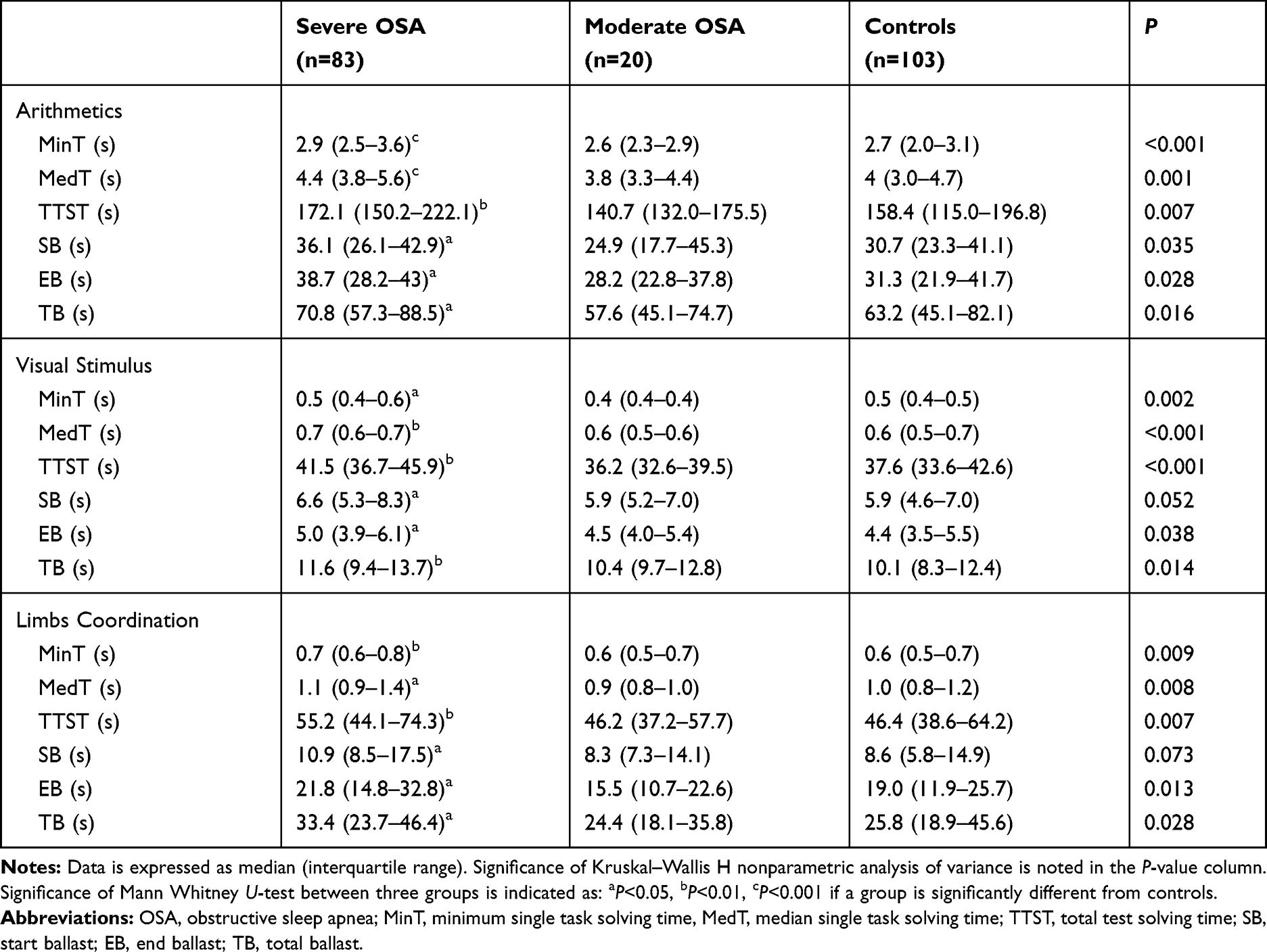 |
Table 7 Psychomotor Changes in Moderate and Severe OSA Patients Compared to Controls |
Discussion
The findings of the current study indicate prolonged reaction times in the perception of visual stimulus, solving simple arithmetic operations, and in tasks requiring psychomotor coordination of upper and lower limbs in male patients with severe OSA compared to control participants. Stability in the test solving time was decreased in severe OSA patients toward the end of the test in all applied tests. Psychomotor performance was not associated with obesity nor excessive daytime sleepiness when age was accounted for.
The results of our study are in accordance with prolonged reaction time in PVT5,14–16 and MDT20 in OSA patients compared to controls. The impairments of OSA patients in visual discrimination task measuring the speed of perception to visual stimulus correspond to the similar findings of prolonged reaction time in MDT of the Vienna Test System.20 Evidence supports the finding that impaired psychomotor performance appears in OSA patients, but the type and degree of impairment are discrepant.5 Inconsistencies in previously published findings of neuropsychological correlates of OSA,6,11,28,36,37 might be explained by the size and population studied, as well as the sensitivity of applied tests. One of the advantages of the CRD test in providing new answers in this area is the availability of data on the stability of psychomotor functioning during testing.22 The findings of decreased stability of performance toward the end of the test, assessed with ballast measures of the CRD test, indicated that OSA patients get considerably slower toward the end of tasks compared to control participants. This form of deceleration was pronounced in the speed of perception, the speed of solving simple arithmetic operations, and the speed of psychomotor limbs coordination. Considering that previous findings report OSA patients’ impairments based on averaged, summed, or minimal reaction time of OSA patients in different tests applied, the information on the stability of reaction time during the process of testing is providing novel insights on psychomotor performance in OSA. These findings are pronouncing the possible effect of tiredness on the test solving procedure. It has been reported that not all OSA patients are susceptible to psychomotor impairments.5,6 Different underlying factors in the pathophysiology of OSA might contribute to the impaired stability, such as hypoxemia, disrupted sleep architecture, psychological parameters, and reduced evoked potentials of both motor and sensory nerves.38
Psychomotor performance seems to be a subject of great significance, due to its’ possible contribution to the higher incidence of motor vehicle crashes in OSA patients.8 Our study established impaired performance among OSA patients in the test measuring speed of perception of the light signal position, which might be an important factor for driving capacity. Executive functioning and processing speed have also been indicated as important in carrying out complex behaviors associated with safe driving.39 The test of convergent thinking and limbs coordination task has been investigated in the current study in order to provide additional information regarding executive functioning and processing speed. Specifically, the test of convergent thinking included the arithmetic operation tasks that require three mental operations (ie, information storage, data organization, and executive control).40,41 We established deterioration in the minimal reaction time necessary to perform arithmetic operations for OSA patients, in comparison to controls. This is in accordance with previous meta-analytic research reporting unambiguous deficits in the executive function in OSA patients.6,28,29 However, it should be further investigated if the impaired performance on the CRD-series might be an indicator for the higher incidence of motor vehicle crashes in OSA patients.8
The question arising from the current findings is the underlying mechanism of psychomotor impairment in OSA patients. It has been reported that one of the contributing factors associated with impaired neuropsychological performance is hypoxemia.3,42 Our findings have indicated AHI, as a measure of OSA severity and consequent hypoxemia, to be associated only with impairment in the visual discrimination task. Additionally, when included in the regression model, AHI was not contributing significantly to the reported results. Indeed, it has been reported that, even in clinically healthy subjects, respiratory indices such as AHI and ODI also increase with age,43 emphasizing age as a possible mediator of neuropsychological impairments, which was the case in our results. Potentially protective mechanisms show deterioration with age, and the elderly population is therefore more vulnerable to hypoxemia.44 Increased arousal45 and decreased slow wave sleep have also been well established46 in older age, additionally marking age as a factor aggravating psychomotor consequences of sleep breathing disorders. Our results contribute to the already established findings36 of decrements of psychomotor performance with age, particularly in the psychomotor limbs coordination tests and speed of solving simple arithmetic operations. Future studies should include larger samples of patients in order to investigate this relationship of advanced age with possible deficits in psychomotor performance of OSA patients.
In addition, the older population less commonly report daytime sleepiness,47 whereas younger people with OSA tend to report sleepiness more often. We established the association of more pronounced daytime sleepiness with shorter reaction time in our OSA patients, when age is not controlled for. Indeed, it has been reported that OSA has a more prominent effect on daytime sleepiness in young or middle-aged patients than in elderly patients,47 which might explain the possible moderating effect of age on this relationship. This was additionally confirmed with our results showing the lack of association of daytime sleepiness and psychomotor performance when age was accounted for.
Age and BMI, which have been established as important confounding factors for explaining differences found in outcome measures,36 were controlled in the current study among the two groups. In addition, we provided a description of the study population regarding confounding factors associated with OSA pathophysiology and psychomotor performance. Specifically, comorbidities of OSA that might have an influence on neuropsychological assessment, such as psychiatric disorders, coronary heart disease, diabetes mellitus, hypertension, and thyroid disorders1,36 have been reported for all OSA patients and the control group. Even though reducing the sample in order to control for such comorbidities might provide an additional explanation to the aforementioned association, this might result in the selection of a specific, less prevalent phenotype of OSA patients such as patients without significant comorbidities and obesity.48 Reducing the sample of OSA patients to such a specific phenotype may have been implausible, due to the reports of a typical OSA patient being overweight or obese, and having a common comorbid diagnosis of systemic hypertension, type 2 diabetes, and dyslipidemia.49
Possible limitations must be taken into account when interpreting the results of the present study. Primarily, PSG/PG was not performed among control participants. Indeed, PSG/PG is an expensive and time-consuming procedure and it was shown that the STOP questionnaire reliably and accurately identifies subjects in the general population with increased risk of OSA.33,50 Considering the high sensitivity of the STOP questionnaire,51,52 which was also established in our previous study,27 we decided not to perform an expensive and time-consuming procedure such as PSG/PG in all control group respondents, in order to minimize medical costs and unnecessary medical procedures in patients not at risk. Accordingly, if the assessment with the use of STOP questionnaire resulted in asymptomatic undiagnosed OSA patients included in the control group, the difference between controls and cases would be even more diminished and more difficult to find. However, it should be emphasized that even after the selection process based on STOP questionnaire and matching of controls and cases using age, BMI and education background, there is a limitation regarding possible uncontrolled baseline differences among the two groups that might have influenced the results.
Since the unique female physiology might include various confounding variables such as the reproductive hormonal status dependent on the menstrual cycle, pregnancy, perimenopausal and postmenopausal statuses,53 we did not include females in our study. Furthermore, a previously published finding indicates a decreased diagnostic accuracy of screening questionnaires for OSA in females.52 A crucial aspect of proper screening for OSA in the female population is the selection of the right screening instruments in selected samples. Therefore, including female participants in our study might have decreased the sensitivity of the screening tool and the selection of the control group.
Conclusion
Our findings add to the growing number of neuropsychological correlates of OSA, with the advantage of including psychomotor tests assessing upper and lower limbs coordination, as well as calculating measures of variability that provide information on the stability in reaction time during a specific task. Precisely, we emphasized a prolonged reaction time of severe male OSA patients in comparison to control subjects toward the end of the tests in the current study. Furthermore, we established a deterioration of the total test solving time in male OSA patients in comparison to controls in the speed of perception to visual stimulus, and speed of complex psychomotor upper and lower limbs coordination. Daytime sleepiness assessed with Epworth sleepiness scale was not associated with the deterioration in psychomotor performance of OSA patients, when age was accounted for. Therefore, the current study provided evidence of specific psychomotor dysfunction and impaired stability in reaction time in male OSA patients, and adds to the knowledge of neuropsychological correlates of OSA.
Abbreviations
OSA, obstructive sleep apnea; EDS, excessive daytime sleepiness; CRD, Complex Reactionmeter Drenovac; CPAP, continuous positive airway pressure; MAD, mandibular advancement device; BMI, body mass index; PSG, polysomnography; PG, polygraphy; AHI, apnea-hypopnea index; ESS, Epworth sleepiness scale; CRD 311, speed of perception to visual stimulus; CRD 411, speed of complex psychomotor limbs coordination; CRD 11, speed of solving simple arithmetic operations; MinT, minimum (best) single task solving time; MedT, median time for task solving; TTST, total test solving time; TB, total ballast; SB, start ballast; EB, end ballast; ODI, oxygen index desaturation.
Ethics Approval
Ethical standards of the institutional research committee and the 1964 Helsinki declaration with its later amendments were applied to the protocol of the study. Ethics approval was obtained from the Ethics Committee for Biomedical Research at the University of Split School of Medicine.
Informed Consent
Following Ethics Committee for Biomedical Research and institutional approvals at the University of Split School of Medicine, all participants were informed about the procedures and the aims of the study, and signed the informed consent forms.
Acknowledgments
The authors are thankful for the technical assistance of Jelena Baricevic. The authors acknowledge Assistant Professor Shelly Pranic, PhD, for her assistance with proofreading the manuscript. An abstract of this paper was presented at the ESRS meeting in Basel 2018 as a conference talk with interim findings. The abstract is cited as: Pavlinac Dodig, I., Pecotic, R., Valic, M., Lusic, L. and Dogas, Z. Moderate and severe OSA in males impair psychomotor reaction times assessed by CRD-series testing. 24th Congress of the European Sleep Research Society, Basel, Switzerland, 25–28 September 2018. DOI: 10.1111/jsr.12751.
Author Contributions
All authors made substantial contributions to conception and design, acquisition of data, or analysis and interpretation of data; took part in drafting the article or revising it critically for important intellectual content; gave final approval of the version to be published; and agree to be accountable for all aspects of the work.
Funding
Croatian Science Foundation (HRZZ) supported this research via grant TIHO2_SLEEP_BREATH 5935, which was obtained by Professor Zoran Dogas.
Disclosure
The authors declare they have no conflict of interest. No competing interests were reported by the authors for any financial interests or commercial associations held by the authors or their family members.
References
1. Dewan NA, Nieto FJ, Somers VK. Intermittent hypoxemia and OSA: implications for comorbidities. Chest. 2015;147(1):266–274. doi:10.1378/chest.14-0500
2. Decary A, Rouleau I, Montplaisir J. Cognitive deficits associated with sleep apnea syndrome: a proposed neuropsychological test battery. Sleep. 2000;23(3):369–381. doi:10.1093/sleep/23.3.1d
3. Quan SF, Wright R, Baldwin CM, et al. Obstructive sleep apnea-hypopnea and neurocognitive functioning in the sleep heart health study. Sleep Med. 2006;7(6):498–507. doi:10.1016/j.sleep.2006.02.005
4. El-Ad B, Lavie P. Effect of sleep apnea on cognition and mood. Int Rev Psychiatry. 2005;17(4):277–282. doi:10.1080/09540260500104508
5. Quan SF, Chan CS, Dement WC, et al. The association between obstructive sleep apnea and neurocognitive performance–the Apnea Positive Pressure Long-term Efficacy Study (APPLES). Sleep. 2011;34(3):303B–314B. doi:10.1093/sleep/34.3.303
6. Bucks RS, Olaithe M, Eastwood P. Neurocognitive function in obstructive sleep apnoea: a meta-review. Respirology. 2013;18(1):61–70. doi:10.1111/j.1440-1843.2012.02255.x
7. Hirsch Allen AJ, Park JE, Daniele PR, Fleetham J, Ryan CF, Ayas NT. Obstructive sleep apnoea and frequency of occupational injury. Thorax. 2016;71(7):664–666. doi:10.1136/thoraxjnl-2015-207994
8. Tregear S, Reston J, Schoelles K, Phillips B. Obstructive sleep apnea and risk of motor vehicle crash: systematic review and meta-analysis. J Clin Sleep Med. 2009;5(6):573–581. doi:10.5664/jcsm.27662
9. Lee IS, Bardwell W, Ancoli-Israel S, Natarajan L, Loredo JS, Dimsdale JE. The Relationship between psychomotor vigilance performance and quality of life in obstructive sleep apnea. J Clin Sleep Med. 2011;15;7(3):254–260. doi:10.5664/JCSM.1064
10. Davies CR, Harrington JJ. Impact of obstructive sleep apnea on neurocognitive function and impact of continuous positive air pressure. Sleep Med Clin. 2016;11(3):287–298. doi:10.1016/j.jsmc.2016.04.006
11. Jonas DE, Amick HR, Feltner C, et al. Screening for obstructive sleep apnea in adults: evidence report and systematic review for the US preventive services task force. JAMA. 2017;317(4):415–433. doi:10.1001/jama.2016.19635
12. D’Rozario AL, Field CJ, Hoyos CM, et al. Impaired neurobehavioural performance in untreated obstructive sleep apnea patients using a novel standardised test battery. Front Surg. 2018;5:35. doi:10.3389/fsurg.2018.00035
13. Tulek B, Atalay NB, Kanat F, Suerdem M. Attentional control is partially impaired in obstructive sleep apnea syndrome. J Sleep Res. 2013;22(4):422–429. doi:10.1111/jsr.2013.22.issue-4
14. Kim H, Thomas RJ, Yun C-H, et al. Association of mild obstructive sleep apnea with cognitive performance, excessive daytime sleepiness, and quality of life in the general population: the Korean Genome and Epidemiology Study (KoGES). Sleep. 2017;40:5. doi:10.1093/sleep/zsx012
15. Batool-Anwar S, Kales SN, Varvarigou V, DeYoung P, Malhotra A, Patel S. Obstructive sleep apnea and psychomotor vigilance task performance. Nature and Science of Sleep. 2014;6:65–71. doi:10.2147/NSS
16. Li Y, Vgontzas A, Kritikou I, et al. Psychomotor vigilance test and its association with daytime sleepiness and inflammation in sleep apnea: clinical implications. J Clin Sleep Med. 2017;13(9):1049–1056. doi:10.5664/jcsm.6720
17. Alakuijala A, Maasilta P, Bachour A. The Oxford Sleep Resistance test (OSLER) and the Multiple Unprepared Reaction Time Test (MURT) detect vigilance modifications in sleep apnea patients. J Clin Sleep Med. 2014;10(10):1075–1082. doi:10.5664/jcsm.4104
18. Landry S, Anderson C, Conduit R. The effects of sleep, wake activity and time-on-task on offline motor sequence learning. Neurobiol Learn Mem. 2016;127:56–63. doi:10.1016/j.nlm.2015.11.009
19. Jackson ML, McEvoy RD, Banks S, Barnes M. Neurobehavioral impairment and CPAP treatment response in mild-moderate obstructive sleep apneas. J Clin Sleep Med. 2018;14(1):47–56. doi:10.5664/jcsm.6878
20. Devita M, Montemurro S, Zangrossi A, et al. Cognitive and motor reaction times in obstructive sleep apnea syndrome: a study based on computerized measures. Brain Cogn. 2017;117:26–32. doi:10.1016/j.bandc.2017.07.002
21. Kim H, Dinges DF, Young T. Sleep-disordered breathing and psychomotor vigilance in a community-based sample. Sleep. 2007;30(10):1309–1316. doi:10.1093/sleep/30.10.1309
22. Drenovac M. An analysis of some attributes of the dynamics of mental processing. Rev Psychol. 2001;8:61–67.
23. Drenovac M. CRD Series of Psychodiagnostic Tests [In Croatian]. Zagreb, Croatia: AKD; 1994.
24. Radic J, Ljutic D, Radic M, Kovacic V, Dodig-Curkovic K, Sain M. Kidney transplantation improves cognitive and psychomotor functions in adult hemodialysis patients. Am J Nephrol. 2011;34(5):399–406. doi:10.1159/000330849
25. Radic J, Ljutic D, Radic M, Kovacic V, Sain M, Dodig-Curkovic K. Is there differences in cognitive and motor functioning between hemodialysis and peritoneal dialysis patients? Ren Fail. 2011;33(6):641–649. doi:10.3109/0886022X.2011.586480
26. Galic T, Bozic J, Pecotic R, Ivkovic N, Valic M, Dogas Z. Improvement of cognitive and psychomotor performance in patients with mild to moderate obstructive sleep apnea treated with mandibular advancement device: a prospective 1-year study. J Clin Sleep Med. 2016;12(2):177–186. doi:10.5664/jcsm.5480
27. Pecotic R, Dodig IP, Valic M, et al. Effects of CPAP therapy on cognitive and psychomotor performances in patients with severe obstructive sleep apnea: a prospective 1-year study. Sleep Breath. 2019;23(1):41–48. doi:10.1007/s11325-018-1642-6
28. Stranks EK, Crowe SF. The cognitive effects of obstructive sleep apnea: an updated meta-analysis. Arch Clin Neuropsychol. 2016;31(2):186–193. doi:10.1093/arclin/acv087
29. Olaithe M, Bucks RS. Executive dysfunction in OSA before and after treatment: a meta-analysis. Sleep. 2013;36(9):1297–1305. doi:10.5665/sleep.2950
30. American Academy of Sleep Medicine. International Classification of Sleep Disorders.
31. Fischer J, Dogas Z, Bassetti CL, et al. Executive Committee (EC) of the Assembly of the National Sleep Societies (ANSS); Board of the European Sleep Research Society (ESRS), Regensburg, Germany. Standard procedures for adults in accredited sleep medicine centres in Europe. J Sleep Res. 2012;21(4):357–368. doi:10.1111/jsr.2012.21.issue-4
32. Rudan I, Marusic A, Jankovic S, et al. “10001 Dalmatians:” Croatia launches its national biobank. Croat Med J. 2009;50(1):4–6. doi:10.3325/cmj.2009.50.4
33. Pecotic R, Dodig IP, Valic M, Ivkovic N, Dogas Z. The evaluation of the croatian version of the Epworth sleepiness scale and STOP questionnaire as screening tools for obstructive sleep apnea syndrome. Sleep Breath. 2012;16(3):793–802. doi:10.1007/s11325-011-0578-x
34. Nevado-Holgado AJ, Kim CH, Winchester L, Gallacher J, Lovestone S. Commonly prescribed drugs associate with cognitive function: a cross-sectional study in UK biobank. BMJ Open. 2016;30(11):6.
35. Bobic J, Pavicevic L, Gomzi M. The difference in complex psychomotor reaction time between patients with and without signs of cerebral circulatory disorders. Coll Antropol. 2002;26(2):515–520.
36. Lal C, Strange C, Bachman D. Neurocognitive impairment in obstructive sleep apnea. Chest. 2012;141(6):1601–1610. doi:10.1378/chest.11-2214
37. Kilpinen R, Saunamaki T, Jehkonen M. Information processing speed in obstructive sleep apnea syndrome: a review. Acta Neurol Scand. 2014;129(4):209–218. doi:10.1111/ane.2014.129.issue-4
38. Mihalj M, Lusic L, Dogas Z. Reduced evoked motor and sensory potential amplitudes in obstructive sleep apnea patients. J Sleep Res. 2016;25(3):287–295. doi:10.1111/jsr.2016.25.issue-3
39. Wolfe PL, Lehockey KA. Neuropsychological Assessment of driving capacity. Arch Clin Neuropsychol. 2016;31(6):517–529. doi:10.1093/arclin/acw050
40. Cragg L, Gilmore C. Skills underlying mathematics: the role of executive function in the development of mathematics proficiency. Trends Neurosci Educ. 2014;3(2):63–68. doi:10.1016/j.tine.2013.12.001
41. Abd Hamid AI, Yusoff AN, Mukari SZ, Mohamad M. Brain activation during addition and subtraction tasks in-noise and in-quiet. Malays J Med Sci. 2011;18(2):3–15.
42. Adams N, Strauss M, Schluchter M, Redline S. Relation of measures of sleep-disordered breathing to neuropsychological functioning. Am J Respir Crit Care Med. 2001;163(7):1626–1631. doi:10.1164/ajrccm.163.7.2004014
43. Mitterling T, Hogl B, Schonwald SV, et al. Sleep and respiration in 100 healthy caucasian sleepers–a polysomnographic study according to american academy of sleep medicine standards. Sleep. 2015;38(6):867–875. doi:10.5665/sleep.4730
44. Sharma G, Goodwin J. Effect of aging on respiratory system physiology and immunology. Clin Interv Aging. 2006;1(3):253–260. doi:10.2147/ciia.2006.1.issue-3
45. Boselli M, Parrino L, Smerieri A, Terzano MG. Effect of age on EEG arousals in normal sleep. Sleep. 1998;21(4):351–357.
46. Edwards BA, O’Driscoll DM, Ali A, Jordan AS, Trinder J, Malhotra A. Aging and sleep: physiology and pathophysiology. Semin Respir Crit Care. 2010;31(5):618–633. doi:10.1055/s-0030-1265902
47. Iannella G, Vicini C, Colizza A, et al. Aging effect on sleepiness and apneas severity in patients with obstructive sleep apnea syndrome: a meta-analysis study. Eur Arch Otorhinolaryngol. 2019;276(12):3549–3556. doi:10.1007/s00405-019-05616-0
48. Vavougios GD, George DG, Pastaka C, Zarogiannis SG, Gourgoulianis KI. Phenotypes of comorbidity in OSAS patients: combining categorical principal component analysis with cluster analysis. J Sleep Res. 2016;25(1):31–38. doi:10.1111/jsr.12344
49. Lévy P, Kohler M, McNicholas WT, et al. Obstructive sleep apnoea syndrome. Nat Rev Dis Primers. 2015;1:15015. doi:10.1038/nrdp.2015.15
50. Silva GE, Vana KD, Goodwin JL, Sherrill DL, Quan SF. Identification of patients with sleep disordered breathing: comparing the four-variable screening tool, STOP, STOP-BANG, and Epworth sleepiness scales. J Clin Sleep Med. 2011;7(5):467–472. doi:10.5664/JCSM.1308
51. Amra B, Rahmati B, Soltaninejad F, Feizi A. Screening questionnaires for obstructive sleep apnea: an updated systematic review. Oman Med J. 2018;33(3):184–192. doi:10.5001/omj.2018.36
52. Chiu HY, Chen PY, Chuang LP, et al. Diagnostic accuracy of the Berlin questionnaire, STOP-BANG, STOP, and Epworth sleepiness scale in detecting obstructive sleep apnea: a bivariate meta-analysis. Sleep Med Rev. 2017;36:57–70. doi:10.1016/j.smrv.2016.10.004
53. Baker FC, Lampio L, Saaresranta T, Polo-Kantola P. Sleep and sleep disorders in the menopausal transition. Sleep Med Clin. 2018;13(3):443–456. doi:10.1016/j.jsmc.2018.04.011
 © 2020 The Author(s). This work is published and licensed by Dove Medical Press Limited. The
full terms of this license are available at https://www.dovepress.com/terms
and incorporate the Creative Commons Attribution
- Non Commercial (unported, 3.0) License.
By accessing the work you hereby accept the Terms. Non-commercial uses of the work are permitted
without any further permission from Dove Medical Press Limited, provided the work is properly
attributed. For permission for commercial use of this work, please see paragraphs 4.2 and 5 of our Terms.
© 2020 The Author(s). This work is published and licensed by Dove Medical Press Limited. The
full terms of this license are available at https://www.dovepress.com/terms
and incorporate the Creative Commons Attribution
- Non Commercial (unported, 3.0) License.
By accessing the work you hereby accept the Terms. Non-commercial uses of the work are permitted
without any further permission from Dove Medical Press Limited, provided the work is properly
attributed. For permission for commercial use of this work, please see paragraphs 4.2 and 5 of our Terms.