
Back to Journals » International Journal of Nanomedicine » Volume 21
Protein Lactylation in Central Nervous System Diseases: Molecular Mechanisms and Targeted Therapeutic Strategies
Authors Cheng Z, Ou G, Zhang G, Ahmed W, Han Y, Zhang D, Chai T, Hu M, Zhang Q  , Lei J, Lei L , Zhu Z
Received 29 April 2026
Accepted for publication 9 July 2026
Published 20 July 2026 Volume 2026:21 621038
DOI https://doi.org/10.2147/IJN.S621038
Checked for plagiarism Yes
Review by Single anonymous peer review
Peer reviewer comments 2
Editor who approved publication: Professor Eng San Thian
Ziyan Cheng,1,* Guangxin Ou,2,* Guilong Zhang,1,* Waqas Ahmed,3,* Yongquan Han,1 Dongshan Zhang,1 Tianran Chai,1 Meng Hu,1 Qiankun Zhang,4 Jiaxin Lei,5 Lanjie Lei,6 Zhihan Zhu1
1Department of Neurosurgery, The Second Affiliated Hospital of Guangzhou Medical University, Guangzhou, Guangdong, People’s Republic of China; 2Department of Spinal Surgery, People’s Hospital of Yangjiang, Yangjiang, Guangdong, People’s Republic of China; 3School of Medicine, Southeast University, Nanjing, Jiangsu, People’s Republic of China; 4Guangzhou First People’s Hospital, School of Medicine, South China University of Technology, Guangzhou, Guangdong, People’s Republic of China; 5Hanzhong Psychiatric Hospital, Hanzhong, Shanxi, People’s Republic of China; 6Key Laboratory of Artificial Organs and Computational Medicine in Zhejiang Province, Shulan International Medical College, Institute of Translational Medicine, Zhejiang Shuren University, Hangzhou, Zhejiang, People’s Republic of China
*These authors contributed equally to this work
Correspondence: Zhihan Zhu, Department of Neurosurgery, The Second Affiliated Hospital of Guangzhou Medical University, Guangzhou, Guangdong, People’s Republic of China, Email [email protected] Lanjie Lei, Key Laboratory of Artificial Organs and Computational Medicine in Zhejiang Province, Shulan International Medical College, Institute of Translational Medicine, Zhejiang Shuren University, Hangzhou, Zhejiang, People’s Republic of China, Email [email protected]
Abstract: Lactate, once considered merely a metabolic byproduct, is now recognized as a cornerstone of central nervous system (CNS) homeostasis, serving as both a vital energy substrate and signaling molecule. The identification of lysine lactylation (Kla) has established this modification as a key epigenetic link between cellular metabolism and genomic regulation. This review examines the molecular mechanisms underlying protein lactylation, including enzymatic regulation by writers, erasers, and readers as well as non-enzymatic mechanisms. The multifaceted roles of Kla are explored in the context of CNS disorders, ranging from malignancies, acute injuries, and neurodegenerative diseases. The review further examines Kla’s role in neuroinflammation, metabolic reprogramming, and neuroplasticity, highlighting its potential as a sensitive biomarker. Potential therapeutic strategies are also considered, including metabolic inhibitors and nanocarriers capable of crossing the blood-brain barrier (BBB) to restore metabolic and epigenetic balance.
Keywords: lactate, lactylation, central nervous system diseases, epigenetic regulation, therapeutic strategies
Introduction
Metabolites, as fundamental products of cellular life, serve functions that extend beyond energy provision and biosynthesis. With the advancement and widespread acceptance of the metabolism-epigenetics axis concept, the direct involvement of metabolites in transcriptional regulation has become increasingly evident. The study of lactate exemplifies a significant paradigm shift in the understanding of metabolites. For nearly two centuries following its discovery, lactate was regarded solely as a byproduct of anaerobic glycolysis. The emergence of the lactate shuttle hypothesis led to the recognition that lactate serves as an intercellular carrier of both energy and information.1,2 In the central nervous system (CNS), where metabolic activity and energy demand are particularly high, astrocyte-neuron lactate coupling plays a critical role in supporting high-order brain functions.3 Recent discoveries have demonstrated that lactate imposes a distinct metabolic imprint on proteins and can directly activate previously dormant genes by altering chromosomal conformation.
In 2019, Zhao et al identified lactylation, a novel lactate-mediated post-translational modification (PTM). This modification of histone lysine residues acts as a crucial regulator of transcriptional activity and directly influences macrophage phenotypic polarization.4 Subsequent studies have employed multi-omics integration technologies to investigate lactylation from new perspectives. These efforts have successfully identified numerous lactylation modification sites and elucidated their roles in the development of various diseases.5,6 Moreover, lactylation is not restricted to histones; it also regulates a range of non-histone proteins, thereby participating in the regulation of cellular processes at a deeper level.7,8 Physiologically, lactylation serves as a key regulator of activities, such as embryonic development and neural plasticity. However, disruption of metabolic homeostasis can trigger severe pathological responses. For example, in the tumor microenvironment (TME), lactate accumulation contributes to the formation of an immunosuppressive barrier. The pathogenic role of lactylation in the CNS is increasingly recognized, including its promotion of microglial inflammatory storms in Alzheimer’s disease (AD), facilitation of glioma cell immune evasion, and contribution to psychological stress-related vascular damage.9–11 Protein lactylation has thus emerged as a key regulator linking metabolic states to pathological processes.
While several recent reviews have summarized the fundamental mechanisms of lactylation in general health and its emerging roles in CNS disorders,1,5–7 these studies have predominantly focused on molecular pathways and cellular functions. In this context, this review systematically examines the multifunctional roles of lactate in the CNS and provides a comprehensive explanation of the molecular mechanisms and pathological implications of protein lactylation in various neurological disorders (Figure 1).12,13 However, a critical gap remains in the systematic translation of these metabolic-epigenetic mechanisms into clinical interventions. Unlike previous reviews, this study bridges this gap by scrutinizing nascent targeted interventions, specifically metabolic reprogramming and exosome-mediated nanodelivery systems engineered to surmount the blood-brain barrier (BBB).14,15 By bridging the gap between metabolic-epigenetic mechanisms and therapeutic applications, this review provides a theoretical foundation and practical insights for future diagnostic and therapeutic innovations.
 |
Figure 1 Beyond its status as a byproduct, lactate operates as a cornerstone bioenergetic fuel and signaling mediator in CNS homeostasis. Lysine lactylation (Kla) establishes a critical metabolic-epigenetic bridge, orchestrating neuroinflammation and plasticity. The black arrows indicate the metabolic pathway of lactate production via glycolysis and its subsequent utilization for lactylation modifications. This study delineates Kla’s multifaceted involvement in CNS pathologies and its biomarker potential, advocating for tailored therapeutic frameworks utilizing metabolic inhibitors and nanodrug delivery systems to recalibrate the metabolic-epigenetic landscape. |
Overview of Lactate and Lactylation
Three isomers of lactic acid are present in the human body: L-lactic acid, D-lactic acid, and DL-lactic acid.16 Under hypoxic conditions or Adenosine triphosphate (ATP) deficiency, glycolytic flux converts carbohydrates and amino acids (AAs) into substantial stores of L-lactate. In addition to serving as a metabolic byproduct, L-lactate serves as a primary neuronal oxidative substrate and plays a role in protein synthesis, synaptic remodeling, and axonal excitability during memory formation.17 In contrast, D-lactate is present at micromolar concentrations, approximately 1% of L-lactate, and is derived from endogenous metabolism and the gastrointestinal (GI) microbiota.18 Lactate also functions in signaling; by activating the G protein-coupled receptor HCA1 (GPR81),19–21 lactate suppresses cyclic AMP (cAMP), thereby regulating neuroprotection, angiogenesis, and immune responses.22–24
Hypoxia inhibits the tricarboxylic acid cycle (TCA), redirecting pyruvate toward lactate synthesis via lactate dehydrogenase (LDH) and bypassing oxidative phosphorylation (OXPHOS).25 Simultaneously, monocarboxylate transporters (MCTs) and sodium-coupled monocarboxylate transporters facilitate lactate uptake, thereby enhancing endogenous flux and contributing to elevated intracellular lactate concentrations.26 However, lactate metabolism in the CNS exhibits several distinctive features. Although the CNS constitutes only 2% of total body weight, it consumes approximately 25% of the body’s glucose-derived energy. The astrocyte-neuron lactate shuttle (ANLS) hypothesis proposes that neuronal activity stimulates astrocytic glucose uptake and its glycolytic conversion to lactate via LDH.27 MCT family primarily mediates transmembrane lactate transport. MCT1 is present in astrocytes, microvascular endothelial cells (MECs), ependymal cells, and oligodendrocytes,28 whereas MCT4 is predominantly localized to astrocytes and mediates lactate efflux. In contrast, MCT2 is expressed in neurons and is essential for efficient lactate uptake.29 This lactate transport mechanism ensures that glutamatergic neurotransmission is closely linked to cellular bioenergetics.30
Excessive lactate accumulation can result from lactate metabolism, primarily due to the induction of a novel acylation process called lactylation. In cellular environments, lactate metabolism occurs via two principal pathways. The first involves lactate oxidation to produce pyruvate, which subsequently serves as a substrate for the mitochondrial TCA to produce ATP. The second pathway involves the conversion of lactate to lactyl-CoA under specific conditions, which then serves as a substrate for the lactylation of histone and non-histone proteins.31 The 2019 identification of Kla by Zhao et al established lactate as a key metabolic substrate for histone epigenetic regulation.4 Post-translational modifications (PTMs) are covalent modifications that occur in proteins after translation. These modifications regulate protein activity, subcellular localization, folding, and interactions with other biomacromolecules, including proteins, nucleic acids, and lipids.32 PTMs are mediated through two pathways: an enzymatic reaction utilizing L-lactyl-CoA as a substrate and a non-enzymatic reaction involving D-lactyl-GSH derived from methylglyoxal (MG) metabolism pathways.33,34 During periods of heightened glycolytic activity, lactate can be enzymatically converted into high-energy lactyl-CoA, which covalently attaches the lactyl group to the lysine residues of proteins.35 In the non-enzymatic pathway, the glycolytic byproduct MG reacts with glutathione to form Lactoylglutathione (LGSH). LGSH facilitates lactoyl modification by transferring its lactate moiety to lysine residues on proteins.36 This modification neutralizes the inherent positive charge of lysine residues, altering the protein conformation and interactions. This process is tightly regulated according to the cellular metabolic state. Under conditions such as hypoxia, inflammation, or cancer, intracellular lactate levels rise significantly, leading to increased protein lactoylation (Figure 2).
 |
Figure 2 The astrocyte-neuron lactate shuttle (ANLS) regulates energy metabolism and protein lactylation in the CNS. In this mechanism, lactate produced by astrocytic glycolysis is exported through monocarboxylate transporters 1/4 (MCT1/4) transporters and subsequently taken up by neuronal MCT2. Imported lactate is oxidized to pyruvate, which then enters the tricarboxylic acid cycle (TCA). Furthermore, lactate acts as a substrate for both histone and non-histone protein lactylation, a type of post-translational modification (PTM), via either an enzymatic pathway involving lactyl-CoA-mediated lysine modification or a non-enzymatic pathway utilizing methylglyoxal-derived D-lactyl-GSH. The solid arrows represent the lactate metabolism processes within central nervous system cells (neurons, astrocytes, and microglia), as well as the export of lactate produced by astrocytic glycolysis via MCT1/4 and its subsequent uptake by neuronal MCT2. The dotted arrows indicate the release of lactate produced by microglial glycolysis through MCT1 and its subsequent uptake by neuronal MCT2. |
Techniques Related to Lactylation
Recent advances in epigenetic research have outpaced conventional sequencing methods in resolving the heterogeneous of the CNS microenvironment. As a result, current epigenomic studies increasingly focus on single-cell and spatial resolution techniques to accurately map cell-specific lactylation landscapes.37
In contrast to conventional chromatin immunoprecipitation sequencing (ChIP-seq), which is often limited by high input requirements and background noise, cleavage under targets and tagmentation (CUT&Tag) uses Tn5 transposase-mediated in situ cleavage to achieve higher signal-to-noise ratios with minimal input material. As a result, single-cell cleavage under targets and tagmentation (scCUTandTag) and spatial epigenomics facilitate the precise resolution of epigenetic heterogeneity in ischemic penumbrae, enabling the identification of microglia and progenitor cell types while preserving the original tissue architecture.38
Liquid chromatography-tandem mass spectrometry is the primary method for identifying histone and non-histone lactylation sites; however, isobaric interference remains a significant limitation.39 The small mass differences between lactylation and other post-translational modifications often lead to false positives during fragmentation, highlighting the need for more precise mass spectrometry techniques and more specific pan-lactylation antibodies.
Furthermore, the abundance of repetitive deoxyribonucleic acid (DNA) elements involved in epigenetic regulation within the CNS poses a significant challenge for conventional short-read sequencing approaches, which often struggle to map these regions accurately Long-read single-molecule sequencing is therefore crucial for uncovering lactylation patterns in non-coding and repetitive sequences.40
Core Molecular Mechanisms of Lactylation
The process of protein lactylation is precisely regulated by a set of key enzymes that operate according to established enzymatic principles. These enzymes include lactyltransferases, which catalyze the insertion of lactyl-CoA into proteins, and delactylases, which remove lactyl-CoA from proteins. Additionally, enzymes capable of converting lactate into lactyl-CoA have been proposed; however, they have not been accurately identified in mammals.41 In protein lactylation, these regulatory enzymes are often metaphorically described as writers, erasers, and readers in epigenetics (Figure 3).
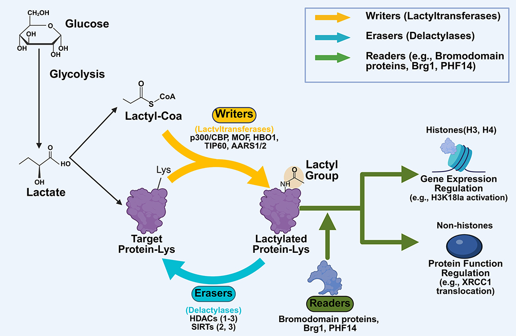 |
Figure 3 Core molecular mechanisms of protein lactylation. |
Writers
Lactyltransferases function as “writers” by catalyzing the addition of lactyl groups to lysine residues. Within the lysine acetyltransferase (KAT) family, p300 was the first enzyme identified to mediate Kla.4,42,43 The p300/CBP complex plays a central role in modifying histones, transcription factors (TFs), and other nuclear substrates. In response to lactyl-CoA availability, p300 catalyzes histone lactylation at residues such as H3K18 and H4K12, promoting chromatin relaxation and transcriptional activation.44 Other KATs, including GCN5, MOF, Tip60, HBO1, and NAA10, also contribute to histone Kla.45–47
Preliminary evidence suggests that alanyl-tRNA synthetases 1 and 2 (AARS1/2) may function as lactyltransferases through an unconventional enzymatic pathway. Unlike CoA-dependent KATs, it is proposed that these enzymes synthesize L-lactyl-AMP directly from free lactate and ATP to mediate Kla; however, this specific mechanism requires further validation.48–50 Notably, certain enzymes exhibit context-dependent catalytic functions. For example, histone deacetylase 6 (HDAC6), typically recognized as a delactylase, can act as a lactyltransferase for α-tubulin, promoting microtubule dynamics and supporting hippocampal neuron growth.51 Despite the identification of multiple lactyltransferases, the precise molecular mechanisms underlying their enzymatic activities remain incompletely understood (Table 1).
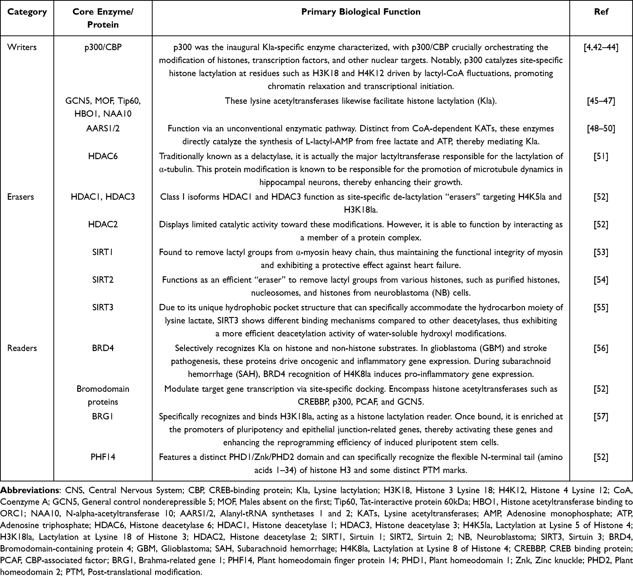 |
Table 1 Key Epigenetic Regulators of Lactylation in the CNS |
Erasers
The eraser enzymes, specifically delactylases, remove lactyl groups from lysine residues. These enzymes are primarily composed of histone deacetylases (HDACs), which are classified into four classes: I–IV. HDACs are further categorized into zinc-dependent enzymes (classes I, II, and IV including HDAC1-11) and NAD-dependent enzymes (class III, comprising sirtuin proteins [SIRT] 1–7).58 Most class I enzymes, such as HDAC1-3 and HDAC8, are believed to function as delactylases. Among these, HDAC1 and HDAC3 act as site-specific de-lactylation “erasers” targeting H4K5la and H3K18la, whereas HDAC2 exhibits limited catalytic activity toward these modifications.52 However, HDAC2 can function through interactions within protein complexes. In contrast, other HDAC family enzymes demonstrate variable catalytic activities and function as both delactylases and deacetylases.6 Recent studies have identified SIRT1, SIRT2, and SIRT3, which are NAD-dependent enzymes, as important erasers. SIRT1 removes lactyl groups from α-myosin heavy chain, thereby maintaining myosin functional integrity and providing a protective effect against heart failure (HF).53 SIRT2 efficiently removes lactyl groups from purified histones, nucleosomes, and histones from neuroblastoma (NB) cells.54 SIRT3, owing to its unique hydrophobic pocket structure that accommodates the hydrocarbon moiety of lysine lactate, exhibits distinct binding mechanisms compared to other deacetylases and demonstrates more efficient deacetylation activity for water-soluble hydroxyl modifications (Table 1).55
Readers
A reader protein can specifically recognize and bind lactylation sites, thereby enabling the recruitment of effector proteins to transmit signals. Although research in this area is still in its early stages, several key proteins have been identified as readers. Bromodomain-containing protein 4 (BRD4), a 110-residue protein module, selectively recognizes Kla on both histone and non-histone substrates. Bromodomain (BRD)-containing proteins modulate target gene transcription via site-specific docking. In the pathogenesis of glioblastoma (GBM) and stroke, these proteins promote the expression of oncogenic and inflammatory genes. BRD proteins include histone acetyltransferases such as CREBBP, p300, PCAF, and GCN5.57
The chromatin-remodeling protein BRG1 specifically recognizes and binds H3K18la, thereby acting as a histone lactylation reader. Upon binding to H3K18la, BRG1 accumulates at the promoters of pluripotency and epithelial junction-related genes, thereby activating these genes and inducing phenotypic changes in cells. This process enhances the reprogramming efficiency of induced pluripotent stem cells.59 Another protein proposed to act as a lactylation reader is plant homeodomain finger protein 14 (PHF14), which contains a distinct PHD1/Znk/PHD2 domain. PHF14 is suggested to specifically recognize the flexible N-terminal tail (amino acids 1–34) of histone H3 and certain PTM marks.52 Currently, BRG1 and PHF14 are among the most prominent proteins identified as lactylation readers. Although the molecular mechanisms underlying their roles in transmitting protein lactylation signals remain unclear, ongoing research is advancing targeted therapeutic strategies (Table 1).
Lactylation of the CNS
The brain exhibits a high energy demand due to its complex activities within the CNS. Although the brain accounts for only 2% of the total body weight, it consumes approximately 20% of the body’s oxygen and glucose at rest.60 This substantial energy consumption primarily supports the maintenance of membrane potential gradients, synaptic processes, and neurotransmitter cycling. The CNS requires a continuous supply of ATP, with glucose accounting for 95% of cerebral metabolic activity, mainly via OXPHOS and glycolysis.61 Under normal physiological conditions, OXPHOS sufficiently meets the brain’s energy requirement.61 However, during acute stimulation, even with adequate oxygen availability, the brain may metabolize glucose to lactate to rapidly generate energy. This process, known as aerobic glycolysis or the Warburg effect, was initially described in cancer cells.62 During periods of elevated neuronal energy demand, such as increased synaptic activity, astrocytes supply neurons with energy as lactate via the ANLS.36
Lactate, the end product of glycolytic metabolism, serves as an energy substrate for neuronal activity in the brain and functions as an intercellular signaling molecule. It acts as a link between metabolic reprogramming and epigenetic regulation. Currently, lactate-induced lactylation is primarily observed in the cerebral cortex and hippocampus, whereas Kla is mainly detected in glutamatergic neurons, GABAergic neurons, astrocytes, and other cell types.
Neuronal Lactylation
Neurons can stimulate glycolysis in astrocytes by releasing glutamate, which increases lactate production from glucose. Lactate produced by astrocytes then serves as an energy source for neurons. Elevated lactate levels also promote lactylation.63 Neuronal excitation, such as potassium stimulation and electroshock, has been shown to induce lactylation in a time-dependent manner. Stress-induced neuronal excitation specifically stimulates histone H1 lactylation. The Kla signal is detectable throughout the neuronal cytoplasm. Experimental evidence suggests that increased histone H1 lactylation is linked to reduced social behavior.64 In addition, elevated lactate levels resulting from exercise or stress can induce lactylation of synaptic proteins in the cortex, including synaptosomal-associated protein 91 (SNAP91). Lactylation of these proteins enhances neural network activity in cortical regions and plays a crucial role in suppressing anxiety in mice exposed to chronic stress.65
Abnormal lactylation in neurons impairs lactate utilization, disrupting neuronal energy metabolism and contributing to dysfunction. Evidence suggests a significant association between lactylation and neurodegenerative diseases.66 In AD, aberrant neuronal lactylation has been observed. Specifically, lactylation of Tau protein at K331 promotes phosphorylation and cleavage while reducing ubiquitination-mediated degradation. Notably, Tau lactylation at K677 triggers Nuclear receptor coactivator 4 (NCOA4)-mediated ferritinophagy and ferroptosis, exacerbating AD pathology.67,68 In IS, K450 lactylation stabilizes NCOA4 in hippocampal neurons, thereby driving pathological ferritinophagy and glycolysis and increasing infarct volume.45 Similarly, downregulation of HDAC6 during ischemia-reperfusion (I/R) induces aberrant lactylation, disrupts calcium homeostasis, and exacerbates neuronal injury.69 Although MCT2 mediates neuronal lactate uptake, its inhibition reduces pathological lactylation but also diminishes lactate-induced synaptic protein expression (eg, PSD95, GAP43), ultimately impairing memory consolidation.70 Collectively, these findings position lactylation as a pivotal regulator of neuronal survival and plasticity, rather than a mere metabolic byproduct, across various neurodegenerative conditions (Table 2).
 |
Table 2 Mechanisms and Outcomes of Lactylation in Specific CNS Cell Types |
Lactylation in Astrocytes
Astrocytes originate from neural precursor cells, which also give rise to neurons. These cells perform essential functions, including neuronal support and protection, energy provision, clearance of environmental toxins, modulation of signaling, and tissue repair. As the primary glycogen reservoirs in the CNS, astrocytes regulate lactate synthesis and secretion via ANLS to maintain neuronal energy homeostasis. Consequently, even under resting conditions, astrocytes exhibit significantly higher lactate concentrations than neurons.77
Lactylation in astrocytes plays a crucial role in regulating neuronal activity and neuroinflammation.75 Under pathological conditions, such as spinal cord injury, an astrocytic UCHL1/PFKFB3/H4K8la positive feedback loop impairs neuronal energy supply and drives neuronal ferroptosis.71 Similarly, in subarachnoid hemorrhage (SAH), BRD4 recognition of H4K8la directly activates inflammatory gene transcription.56 Conversely, astrocytic lactylation can also be neuroprotective; for instance, in cerebral I/R models, ADP-ribosylation factor 1 (ARF1) lactylation facilitates the transfer of healthy mitochondria from astrocytes to damaged neurons.72 Furthermore, fluoxetine-induced lactate release from cortical astrocytes implicates lactylation in mood regulation.78 Thus, the impact of astrocytic lactylation is defined by its specific downstream molecular targets rather than a generalized inflammatory response (Table 2).
Lactylation in Microglia
Microglia, the smallest glial cells in the CNS, function as resident phagocytes that maintain immune homeostasis and mediate local responses.79 Under pathological conditions, microglia undergo metabolic reprogramming and shift their primary energy metabolism from OXPHOS to aerobic glycolysis. This shift results in substantial lactate production in microglial cells. Lactate plays a crucial role in the production of various inflammatory cytokines, including TNF-α, IL-6, and IL-1β.66 Crucially, microglial lactylation functions as a potent epigenetic regulator, that precisely orchestrates gene transcription to dictate cellular states. In Alzheimer’s disease, glycolytic lactate induces H4K12la-mediated upregulation of pyruvate kinase M2 (PKM2), which chronically sustains microglial hyperactivation.9 Conversely, in ischemic injury, LDHA-driven histone lactylation activates HMGB1-mediated pyroptosis, whereas PLBD1 lactylation at K155 exacerbates inflammatory damage.73 Despite these pro-inflammatory effects, lactate also exhibits a protective role. Exogenous or exercise-induced lactate accumulation can shift microglia from a pro-inflammatory to an anti-inflammatory phenotype via histone lactylation, thus relieving cognitive dysfunction.75 In sevoflurane-induced models, histone lactylation-mediated YTHDF3 upregulation directly suppresses pyroptosis.45 These contrasting outcomes highlight that microglial lactylation operates through distinct mechanistic axes, depending on the metabolic microenvironment (Table 2).
Lactylation in CNS Diseases
Lactylation in CNS Tumors
The Warburg effect refers to the neoplastic shift toward aerobic glycolysis under normoxic conditions, which enforces glycolytic dependency and creates a lactate-rich microenvironment. This elevated-lactate microenvironment supports rapid biosynthesis in tumor cells, providing the necessary substrates for tumor growth and proliferation.80 In normal tissues, lactylation primarily maintains metabolic homeostasis; however, histone lactylation significantly contributes to oncogene expression and tumor progression. Lactylation also regulates processes within the TME, including immune state transitions and tumor malignancy.6 Furthermore, lactylation coordinates glycolytic flux and macrophage polarization, modulating gene expression via epigenetic mechanisms to promote tumorigenesis and immune evasion. Given its critical role, recent studies have proposed targeting lactylation-dependent glycolysis as a potential antitumor strategy.81
GBM, the most prevalent primary CNS malignancy, is typically managed with postoperative chemoradiotherapy. However, the therapeutic efficacy is significantly limited in aldehyde dehydrogenase 1 family member A3 (ALDH1A3)-high variants, which are resistant to standard protocols. The interaction between ALDH1A3 and PKM2 promotes PKM2 tetramerization, thereby increasing lactate production and protein lactylation in GBM stem cells. Proteomic analysis has shown that x-ray repair cross-complementing protein 1 (XRCC1) K247 lactylation enhances its binding affinity for importin-α (also known as Karyopherin-α), facilitating nuclear translocation and promoting DNA damage repair in tumor cells. Furthermore, disruption of the ALDH1A3–PKM2 complex sensitizes high-ALDH1A3 GBM cells to chemoradiotherapy.82 Additionally, studies have demonstrated that histone lactylation enhances the immunosuppressive function of monocyte-derived macrophages (MDMs). Notably, protein kinase RNA-like endoplasmic reticulum kinase (PERK) deficiency in MDMs inhibits histone lactylation, leading to increased intratumoral T-cell infiltration and significantly inhibiting tumor progression.83 Currently, chimeric antigen receptor T (CAR-T) cell therapy is being explored as a novel treatment for GBM.84 Recent findings indicate that intracellular lactate-induced H3K18la directly increases the activity of CD39 (Figure 4), CD73, and CCR8 gene promoters. The inhibition of lactate production alleviates immunosuppression in the TME and reduces the number of tumor-infiltrating CAR-Treg cells, thereby supporting the improved application of CAR-T cell therapy.85
 |
Figure 4 Lactylation in CNS diseases. |
Lactylation in Acute Brain Injury (ABI)
ABI encompasses a heterogeneous group of complex disorders characterized by acute cerebral tissue damage or dysfunction. This classification includes traumatic brain injury (TBI), cerebrovascular accidents (CVA), hypoxic-ischemic injuries (HII), and inflammatory sequelae. At the onset of ABI, lactate becomes the brain’s primary energy source. Lactate-induced lactylation then plays a crucial role in both the progression and recovery from the injury.86
During I/R, elevated lactate levels exacerbate BBB disruption and neuronal loss. Ischemic-hypoxic stress induces upregulation of LDHA, thereby enhancing glycolytic flux and promoting the release of pro-inflammatory cytokines, including TNF-α, IL-6, and IL-1. This process amplifies damage to inflammatory cells.87 In addition to promoting cytokine production, accumulated lactate serves as a key substrate for protein lactylation. A comprehensive study of protein lactylation in cerebral endothelial cells (ECs) from rats subjected to I/R identified 54 upregulated and 54 downregulated lactylation sites. These findings suggest that lactylation resulting from elevated lactate levels during the I/R phase is associated with key cellular processes, including energy metabolism, neuronal injury, and repair.88 Lactylation is a process that involves multiple overlapping signaling pathways. First, LDHA-mediated lactylation of the HMGB1 promoter region activates the NLRP3 inflammasome and induces pyroptosis. The knockdown of LDHA significantly reduces infarct volume.74 Second, oxygen and glucose deprivation induce lactylation at the K450 site of NCOA4, increasing protein stability and triggering ferritinophagy and glycolytic flux in hippocampal neurons, which contributes to brain injury.8
Lactylation exhibits a dual role in hemorrhagic stroke. In experimental intracerebral hemorrhage, methyltransferase-like 3 (METTL3)-mediated lactylation is significantly increased, thereby enhancing protein stability and expression. This process promotes N6-methyladenosine (m6A) modification of the transferrin receptor (TfR) messenger RNA (mRNA), leading to ferroptosis and perihematomal neuronal injury.89 In contrast, histone lactylation provides neuroprotection in SAH by suppressing pro-inflammatory A1 astrocyte polarization, thereby mitigating neuroinflammation and secondary neuronal loss. This neuroprotective mechanism depends on BRD4 activity in astrocytes. Silencing BRD4 reduces lactylation of H4K8la, which in turn increases the polarization of astrocytes towards the A1 phenotype and exacerbates neuronal death.56
In TBI, trauma-induced ischemia and hypoxia lead to a surge in lactate levels. Lactate serves a dual function: it contributes to energy metabolism and acts as a key immunoregulatory molecule in pathological repair processes through histone lactylation. Lactylation regulates the metabolic transformation of macrophages and microglia. Microglia exhibit functional duality, with the M1 phenotype responsible for pathogen clearance and the M2 phenotype providing neuroprotection. During early brain hypoxia, glycolytic lactate catalyzes M1 polarization and subsequent pro-inflammatory signaling. However, lactylation can also promote the anti-inflammatory M2 phenotype in macrophages via a positive feedback mechanism, stimulating glycolysis and microglial activation. This process aids in resolving inflammation and clearing damaged cells, thereby restoring microenvironmental homeostasis in the brain following injury.76
In addition, lactate activates its receptor, GPR81, which contributes to neuroprotective effects. Following TBI, GPR81 expression in brain tissue is significantly upregulated, initiating the cAMP-PKA signaling cascade and promoting the transcription and translation of brain-derived neurotrophic factor (BDNF). This process suggests that increased lactate concentration in the brain following TBI is associated with changes in histone lactylation levels and the regulation of gene expression, such as BDNF (Figure 4), thereby exerting neuroprotective and regenerative effects.90
Lactylation in Neurodegenerative Diseases
AD, characterized by progressive cognitive decline,91 is an age-related neurodegenerative disorder primarily driven by amyloid-beta (Aβ) accumulation, tau hyperphosphorylation, and synaptic loss. Aβ plaques and neurofibrillary tangles are the definitive diagnostic hallmarks of AD.92 Recent research has established a link between lactylation and Aβ plaque accumulation. Elevated lactylation levels have been observed in tissues from both the 5 familial Alzheimer’s disease mutations (5xFAD) mice model and AD patients, with H4K12 lactylation being particularly prominent. Increased lactylation was correlated with Aβ plaque formation. H4K12 lactylation upregulates glycolytic gene transcription, promoting PKM2-mediated conversion of pyruvate to lactate. This metabolic shift creates a self-reinforcing feedback loop via H4K12 lactylation, driving microglial hyperactivation and chronic neuroinflammation, which ultimately exacerbates AD pathology.9 Beyond its association with Aβ plaque formation, lactylation is also implicated in tau protein pathology. Specifically, tau lactylation at K677 promotes ferritinophagy-mediated ferroptosis by enhancing Tau-NCOA4 binding, thereby accelerating cognitive decline.68 In studies examining AD-associated genes, researchers used Gene Expression Omnibus (GEO) datasets GSE85426 and GSE97760 to identify potential target genes by intersecting results from weighted gene co-expression network analysis and the differentially expressed genes (DEGs) from AD controls. This analysis identified ARID5B, SESN1, and XPA as histone lactylation-associated targets. SESN1 and XPA modulate AD progression through cellular senescence, and XPA further mitigates proteopathy by facilitating lysosomal clearance of Aβ and tau.93
Parkinson’s disease (PD), a common neurodegenerative disorder in older adults, results from the progressive loss of dopaminergic neurons in the substantia nigra (SN) and striatum. Notably, recent evidence has linked late-onset PD with increased cerebrospinal fluid (CSF) lactate levels.94 Elevated lactate promotes H3K9 lactylation (H3K9la) enrichment at the SLC7A11 promoter, thereby enhancing its transcriptional activity. This process leads to increased microglial activation and heightened neuroinflammatory damage (Figure 4). Inhibition of this pathway has been shown to reduce dopaminergic neuron apoptosis and attenuate the inflammatory microglial state.95
Lactylation in Other CNS Disorders
Schizophrenia (SCZ) is a highly polygenic neuropsychiatric disorder characterized by pervasive cognitive impairments, particularly in learning, attention, and executive control, with significant disruptions in working memory. Clinical manifestations include negative symptoms, such as affective flattening, social disengagement, and avolition, as well as positive symptoms, including hallucinations and delusions. Notably, cognitive dysfunction affects more than 80% of individuals with SCZ.96 Pathologically, SCZ is associated with cellular hypermetabolism and elevated senescence-associated secretory phenotypes. The aryl hydrocarbon receptor, a regulator of cellular senescence, has been shown to inhibit premature senescence of neurons and glial cells, preventing excessive lactylation, particularly in neurons. However, studies have indicated that senescent microglia exhibiting H3K18 lactylation at histone sites can lead to the clearance of normal synapses and neurons.97
HMGB1, a highly conserved non-histone chromosomal protein, is involved in gene transcription and nucleosome stability. Elevated HMGB1 levels serve as a significant marker of SCZ.98 Hippocampal dysfunction is a major contributor to SCZ, with increased HMGB1 levels being associated with abnormal hippocampal neuron function.99 Research has shown that in MK801-induced SCZ, HMGB1 expression is upregulated in hippocampal neurons. Moreover, elevated HMGB1 can induce apoptosis in hippocampal neurons.100 Lactate accumulation and increased levels of histone H3K9la and H3K18la further promote HMGB1 in hippocampal neurons. The administration of the glycolytic inhibitor 2-deoxy-D-glucose reduces cerebral lactate and H3K9/18 lactylation, thereby ameliorating behavioral deficits in mice.101
Similar to the metabolic disorders observed in SCZ, dysregulation of lactate metabolism is also significantly implicated in the pathogenesis of post-traumatic stress disorder (PTSD). Metabolomic studies have identified lactate as a key substrate in the development of PTSD. In stressful microenvironments, hyper-lactylation of HIF-1α and neuroprotective proteins (notably membrane-associated transient spine proteins) triggers premature senescence, driving the severe depressive and anxious symptomatology characteristic of PTSD. Lactylation modification is recognized as a critical mechanism underlying the pathogenesis of PTSD.102,103 Additionally, abnormalities in glycolytic metabolic pathways and the negative interaction between histone lactylation and dysregulated HIF-1α exacerbate glucose metabolic disorders and neuroinflammation in microglia, thereby worsening PTSD symptoms.11
In the context of depression, functional impairments, such as a marked reduction in both the quantity and density of astrocytes within the prefrontal cortex, are recognized as major pathogenetic mechanisms of the disease.104 Lactate demonstrates unique neuroprotective and antidepressant potential in mood regulation. Studies have shown that certain antidepressants induce lactate release from cortical astrocytes, whereas lactate supplementation reverses depressive-like behaviors, including behavioral despair.78,105,106 The antidepressant effect of exogenously administered lactate is highly dependent on adult neurogenesis in models of corticosterone-induced depression.107 Biochemically, the conversion of lactate into pyruvate, accompanied by NADH generation, provides critical neuroprotection by inhibiting the formation of reactive oxygen species.107 Compared to pyruvate alone, lactate exhibits irreplaceable antidepressant effects, highlighting the essential role of NADH produced in LDH-catalyzed biochemical reactions in mediating these effects.107,108
Furthermore, external stimuli, such as chronic stress, significantly influence neural activity and the generation of antidepressant effects. For example, chronic stress increases neural activity in the brain,109 and leads to elevated levels of protein Kla through metabolic reprogramming.110 In the prefrontal cortex of mice exposed to chronic stress, histone H1 lactylation is substantially increased, and Kla levels in this region are strongly correlated with anxiety-like behavior. These findings further support the critical role of lactylation in promoting neuronal hyperexcitability and emotional behavioral dysregulation.64
In ABI resulting from ischemia or hypoxia, the effects of the lactylation mechanism are dual and highly dependent on the specific cell types involved. During the acute stage of neonatal hypoxic-ischemic encephalopathy (HIE), microglial cells become hyperactivated and release large quantities of pro-inflammatory cytokines, exacerbating brain injury. However, histone lactylation has been shown to exert a protective effect against HIE by precisely regulating microglial polarization. This regulation shifts microglial cells from a pro-inflammatory, destructive phenotype to an anti-inflammatory phenotype that promotes tissue repair, thereby restoring the microenvironment of the damaged brain.111
In the microenvironment of high-altitude cerebral edema (HACE), lactylation modification predominantly contributes to harmful pathological processes. High-resolution lactylome analysis has facilitated the identification of lactylated proteins in HACE mouse models. Under severe hypoxia in HACE, proteins modified by Kla are overexpressed and play critical roles in key functional pathways, including the nucleosome remodeling and deacetylase (NuRD) complex, ribosome biogenesis complex, and DNA replication complex. Extensive abnormal lactylation does not confer protective effects; rather, it significantly exacerbates hypoxia-induced neuroinflammatory storms, accelerating the pathological progression of HACE. This study provides valuable molecular insights into the hypoxic pathological mechanisms underlying HACE.112
In addition to the previously discussed diseases, the disruption of the lactate network plays a significant role in the pathogenesis of chronic neurodegenerative diseases, such as multiple sclerosis (MS) and Huntington’s disease (HD). MS is an incurable disorder characterized by inflammation of the CNS.113 Elevated CSF lactate concentrations have been observed in individuals with MS compared to healthy controls.114 These findings suggest that lactate levels in CSF or serum could serve as sensitive biomarkers for accurately assessing disease progression.115
In HD, an autosomal dominant disorder characterized by chorea, dystonia, incoordination, and dementia,116 mutant cells undergo significant metabolic reprogramming, particularly regarding energy substrate preference. Research indicates that inhibition of glucose transport leads to a compensatory increase in lactate uptake, whereas overexpression of glucose transporter-3 (GLUT3) disrupts lactate homeostasis in HD cellular models.117 These findings enhance our understanding of lactate’s role in CNS disorders and underscore the considerable clinical translational value of lactate and its homeostatic alterations as potential biomarkers and therapeutic targets for these complex conditions.
In summary, lactate has evolved beyond its traditional role as a secondary product of energy metabolism to become part of a highly complex metabolic-epigenetic regulatory axis within the CNS. Through the recently identified post-translational regulatory mechanism of lactylation, lactate plays a central role in a wide range of physiological and pathological processes, including synaptic reorganization during learning and memory, tissue repair, cytokine storms following acute injury, chronic neurodegeneration, and cancer immune evasion (Figure 4).
Therapeutic Strategies Targeting Lactylation
Therapeutic Strategies Targeting Lactate and Metabolic Pathways
The microenvironment of malignancies and neuroinflammatory conditions is characterized by elevated glycolytic metabolism and lactate production.118 Cancer cells employ metabolic adaptations, such as protein lactylation, to facilitate immune evasion and promote the resolution of inflammation. Current therapeutic strategies to counteract these metabolic alterations focus on two main strategies: inhibition of lactate production and blockade of lactate transport.119 Inhibition of lactate production targets LDH; however, suppressing only one LDH subunit is insufficient to reduce lactate secretion due to compensatory mechanisms. Therefore, simultaneous knockdown of both LDHA and LDHB is required for effective inhibition. Diclofenac has demonstrated potential as a glycolysis inhibitor, independent of its cyclooxygenase (COX) inhibitory effect, and may enhance the efficacy of anti-PD-1 therapy.120 The second strategy targets MCTs to block lactate efflux. AZD3956 has shown promise as a clinical candidate for reducing lactate levels in the TME, thereby facilitating immune cell infiltration. Additionally, diclofenac has shown potential as a physical antagonist of MCT4, further inhibiting lactate efflux.121
In addition to their teratogenic effects, immunomodulatory drugs exhibit significant anti-angiogenic and anti-invasive properties.122 At the molecular level, these agents target the CD147-MCT1 interaction, which is essential for anaerobic glycolysis. Binding to cereblon destabilizes this complex and impairs lactate efflux, a process that is particularly detrimental in hypoxic microenvironments characteristic of myeloid malignancies. Moreover, inhibition of CD147 mitigates T-cell suppression and enhances antitumor immune responses.123
Hyperactive glycolysis drives malignant cell proliferation and contributes to therapeutic resistance in multiple myeloma (MM) and non-small cell lung cancer (NSCLC). Hypoxic and acidic microenvironments act as biochemical barriers that impede therapeutic penetration.124 These conditions induce overexpression of P-glycoprotein (P-gp), which promotes the efflux of chemotherapeutic agents, such as doxorubicin (DOX) and paclitaxel (PTX). In addition to maintaining BBB integrity and modulating neuroinflammation, P-gp is a key mediator of multidrug resistance. To address this challenge, various therapeutic approaches targeting anti-glycolytic pathways and lactate metabolism have been explored. Clinically approved drugs, such as esketamine, have demonstrated significant neuroprotective potential in ABI by modulating glycolysis.125 Furthermore, inhibition of lactate metabolism has emerged as a promising approach for developing low-toxicity therapeutics for CNS disorders and cancers.
Targeted Drug Delivery Systems
Targeting the CNS glycolysis-lactylation pathway with unencapsulated small molecules or nucleic acids faces significant translational barriers due to the BBB. The BBB, composed of MECs with tight junctions (TJs), a basement membrane, pericytes, and astrocyte end feet, serves as a selective filter that restricts the passage of hydrophilic and large lipophilic agents (>400 Da), while maintaining neural homeostasis through efflux transporters such as P-gp.126 Furthermore, non-selective metabolic suppression poses risks to peripheral homeostasis and increases the likelihood of lactic acidosis. To overcome these challenges, engineered nano-drug delivery systems that facilitate efficient BBB penetration and pathology-responsive release are crucial for the clinical applicability of lactylation-targeted therapies.127
Overview of Nanocarriers
Nanodelivery platforms ranging from 30–200 are employed to encapsulate therapeutics for complex CNS diseases through physical entrapment, covalent conjugation, or electrostatic adsorption. The precise engineering of the physiochemical properties of these nano systems, combined with ligand conjugation, enhances their ability to cross the BBB via receptor-mediated transport (RMT), adsorptive-mediated transport, and carrier-mediated transport pathways.127 These nanodelivery systems are classified by origin and structure into liposomes, synthetic nanoparticles, or exosomes.
Liposomes, prominent biomimetic nano-platforms, use phospholipid-cholesterol scaffolds to co-encapsulate both hydrophilic and lipophilic therapeutic agents.128 Polyethylene glycol (PEG) modification reduces immune clearance and prolongs systemic circulation, while surface ligands attachment facilitates BBB traversal via RMT. However, liposomes exhibit limited diffusion within dense neural parenchyma and may induce accelerated blood clearance or complement activation upon repeated administration.129 Synthetic nanoparticles, including polymeric materials such as poly(lactide-co-glycolide) PLGA and polyethylenimine (PEI), as well as inorganic materials like silica and gold, offer significant physicochemical tunability.130 This tunability arises from the ability to precisely control structural parameters, such as molecular weight, cross-linking, and porosity, thereby optimizing cargo capacity. Furthermore, cationic surface engineering enhances adsorptive-mediated transcytosis through electrostatic interactions with the anionic BBB. Despite these advantages, the clinical translation of synthetic nanoparticles is hindered by rapid clearance via the reticuloendothelial system and concerns about their metabolism and potential chronic neurotoxicity from foreign substances.131
Exosomes are native extracellular vesicles (EVs), ranging in size from 30–150 nm, secreted via the fusion of multivesicular bodies with the plasma membrane. They are considered the preferred nanocarriers for targeting the CNS. Due to their intrinsic biocompatibility and permeability, exosomes offer significant advantages over artificial nano systems for delivering complex metabolic and epigenetic regulators, while also supporting homeostatic intercellular communication.132 The exosomal membrane is enriched with cholesterol, sphingomyelin, ceramide, and phosphatidylserine, organized into lipid rafts that enhance structural stability and enzymatic protection. This unique composition enables exosomes to efficiently fuse with substrates, including activated microglia, injured neurons, and brain MECs. Consequently, exosomes can deliver lactylated therapeutic macromolecules, such as mRNA, microRNA (miRNA), and proteins, directly into the cytoplasm of the targeted cells, bypassing the lysosomal compartment and preserving the bioactivity of the delivered macromolecules.133 Exosomes also possess inherent properties that facilitate immune evasion and penetration of the BBB. For example, tetraspanins such as CD9, CD63, and CD81 on the exosome surface facilitate cell recognition and signal transduction.134 Notably, exosomes derived from Mesenchymal stem cells (MSCs) can interact with CD47 and SIRPα on macrophages, enabling immune evasion through an innate mechanism of long-term systemic circulation without the need for artificial PEGylation.135 Exosomes cross the BBB via RMT, preserving TJs integrity and minimizing neuroinflammation (Figure 5).
 |
Figure 5 Engineered exosomes for targeted drug delivery across the BBB. |
Advanced bioengineering techniques harness the versatility of exosomes to develop precision platforms for regulating CNS lactate metabolism. Both exogenous loading and endogenous assembly methods for exosomes have been established. Exogenous methods utilize physicochemical stressors including electroporation, sonication, extrusion, and thermal cycling to induce transient membrane poration, enabling the encapsulation of poorly soluble glycolysis inhibitors such as shikonin and other large nucleic acids. In contrast, endogenous methods involve genetic engineering to program exosomes. This is achieved by stable transfection of MSCs with plasmids encoding small interfering RNA (siRNA) targeting LDH and PKM2, directing the cellular sorting machinery to load therapeutic cargo during exosomes biogenesis.136 Co-transfection with these plasmids enables the secretion of exosomes containing brain-penetrating ligands, such as RVG, while preserving the integrity of nucleic acid-based cargo and ensuring high specificity for distinct microglial populations. Due to their low immunogenicity and inherent ability to cross biological barriers, engineered exosomes stand out as a leading platform for CNS-targeted metabolic and epigenetic therapies.137 Recent systematic reviews and longitudinal large animal studies have validated the safety, efficacy, and negligible neurotoxicity of engineered exosomes, facilitating their translation toward CNS therapeutics.138,139
Therapeutic Applications of Nanodrug Delivery Systems Targeting Lactylation
Engineered exosomes and surface-modified liposomal platforms provide a multidimensional glycolysis-lactate-epigenetics intervention framework for CNS disorders. Beyond overcoming anatomical delivery barriers, these systems coordinate molecular and microenvironmental remodeling, enhancing the therapeutic efficacy of conventional free-drug regimens.
The pathogenesis of complex CNS disorders involves integrated, multi-pathway networks rather than linear cascades. In AD models, microglia exhibit a self-amplifying pathological circuit characterized by aberrant glycolysis, increased H4K12la histone lactylation, and upregulation of PKM2 or pro-inflammatory factors.140 Single-target interventions often trigger compensatory mechanisms; in contrast, spatial compartmentalization offered by nanocarriers enables the simultaneous delivery of multifunctional metabolic modulators. Engineered amphiphilic architectures enable nanocarriers to concurrently sequester hydrophobic glycolytic or lactate transport inhibitors while housing macromolecular payloads, such as p300-silencing siRNA.141 Upon crossing the BBB and internalization by target cells, including microglia within plaques, these nanocarriers achieve potent intracellular synergy: metabolic inhibitors disrupt lactate pools by blocking efflux and conversion, while epigenetic regulators suppress nuclear histone lactylation.142 This spatiotemporally coordinated dual-inhibition strategy simultaneously addresses the glycolysis-lactylation-inflammation axis, improves pharmacological efficacy at lower dosages, and promotes robust disease reversal.
Localized acidosis (pH 6.0–6.8), driven by increased anaerobic glycolysis and lactate accumulation, is a hallmark of pathological microenvironments, such as the ischemic penumbra, neuroinflammatory amyloid deposits, and proliferative regions of GBM.143 This acidic milieu serves as an endogenous trigger for stimuli-responsive drug delivery. Recent advances in materials engineering have enabled the integration of acid-labile groups such as hydrazones, acetals, and orthoesters into polymer backbones, as well as the conjugation of lipid-based carriers with cationic polymers to exploit the proton sponge effect.144 These nanovehicles remain structurally stable at physiological pH (7.4), minimizing premature leakage of therapeutic agents. Upon ligand-mediated translocation across the BBB, the acidic lactate-rich environment triggers rapid physicochemical transformations in the nanovehicles, releasing metabolites and epigenetic modulators. Simultaneously, the proton sponge effect facilitates efficient endosomal escape of macromolecular therapeutics. This metabolite-responsive release strategy enhances bioavailability and enables precise, spatially targeted intervention.145
Systemic interventions are limited by the essential role of glycolysis in both pathological hyperproliferative states and normal physiological processes, such as neuronal function and muscular contraction. Widespread inhibition of glycolytic enzymes or MCTs frequently induces severe adverse effects such as lactic acidosis or metabolic collapse, contributing to low success rates in early clinical trials. Targeted nano-delivery systems, such as surface-engineered exosomes, utilize receptor-ligand specificity to deliver metabolic or epigenetic modulators to define specific cell populations within the CNS. In neuroinflammatory disorders, these platforms selectively target pro-inflammatory (M1) microglia in lesioned regions. The released therapeutic agents suppresses glycolytic metabolism and epigenetic histone lactylation (eg, H4K12la), leading to transcriptional silencing of pro-inflammatory cytokines (IL-1β, IL-6, TNF-α).146 This coordinated metabolic and epigenetic modulation shifts M1 microglia from glycolytic toward mitochondrial OXPHOS, reprogramming them toward an anti-inflammatory M2 phenotype. By confining intervention to localized immune dysregulation, such approaches minimize systemic toxicity and establish a novel paradigm for managing complex CNS metabolic and immune dysfunction.
Perspectives
This article summarizes the production and utilization of lactate, as well as its roles and underlying mechanisms in the CNS. The effects of lactate on brain function are multifaceted and complex. Kla represents a transformative paradigm for understanding CNS pathophysiology. Although initial studies have begun to delineate its regulatory landscape and interactions with PTM, systematic exploration of this modification remains in its early stages. Further basic and clinical research is required to establish a robust theoretical framework for the treatment of CNS disorders. Future directions for the clinical translation of lactylation mechanisms include the development of lactylation-based inflammatory biomarkers and the application of targeted exosome-based therapies to facilitate delivery across the BBB.
Lactylation may represent a more sensitive diagnostic marker than conventional inflammatory indicators, such as CRP or IL-6. Traditional markers are limited by their inability to capture complex metabolic changes in the brain. In contrast, lactylation, as a direct product of immune cell metabolism, provides immediate insight into immune cell activation. In clinical settings, neuroinflammation can increase BBB permeability, allowing lactylated protein fragments, such as H4K12la, to enter the CSF or bloodstream. This enables minimally invasive liquid biopsy for quantitative assessment of CNS inflammation. Importantly, changes in lactylation levels have been observed to precede structural pathology in diseases such as AD and stroke, potentially allowing earlier diagnosis and timely therapeutic intervention.
Therapeutically, the BBB limits the entry of conventional drugs into the CNS. Exosome-based delivery systems offer a promising approach for lactylation-targeted therapy. As naturally derived nanovesicles with lower immunogenicity, exosomes inherently traverse the BBB, making them an ideal platform for CNS interventions. Through bioengineering, exosomes can be loaded with lactylation pathway modulators, such as p300 inhibitors or glycolysis inhibitors, and surface-functionalized to selectively target pro-inflammatory microglia. This strategy enables precise peri-lesional microglia targeting, disrupting the “glycolysis-lactylation-inflammation” axis, and preserves baseline neuronal metabolism. Consequently, exosome-based therapeutics provide a versatile and effective solution for managing complex CNS disorders while enabling precise regulation of brain micro-environmental metabolism.
Abbreviations
CNS, Central nervous system; Kla, Lysine lactylation; BBB, Blood-brain barrier; ANLS, Astrocyte-neuron lactate shuttle; PTM, Post-translational modification; AD, Alzheimer’s disease; cAMP, cyclic adenosine monophosphate; TCA, Tricarboxylic acid cycle; LDH, Lactate dehydrogenase; MCT, Monocarboxylate transporter; LGSH, Lactoylglutathione; KAT, Lysine acetyltransferase; AARS1/2, Alanyl-tRNA synthetases 1 and 2; HDACs, Histone deacetylases; SIRT, Sirtuin proteins; NB, Neuroblastoma; PHF14, Plant homeodomain finger protein 14; NCOA4, Nuclear receptor coactivator 4; PKM2, Pyruvate kinase M2; LDHA, Lactate dehydrogenase A; GBM, Glioblastoma; MDMs, Monocyte-derived macrophages; ABI, Acute brain injury; I/R, Ischemia-reperfusion; SAH, Subarachnoid hemorrhage; TBI, Traumatic brain injury; BDNF, Brain-derived neurotrophic factor; PD, Parkinson’s disease; SCZ, Schizophrenia; HMGB1, High mobility group box 1; PTSD, Post-traumatic stress disorder; HIF-1α, Hypoxia-inducible factor 1-alpha; HIE, Neonatal hypoxic-ischemic encephalopathy; HACE, High-altitude cerebral edema; MS, Multiple sclerosis; HD, Huntington’s disease; P-gp, P-glycoprotein; RMT, Receptor-mediated transport; MSCs, Mesenchymal stem cells.
Data Sharing Statement
Data sharing is not applicable to this article, as no new datasets were generated or analyzed in the current study. All information and data discussed in this review are derived from the published literature cited in the reference list. The materials and conclusions are based on publicly available sources, ensuring the accessibility and verifiability of the synthesized knowledge.
Acknowledgments
This work was supported by the National Natural Science Foundation of China (82171459). We are grateful to all members of the laboratory for their valuable comments and suggestions during the preparation of this review. We also thank Guilong Zhang for technical assistance and language editing of the manuscript. All figures were created with BioRender.com.
Author Contributions
All authors made a significant contribution to the work reported, whether that is in the conception, study design, execution, acquisition of data, analysis and interpretation, or in all these areas; took part in drafting, revising or critically reviewing the article; gave final approval of the version to be published; have agreed on the journal to which the article has been submitted; and agree to be accountable for all aspects of the work.
Funding
This work was supported by the National Natural Science Foundation of China (82171459).
Disclosure
The authors declare no competing interests in this work.
References
1. Wang Y, Li P, Xu Y, et al. Lactate metabolism and histone lactylation in the central nervous system disorders: impacts and molecular mechanisms. J Neuroinflammation. 2024;21(1):308. doi:10.1186/s12974-024-03303-4
2. Yang C, Pan RY, Guan F, Yuan Z. Lactate metabolism in neurodegenerative diseases. Neural Regen Res. 2024;19(1):69–22. doi:10.4103/1673-5374.374142
3. Yin R, Jing G, Tian Y, Ma M, Zhang M. The impact of lactate on diabetic cognitive dysfunction: insights from energy metabolism to epigenetic modulation. Biochim Biophys Acta BBA - Mol Basis Dis. 2025;1871(4):167749. doi:10.1016/j.bbadis.2025.167749
4. Zhang D, Tang Z, Huang H, et al. Metabolic regulation of gene expression by histone lactylation. Nature. 2019;574(7779):575–580. doi:10.1038/s41586-019-1678-1
5. Yang Y, Deng L, Zhang H, et al. Function and mechanism of lactylation in health and diseases: a comprehensive review. Int J Surg. 2025;111(8):5387–5402. doi:10.1097/JS9.0000000000002623
6. Tian J, Zhang T, Zhang R, et al. Lactylation in CNS disorders: mechanisms, cellular function, and disease relevance. Front Cell Dev Biol. 2025;13:1566921. doi:10.3389/fcell.2025.1566921
7. Liu J, Zhao F, Qu Y. Lactylation: a Novel Post-Translational Modification with Clinical Implications in CNS Diseases. Biomolecules. 2024;14(9):1175. doi:10.3390/biom14091175
8. He X, Wang Z, Ge Q, Sun S, Li R, Wang B. Lactylation of nuclear receptor coactivator 4 promotes ferritinophagy and glycolysis of neuronal cells after cerebral ischemic injury. NeuroReport. 2024;35(14):895–903. doi:10.1097/WNR.0000000000002080
9. Pan RY, He L, Zhang J, et al. Positive feedback regulation of microglial glucose metabolism by histone H4 lysine 12 lactylation in Alzheimer’s disease. Cell Metab. 2022;34(4):634–648.e6. doi:10.1016/j.cmet.2022.02.013
10. Zhang J, Li Z, Yin J, et al. SPP1+ macrophages polarized by lactate confer the progression of hypoxic adaptive tumor cells in brain. Neuro Oncol. 2026;28(1):70–85. doi:10.1093/neuonc/noaf208
11. Liu G, Zhang H. The role of lactylation in regulating microglial inflammation in PTSD. Discov Med. 2024;36(186):1345. doi:10.24976/Discov.Med.202436186.124
12. Ran T, Shen Y, Peng D, et al. Dual roles of lactate and lactylation modification in the nervous system: neuroprotection and neuroinjury. Front Aging Neurosci. 2026;18:1713583. doi:10.3389/fnagi.2026.1713583
13. Wang H, Shao K, Zhou R, et al. Lactylation at the crossroads of metabolism and epigenetics in neuroinflammation. Front Immunol. 2026;17:1784112. doi:10.3389/fimmu.2026.1784112
14. Bao X, Chen L, Yu H, et al. Exosome-based modulation of ferroptosis in neurological disorders: mechanisms, therapeutic potential, and translational challenges. Front Immunol. 2025;16:1677808. doi:10.3389/fimmu.2025.1677808
15. Saraswathi TS, Mothilal M, Bukke SPN, Thalluri C, Chettupalli AK. Recent advances in potential drug nanocarriers for CNS disorders: a review. Biomed Eng OnLine. 2025;24(1):137. doi:10.1186/s12938-025-01474-6
16. Zhang D, Gao J, Zhu Z, et al. Lysine l-lactylation is the dominant lactylation isomer induced by glycolysis. Nat Chem Biol. 2025;21(1):91–99. doi:10.1038/s41589-024-01680-8
17. Li X, Yang Y, Zhang B, et al. Lactate metabolism in human health and disease. Signal Transduct Target Ther. 2022;7(1):305. doi:10.1038/s41392-022-01151-3
18. Levitt MD, Levitt DG. Quantitative evaluation of d-lactate pathophysiology: new insights into the mechanisms involved and the many areas in need of further investigation. Clin Exp Gastroenterol. 2020;13:321–337. doi:10.2147/CEG.S260600
19. Chen Y, Xiao D, Li X. Lactylation and central nervous system diseases. Brain Sci. 2025;15(3):294. doi:10.3390/brainsci15030294
20. Pucino V, Cucchi D, Mauro C. Lactate transporters as therapeutic targets in cancer and inflammatory diseases. Expert Opin Ther Targets. 2018;22(9):735–743. doi:10.1080/14728222.2018.1511706
21. Blad CC, Ahmed K, AdP I, Offermanns S. Biological and Pharmacological Roles of HCA Receptors. Adv Pharmacol. 2011;62(2011):219–250. doi:10.1016/B978-0-12-385952-5.00005-1
22. Porporato PE, Payen VL, De saedeleer CJ, et al. Lactate stimulates angiogenesis and accelerates the healing of superficial and ischemic wounds in mice. Angiogenesis. 2012;15(4):581–592. doi:10.1007/s10456-012-9282-0
23. Schurr A, Payne RS, Miller JJ, Tseng MT, Rigor BM. Blockade of lactate transport exacerbates delayed neuronal damage in a rat model of cerebral ischemia. Brain Res. 2001;895(1–2):268–272. doi:10.1016/S0006-8993(01)02082-0
24. Hoque R, Farooq A, Ghani A, Gorelick F, Mehal WZ. Lactate reduces liver and pancreatic injury in toll-like receptor– and inflammasome-mediated inflammation via gpr81-mediated suppression of innate immunity. Gastroenterology. 2014;146(7):1763–1774. doi:10.1053/j.gastro.2014.03.014
25. Chen H, Li Y, Li H, et al. NBS1 lactylation is required for efficient DNA repair and chemotherapy resistance. Nature. 2024;631(8021):663–669. doi:10.1038/s41586-024-07620-9
26. Fang Y, Li Z, Yang L, et al. Emerging roles of lactate in acute and chronic inflammation. Cell Commun Signal. 2024;22(1):276. doi:10.1186/s12964-024-01624-8
27. Pellerin L, Magistretti PJ. Glutamate uptake into astrocytes stimulates aerobic glycolysis: a mechanism coupling neuronal activity to glucose utilization. Proc Natl Acad Sci. 1994;91(22):10625–10629. doi:10.1073/pnas.91.22.10625
28. Domènech-Estévez E, Baloui H, Repond C, et al. Distribution of monocarboxylate transporters in the peripheral nervous system suggests putative roles in lactate shuttling and myelination. J Neurosci. 2015;35(10):4151–4156. doi:10.1523/JNEUROSCI.3534-14.2015
29. Pierre K, Pellerin L. Monocarboxylate transporters in the central nervous system: distribution, regulation and function. J Neurochem. 2005;94(1):1–14. doi:10.1111/j.1471-4159.2005.03168.x
30. Lee HG, Wheeler MA, Quintana FJ. Function and therapeutic value of astrocytes in neurological diseases. Nat Rev Drug Discov. 2022;21(5):339–358. doi:10.1038/s41573-022-00390-x
31. Zhu R, Ye X, Lu X, et al. ACSS2 acts as a lactyl-CoA synthetase and couples KAT2A to function as a lactyltransferase for histone lactylation and tumor immune evasion. Cell Metab. 2025;37(2):361–376.e7. doi:10.1016/j.cmet.2024.10.015
32. Han ZJ, Feng YH, Gu BH, Li YM, Chen H. The post-translational modification, SUMOylation, and cancer (Review). Int J Oncol. 2018. doi:10.3892/ijo.2018.4280
33. Gaffney DO, Jennings EQ, Anderson CC, et al. Non-enzymatic lysine lactoylation of glycolytic enzymes. Cell Chem Biol. 2020;27(2):206–213.e6. doi:10.1016/j.chembiol.2019.11.005
34. Xin Q, Wang H, Li Q, et al. Lactylation: a passing fad or the future of posttranslational modification. Inflammation. 2022;45(4):1419–1429. doi:10.1007/s10753-022-01637-w
35. Wu X, Xu M, Geng M, et al. Targeting protein modifications in metabolic diseases: molecular mechanisms and targeted therapies. Signal Transduct Target Ther. 2023;8(1):220. doi:10.1038/s41392-023-01439-y
36. Gong H, Zhong H, Cheng L, Li LP, Zhang DK. Post-translational protein lactylation modification in health and diseases: a double-edged sword. J Transl Med. 2024;22(1):41. doi:10.1186/s12967-023-04842-9
37. Zhang B, Li F, Shi Y, et al. Single‐cell
38. Bartosovic M, Castelo-Branco G. Multimodal chromatin profiling using nanobody-based single-cell CUT&Tag. Nat Biotechnol. 2023;41(6):794–805. doi:10.1038/s41587-022-01535-4
39. Liu Y, Guo X, Hu X, et al. Lysine lactylation in diseases: beyond histone lactylation. Cell Death Dis. 2025;17(1):55. doi:10.1038/s41419-025-08223-6
40. Ewing AD, Smits N, Sanchez-Luque FJ, et al. Nanopore sequencing enables comprehensive transposable element epigenomic profiling. Mol Cell. 2020;80(5):915–928.e5. doi:10.1016/j.molcel.2020.10.024
41. Izzo LT, Wellen KE. Histone lactylation links metabolism and gene regulation. Nature. 2019;574(7779):492–493. doi:10.1038/d41586-019-03122-1
42. Yu X, Yang J, Xu J, et al. Histone lactylation: from tumor lactate metabolism to epigenetic regulation. Int J Biol Sci. 2024;20(5):1833–1854. doi:10.7150/ijbs.91492
43. Rho H, Hay N. Protein lactylation in cancer: mechanisms and potential therapeutic implications. Exp Mol Med. 2025;57(3):545–553. doi:10.1038/s12276-025-01410-7
44. Liu R, Ren X, Park YE, et al. Nuclear GTPSCS functions as a lactyl-CoA synthetase to promote histone lactylation and gliomagenesis. Cell Metab. 2025;37(2):377–394.e9. doi:10.1016/j.cmet.2024.11.005
45. Long X, Sun W, Li Q. Protein lactylation modulating regulated cell death: a novel therapeutic target in neurological diseases. Mol Neurobiol. 2026;63(1):40. doi:10.1007/s12035-025-05360-z
46. Zhang YM, Yang F, Li Q, Zhang JN. Role of histone lactylation in neurological disorders. Int J Mol Sci. 2025;26(16):7949. doi:10.3390/ijms26167949
47. Wang N, Wang W, Wang X, et al. Histone lactylation boosts reparative gene activation post–myocardial infarction. Circ Res. 2022;131(11):893–908. doi:10.1161/CIRCRESAHA.122.320488
48. Li H, Liu C, Li R, et al. AARS1 and AARS2 sense l-lactate to regulate cGAS as global lysine lactyltransferases. Nature. 2024;634(8036):1229–1237. doi:10.1038/s41586-024-07992-y
49. Ren H, Zhang D. Lactylation constrains OXPHOS under hypoxia. Cell Res. 2024;34(2):91–92. doi:10.1038/s41422-023-00872-6
50. Yang Z, Zheng Y, Gao Q. Lysine lactylation in the regulation of tumor biology. Trends Endocrinol Metab. 2024;35(8):720–731. doi:10.1016/j.tem.2024.01.011
51. Sun S, Xu Z, He L, et al. Metabolic regulation of cytoskeleton functions by HDAC6-catalyzed α-tubulin lactylation. Nat Commun. 2024;15(1):8377. doi:10.1038/s41467-024-52729-0
52. Xu H, Wu M, Ma X, Huang W, Xu Y. Function and mechanism of novel histone posttranslational modifications in health and disease. BioMed Res Int. 2021;2021(1):6635225. doi:10.1155/2021/6635225
53. Zhang N, Zhang Y, Xu J, et al. α-myosin heavy chain lactylation maintains sarcomeric structure and function and alleviates the development of heart failure. Cell Res. 2023;33(9):679–698. doi:10.1038/s41422-023-00844-w
54. Zu H, Li C, Dai C, et al. SIRT2 functions as a histone delactylase and inhibits the proliferation and migration of neuroblastoma cells. Cell Discov. 2022;8(1):54. doi:10.1038/s41421-022-00398-y
55. Xu K, Zhang K, Wang Y, Gu Y. Comprehensive review of histone lactylation: structure, function, and therapeutic targets. Biochem Pharmacol. 2024;225:116331. doi:10.1016/j.bcp.2024.116331
56. Zhang F, Zhou J, Lu P, et al. Lactylation of histone by BRD4 regulates astrocyte polarization after experimental subarachnoid hemorrhage. J Neuroinflammation. 2024;21(1):186. doi:10.1186/s12974-024-03185-6
57. Cochran AG, Conery AR, Sims RJ. Bromodomains: a new target class for drug development. Nat Rev Drug Discov. 2019;18(8):609–628. doi:10.1038/s41573-019-0030-7
58. Ichiro IS, Guarente L. NAD+ and sirtuins in aging and disease. Trends Cell Biol. 2014;24(8):464–471. doi:10.1016/j.tcb.2014.04.002
59. Hu X, Huang X, Yang Y, et al. Dux activates metabolism-lactylation-MET network during early iPSC reprogramming with Brg1 as the histone lactylation reader. Nucleic Acids Res. 2024;52(10):5529–5548. doi:10.1093/nar/gkae183
60. Bonvento G, Bolaños JP. Astrocyte-neuron metabolic cooperation shapes brain activity. Cell Metab. 2021;33(8):1546–1564. doi:10.1016/j.cmet.2021.07.006
61. Cunnane SC, Trushina E, Morland C, et al. Brain energy rescue: an emerging therapeutic concept for neurodegenerative disorders of ageing. Nat Rev Drug Discov. 2020;19(9):609–633. doi:10.1038/s41573-020-0072-x
62. Zhang S, Lachance BB, Mattson MP, Jia X. Glucose metabolic crosstalk and regulation in brain function and diseases. Prog Neurobiol. 2021;204:102089. doi:10.1016/j.pneurobio.2021.102089
63. Xiong XY, Pan XR, Luo XX, et al. Astrocyte-derived lactate aggravates brain injury of ischemic stroke in mice by promoting the formation of protein lactylation. Theranostics. 2024;14(11):4297–4317. doi:10.7150/thno.96375
64. Hagihara H, Shoji H, Otabi H, et al. Protein lactylation induced by neural excitation. Cell Rep. 2021;37(2):109820. doi:10.1016/j.celrep.2021.109820
65. Yan L, Wang Y, Hu H, et al. Physical exercise mediates cortical synaptic protein lactylation to improve stress resilience. Cell Metab. 2024;36(9):2104–2117.e4. doi:10.1016/j.cmet.2024.07.018
66. Yang H, Mo N, Tong L, et al. Microglia lactylation in relation to central nervous system diseases. Neural Regen Res. 2025;20(1):29–40. doi:10.4103/NRR.NRR-D-23-00805
67. Zhang X, Liu Y, Rekowski MJ, Wang N. Lactylation of tau in human Alzheimer’s disease brains. Alzheimers Dement. 2025;21(2):e14481. doi:10.1002/alz.14481
68. An X, He J, Xie P, et al. The effect of tau K677 lactylation on ferritinophagy and ferroptosis in Alzheimer’s disease. Free Radic Biol Med. 2024;224:685–706. doi:10.1016/j.freeradbiomed.2024.09.021
69. Cao J, Hong Z, Zeng F, et al. Lactate-derived HDAC6 Lactylation as a new target for neuronal protection in cerebral ischemic reperfusion injury. Neuroscience. 2022. doi:10.1101/2022.11.27.518054
70. Wu Y, Hu H, Liu W, et al. Hippocampal lactate-infusion enhances spatial memory correlated with monocarboxylate transporter 2 and lactylation. Brain Sci. 2024;14(4):327. doi:10.3390/brainsci14040327
71. Xiong J, Ge X, Pan D, et al. Metabolic reprogramming in astrocytes prevents neuronal death through a UCHL1/PFKFB3/H4K8la positive feedback loop. Cell Death Differ. 2025;32(7):1214–1230. doi:10.1038/s41418-025-01467-x
72. Wang MY, Zhou Y, Li WL, Zhu LQ, Liu D. Friend or foe: lactate in neurodegenerative diseases. Ageing Res Rev. 2024;101:102452. doi:10.1016/j.arr.2024.102452
73. Zhou F, Chen G, Li X, Yu X, Yang Y. Lactylation of PLBD1 facilitates brain injury induced by ischemic stroke. J Integr Neurosci. 2025;24(2):25949. doi:10.31083/JIN25949
74. Yao X, Li C. Lactate dehydrogenase A mediated histone lactylation induced the pyroptosis through targeting HMGB1. Metab Brain Dis. 2023;38(5):1543–1553. doi:10.1007/s11011-023-01195-6
75. Cai Y, Guo H, Han T, Wang H. Lactate: a prospective target for therapeutic intervention in psychiatric disease. Neural Regen Res. 2024;19(7):1473–1479. doi:10.4103/1673-5374.387969
76. Ivashkiv LB. The hypoxia–lactate axis tempers inflammation. Nat Rev Immunol. 2020;20(2):85–86. doi:10.1038/s41577-019-0259-8
77. Kasparov S. Are astrocytes the pressure-reservoirs of lactate in the brain? Cell Metab. 2016;23(1):1–2. doi:10.1016/j.cmet.2015.11.001
78. Allaman I, Fiumelli H, Magistretti PJ, Martin JL. Fluoxetine regulates the expression of neurotrophic/growth factors and glucose metabolism in astrocytes. Psychopharmacology. 2011;216(1):75–84. doi:10.1007/s00213-011-2190-y
79. Kabba JA, Xu Y, Christian H, et al. Microglia: housekeeper of the central nervous system. Cell Mol Neurobiol. 2018;38(1):53–71. doi:10.1007/s10571-017-0504-2
80. Liberti MV, Locasale JW. The warburg effect: how does it benefit cancer cells? Trends Biochem Sci. 2016;41(3):211–218. doi:10.1016/j.tibs.2015.12.001
81. Dai SK, Liu PP, Li X, Jiao LF, Teng ZQ, Liu CM. Dynamic profiling and functional interpretation of histone lysine crotonylation and lactylation during neural development. Development. 2022;149(14):dev200049. doi:10.1242/dev.200049
82. Li G, Wang D, Zhai Y, et al. Glycometabolic reprogramming-induced XRCC1 lactylation confers therapeutic resistance in ALDH1A3-overexpressing glioblastoma. Cell Metab. 2024;36(8):1696–1710.e10. doi:10.1016/j.cmet.2024.07.011
83. De Leo A, Ugolini A, Yu X, et al. Glucose-driven histone lactylation promotes the immunosuppressive activity of monocyte-derived macrophages in glioblastoma. Immunity. 2024;57(5):1105–1123.e8. doi:10.1016/j.immuni.2024.04.006
84. Maalej KM, Merhi M, Inchakalody VP, et al. CAR-cell therapy in the era of solid tumor treatment: current challenges and emerging therapeutic advances. Mol Cancer. 2023;22(1):20. doi:10.1186/s12943-023-01723-z
85. Sun T, Liu B, Li Y, et al. Oxamate enhances the efficacy of CAR-T therapy against glioblastoma via suppressing ectonucleotidases and CCR8 lactylation. J Exp Clin Cancer Res. 2023;42(1):253. doi:10.1186/s13046-023-02815-w
86. Brooks GA. The science and translation of lactate shuttle theory. Cell Metab. 2018;27(4):757–785. doi:10.1016/j.cmet.2018.03.008
87. Xiang WF, Mu G, hang YZ, et al. Lactylation: a promising therapeutic target in ischemia-reperfusion injury management. Cell Death Discov. 2025;11(1):100. doi:10.1038/s41420-025-02381-4
88. Yao Y, Bade R, Li G, et al. Global-scale profiling of differential expressed lysine-lactylated proteins in the cerebral endothelium of cerebral ischemia–reperfusion injury rats. Cell Mol Neurobiol. 2023;43(5):1989–2004. doi:10.1007/s10571-022-01277-6
89. Zhang L, Wang X, Che W, Zhou S, Feng Y. METTL3 silenced inhibited the ferroptosis development via regulating the TFRC levels in the Intracerebral hemorrhage progression. Brain Res. 2023;1811:148373. doi:10.1016/j.brainres.2023.148373
90. Müller P, Duderstadt Y, Lessmann V, Müller NG. Lactate and BDNF: key mediators of exercise induced neuroplasticity? J Clin Med. 2020;9(4):1136. doi:10.3390/jcm9041136
91. Gao Y, Tan L, Yu JT, Tan L. Tau in alzheimer’s disease: mechanisms and therapeutic strategies. Curr Alzheimer Res. 2018;15(3):283–300. doi:10.2174/1567205014666170417111859
92. Kaur D, Sharma V, Deshmukh R. Activation of microglia and astrocytes: a roadway to neuroinflammation and Alzheimer’s disease. Inflammopharmacology. 2019;27(4):663–677. doi:10.1007/s10787-019-00580-x
93. Guo S, Zhu W, Bian Y, et al. Developing diagnostic biomarkers for Alzheimer’s disease based on histone lactylation-related gene. Heliyon. 2024;10(18):e37807. doi:10.1016/j.heliyon.2024.e37807
94. Schirinzi T, Di Lazzaro G, Sancesario GM, et al. Young-onset and late-onset Parkinson’s disease exhibit a different profile of fluid biomarkers and clinical features. Neurobiol Aging. 2020;90:119–124. doi:10.1016/j.neurobiolaging.2020.02.012
95. Qin Q, Wang D, Qu Y, et al. Enhanced glycolysis-derived lactate promotes microglial activation in Parkinson’s disease via histone lactylation. Npj Park Dis. 2025;11(1):3. doi:10.1038/s41531-024-00858-0
96. Stępnicki P, Kondej M, Kaczor AA. Current concepts and treatments of schizophrenia. Molecules. 2018;23(8):2087. doi:10.3390/molecules23082087
97. Sfera A, Imran H, Sfera DO, Anton JJ, Kozlakidis Z, Hazan S. Novel insights into psychosis and antipsychotic interventions: from managing symptoms to improving outcomes. Int J Mol Sci. 2024;25(11):5904. doi:10.3390/ijms25115904
98. Al-Dujaili AH, Mousa RF, Al-Hakeim HK, Maes M. High mobility group protein 1 and dickkopf-related protein 1 in schizophrenia and treatment-resistant schizophrenia: associations with interleukin-6, symptom domains, and neurocognitive impairments. Schizophr Bull. 2021;47(2):530–541. doi:10.1093/schbul/sbaa136
99. Chung JW, Seo JH, Baek SB, Kim CJ, Kim TW. Treadmill exercise inhibits hippocampal apoptosis through enhancing <italic>N< /italic>-methyl-D-aspartate receptor expression in the MK-801-induced schizophrenic mice. J Exerc Rehabil. 2014;10(4):218–224. doi:10.12965/jer.140144
100. Xu B, cheng ZS, Li SZ, et al. HMGB1-mediated differential response on hippocampal neurotransmitter disorder and neuroinflammation in adolescent male and female mice following cold exposure. Brain Behav Immun. 2019;76:223–235. doi:10.1016/j.bbi.2018.11.313
101. Xie J, Hong S, Zhang X, Li Y, Xie R. Inhibition of glycolysis prevents behavioural changes in mice with MK801-induced SCZ model by alleviating lactate accumulation and lactylation. Brain Res. 2023;1812:148409. doi:10.1016/j.brainres.2023.148409
102. Kozlakidis Z, Shi P, Abarbanel G, Klein C, Sfera A. Recent Developments in Protein Lactylation in PTSD and CVD: novel Strategies and Targets. BioTech. 2023;12(2):38. doi:10.3390/biotech12020038
103. Sara JDS, Toya T, Ahmad A, et al. Mental stress and its effects on vascular health. Mayo Clin Proc. 2022;97(5):951–990. doi:10.1016/j.mayocp.2022.02.004
104. Rajkowska G, Stockmeier C. Astrocyte pathology in major depressive disorder: insights from human postmortem brain tissue. Curr Drug Targets. 2013;14(11):1225–1236. doi:10.2174/13894501113149990156
105. Carrard A, Elsayed M, Margineanu M, et al. Erratum: peripheral administration of lactate produces antidepressant-like effects. Mol Psychiatry. 2018;23(2):488. doi:10.1038/mp.2016.237
106. Karnib N, El-Ghandour R, El hayek L, et al. Lactate is an antidepressant that mediates resilience to stress by modulating the hippocampal levels and activity of histone deacetylases. Neuropsychopharmacology. 2019;44(6):1152–1162. doi:10.1038/s41386-019-0313-z
107. Carrard A, Cassé F, Carron C, et al. Role of adult hippocampal neurogenesis in the antidepressant actions of lactate. Mol Psychiatry. 2021;26(11):6723–6735. doi:10.1038/s41380-021-01122-0
108. Golden SA, Covington HE, Berton O, Russo SJ. A standardized protocol for repeated social defeat stress in mice. Nat Protoc. 2011;6(8):1183–1191. doi:10.1038/nprot.2011.361
109. Díaz-García CM, Mongeon R, Lahmann C, Koveal D, Zucker H, Yellen G. Neuronal stimulation triggers neuronal glycolysis and not lactate uptake. Cell Metab. 2017;26(2):361–374.e4. doi:10.1016/j.cmet.2017.06.021
110. Sada N, Lee S, Katsu T, Otsuki T, Inoue T. Targeting LDH enzymes with a stiripentol analog to treat epilepsy. Science. 2015;347(6228):1362–1367. doi:10.1126/science.aaa1299
111. Zhou Y, Yang L, Liu X, Wang H. Lactylation may be a novel posttranslational modification in inflammation in neonatal hypoxic-ischemic encephalopathy. Front Pharmacol. 2022;13:926802. doi:10.3389/fphar.2022.926802
112. Jiang X, Gao J, Fei X, et al. Global profiling of protein lactylation in microglia in experimental high-altitude cerebral edema. Cell Commun Signal. 2024;22(1):374. doi:10.1186/s12964-024-01748-x
113. Reich DS, Lucchinetti CF, Calabresi PA. Multiple Sclerosis. N Engl J Med. 2018;378(2):169–180. doi:10.1056/NEJMra1401483
114. Albanese M, Zagaglia S, Landi D, et al. Cerebrospinal fluid lactate is associated with multiple sclerosis disease progression. J Neuroinflammation. 2016;13(1):36. doi:10.1186/s12974-016-0502-1
115. Ghareghani M, Ghanbari A, Dokoohaki S, et al. Methylprednisolone improves lactate metabolism through reduction of elevated serum lactate in rat model of multiple sclerosis. Biomed Pharmacother. 2016;84:1504–1509. doi:10.1016/j.biopha.2016.11.042
116. Wyant KJ, Ridder AJ, Dayalu P. Huntington’s disease—update on treatments. Curr Neurol Neurosci Rep. 2017;17(4):33. doi:10.1007/s11910-017-0739-9
117. Solís‐Maldonado M, Miró MP, Acuña AI, et al. Altered lactate metabolism in Huntington’s disease is dependent on
118. Hao Y, Zhang Y, Zhang Y, et al. Lactate and gastric cancer immunotherapy from regulatory mechanisms to therapeutic strategies: a critical review. Eur J Med Res. 2025;30(1):1124. doi:10.1186/s40001-025-03383-9
119. Yang B, Li L, Shi D, Zhong T, Xiong H. Lactylation and antitumor immunity. Front Immunol. 2025;16:1690068. doi:10.3389/fimmu.2025.1690068
120. Chirasani SR, Leukel P, Gottfried E, et al. Diclofenac inhibits lactate formation and efficiently counteracts local immune suppression in a murine glioma model. Int, J, Cancer. 2013;132(4):843–853. doi:10.1002/ijc.27712
121. Leidgens V, Seliger C, Jachnik B, et al. Ibuprofen and Diclofenac Restrict Migration and Proliferation of Human Glioma Cells by Distinct Molecular Mechanisms. PLoS One. 2015;10(10):e0140613. doi:10.1371/journal.pone.0140613
122. Jiang Y, Ni S, Xiao B, Jia L. Function, mechanism and drug discovery of ubiquitin and ubiquitin-like modification with multiomics profiling for cancer therapy. Acta Pharm Sin B. 2023;13(11):4341–4372. doi:10.1016/j.apsb.2023.07.019
123. Chen AN, Luo Y, Yang YH, et al. Lactylation, a novel metabolic reprogramming code: current status and prospects. Front Immunol. 2021;12:688910. doi:10.3389/fimmu.2021.688910
124. Abbruzzese C, Matteoni S, Matarrese P, et al. Chlorpromazine affects glioblastoma bioenergetics by interfering with pyruvate kinase M2. Cell Death Dis. 2023;14(12):821. doi:10.1038/s41419-023-06353-3
125. Liu Y, Gong Z, Zhang L, et al. Esketamine attenuates traumatic brain injury by modulating STAT3-mediated Glycolysis and immune responses. BMC Neurosci. 2025;26(1):21. doi:10.1186/s12868-025-00941-z
126. Gao C, Xu Y, Liang Z, et al. A novel PGAM5 inhibitor LFHP-1c protects blood–brain barrier integrity in ischemic stroke. Acta Pharm Sin B. 2021;11(7):1867–1884. doi:10.1016/j.apsb.2021.01.008
127. Dabbagh F, Schroten H, Schwerk C. In vitro models of the blood–cerebrospinal fluid barrier and their applications in the development and research of (neuro)pharmaceuticals. Pharmaceutics. 2022;14(8):1729. doi:10.3390/pharmaceutics14081729
128. Khan MS, Baskoy SA, Yang C, et al. Lipid-based colloidal nanoparticles for applications in targeted vaccine delivery. Nanoscale Adv. 2023;5(7):1853–1869. doi:10.1039/D2NA00795A
129. Naqvi S, Panghal A, Flora SJS. Nanotechnology: a promising approach for delivery of neuroprotective drugs. Front Neurosci. 2020;14:494. doi:10.3389/fnins.2020.00494
130. Narayanan KB. Nanotopographical features of polymeric nanocomposite scaffolds for tissue engineering and regenerative medicine: a review. Biomimetics. 2025;10(5):317. doi:10.3390/biomimetics10050317
131. Zhuang L, Lian Y, Zhu T. Multifunctional gold nanoparticles: bridging detection, diagnosis, and targeted therapy in cancer. Mol Cancer. 2025;24(1):228. doi:10.1186/s12943-025-02431-6
132. Liu Q, Zou J, Chen Z, He W, Wu W. Current research trends of nanomedicines. Acta Pharm Sin B. 2023;13(11):4391–4416. doi:10.1016/j.apsb.2023.05.018
133. Liu YY, Li Y, Wang L, et al. Mesenchymal stem cell-derived exosomes regulate microglia phenotypes: a promising treatment for acute central nervous system injury. Neural Regen Res. 2022. doi:10.4103/1673-5374.363819
134. Zhang Y, Li Z, Guan H, Qiu Z, Zou C. Engineering exosomes for Alzheimer’s disease: multi‐target therapeutic strategies from pathogenesis to clinical translation. Clin Transl Med. 2025;15(12):e70548. doi:10.1002/ctm2.70548
135. Kibria G, Ramos EK, Wan Y, Gius DR, Liu H. Exosomes as a drug delivery system in cancer therapy: potential and challenges. Mol Pharm. 2018;15(9):3625–3633. doi:10.1021/acs.molpharmaceut.8b00277
136. Sanadgol N, Abedi M, Hashemzaei M, et al. Exosomes as nanocarriers for brain-targeted delivery of therapeutic nucleic acids: advances and challenges. J Nanobiotechnol. 2025;23(1):453. doi:10.1186/s12951-025-03528-2
137. Muolokwu CE, Chaulagain B, Gothwal A, et al. Functionalized nanoparticles to deliver nucleic acids to the brain for the treatment of Alzheimer’s disease. Front Pharmacol. 2024;15:1405423. doi:10.3389/fphar.2024.1405423
138. Williams AM, Dennahy IS, Bhatti UF, et al. Mesenchymal stem cell-derived exosomes provide neuroprotection and improve long-term neurologic outcomes in a swine model of traumatic brain injury and hemorrhagic shock. J Neurotrauma. 2019;36(1):54–60. doi:10.1089/neu.2018.5711
139. Zhang ZG, Buller B, Chopp M. Exosomes — beyond stem cells for restorative therapy in stroke and neurological injury. Nat Rev Neurol. 2019;15(4):193–203. doi:10.1038/s41582-018-0126-4
140. Tang J, Zhang Y, Wang Y, et al. Histone lactylation as an epigenetic regulator in alzheimer’s disease pathophysiology: a narrative review. J Inflamm Res. 2025;18:15701–15716. doi:10.2147/JIR.S557031
141. Cai S, Li S, Liu J. Nanomedicine-based lactate metabolism and lactylation regulation for exploring new therapeutic strategies in cancer. Mater Today Bio. 2026;37:102893. doi:10.1016/j.mtbio.2026.102893
142. Chen L, Guan Y, Wang S, Han X, Guo F, Wang Y. Engineered nanoplatforms for brain-targeted co-delivery of phytochemicals in Alzheimer’s disease: rational design, blood-brain barrier penetration, and multi-target therapeutic synergy. Neurotherapeutics. 2025;22(6):e00722. doi:10.1016/j.neurot.2025.e00722
143. Wang J, Chen Y, Chen S, Mu Z, Chen J. How endothelial cell metabolism shapes blood–brain barrier integrity in neurodegeneration. Front Mol Neurosci. 2025;18:1623321. doi:10.3389/fnmol.2025.1623321
144. Tran TH, Rastogi R, Shelke J, Amiji MM. Modulation of Macrophage Functional Polarity towards Anti-Inflammatory Phenotype with Plasmid DNA Delivery in CD44 Targeting Hyaluronic Acid Nanoparticles. Sci Rep. 2015;5(1):16632. doi:10.1038/srep16632
145. Luiz MT, Dutra JAP, Tofani LB, et al. Targeted liposomes: a nonviral gene delivery system for cancer therapy. Pharmaceutics. 2022;14(4):821. doi:10.3390/pharmaceutics14040821
146. Song M, Liu B, Wang H, Sun W. Lactate metabolism and lactylation modification: new opportunities and challenges in cardiovascular disease. MedComm. 2025;6(7):e70269. doi:10.1002/mco2.70269
 © 2026 The Author(s). This work is published and licensed by Dove Medical Press Limited. The
full terms of this license are available at https://www.dovepress.com/terms
and incorporate the Creative Commons Attribution
- Non Commercial (unported, 4.0) License.
By accessing the work you hereby accept the Terms. Non-commercial uses of the work are permitted
without any further permission from Dove Medical Press Limited, provided the work is properly
attributed. For permission for commercial use of this work, please see paragraphs 4.2 and 5 of our Terms.
© 2026 The Author(s). This work is published and licensed by Dove Medical Press Limited. The
full terms of this license are available at https://www.dovepress.com/terms
and incorporate the Creative Commons Attribution
- Non Commercial (unported, 4.0) License.
By accessing the work you hereby accept the Terms. Non-commercial uses of the work are permitted
without any further permission from Dove Medical Press Limited, provided the work is properly
attributed. For permission for commercial use of this work, please see paragraphs 4.2 and 5 of our Terms.
Recommended articles


Lactate and Lactylation in Sepsis: A Comprehensive Review

Liu S, Yang T, Jiang Q, Zhang L, Shi X, Liu X, Li X
Journal of Inflammation Research 2024, 17:4405-4417
Published Date: 8 July 2024